plasma nitriding u nder low temperatu re improves the
TRANSCRIPT
See discussions, stats, and author profiles for this publication at: https://www.researchgate.net/publication/331415470
Plasma nitriding under low temperature improves the endothelial cell
biocompatibility of 316L stainless steel
Article in Biotechnology Letters · February 2019
DOI: 10.1007/s10529-019-02657-7
CITATIONS
5READS
212
10 authors, including:
Some of the authors of this publication are also working on these related projects:
Expression of leptin receptor in ovary of preás (galea spixii) View project
Atmospheric plasma View project
Janine Braz
Universidade Federal do Rio Grande do Norte
17 PUBLICATIONS 18 CITATIONS
SEE PROFILE
Gabriel M. Martins
2 PUBLICATIONS 5 CITATIONS
SEE PROFILE
Vladimir Galdino Sabino
Universidade Federal do Rio Grande do Norte
1 PUBLICATION 5 CITATIONS
SEE PROFILE
J.O. Vitoriano
36 PUBLICATIONS 112 CITATIONS
SEE PROFILE
All content following this page was uploaded by Clodomiro Alves Junior on 20 March 2019.
The user has requested enhancement of the downloaded file.

ORIGINAL RESEARCH PAPER
Plasma nitriding under low temperature improvesthe endothelial cell biocompatibility of 316L stainless steel
Janine K. F. S. Braz . Gabriel M. Martins . Vladimir Sabino . Jussier O. Vitoriano .
Carlos Augusto G. Barboza . Ana Katarina M. C. Soares . Hugo A. O. Rocha .
Moacir. F. Oliveira . Clodomiro Alves Junior . Carlos Eduardo B. Moura
Received: 29 November 2018 / Accepted: 22 February 2019
� Springer Nature B.V. 2019
Abstract
Objectives To evaluate the effects of the surface
modification of 316L stainless steel (SS) by low-
temperature plasma nitriding on endothelial cells for
stent applications.
Results X-ray diffraction (XRD) confirmed the
incorporation of nitrogen into the treated steel. The
surface treatment significantly increased SS roughness
and hydrophilic characteristics. After 4 h the cells
adhered to the nitride surfaces and formed clusters.
During the 24 h incubation period, cell viability on the
nitrided surface was higher compared to the polished
surface. Nitriding reduced late apoptosis of rabbit
aorta endothelial cell (RAEC) on the SS surface.
Conclusion Low temperature plasma nitriding
improved the biocompatible of stainless steel for use
in stents.
Keywords Biomaterial � Intravascular devices �Metal surfaces � Nitrited � Stents
Introduction
316L stainless steel is one of the most frequently
applied metals in the manufacturing of cardiovascular
stents, because of its mechanical strength, low amount
of impurities and low magnetic permeability (Chi-
chareon et al. 2019). However, in recent years stainless
steel usage has been reduced due to the dissolution of
steel in body fluids, which can lead to the activations
of the coagulation cascade and consequent risk of
thrombosis, which complicates the tissue integration
process (Butruk-Raszeja et al. 2016). The release of
these metal ions may also be due to wear, but is most
frequently caused by corrosion (Morais et al. 2007).
Corrosion and alterations of stainless steel (316L)
cardiovascular stents properties make it difficult to
adapt the material to the tissues (Fox et al. 2019). In
addition, blood can induce corrosion by passive
oxidation of the stent surface, increasing the risk of
J. K. F. S. Braz � G. M. Martins � M. F. Oliveira �C. E. B. Moura (&)
Departamento de Ciencias Animais, Universidade Federal
Rural do Semi-Arido, UFERSA, Av. Francisco Mota, 572
–Bairro Costa e Silva, Mossoro, RN CEP: 59.625-900,
Brazil
e-mail: [email protected]
V. Sabino � C. A. G. BarbozaDepartamento de Morfologia, Universidade Federal do
Rio Grande do Norte, Natal, RN, Brazil
J. O. Vitoriano � C. Alves JuniorLaboratorio de Processamento a Plasma, LABPLASMA,
Universidade Federal Rural do Semi-Arido, UFERSA,
Mossoro, RN, Brazil
A. K. M. C. Soares � H. A. O. RochaDepartamento de Bioquımica, Universidade Federal do
Rio Grande do Norte, Natal, RN, Brazil
123
Biotechnol Lett
https://doi.org/10.1007/s10529-019-02657-7(0123456789().,-volV)( 0123456789().,-volV)
ions being released into the bloodstream (toxic and
carcinogenic) and forming thrombi (Kathuria 2006;
Talha et al. 2019). Stents should display flexibility and
elasticity, and promote biocompatible biological
responses by recruiting growth and chemotactic
factors (Schwartz et al. 2008). These aspects can be
evaluated by in vitro studies using the endothelial cell
model (Arslan et al. 2008).
However, it is possible to increase metallic resis-
tance to corrosion to ensure greater biocompatibility
efficiency. Plasma nitriding improves functionaliza-
tion, chemical restructuring, surface compatibilization
and the activation of organic and inorganic surfaces of
the treated material, such as austenitic stainless steel
(Alves et al. 2006; Samanta et al. 2017). With this, it is
possible to improve the physical and chemical prop-
erties of the material by the formation of a film by
ionic bombardment, for example, of nitrogen ions (Lu
et al. 2009). The increase in stainless steel nitrogen
concentrations reduces the toxicity of this metal for
application to cardiovascular devices (Su et al. 2018).
The plasma nitridingmethod is one of the most applied
method for modifying stainless steel mechanical and
chemical properties (Trabzon and Igdil 2006; Samanta
et al. 2017). Plasma nitriding significantly improves
the tribological properties of stainless steel (friction,
wear and lubrication) and maintains its passive nature
at low temperatures (Zhao et al. 2016; Lin et al. 2016),
due to increases in hardness and corrosion resistance
to body fluids (Arslan et al. 2008).
Some authors state that a decrease in stainless steel
corrosion rates after plasma nitriding at low temper-
atures is detected (Zhao et al. 2016; Kao et al. 2017).
However, cellular biocompatibility evaluations were
not performed. In addition, stainless steel is exposed to
plasma for an extended period ranging from 4 to
168 h, thus leading to high production costs (Braceras
et al. 2018). In this context, this study aimed to assess
the effect of low temperature plasma nitriding of 316L
stainless steel on endothelial cell viability.
Materials and methods
Stainless steel discs
A total of 30 stainless steel discs at 19 mm diameter
and 3 mm thickness were used. Their surfaces were
gradually sanded with silicon carbide (SiC) 220, 440,
600, 1500 and 2000 MESH granulometries and
polished using an aluminum oxide solution for
30 min. Subsequently, the disks were immersed in
0.5% of enzymatic detergent (DEIV) solution in
double-distilled water and ultrasound treated for
10 min. The samples were then washed in ethanol
and double-distilled water and submitted to ultrasound
treatment for another 10 min. Then, the surfaces to be
treated were subjected to a nitriding atmosphere (36N2
and 24H2) in a 200 9 300 mm hermetic cylindrical
chamber (diameter and height) under a pressure of
1 mbar at 450 �C for 1 h.
Surface characterization
Surface nanotopography analysis was based on rough-
ness parameters (Ra, Rp, Rz and Rp/Rz), obtained
using an atomic force microscope (AFM, SPM 9700,
Shimadzu). Wettability was evaluated through the
sessile drop method (Silva et al. 2015), which consists
in measuring the angle formed by a drop of 20 lL of
deionized water pipetted onto the samples (polished
and nitrided). Then images were captured by the
goniometer video camera and the Suftens program
was used to obtain the contact angles. The stainless
steel surfaces were chemically evaluated by the
Grazing Incidence X-ray Diffraction (GIXRD) tech-
nique, at a flat and fixed 2h incidence angle sweep
detection in the diffractometer.
Rabbit aorta endothelial cell culture (RAEC)
Rabbit aortic endothelial cells (RAEC) were cultured
in HAM-F12 medium, supplemented with fetal bovine
serum (10%), and penicillin/streptomycin antibiotics
(100 mU/mL; 100 lg/mL, respectively), and subse-
quently incubated at 37 �C in a 5%CO2 chamber, with
exchanges of culture medium every 72 h.
Cellular morphology
Endothelial cells (5 9 104) were cultured on the
stainless steel discs (polished and nitrided) for 4 h to
describe cellular morphology. The disks were then
washed with a phosphate buffer solution (PBS), fixed
with 2.5% glutaraldehyde in PBS, pH 7.0, and
postfixed with osmium tetroxide. The samples were
then serially dehydrated in increasing concentrations,
and plated with gold (Q Plus Series, Quorum
123
Biotechnol Lett

Technologies Ltd., Laughton, England). Images were
captured by Scanning Electron Microscope (SEM)
(SEM-SSX 550 Superscan, Shimadzu Corporation,
Tokyo, Japan)and analyzed using theImage Pro-Plus�
software (Version 4.5.0.29). Cell morphology was
evaluated by capturing 30 cells per surface to obtain
the Form Factor (FF), which consists of the product of
the division between area and cellular perimeter:
FF = (area/perimeter2) 9 4p (Shah et al. 1999).
MTT assay
The RAEC (2 9 103 cells/disk) was grown on the
stainless steel surfaces for 24 h, followed by dilution
of 1 mL of 3-[4,5-dimethylthiazol-2-yl]-2,5-diphenyl-
tetrazoliumbromide (MTT, Invitrogen, Life Technolo-
gies, Carlsbad, CA, USA) in the culture medium
(1 mg/mL). After 3 h of incubation, the formazan
crystals produced by MTT reduction were dissolved
after adding 1 mL of ethanol to each well for 15 min
under constant stirring. Then, 100 lL from each well
were transferred to 96-well culture plates and quan-
tified by absorbance spectrophotometry at 570 nm
using a microplate reader (Quant MKX200, BioTek
Instruments, Winooski, VT, USA).
Apoptosis assay
A FITC/Annexin V Dead Cell Apoptosis Kit with
FITC Annexin and PI (Invitrogen, USA) was used for
apoptosis detection. The RAEC (2 9 103 cells/disc)
was cultured on the two different surfaces. After 24 h,
adherent cells were released by viokase, washed twice
in ice-cold PBS and then incubated with 5 lL of
annexin V-FITC and 1 lL of propidium iodide (PI) at
100 lg/mL PBS at room temperature for 20 min,
protected from light. The apoptosis percentage was
determined every 10,000 events using a flow cytome-
ter (BD Facscanto II), atemission and fluorescence
wavelengths of 530 nm and 570 nm. The obtained
data were analyzed using the FlowJo Analysis soft-
ware version 9.3.2 (Tree Star Incorporation, OR,
USA).
Statistical analyses
The experiments were performed in duplicate for each
surface. Student’s t test was applied to the RAEC Form
Factor, roughness and surface wettability parameters.
The MTT data and apoptosis assay were submitted to
an analysis of variance (ANOVA) assessment, fol-
lowed by a post hoc student’s t-test. The analyses were
performed with the Graph Pad Instat software, version
3.5, assuming p\ 0.05.
Results
Surface characterization
The roughness profiles are displayed in Fig. 1A–D.
Plasma nitriding generated peaks on the treated
surface compared to the polished surface. The Ra,
Rp and Rz roughness parameters (Table 1) were
obtained based on these profiles. Plasma nitriding
significantly increased all analyzed stainless steel
roughness parameters (Ra, Rp and Rz) compared to
the polished surface (Table 1). In addition, the shapes
of the surface peaks were evaluated by the Rp/Rz ratio,
which indicated no significant difference between
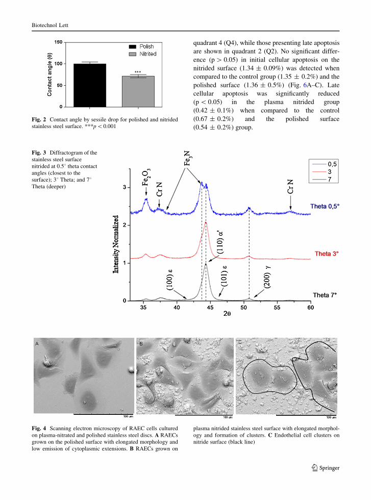
samples. However, the contact angle of the nitrided
surface was significantly lower when compared to the
polished surface (71.81� ± 2.10 versus
100.13� ± 2.49, p\ 0.05) (Fig. 2). Thus, plasma
nitriding increased surface hydrophilicity.
Next, GIXRD confirmed nitrogen incorporation to the
treated steel, with the formation of small chrome nitrite
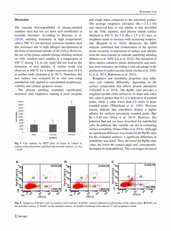
(CrN) peaks (Fig. 3).Adherent cellswere detected on the
samples after 4 h. Cell morphology on the nitrided
stainless steel was elliptical with projections (Fig. 4A,
B). Despite the morphological similarity on the different
surfaces, confirmed by the results of the form factor
(0.37 ± 0.1 vs. 0.40 ± 0.1; p[0.05 for nitrided and
polished, respectively) cell clusters were observed on the
nitrided surface (Fig. 4C).
Cellular viability
Cell viability on the nitrided surface detected via the
MTT assay was significantly higher after 24 h in
comparison to the polished surface (1.73 9 104 cells
vs. 4.9 9 103; p = 0.022) (Fig. 5).
Cell death
Cellular apoptosis was quantified by flow cytometry.
Living cells are reported in quadrant 3 (Q3). Cells
displaying recent apoptosis are represented in
123
Biotechnol Lett

Fig. 1 Nanotipography of stainless steel by AFM. A–B Surface of polished stainless steel. C–D Surface of plasma nitrided stainless
steel. Area = 10 9 10 lm
Table 1 Roughness parameters (nm) of the polished and nitrided plasma stainless steel
Surface Ra Rp Rz Rp/Rz
Polished 0.9 ± 0.05a 3.9 ± 1.67a 5.7 ± 0.61a 0.7 ± 0.2
Nitrided 2.4 ± 0.6b 10.1 ± 3.3b 15.9 ± 5.8b 0.6 ± 0.1
Data are expressed as average ± standard deviation. Averages with different pairs of lower case letters on the same line (a–b)
(p\ 0.001)
123
Biotechnol Lett
quadrant 4 (Q4), while those presenting late apoptosis
are shown in quadrant 2 (Q2). No significant differ-
ence (p[ 0.05) in initial cellular apoptosis on the
nitrided surface (1.34 ± 0.09%) was detected when
compared to the control group (1.35 ± 0.2%) and the
polished surface (1.36 ± 0.5%) (Fig. 6A–C). Late
cellular apoptosis was significantly reduced
(p\ 0.05) in the plasma nitrided group
(0.42 ± 0.1%) when compared to the control
(0.67 ± 0.2%) and the polished surface
(0.54 ± 0.2%) group.
Fig. 2 Contact angle by sessile drop for polished and nitrided
stainless steel surface. ***p\0.001
Fig. 3 Diffractogram of the
stainless steel surface
nitrided at 0.58 theta contactangles (closest to the
surface); 38 Theta; and 78Theta (deeper)
Fig. 4 Scanning electron microscopy of RAEC cells cultured
on plasma-nitrated and polished stainless steel discs. A RAECs
grown on the polished surface with elongated morphology and
low emission of cytoplasmic extensions. B RAECs grown on
plasma nitrided stainless steel surface with elongated morphol-
ogy and formation of clusters. C Endothelial cell clusters on
nitride surface (black line)
123
Biotechnol Lett
Discussion
The vascular biocompatibility of plasma-nitrided
stainless steel has not yet been well established in
scientific literature. According to Braceras et al.
(2018), nitriding treatments at high temperatures
(above 500 �C) can decrease corrosion stainless steel
due resistance due to high nitrogen incorporation in
the form of chromium nitrides (CrN, CrN2). However,
the use of the planar cathode plasma nitriding method
on 316L stainless steel samples at a temperature of
450 �C during 1 h in our study did not lead to the
formation of iron nitrides. A similar result was
observed at 400 �C for a longer exposure time of 8 h
in another study (Samanta et al. 2017). Therefore, this
new surface was evaluated by in vitro tests using
endothelial cells applied to consolidated morphology,
viability and cellular apoptosis assays.
The plasma nitriding treatment significantly
increased steel roughness, making it more irregular
and rough when compared to the untreated surface.
The average roughness elevation (Ra = 2.4 ± 0.6
nm) observed here in was similar to that described
for the 316L stainless steel plasma nitride surface
obtained at 430 �C for 5 h (Ra = 2.5 ± 0.1 nm), as
roughness tends to increase with increasing tempera-
ture (Borgioli et al. 2016). Moreover, the XRD
analysis confirmed that condensation of the ejected
atoms occurred, or deposition of surface iron nitrides
from the mass transfer of cathodic sputtering nitriding
(Ribeiro et al. 2008; Lin et al. 2018). The formation of
these nitrites enhances plastic deformation and stain-
less steel resistance, providing a vital advantage in the
production of cardiovascular stents (Arslan et al. 2008;
Li et al. 2014; Kahraman et al. 2018).
Roughness and wettability properties may influ-
ence cell viability differently, depending on the
surface composition that affects protein adsorption
(Vilardell et al. 2018). The Rp/Rz ratio provides a
roughness profile of the surface by its shape and, when
this value is greater than 0.5, it is indicative of pointed
peaks, while a value lower than 0.5 refers to more
rounded peaks (Whitehead et al. 1995). Previous
reports indicate that osteoblasts display a higher
affinity for surfaces presenting rounded peaks (Rp/
Rz = 0.45 nm) (Silva et al. 2015). However, this
behavior had not yet been described for endothelial
cells. In addition, this variable can aid in estimating
surface wettability (Nunes Filho et al. 2018). Although
no significant difference was found for the Rp/Rz ratio
for the evaluated surfaces, a significant difference in
wettability was noted. Thus, the lower the Rp/Rz ratio
value, the lower the contact angle and, consequently,
the higher its hydrophilicity. This can trigger increased
Fig. 5 Cell viability by MTT after 24 hours of culture in
contact with polystyrene, polished and nitrided surfaces. (a–b) p
\0.05
Fig. 6 Apoptosis of RAEC cells on stainless steel surfaces. A RAEC control cultured on polystyrene of the culture plate. B RAEC on
the polished surface. C RAEC on the nitrided surface, all double-incubated with annexin-V and propidium iodide
123
Biotechnol Lett

proliferation and cell differentiation (Vilardell et al.
2018).
Both focal adhesion and the cell spreading area are
important parameters used to assess cell-biomaterial
interactions (Turner et al. 2004). Here in, the endothe-
lial cells showed adhesion and spreading in the first
4 h after incubation on the nitrided surface. Therefore,
it is probable that the chemical and physical changes
due to plasma nitriding stimulated protein adsorption
on the surface, being important for the activation of
cell adhesion proteins (Ferraz et al. 2014; Moura et al.
2016; Talha et al. 2019). Both roughness and nitriding
conditions play an important role in promoting
adhesion (Martinesi et al. 2013; Jayalakshmi et al.
2018). According to van Wachem et al. (1985) the
initial adhesion of human umbilical cord vein endothe-
lial cells to surfaces requires high clustered cell
density, which ensures cellular spreading and prolif-
eration on the polymer surface. This implies that the
applied metal nitriding stimulated the colonization of
endothelial cells, an important feature to increase
vascularization and re-endothelialization, which aid in
functionalizing stainless steel implants (Offner et al.
2017).
Surface cell adhesion does not necessarily imply
that the cells maintain their viability (Popat et al.
2007). However, a significant increase in cell viability
on the treated surface was observed 24 h after
adhesion, indicating that plasma nitriding indirectly
improves biocompatibility (Arslan et al. 2008). How-
ever, some authors observed endothelial cell prolifer-
ation on 316L stainless steel only 72 h after using
different plasma nitriding conditions at low tempera-
tures (400 �C for 5 h) (Martinesi et al. 2013). Thus, the
nitriding condition used in our study reduced cytotox-
icity in the first hours of adhesion and favored greater
proliferation of viable cells.
The plasma nitriding carried out under the condi-
tions applied in the present study reduced the late
apoptosis of endothelial cells. It is possible that the
treatment reduced the release of nickel ions, which
raises the cytotoxic effect of the surface, since it is then
necessary to add high nitrogen concentrations to
stainless steel to produce a nickel-free metal (Lo
et al. 2009). In the present study, the application of low
temperature plasma nitriding on stainless steel,
besides promoting better adhesion and greater viabil-
ity of endothelial cells, also reduced the cytotoxic
effect of the stainless steel in the first 24 h. Nitrogen
incorporation, carried out at 450 �C for only 1 h, was
able to increase stainless steel corrosion resistance.
Therefore, this treatment is a possible candidate for
use in cardiovascular stainless steel devices.
Acknowledgements This study was financed in part by the
Coordenacao de Aperfeicoamento de Pessoal de Nıvel
Superior—Brasil (CAPES)—Finance Code 001. The authors
wish to acknowledge the professional efforts of team of the
Laboratory of Structural Characterization of Materials at UFRN
and Dr. Helena B. Nader of UNIFESP, Sao Paulo, Brazil for
contributing with the endothelial rabbit aorta cell line.
Compliance with ethical standards
Conflict of interest All authors declares that they have no
conflict of interest.
Ethical approval This article does not contain any studies
with human participants or animals performed by any of the
authors.
References
Alves C, Guerra Neto CLB, Morais GHS et al (2006) Nitriding
of titanium disks and industrial dental implants using hol-
low cathode discharge. Surf Coat Technol 200:3657–3663.
https://doi.org/10.1016/J.SURFCOAT.2005.08.005
Arslan E, Igdil MC, Yazici H et al (2008) Mechanical properties
and biocompatibility of plasma-nitrided laser-cut 316L
cardiovascular stents. J Mater Sci 19:2079–2086. https://
doi.org/10.1007/s10856-007-3302-4
Borgioli F, Galvanetto E, Bacci T (2016) Low temperature
nitriding of AISI 300 and 200 series austenitic stainless
steels. Vacuum 127:51–60. https://doi.org/10.1016/j.
vacuum.2016.02.009
Braceras I, Ibanez I, Dominguez-Meister S et al (2018) Plasma
nitriding of the inner surface of stainless steel tubes. Surf
Coat Technol 1:4. https://doi.org/10.1016/j.surfcoat.2018.
04.057
Butruk-Raszeja B, Dresler M, Kuzminska A, Ciach T (2016)
Endothelialization of polyurethanes: surface silanization
and immobilization of REDV peptide. Elsevier,
Amsterdam
Chichareon P, Katagiri Y, Asano T et al (2019) Mechanical
properties and performances of contemporary drug-eluting
stent: focus on the metallic backbone. Expert Rev Med
Devices 17434440(2019):1573142. https://doi.org/10.
1080/17434440.2019.1573142
De Morais LS, Guimaraes GS, Elias CN (2007) Liberacao de
ıons por biomateriais metalicos. Maringa 12:48–53. https://
doi.org/10.1590/S1415-54192007000600006
Ferraz EP, Sa JC, de Oliveira PT et al (2014) The effect of
plasma-nitrided titanium surfaces on osteoblastic cell
adhesion, proliferation, and differentiation. J Biomed
Mater Res Part A 102:991–998. https://doi.org/10.1002/
jbm.a.34761
123
Biotechnol Lett
Fox KE, Tran NL, Nguyen TA et al (2019) Surface modification
of medical devices at nanoscale—recent development and
translational perspectives. Biomater Transl Med. https://
doi.org/10.1016/b978-0-12-813477-1.00008-6
Jayalakshmi M, Bhat BR, Bhat KU (2018) Enhanced cell
adhesion on severe peened-plasma nitrided 316L stainless
steel. AIP Publishing, New York, p 020086
Kahraman F, Gencer GM, Kahraman AD et al (2018) Low-
temperature nitriding behavior of compressive deformed
AISI 316Ti austenitic stainless steels. Surf Rev Lett.
https://doi.org/10.1142/s0218625x18501883
Kao W-H, Su Y-L, Horng J-H, Hsieh Y-T (2017) Improved
tribological properties, electrochemical resistance and
biocompatibility of AISI 316L stainless steel through
duplex plasma nitriding and TiN coating treatment. J Bio-
mater Appl 32:12–27. https://doi.org/10.1177/
0885328217712109
Kathuria YP (2006) The potential of biocompatible metallic
stents and preventing restenosis. Mater Sci Eng A
417:40–48. https://doi.org/10.1016/J.MSEA.2005.11.007
Li Y, Zhang S, He Y et al (2014) Characteristics of the nitrided
layer formed on AISI 304 austenitic stainless steel by high
temperature nitriding assisted hollow cathode discharge.
Mater Des 64:527–534. https://doi.org/10.1016/J.
MATDES.2014.08.023
Lin N, Liu Q, Zou J et al (2016) Surface texturing-plasma
nitriding duplex treatment for improving tribological per-
formance of AISI 316 stainless steel. Materials (Basel)
9:875. https://doi.org/10.3390/ma9110875
Lin K, Li X, Dong H et al (2018) Nitrogen mass transfer and
surface layer formation during the active screen plasma
nitriding of austenitic stainless steels. Vacuum
148:224–229. https://doi.org/10.1016/J.VACUUM.2017.
11.022
Lo KH, Shek CH, Lai JKL (2009) Recent developments in
stainless steels. Mater Sci Eng R 65:39–104. https://doi.
org/10.1016/j.mser.2009.03.001
Lu B, Malcuit C, Wang S et al (2009) Long-term safety and
function of RPE from human embryonic stem cells in
preclinical models of macular degeneration. Stem Cells
27:2126–2135. https://doi.org/10.1002/stem.149
Martinesi M, Stio M, Treves C, Borgioli F (2013) Biocompat-
ibility studies of low temperature nitrided and collagen-I
coated AISI 316L austenitic stainless steel. J Mater Sci
24:1501–1513. https://doi.org/10.1007/s10856-013-4902-
9
Moura CEB, Silva NB, Sa JC et al (2016) MC3T3-E1 cells
behavior on surfaces bombarded by argon ions in planar
cathode discharge. Artif Organs 40:497–504. https://doi.
org/10.1111/aor.12597
Nunes Filho A, de Aires M, Braz DC et al (2018) Titanium
surface chemical composition interferes in the Pseu-
domonas aeruginosa biofilm formation. Artif Organs
42:193–199. https://doi.org/10.1111/aor.12983
Offner D, Wagner Q, Idoux-Gillet Y et al (2017) Hybrid col-
lagen sponge and stem cells as a new combined scaffold
able to induce the re-organization of endothelial cells into
clustered networks. Biomed Mater Eng 28:S185–S192.
https://doi.org/10.3233/BME-171640
Popat KC, Leoni L, Grimes CA, Desai TA (2007) Influence of
engineered titania nanotubular surfaces on bone cells.
Biomaterials 28:3188–3197. https://doi.org/10.1016/J.
BIOMATERIALS.2007.03.020
Ribeiro KJB, de Sousa RRM, de Araujo FO et al (2008)
Industrial application of AISI 4340 steels treated in
cathodic cage plasma nitriding technique. Mater Sci Eng A
479:142–147. https://doi.org/10.1016/j.msea.2007.06.033
Samanta A, Chakraborty H, Bhattacharya M et al (2017) Nan-
otribological response of a plasma nitrided bio-steel.
J Mech Behav Biomed Mater 65:584–599. https://doi.org/
10.1016/j.jmbbm.2016.09.017
Schwartz RS, Edelman E, Virmani R et al (2008) Drug-eluting
stents in preclinical studies: updated consensus recom-
mendations for preclinical evaluation. Circ Cardiovasc
Interv 1:143–153. https://doi.org/10.1161/CIRCINTER
VENTIONS.108.789974
Shah A, Sinha R, Hickok N, Tuan R (1999) High-resolution
morphometric analysis of human osteoblastic cell adhesion
on clinically relevant orthopedic alloys. Bone 24:499–506.
https://doi.org/10.1016/S8756-3282(99)00077-0
Silva MAM, Valentim RAM, Guerra PVA et al (2015) Influ-
encia da topografia na molhabilidade em superfıcies de
titanio tratadas por plasma. Rev Bras Inov Technol Saude.
https://doi.org/10.18816/r-bits.v5i2.7247Su Y, Luo C, Zhang Z et al (2018) Bioinspired surface functional-
ization of metallic biomaterials. J Mech Behav BiomedMater
77:90–105. https://doi.org/10.1016/j.jmbbm.2017.08.035
Talha M, Ma Y, Kumar P et al (2019) Role of protein adsorption
in the bio corrosion of metallic implants—a review. Col-
loids Surf B 176:494–506. https://doi.org/10.1016/j.
colsurfb.2019.01.038
Trabzon L, Igdil MC (2006) On the materials properties of thin
film plasma-nitrided austenitic stainless steel. Surf Coat
Technol 200:4195–4200. https://doi.org/10.1016/J.
SURFCOAT.2004.12.012
Turner N, Armitage M, Butler R, Ireland G (2004) An in vitro
model to evaluate cell adhesion to metals used in implan-
tation shows significant differences between palladium and
gold or platinum. Cell Biol Int 28:541–547. https://doi.org/
10.1016/j.cellbi.2004.04.009
van Wachem PB, Beugeling T, Feijen J et al (1985) Interaction
of cultured human endothelial cells with polymeric sur-
faces of different wettabilities. Biomaterials 6:403–408.
https://doi.org/10.1016/0142-9612(85)90101-2
Vilardell AM, Cinca N, Garcia-Giralt N et al (2018)
Osteoblastic cell response on high-rough titanium coatings
by cold spray. J Mater Sci 29:1–10. https://doi.org/10.
1007/s10856-018-6026-8
Whitehead SA, Shearer AC, Watts DC, Wilson NHF (1995)
Comparison of methods for measuring surface roughness
of ceramic. J Oral Rehabil 22:421–427. https://doi.org/10.
1111/j.1365-2842.1995.tb00795.x
Zhao G-H, Aune RE, Espallargas N (2016) Tribocorrosion
studies of metallic biomaterials: the effect of plasma
nitriding and DLC surface modifications. J Mech Behav
Biomed Mater 63:100–114. https://doi.org/10.1016/J.
JMBBM.2016.06.014
Publisher’s Note Springer Nature remains neutral with
regard to jurisdictional claims in published maps and
institutional affiliations.
123
Biotechnol Lett
View publication statsView publication stats