plasmenylethanolamine is the arachidonic ii stimulation · proc. natl. acad. sci. usa vol. 86, pp....
TRANSCRIPT

Proc. Natl. Acad. Sci. USAVol. 86, pp. 3479-3483, May 1989Biochemistry
Plasmenylethanolamine is the major storage depot for arachidonicacid in rabbit vascular smooth muscle and is rapidly hydrolyzedafter angiotensin II stimulation
(plasmalogens/phosphatidylethanolamine/phosphatidylcholine)
DAVID A. FORD*t AND RICHARD W. GROSS*tDepartments of *Medicine and tChemistry, Washington University, Saint Louis, MO 63110
Communicated by William D. Phillips, January 3, 1989
ABSTRACT The present study demonstrates that rabbitaortic intimal smooth muscle cells contain the majority of theirendogenous arachidonic acid mass in plasmenylethanolaminemolecular species. To demonstrate the potential significance ofthese plasmenylethanolamines as substrates for the smoothmuscle cell phospholipases that are activated during agoniststimulation, aortic rings were prelabeled with [3H]arachidonicacid and stimulated with angiotensin II. Although the specificactivities of the choline and inositol glycerophospholipid poolswere similar after the labeling interval, ethanolamine glycero-phospholipids had a specific activity of only 20% of the specificactivity of choline and inositol glycerophospholipids. Despitethe marked disparity in the specific activities of these threephospholipid classes, angiotensin II stimulation resulted insimilar fractional losses (35-41%) of [3lljarachidonic acidfrom vascular smooth muscle choline, ethanolamine, andinositol glycerophospholipid classes. Reverse-phase HPLCdemonstrated that >60% of the [3H]arachidonic acid releasedfrom ethanolamine glycerophospholipids after angiotensin IIstimulation originated from plasmenylethanolamine molecularspecies. Taken together, the results demonstrate that the majorphospholipid storage depot for arachidonic acid in vascularsmooth muscle cells are plasmenylethanolamine molecularspecies which are important substrates for the phospholipase(s)that are activated during agonist stimulation.
Icosanoids are important regulators of vascular smoothmuscle cell contractile state, hypertrophy, and proliferation(1-6). Accordingly, significant attention has focused on thebiochemical events responsible for the liberation of arachi-donic acid from endogenous smooth muscle cell lipids duringsignal transduction. Arachidonic acid release from cellularphospholipids may occur directly in a single catalytic step bythe action of phospholipase A2 or by a multistep sequentialpathway initiated by phospholipase C and culminated by thesequential actions of diglyceride and monoglyceride lipases(7-9). Although recent studies demonstrated that radiola-beled arachidonic acid was released from choline, ethanol-amine, and inositol glycerophospholipid in cultured smoothmuscle cell pools after agonist stimulation, quantification ofthe amount of released radiolabeled arachidonic acid fromeach pool suggested that different phospholipid classes wereprimarily responsible for radiolabeled arachidonic acid re-lease (10-13). Since the biochemical and physiologic prop-erties of cultured smooth muscle cells differ substantiallyfrom properties of vascular smooth muscle cells in intacttissue, the present study was performed to assess the distri-bution of arachidonic acid mass in phospholipids and thefractional turnover of individual phospholipid molecularspecies in intact vascular smooth muscle. The results dem-
onstrate that the majority of arachidonic acid mass in vas-cular smooth muscle is present in plasmenylethanolaminemolecular species and that substantial amounts of arachi-donic acid in smooth muscle plasmenylethanolamines arerapidly mobilized after angiotensin II stimulation.
MATERIALS AND METHODSExtraction of Aortic Intimal Smooth Muscle Glycerophos-
pholipids. The intimal layer of aortic smooth muscle wasprepared according to the method of Ross (14) from NewZealand rabbit and mongrel dog aorta denuded of endothe-lium. The intimal layer was frozen at liquid nitrogen temper-ature and pulverized. An aliquot of the powdered tissue wastaken for subsequent protein analysis and the remainingtissue was extracted by the method of Bligh and Dyer (15).
Preparation of Aortic Rings, [3H]Arachidonic Acid Label-ing, and Angiotensin II Stimulation. Thoracic aortas wereremoved from New Zealand rabbits, denuded of endothelialcells, and extravascular fat was dissected. The aortas werecut into rings (-0.5 cm wide) and incubated for 12 hr at 370Cin 20 ml of modified Krebs-Henseleit buffer at pH 7.3continuously equilibrated with O2/CO2 (95:5, vol/vol) con-taining 118 mM NaCl, 4.7 mM KCl, 3.0 mM CaC12, 1.2 mMMgSO4, 1.2 mM KH2PO4, 25 mM NaHCO3, 0.5 mM NaEDTA, 15 mM glucose, and 20 ,Ci of [3H]arachidonic acid(1 Ci = 37 GBq). The aortic rings next were rinsed twice withmodified Krebs-Henseleit buffer without [3H]arachidonicacid and then were incubated for 15 min with modifiedKrebs-Henseleit buffer containing 100 nM angiotensin II orsaline. Incubations were terminated by freeze clampingblotted tissue. The tissue was pulverized at liquid nitrogentemperature, the wet weight was determined, a sample wastaken to determine the wet/dry weight ratio, and tissue wasextracted by the Bligh and Dyer method (15). During theextraction procedure, 1 x 10⁵ dpm of [14C]lysophosphatidyl-choline and 500 nmol of 1,2-dioctadec-9',12',15'-enoyl-GPE[-sn-glycero(3)phosphoethanolamine] were added as internalstandards.
Phospholipid Separation and Analyses. Phospholipids wereresolved by using an Ultrasphere-Si column (4.5 x 250 mm;5 ,tm; Beckman) as the stationary phase with an initial lineargradient over 10 min from a mobile phase of hexane/isopro-panol/water (48.5:48.5:3, vol/vol) to hexane/isopropanol/water (47.75:47.75:4.5, vol/vol). The composition of thelatter mobile phase was held constant for 8 min followed bya step change to hexane/isopropanol/water (46.5:46.5:7,vol/vol). The mass of each phospholipid class was quanti-
Abbreviations: -GPE, -sn-glycero(3)phosphoethanolamine; -GPC,-sn-glycero(3)phosphocholine.tTo whom reprint requests should be addressed at: Molecular andCellular Cardiovascular Biochemistry, Box 8020, Washington Uni-versity School of Medicine, 660 South Euclid Avenue, Saint Louis,MO 63110.
3479
The publication costs of this article were defrayed in part by page chargepayment. This article must therefore be hereby marked "advertisement"in accordance with 18 U.S.C. §1734 solely to indicate this fact.
3480 Biochemistry: Ford and Gross
tated by comparisons of detector response (flame ionizationdetector) of derivatized (methanol hydrochloride) straight-phase HPLC-purified phospholipids with that of exogenousarachidic acid (internal standard) as described (16). Individ-ual molecular species of ethanolamine and choline glycero-phospholipids were determined by fast atom bombardmentmass spectrometry as described (16). The content of alkylether choline and ethanolamine glycerophospholipids wasdetermined by a modification of the method of Blank et al.(17). The positional specificity of fatty acids and aldehydes incholine glycerophospholipids was analyzed by exploiting theregiospecific cleavage of phospholipids catalyzed by phos-pholipase A2 (16). Choline and ethanolamine glycerophos-pholipid molecular species were resolved by reverse-phaseHPLC (18). The amount of [3H]arachidonic acid present inindividual molecular species of ethanolamine glycerophos-pholipids was quantified by liquid scintillation spectrometryof reverse-phase HPLC column eluents. Small differences inrecovery were corrected by normalization with the integratedarea of the exogenous internal standard 1,2-dioctadec-9',-12',15'-enoyl-GPE.
RESULTSPhospholipid Class and Subclass Analysis of Aortic Smooth
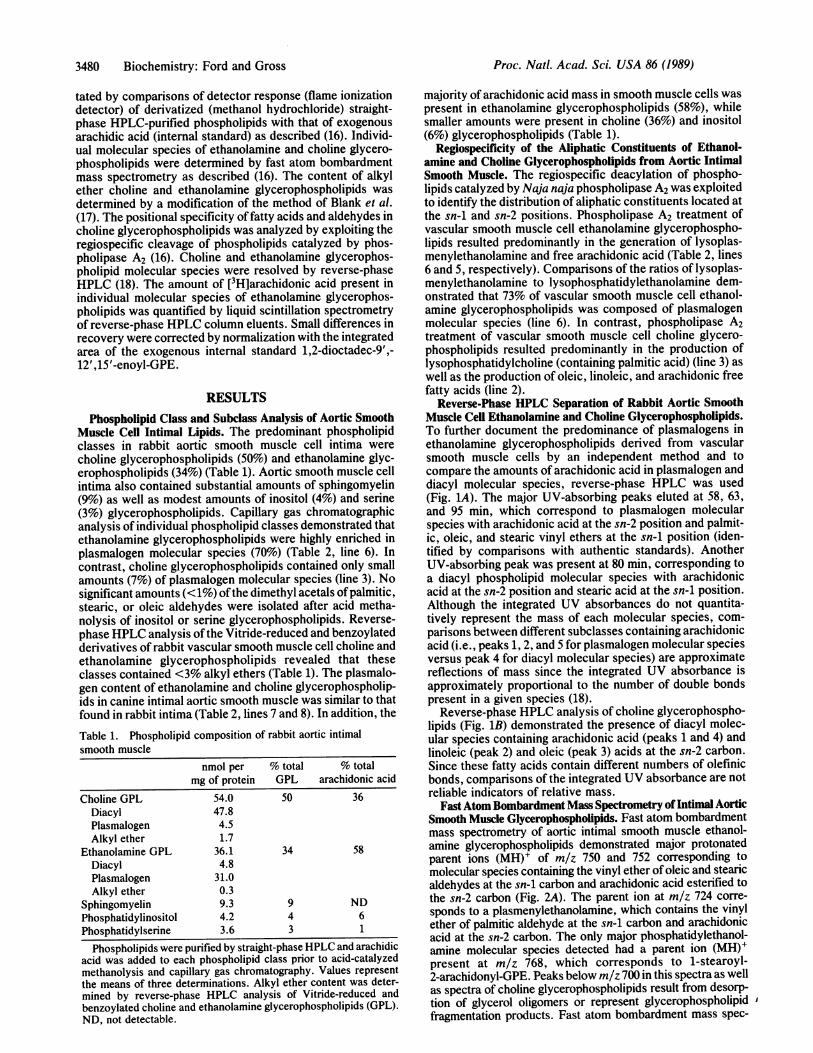
Muscle Cell Intimal Lipids. The predominant phospholipidclasses in rabbit aortic smooth muscle cell intima werecholine glycerophospholipids (50%) and ethanolamine glyc-erophospholipids (34%) (Table 1). Aortic smooth muscle cellintima also contained substantial amounts of sphingomyelin(9%) as well as modest amounts of inositol (4%) and serine(3%) glycerophospholipids. Capillary gas chromatographicanalysis of individual phospholipid classes demonstrated thatethanolamine glycerophospholipids were highly enriched inplasmalogen molecular species (70%) (Table 2, line 6). Incontrast, choline glycerophospholipids contained only smallamounts (7%) of plasmalogen molecular species (line 3). Nosignificant amounts (<1%) ofthe dimethyl acetals ofpalmitic,stearic, or oleic aldehydes were isolated after acid metha-nolysis of inositol or serine glycerophospholipids. Reverse-phase HPLC analysis ofthe Vitride-reduced and benzoylatedderivatives of rabbit vascular smooth muscle cell choline andethanolamine glycerophospholipids revealed that theseclasses contained <3% alkyl ethers (Table 1). The plasmalo-gen content of ethanolamine and choline glycerophospholip-ids in canine intimal aortic smooth muscle was similar to thatfound in rabbit intima (Table 2, lines 7 and 8). In addition, the
Table 1. Phospholipid composition of rabbit aortic intimalsmooth muscle
nmol per % total % totalmg of protein GPL arachidonic acid
Choline GPL 54.0 50 36Diacyl 47.8Plasmalogen 4.5Alkyl ether 1.7
Ethanolamine GPL 36.1 34 58Diacyl 4.8Plasmalogen 31.0Alkyl ether 0.3
Sphingomyelin 9.3 9 NDPhosphatidylinositol 4.2 4 6Phosphatidylserine 3.6 3 1
Phospholipids were purified by straight-phase HPLC and arachidicacid was added to each phospholipid class prior to acid-catalyzedmethanolysis and capillary gas chromatography. Values representthe means of three determinations. Alkyl ether content was deter-mined by reverse-phase HPLC analysis of Vitride-reduced andbenzoylated choline and ethanolamine glycerophospholipids (GPL).ND, not detectable.
majority of arachidonic acid mass in smooth muscle cells waspresent in ethanolamine glycerophospholipids (58%), whilesmaller amounts were present in choline (36%) and inositol(6%) glycerophospholipids (Table 1).
Regiospecificity of the Aliphatic Constituents of Ethanol-amine and Choline Glycerophospholipids from Aortic IntimalSmooth Muscle. The regiospecific deacylation of phospho-lipids catalyzed by Naja naja phospholipase A2 was exploitedto identify the distribution of aliphatic constituents located atthe sn-i and sn-2 positions. Phospholipase A2 treatment ofvascular smooth muscle cell ethanolamine glycerophospho-lipids resulted predominantly in the generation of lysoplas-menylethanolamine and free arachidonic acid (Table 2, lines6 and 5, respectively). Comparisons of the ratios of lysoplas-menylethanolamine to lysophosphatidylethanolamine dem-onstrated that 73% of vascular smooth muscle cell ethanol-amine glycerophospholipids was composed of plasmalogenmolecular species (line 6). In contrast, phospholipase A2treatment of vascular smooth muscle cell choline glycero-phospholipids resulted predominantly in the production oflysophosphatidylcholine (containing palmitic acid) (line 3) aswell as the production of oleic, linoleic, and arachidonic freefatty acids (line 2).
Reverse-Phase HPLC Separation of Rabbit Aortic SmoothMuscle Cell Ethanolamine and Choline Glycerophospholipids.To further document the predominance of plasmalogens inethanolamine glycerophospholipids derived from vascularsmooth muscle cells by an independent method and tocompare the amounts of arachidonic acid in plasmalogen anddiacyl molecular species, reverse-phase HPLC was used(Fig. 1A). The major UV-absorbing peaks eluted at 58, 63,and 95 min, which correspond to plasmalogen molecularspecies with arachidonic acid at the sn-2 position and palmit-ic, oleic, and stearic vinyl ethers at the sn-i position (iden-tified by comparisons with authentic standards). AnotherUV-absorbing peak was present at 80 min, corresponding toa diacyl phospholipid molecular species with arachidonicacid at the sn-2 position and stearic acid at the sn-i position.Although the integrated UV absorbances do not quantita-tively represent the mass of each molecular species, com-parisons between different subclasses containing arachidonicacid (i.e., peaks 1, 2, and 5 for plasmalogen molecular speciesversus peak 4 for diacyl molecular species) are approximatereflections of mass since the integrated UV absorbance isapproximately proportional to the number of double bondspresent in a given species (18).
Reverse-phase HPLC analysis of choline glycerophospho-lipids (Fig. 1B) demonstrated the presence of diacyl molec-ular species containing arachidonic acid (peaks 1 and 4) andlinoleic (peak 2) and oleic (peak 3) acids at the sn-2 carbon.Since these fatty acids contain different numbers of olefinicbonds, comparisons of the integrated UV absorbance are notreliable indicators of relative mass.
Fast Atom Bombardment Mass Spectrometry ofIntimal AorticSmooth Muscle Glycerophospholipids. Fast atom bombardmentmass spectrometry of aortic intimal smooth muscle ethanol-amine glycerophospholipids demonstrated major protonatedparent ions (MH)+ of m/z 750 and 752 corresponding tomolecular species containing the vinyl ether of oleic and stearicaldehydes at the sn-1 carbon and arachidonic acid esterified tothe sn-2 carbon (Fig. 2A). The parent ion at m/z 724 corre-sponds to a plasmenylethanolamine, which contains the vinylether of palmitic aldehyde at the sn-i carbon and arachidonicacid at the sn-2 carbon. The only major phosphatidylethanol-amine molecular species detected had a parent ion (MH)+present at m/z 768, which corresponds to 1-stearoyl-2-arachidonyl-GPE. Peaks below m/z 700 in this spectra as wellas spectra of choline glycerophospholipids result from desorp-tion of glycerol oligomers or represent glycerophospholipid lfragmentation products. Fast atom bombardment mass spec-
Proc. Natl. Acad. Sci. USA 86 (1989)

Proc. Natl. Acad. Sci. USA 86 (1989) 3481
Table 2. Fatty acid and aldehyde profiles of native and phospholipase A2-treated phospholipids of intimal smooth muscle from rabbitaorta and canine aorta
16:0 (D) 16:0 (F) 18:0 (D) 18:0 (F) 18:1 (D) 18:1 (F) 18:2 (F) 20:3 (F) 20:4 (F) 22:4 (F)Rabbit CGP 3 33 1 20 ND 20 9 1 13 1Rabbit PLA2-CGP-FA ND 20 ND 4 ND 31 27 ND 19 NDRabbit PLA2-CGP-LPC 5 56 2 32 ND 5 ND ND ND NDRabbit EGP 11 4 24 11 4 10 2 2 31 2Rabbit PLA2-EGP-FA ND 4 ND 4 ND 20 8 2 57 1Rabbit PLA2-EGP-LPE 15 9 51 14 7 4 2 ND ND NDCanine CGP 2 30 1 26 ND 14 5 1 16 6Canine EGP 5 7 23 15 5 4 1 1 29 11
Choline and ethanolamine glycerophospholipids (CGP and EGP, respectively) from rabbit and canine aortic intima were purified bystraight-phase HPLC, subjected to acid-catalyzed methanolysis, and analyzed by capillary gas chromatography. Products of phospholipaseA2-treated CGP and EGP were separated by TLC and subjected to acid-catalyzed methanolysis. PLA2-CGP(EGP)-FA and PLA2-CGP(EGP)-LPC(LPE) are the fatty acid and lysophospholipid products of phospholipase A2 treatment, respectively. D, dimethyl acetal; F, fattyacid methyl ester; ND, not detectable. Values are expressed in % weight.
trometry of choline glycerophospholipids demonstrated pre-dominant parent ions at m/z 758 and 760 corresponding to1-palmitoyl-2-linoleoyl-GPC [-sn-glycero(3)phosphocholine]and 1-palmitoyl-2-oleoyl-GPC, respectively (Fig. 2B). Aorticintimal smooth muscle inositol glycerophospholipids containedmajor negative ions (M-1)- at m/z 883 and 885 correspondingto 1-oleoyl-2-arachidonyl-GPI [-sn-glycero(3)phospho(1)-L-myo-inositol] (20%o) and 1-stearoyl-2-arachidonyl-GPI (80%o)(data not shown).
Radiolabeling of Aortic Smooth Muscle Phospholipids andAngiotensin 11-Stimulated Release of Incorporated [3H]Arach-idonic Acid. Rabbit aortic rings incorporated substantialamounts of radiolabeled arachidonic acid into choline, eth-anolamine, and inositol glycerophospholipids during the12-hr labeling interval (Table 3). Despite the fact that etha-nolamine glycerophospholipids contained the majority ofarachidonic acid mass in rabbit vascular smooth muscle, thecholine glycerophospholipid pool contained >3 times as
A B
5
Uz4
o 1 1
0 ~2 4
324
3
20 40 60 80 100 120 20 40 60 80 100 120TIME AFTER INJECTION (Min)
FIG. 1. Reverse-phase HPLC of aortic intimal ethanolamine andcholine glycerophospholipids. Intimal ethanolamine and cholineglycerophospholipids (-300 nmol) were purified by straight-phaseHPLC, injected onto an octadecyl silica column, and eluted with amobile phase composed of methanol/acetonitrile/water (90.5:2.5:7,vol/vol) containing 20 mM choline chloride. UV absorbance wasmonitored at 203 nm. The molecular identities of the ethanolamineglycerophospholipids (A) are 1-O-hexadec-1'-enyl-2-icosodec-5',8',11',14'-enoyl-GPE (peak 1), 1-O-octadec-1',9'-enyl-2-icosodec-5',8',11',14'-enoyl-GPE (peak 2), 1-O-hexadec-1'-enyl-2-octadec-9',12'-enoyl-GPE (peak 3), 1-octadecanoyl-2-icosodec-5',8',11',14'-enoyl-GPE (peak 4), and 1-O-octadec-1'-enyl-2-icosodec-5',8',11',14'-enoyl-GPE (peak 5). The molecularidentities of choline glycerophospholipids (B) are 1-hexadecanoyl-2-icosodec-5',8',11',14'-enoyl-GPC (peak 1), 1-hexadecanoyl-2-octadec-9',12'-enoyl-GPC (peak 2), 1-hexadecanoyl-2-octadec-9'-enoyl-GPC (peak 3), and 1-octadecanoyl-2-icosodec-5',8',11',14'-enoyl-GPC (peak 4).
much radiolabeled arachidonic acid after the labeling inter-val. Accordingly, the specific activity of arachidonic acid ineach major glycerophospholipid pool was substantially dif-ferent. Choline and inositol glycerophospholipids had aspecific activity -5-fold that of the specific activity ofethanolamine glycerophospholipids (Table 3). It should benoted that initial studies demonstrated that the phospholipidcomposition of aortic rings was virtually identical to that ofthe intimal layer of aorta. Despite the marked disparities inthe specific activities of these three phospholipid classes,subsequent incubation of prelabeled rabbit aortic rings with100 nM angiotensin II resulted in the release of similarfractional percentages of radiolabeled arachidonic acid fromcholine, ethanolamine, and inositol glycerophospholipidclasses (35-41% loss from each pool) (Table 3). Directquantification of released radiolabeled arachidonic acid dem-onstrated that choline and ethanolamine glycerophospholip-ids together were responsible for >90% of the releasedradiolabeled arachidonic acid. To identify the individualmolecular species that incorporated radiolabeled arachidonic
100.
! 80-
3 60.z
Z 40.
< 20.uj
IC
-
z
4
I-4
t!
A 3
33962
387 4 6611
502 583
752
!7
750
724 768
I . I L L 842
300 400 500 600 700 800 900M/z
B
496
.
t jA760so- 5
22
78620 ii 550 72 7821
A iI 645810 E4s0 500 550 600 650 700
m /r750 800
FIG. 2. Fast atom bombardment mass spectrometry of aorticintimal ethanolamine and choline glycerophospholipids. HPLC-purified intimal ethanolamine (A) and choline (B) glycerophospho-lipids (-300 nmol) were dissolved in 20 ul of chloroform/methanol(1:1, vol/vol) and 2 Il was mixed with 3 ,ul of glycerol on a copperprobe. Fast atom bombardment mass spectrometry was performedas described in Materials and Methods.
Biochemistry: Ford and Gross
A

3482 Biochemistry: Ford and Gross
Table 3. Distribution of [3H]arachidonic acid in aortic phospholipids before and after incubationwith angiotensin II
Control All change RSA
Choline GPL 413,010 ± 48,400 255,220 ± 55,890* -38 1Ethanolamine GPL 130,820 ± 8,210 77,510 ± 4,420t -41 0.20Inositol GPL 63,630 ± 11,350 41,310 ± 9,120* -35 0.93
Defatted and endothelial denuded aortic rings were labeled for 12 hr with [3H]arachidonic acid (1ACi/ml) in 20 ml of modified oxygenated Krebs-Henseleit (K-H) buffer at 370C. Extracellular[3H]arachidonic acid was subsequently removed by two sequential 15-min incubations at 370C in 25 mlof modified oxygenated K-H buffer. The aortic rings were incubated for 15 min in modified K-H buffercontaining either 100 nM angiotensin II (All) or vehicle (saline). Aortic lipids were extracted by theBligh and Dyer (15) method, at which time 1 x 10⁵ dpm of 14C]lysophosphatidylcholine was added(internal standard). Individual phospholipid classes were resolved by straight-phase HPLC andincorporated radiolabel was quantitated by liquid scintillation spectrometry. The values for radiolabelincorporated into each phospholipid pool are the means ± SEM for four separate experiments and areexpressed as cpm/g (dry weight). RSA, relative specific activity (ratio of [3H]arachidonic acid cpm tonmol of arachidonylated phospholipid; the RSA of choline GPL was normalized to 1); GPL,glycerophospholipids.*P < 0.05 for control vs. All-treated aortic rings.tp < 0.01 for control vs. All-treated aortic rings.
acid during the labeling interval and those that released[3H]arachidonic acid after agonist stimulation, reverse-phaseHPLC was used. As expected, radiolabeled arachidonic acidwas predominantly incorporated into 1-palmitoyl-2-arachi-donyl-GPC (75%) with smaller amounts present in 1-stearoyl-2-arachidonyl-GPC as well as other choline glycerophospho-lipid subclasses (data not shown). Angiotensin II stimulationresulted in comparable fractional decreases in each radiola-beled choline glycerophospholipid molecular species. Re-verse-phase HPLC of radiolabeled ethanolamine glycero-phospholipids demonstrated that plasmalogen molecular spe-cies contained the majority of radiolabeled arachidonic acid(70%), while smaller amounts (30%) were present in phos-phatidylethanolamine molecular species (Fig. 3). Surpris-ingly, the distribution of [3H]arachidonic acid in plasmalogenmolecular species was similar in plasmenylethanolaminescontaining the vinyl ether of palmitaldehyde and stearalde-hyde at the sn-i position, although the relative mass distri-
20 -A
3
15k
101
1 2
6
bution of these two molecular species was substantiallydifferent. Analysis of individual radiolabeled molecular spe-cies of ethanolamine glycerophospholipids that contributedto the release of radiolabeled arachidonic acid after angio-tensin II stimulation was made by comparing the amounts of[3H]arachidonic acid in individual molecular species afternormalization to the recovery of internal standard as de-scribed in Materials and Methods (Fig. 3). Statisticallysignificant losses (P < 0.02; n = 3) of arachidonylatedplasmenylethanolamine molecular species (peaks 3, 4, and 6)as well as arachidonylated phosphatidylethanolamine molec-ular species (peak 5) were observed. The fractional percent-age of hydrolysis of each ethanolamine glycerophospholipidsubclass was similar, demonstrating that the phospholi-pase(s) responsible for the release of arachidonic acid fromvascular smooth muscle phospholipids during angiotensin IIstimulation effectively hydrolyzes both phosphatidylethanol-amine and plasmenylethanolamine substrates.
B
6
1 2
20 40 60 80 100 120TIME (Min)
FIG. 3. Reverse-phase HPLC of [3H]arachidonic acid-labeled ethanolamine glycerophospholipids from rabbit aortic rings incubated with andwithout 100 nM angiotensin II. Aortic rings were prelabeled with [3H]arachidonic acid and subsequently incubated with or without 100 nMangiotensin II. Cellular phospholipids were extracted as described in Materials and Methods in the presence of 500 nmol of 1,2-dioctadec-8',11',14'-enoyl-GPE (internal standard). Ethanolamine glycerophospholipids were purified by straight-phase HPLC, injected onto
an octadecyl silica column, and eluted with methanol/acetonitrile/water (90.5:2.5:7,vol/vol) containing 20mM choline chloride. Column eluents
were collected in 1-min fractions and their radioactivity was determined by liquid scintillation spectrometry. The peaks displaying significantamounts of radioactivity are 1-O-hexadec-1'-enyl-2-icosodec-5',8',11',14'-enoyl-GPE (peak 3), 1-O-octadec-1',9'-enyl-2-icosodec-5',8',11',14'-enoyl-GPE (peak 4), 1-octadecanoyl-2-icosodec-5',8',11',14'-enoyl-GPE (peak 5), and 1-O-octadec-1'-enyl-2-icosodec-5',8',11',14'-enoyl-GPE (peak 6). The amount of radioactivity associated with the ethanolamine glycerophospholipid molecular species of aortic
rings incubated without (A) and with (B) angiotensin II can be directly compared since the same mass (0.1 g, dry weight) of aortic rings was
extracted from both incubations and since the radioactivity in column eluents has been normalized to the integrated area of the internal standard
peak (1,2-dioctadec-8',11',14'-enoyl-GPE).
T0x
a-u
Proc. Natl. Acad. Sci. USA 86 (1989)

Proc. Natl. Acad. Sci. USA 86 (1989) 3483
DISCUSSIONThe results of this study demonstrate that ethanolamineglycerophospholipids in aortic smooth muscle cells from bothdogs and rabbits are predominantly composed of plasmalo-gen molecular species. Over 63% of plasmenylethanolaminemolecular species in rabbit aorta contain arachidonic acidesterified to the sn-2 position as ascertained by capillary gaschromatography, fast atom bombardment mass spectrome-try, and reverse-phase HPLC. Although plasmenylethanol-amines are an important storage depot for arachidonic acid inother tissues such as platelets, Madin-Darby kidney cells,and endothelial cells (19-21), the fact that plasmenylethanol-amines represent the major phospholipid storage depot forarachidonic acid in vascular smooth muscle cells has previ-ously gone unrecognized. Furthermore, the loss of arachi-donic acid from plasmenylethanolamine during angiotensin IIstimulation suggests an important biologic role for plasme-nylethanolamine molecular species as substrates for thephospholipases that are activated during signal transductionin smooth muscle cells. Considering the potential role oficosanoids in the development of atherosclerotic plaques,smooth muscle cell hypertrophy, and smooth muscle cellinjury, it seems likely that hydrolysis of arachidonylatedplasmenylethanolamines may contribute to the pathophysi-ologic sequelae of these processes.
Since radiolabeled arachidonic acid is rapidly lost fromplasmenylethanolamine molecular species within minutes ofangiotensin II stimulation of rabbit aortic rings, vascularsmooth muscle cells must contain phospholipases that areactivated after agonist stimulation that can effectively utilizeplasmenylethanolamine as substrate. However, quantitationof the relative contribution of the plasmenylethanolaminepool to arachidonic acid release in these studies, as well asthose of others, requires assumptions about the relationshipbetween radiolabeled ethanolamine glycerophospholipidsand nonradiolabeled ethanolamine glycerophospholipids. Ifthe simplifying assumption is made that the fractional turn-over of radiolabeled ethanolamine glycerophospholipids isrepresentative of the turnover of ethanolamine glycerophos-pholipids in vascular smooth muscle cells in general, then itseems likely that the ethanolamine glycerophospholipid poolis a substantial contributor to the release of arachidonic acidmass during signal transduction. Since significant subcellularcompartmentation of radiolabeled ethanolamine glycero-phospholipid molecular species into metabolically distinctcompartments may occur, these extrapolations, as withextrapolations from any radiolabeling experiment with intactcells, should be interpreted cautiously.
Recently, the importance of alterations in membranedynamics as a modulator of transmembrane ion flux hasbecome increasingly appreciated (22). The molecular dynam-ics of plasmalogen molecular species differ substantially fromthat of their diacyl counterparts (23). Furthermore, the recentdemonstration of plasmalogen selective phospholipases (24,25) has suggested an important role of these moieties as
substrates of the phospholipases that are activated duringsignal transduction. The results of the present study demon-strate that smooth muscle cells contain substantial amountsof arachidonylated plasmalogen molecular species, whichlikely contribute to smooth muscle cell function both throughtheir effects on membrane dynamics and as important sub-strates for the phospholipases activated during angiotensin IIstimulation.
This research was supported in part by National Institutes ofHealth Grant HL 41250 and an American Heart Association Grant-in-Aid. D.A.F. is an American Heart Association Missouri AffiliateFellow. R.W.G. is the recipient of an Established Investigator Awardfrom the American Heart Association. We are grateful for thesupport of the Washington University Mass Spectrometry Resource,which is funded by National Institutes of Health Grant RR 00954.
1. Needleman, P. & Kaley, G. (1978) N. Engl. J. Med. 298, 1122-1128.
2. Katori, M., Harada, Y., Yamashita, Y., Ishibashi, M. &Miyaki, H. (1978) Prostaglandins 15, 697-698.
3. Moncada, S. & Vane, J. R. (1979) Fed. Proc. Fed. Am. Soc.Exp. Biol. 38, 66-71.
4. Hojvat, S. A., Musch, M. W. & Miller, R. (1983) J. Pharmacol.Exp. Ther. 226, 749-755.
5. McIntyre, T. M., Zimmerman, G. A., Satoh, K. & Prescott,S. M. (1985) J. Clin. Invest. 76, 271-280.
6. Bretherton, K. N., Day, A. J. & Skinner, S. L. (1987) Ather-osclerosis 27, 79-87.
7. Bell, R. L., Kennerly, D. A., Stanford, N. & Majerus, P. W.(1979) Proc. Nati. Acad. Sci. USA 76, 3238-3241.
8. Van den Bosch, H. (1980) Biochim. Biophys. Acta 604, 191-246.9. Prescott, S. M. & Majerus, P. W. (1983) J. Biol. Chem. 258,
764-769.10. Derian, C. K. & Moskowitz, M. A. (1986) J. Biol. Chem. 261,
3831-3837.11. Griendling, K. K., Rittenhouse, S. E., Brock, T. A., Ekstein,
L. S., Gimbrone, M. A. & Alexander, R. W. (1986) J. Biol.Chem. 261, 5901-5906.
12. Kawaguchi, H. & Yasuda, H. (1986) Hypertension 8, 192-197.13. Grillone, L. R., Clark, M. A., Godfrey, R. W., Stassen, F. &
Crooke, S. T. (1988) J. Biol. Chem. 263, 2658-2663.14. Ross, R. (1971) J. Cell Biol. 50, 172-186.15. Bligh, E. G. & Dyer, W. J. (1959) Can. J. Biochem. Physiol. 37,
911-917.16. Gross, R. W. (1984) Biochemistry 23, 158-165.17. Blank, M. L., Cress, E. A., Lee, T.-C., Stephens, N., Pianta-
dosi, C. & Snyder, F. (1983) Anal. Biochem. 133, 430-436.18. Gross, R. W. (1985) Biochemistry 24, 1662-1668.19. Rittenhouse-Simmons, S., Russell, F. A. & Deykin, D. (1976)
Biochem. Biophys. Res. Commun. 70, 295-301.20. Daniel, L. W., King, L. & Waite, M. (1981) J. Biol. Chem. 256,
12830-12835.21. Brown, M. L., Jakubonski, J. A., Leventis, L. L. & Deykin,
D. (1987) Biochim. Biophys. Acta 921, 159-166.22. Pitts, B. J. R. & Okhuysen, C. H. (1983) Am. J. Physiol. 247,
H840-H846.23. Pak, J. H., Bork, V. P., Norberg, R. E., Creer, M. H., Wolf,
R. A. & Gross, R. W. (1987) Biochemistry 26, 4824-4830.24. Wolf, R. A. & Gross, R. W. (1985) J. Biol. Chem. 260, 7295-
7303.25. Loeb, L. A. & Gross, R. W. (1986) J. Biol. Chem. 261, 10467-
10470.
Biochemistry: Ford and Gross