playing tetris with dna modificationsemboj.embopress.org/content/embojnl/33/11/1198.full.pdfreview...
TRANSCRIPT
Review
Playing TETris with DNA modificationsBenjamin Delatte, Rachel Deplus & François Fuks*
Abstract
Methylation of the fifth carbon of cytosine was the first epigeneticmodification to be discovered in DNA. Recently, three new DNAmodifications have come to light: hydroxymethylcytosine, formyl-cytosine, and carboxylcytosine, all generated by oxidation ofmethylcytosine by Ten Eleven Translocation (TET) enzymes. Thesemodifications can initiate full DNA demethylation, but they arealso likely to participate, like methylcytosine, in epigenetic signal-ling per se. A scenario is emerging in which coordinated regulationat multiple levels governs the participation of TETs in a wide rangeof physiological functions, sometimes via a mechanism unrelatedto their enzymatic activity. Although still under construction, asophisticated picture is rapidly forming where, according to thefunction to be performed, TETs ensure epigenetic marking tocreate specific landscapes, and whose improper build-up can leadto diseases such as cancer and neurodegenerative disorders.
Keywords DNA modifications; epigenetics; human diseases; hydroxymethyla-
tion; TET proteins
DOI 10.15252/embj.201488290 | Received 20 February 2014 | Revised 20
March 2014 | Accepted 8 April 2014 | Published online 13 May 2014
The EMBO Journal (2014) 33: 1198–1211
See the Glossary for abbreviations used in this article.
Introduction
DNA methylation was observed more than a century ago by W. G.
Ruppel, who discovered in Mycobacterium tuberculosis, the causa-
tive agent of tuberculosis, a non-canonical nucleotide that he
believed to be a methylated pyrimidine (Ruppel, 1898). Since this
initial discovery, the modified base has been identified as 5-methyl-
cytosine (5mC), and a plethora of papers have shown it to play
many roles in both normal physiology and disease. This epigenetic
mark is made de novo by the DNA methyltransferases DNMT3A
and DNMT3B, and is propagated during DNA replication by the
maintenance methyltransferase DNMT1 (Denis et al, 2011). In
diseases such as certain cancers and neuronal disorders, 5mC
patterns are often altered. For example, neoplasia is frequently
characterized by both global hypomethylation, rendering the
genome unstable, and local hypermethylation and repression of
tumour suppressor genes needed to fight cancer (You & Jones,
2012). In 2009, two seminal papers reported the existence of
another modified form of cytosine: 5-hydroxymethylcytosine
(5hmC). This modified base is generated by the Ten Eleven Translo-
cation (TET) enzymes through oxidation of 5mC (Kriaucionis &
Heintz, 2009; Tahiliani et al, 2009). The TET enzyme family
comprises three TETs (TET1, TET2, TET3), which all derive from a
common ancestor that was triplicated in a jawed vertebrate (Pastor
et al, 2013). They use Fe2+ and a-ketoglutarate for catalysis, and
all three TETs can also iteratively oxidize 5-hydroxymethylcytosine
to 5-formylcytosine (5fC) and 5-carboxylcytosine (5caC) (Tahiliani
et al, 2009; Ito et al, 2011; Blaschke et al, 2013; Minor et al, 2013;
Yin et al, 2013). In tissues, the abundance of these DNA modifica-
tions tends to vary in the order 5mC > 5hmC > 5fC > 5caC, where
both 5fC and 5caC abundance are respectively found at nearly one
and two orders of magnitude lower than 5hmC. In two recent
studies, TET-mediated catalysis was further investigated by analysing
the crystal structures of human TET2 and an amoebal homologue
of mammalian TET1 (NgTET1 from Naegleria gruberi). It seems that
the negatively charged DNA binds the basic amino acids located at
the surface of the TET protein and that the enzyme flips the methyl-
cytosine out of the DNA double helix into its double-stranded
b-helix catalytic pocket where it is stabilized by hydrogen bonds.
DNMTs also show such a base-flipping mechanism. Importantly,
the cys-rich DNA binding domain located in the carboxyterminal
portion of human TET2 also plays a role in stabilizing the double-
stranded b-helix domain (Hu et al, 2013; Hashimoto et al, 2014).
Through their catalytic activity, the TETs act as initiators of DNA
demethylation, either via passive dilution of 5hmC with DNA repli-
cation as seen in maturating primordial germ cells (PGCs) and fertil-
ized zygotes, or through the action of DNA repair enzymes (the
base excision repair (BER) machinery), which enzymatically
remove the modified base and replace it with a cytosine (Guo et al,
2011; He et al, 2011; Maiti & Drohat, 2011; Zhang et al, 2012). This
last process can occur through direct removal of 5fC and 5caC by
the TDG glycosylase, followed by DNA repair. Alternatively, an
initial deamination of 5hmC to 5-hydroxymethyluridine (5-hmU) by
AID/APOBEC enzymes followed by BER also leads to complete
DNA demethylation (Guo et al, 2011). Intriguingly, decarboxylation
of 5caC has been observed in mouse embryonic stem cell (mESC)
extracts, but no putative decarboxylase has yet been discovered
(Schiesser et al, 2012). Lastly, Chen et al (2012) reported in 2012
that DNMT3A and DNMT3B are also DNA 5-hydroxymethylcytosine
dehydroxymethylases, and that incubation of these methyl-
transferases with 5hmC-containing PCR products leads to partial
Laboratory of Cancer Epigenetics, Faculty of Medicine, Université Libre de Bruxelles, Brussels, Belgium*Corresponding author. Tel: +32 2 555 62 45; Fax: +32 2 555 62 57; E-mail: [email protected]
The EMBO Journal Vol 33 | No 11 | 2014 ª 2014 The Authors1198
Published online: May 13, 2014
dehydroxymethylation, this activity being increased in an oxidizing
environment.
In summary, many routes of TET-triggered DNA demethylation
(replication dependent, BER, deamination followed by BER, decar-
boxylation or dehydroxymethylation) are opened by a first oxidation
of 5mC to 5hmC. Yet we should bear in mind that gene-rich regions
in some tissues, such as those of the nervous system, display signifi-
cant “stable” enrichment in hydroxymethylcytosine (Kriaucionis &
Heintz, 2009), suggesting that TET-mediated 5hmC formation not
only primes demethylation but also creates a unique epigenetic
landscape in these cells.
Previous reviews discussing DNA modifications created by TET
proteins have notably dealt with general aspects, their genomic
distribution, and their roles in embryonic development (e.g. (Delatte
& Fuks, 2013; Pastor et al, 2013; Wu & Zhang, 2014). The present
review covers more recent discoveries in the exciting world of TETs.
It first offers a glimpse at new TETs modes of action that are now
surfacing thanks to studies providing novel insights into TET regula-
tion, TET partners, and readers of DNA modifications. The focus
then shifts to the roles played by TETs in key physiological
processes and to the involvement of TET malfunction in disease,
with special emphasis on non-cancer pathologies.
Novel insights into the mechanisms of TET-mediatedDNA modifications
While our picture of how TET enzymes contribute to the dynamic
methylation/demethylation cycle is still blurred, researchers have
begun to lift the veil on how TET enzymatic activity is regulated,
how TETs are targeted to precise DNA sequences, and how the 5mC
oxidation signal is further read by “interpreters”. Below, we
describe significant recent advances in this area.
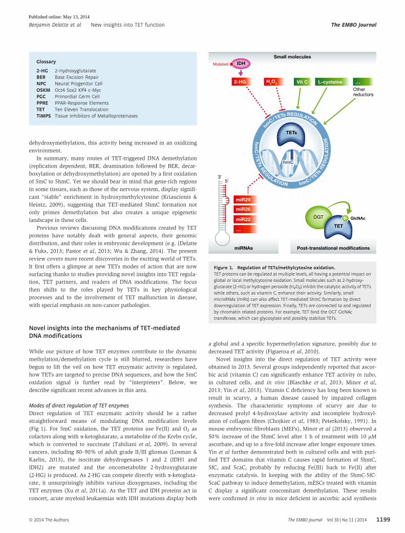
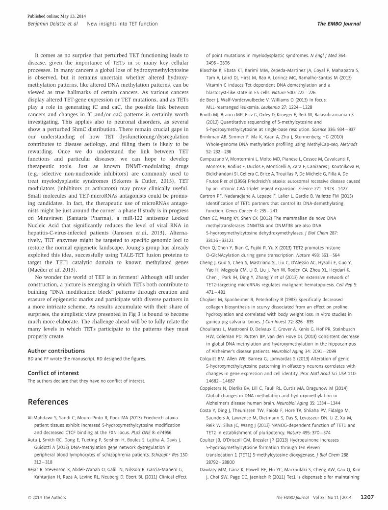
Modes of direct regulation of TET enzymes
Direct regulation of TET enzymatic activity should be a rather
straightforward means of modulating DNA modification levels
(Fig 1). For 5mC oxidation, the TET proteins use Fe(II) and O2 as
cofactors along with a-ketoglutarate, a metabolite of the Krebs cycle,
which is converted to succinate (Tahiliani et al, 2009). In several
cancers, including 80–90% of adult grade II/III gliomas (Losman &
Kaelin, 2013), the isocitrate dehydrogenases 1 and 2 (IDH1 and
IDH2) are mutated and the oncometabolite 2-hydroxyglutarate
(2-HG) is produced. As 2-HG can compete directly with a-ketogluta-rate, it unsurprisingly inhibits various dioxygenases, including the
TET enzymes (Xu et al, 2011a). As the TET and IDH proteins act in
concert, acute myeloid leukaemias with IDH mutations display both
a global and a specific hypermethylation signature, possibly due to
decreased TET activity (Figueroa et al, 2010).
Novel insights into the direct regulation of TET activity were
obtained in 2013. Several groups independently reported that ascor-
bic acid (vitamin C) can significantly enhance TET activity in tubo,
in cultured cells, and in vivo (Blaschke et al, 2013; Minor et al,
2013; Yin et al, 2013). Vitamin C deficiency has long been known to
result in scurvy, a human disease caused by impaired collagen
synthesis. The characteristic symptoms of scurvy are due to
decreased prolyl 4-hydroxylase activity and incomplete hydroxyl-
ation of collagen fibres (Chojkier et al, 1983; Peterkofsky, 1991). In
mouse embryonic fibroblasts (MEFs), Minor et al (2013) observed a
50% increase of the 5hmC level after 1 h of treatment with 10 lMascorbate, and up to a five-fold increase after longer exposure times.
Yin et al further demonstrated both in cultured cells and with puri-
fied TET domains that vitamin C causes rapid formation of 5hmC,
5fC, and 5caC, probably by reducing Fe(III) back to Fe(II) after
enzymatic catalysis. In keeping with the ability of the 5hmC-5fC-
5caC pathway to induce demethylation, mESCs treated with vitamin
C display a significant concomitant demethylation. These results
were confirmed in vivo in mice deficient in ascorbic acid synthesis
5'3'
miR29
H2O2 Vit C L-cysteine …
Otherreductors
miR26
miR22
…
TETs
hmChmC
TET
OGT GlcNAc
hmC/TETs R
EG
UL
AT
IONh
mC
/TE
Ts REGULATION
hmC/TETs REGULATION
Small molecules
Post-translational modificationsmiRNAs
IDH
2-HG
Mutated
Figure 1. Regulation of TETs/methylcytosine oxidation.TET proteins can be regulated at multiple levels, all having a potential impact onglobal or local methylcytosine oxidation. Small molecules such as 2-hydroxy-glutarate (2-HG) or hydrogen peroxide (H2O2) inhibit the catalytic activity of TETswhile others, such as vitamin C, enhance their activity. Similarly, smallmicroRNAs (miRs) can also affect TET-mediated 5hmC formation by directdownregulation of TET expression. Finally, TETs are connected to and regulatedby chromatin related proteins. For example, TET bind the OGT GlcNActransferase, which can glycosylate and possibly stabilize TETs.
1199
Glossary
2-HG 2-HydroxyglutarateBER Base Excision RepairNPC Neural Progenitor CellOSKM Oct4 Sox2 Klf4 c-MycPGC Primordial Germ CellPPRE PPAR-Response ElementsTET Ten Eleven TranslocationTIMPS Tissue Inhibitors of Metalloproteinases
ª 2014 The Authors The EMBO Journal Vol 33 | No 11 | 2014
Benjamin Delatte et al New insights into TET function The EMBO Journal
Published online: May 13, 2014

(Yin et al, 2013), and genome-wide mapping of 5hmC after vitamin
C treatment of mESCs evidenced a global increase in hydroxymethy-
lation and a decrease in 5mC (Blaschke et al, 2013; Yin et al, 2013).
Interestingly, Blaschke et al found vitamin C to both induce DNA
demethylation and increase expression of germline genes, whereas
some regions such as imprinted loci and intracisternal A particle
(IAP) retroelements proved refractory to DNA demethylation.
Furthermore, although various chemical reducing agents (GSH, vita-
min B1, vitamin E) appeared not to affect 5-methylcytosine oxida-
tion patterns, L-cysteine was shown to increase 5hmC by nearly
20% and 5fC by 50% (Blaschke et al, 2013; Yin et al, 2013). Simi-
larly, hydroquinone, a potent reducing agent used for skin depig-
mentation, significantly increases hydroxymethylcytosine (Coulter
et al, 2013). It remains to be seen whether oxidizing agents nega-
tively impact TET activity, perhaps by oxidizing the Fe(II) catalytic
centre. Unpublished data from our lab point to inactivation of the
TET enzymes by hydrogen peroxide treatment, and we have
observed a significant decrease in 5hmC in both cells treated with
various oxidants and GPxI/II-knockout mice (GPx enzymes being
the major natural antioxidants in the cells). As oxidative stress is
involved in various diseases such as cancers and neuronal dis-
orders, it will be important to assess whether alteration of global
and local 5hmC patterns might explain the manifestations of these
life-threatening diseases (B. Delatte & F. Fuks, unpublished data).
Besides regulation by small molecules, recent reports indicate
that TET proteins can be post-transcriptionally regulated by micro-
RNAs (miRNAs or miRs) (Cheng et al, 2013; Fu et al, 2013; Morita
et al, 2013; Song et al, 2013a,b). For example, Morita et al (2013)
have shown that miR-29-family of microRNAs can repress TET1 and
thymine-DNA glycosylase (TDG) without greatly altering DNA
methylation, possibly because they can also downregulate DNMT3A
and DNMT3B. Zhang et al (2013a) have found miR-29 to inhibit
TET1-TET2 and TET3 as well as TDG, this being accompanied by a
small but significant decrease in hydroxymethylcytosine. MiR-26
can also repress all three TETs and TDG, emphasizing a quite
complex regulation of DNA demethylation enzymes by small RNAs
(Fu et al, 2013). Interestingly, transgenic mice overexpressing miR-
26 display a decreased level of 5hmC and an increased proliferation
and differentiation of pancreatic cells in vivo. This suggests that
TETs might play a role in pancreatic differentiation and also in
pancreas-related diseases such as cancer and diabetes (Fu et al,
2013). As miR-26 and many other miRs are deregulated in human
malignancies and can function either as oncogenes or as tumour-
suppressor genes (Fu et al, 2013), and as a decreased hydroxymethyl-
cytosine level is suggested to be a hallmark of cancer (see below;
cf. also Delatte & Fuks, 2013), TET proteins might be directly down-
regulated by miR networks during tumorigenesis. It is noteworthy
in this regard that overexpression of miR-22 in the haematopoietic
or mammary gland compartment inhibits TET-mediated hydroxy-
methylation and promotes leukaemogenesis or breast cancer,
respectively. In fact, myelodysplastic patients displaying a decreased
level of TET2 show poor survival and an inverse correlation
between the TET2 and miR-22 levels. A similar anti-correlation has
been observed in breast cancer patients exhibiting reduced TET1/
TET2 expression (Song et al, 2013a,b). TET2 being a target of
miR-29 and miR-26 has been recently confirmed by Chen et al,
who studied the effects of overexpressing nearly 500 miR constructs
in H293T cells. They observed no deregulation by miR-22, possibly
for technical reasons such as the length of the 30-untranslatedregions used in their experiments. Remarkably, they identified
another ten miR families that regulate TET2, notably miR-101 and
miR-125, which induce abnormal haematopoiesis and a malignant
phenotype in xenograft experiments, directly via TET2 downregula-
tion (Cheng et al, 2013). These reports highlight the possible role of
miRNAs in mediating the post-transcriptional regulation of TETs
during pathogenesis.
Another mode of TET regulation to be considered is post-trans-
lational modifications (PTM). Covalent PTMs (such as acetylation,
methylation, phosphorylation, and glycosylation) are well known
to provide a vast indexing potential and to expand the range of
functions of histones and non-histone proteins. Little has been
published on the possible regulation of TETs by post-translational
modifications. We do know that all three TETs can be modified by
the O-linked N-acetylglucosamine (O-GlcNAc) transferase (OGT)
(Shi et al, 2013; Vella et al, 2013; Ito et al, 2014). This glycosyl-
transferase adds O-GlcNAc moieties on various cytosolic and
nuclear proteins (Hu et al, 2010). Protein O-glycosylation is known
to antagonize phosphorylation, by direct competition for site-occu-
pancy at a single hydroxyl group or via steric hindrance at proxi-
mal sites in the same polypeptide (Wang et al, 2008). As
glycosylation of threonine 535 of TET1 enhances the protein’s
stability (Shi et al, 2013), it is tempting to propose that phosphor-
ylation of this residue in the absence of GlcNAc might flag the
protein for ubiquitin-mediated degradation and thereby cause a
significant decrease in 5mC oxidation marks (Shi et al, 2013). One
might expect TETs, like many other epigenetic enzymes (Denis
et al, 2011), to be decorated by various PTMs affecting their enzy-
matic activity.
In summary, the body of available evidence suggests that
small molecules, microRNAs, and PTMs may regulate hydroxy-
methylcytosine levels by directly modulating TET expression and/or
enzymatic activity. Future studies may bring to light additional
modes of regulation, notably involving direct amino acid modifi-
cations.
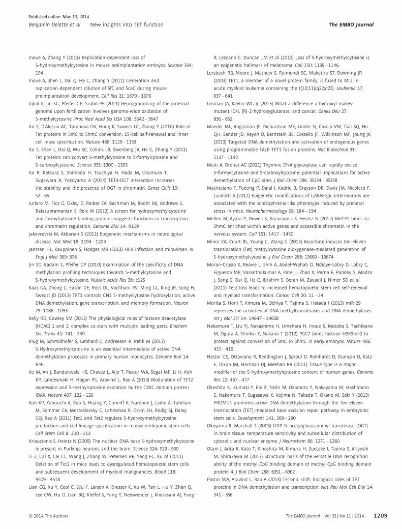
An expanding TET interaction network
Since the original discovery of TETs in 2009, another approach to
elucidating TET functions has been to identify proteins with which
these enzymes interact. The search for TET interactants has brought
to light a plethora of transcription-related factors: transcription
factors/nuclear receptors (CXXC4, NANOG, PPARc, PU.1, EBF1,
PRDM14) and chromatin-associated proteins involved in transcrip-
tional activation (OGT and SET1/COMPASS complex) or repression
(SIN3A/HDACs, NURD). Below we describe in more detail what we
know about these TET partners and how they might help TETs to
perform their functions (Fig 2).
The strongest TET partner identified so far is OGT, which we and
others have recently reported (Chen et al, 2012; Deplus et al, 2013;
Shi et al, 2013; Vella et al, 2013; Ito et al, 2014). Recent evidence of
an epigenetic function for OGT has emerged with the discovery that
it can glycosylate histone H2B on serine 112, causing its ubiquitiny-
lation and the subsequent trimethylation of histone H3 at lysine 4
(H3K4me3). This modification is known to “decorate” active genes
and to be mainly deposited by the SET1/COMPASS complex (Fujiki
et al, 2011, 2013). Depending on TETs and OGT abundance and on
the cell context, TET1, TET2, and TET3 all seem able to interact
The EMBO Journal Vol 33 | No 11 | 2014 ª 2014 The Authors
The EMBO Journal New insights into TET function Benjamin Delatte et al
1200
Published online: May 13, 2014
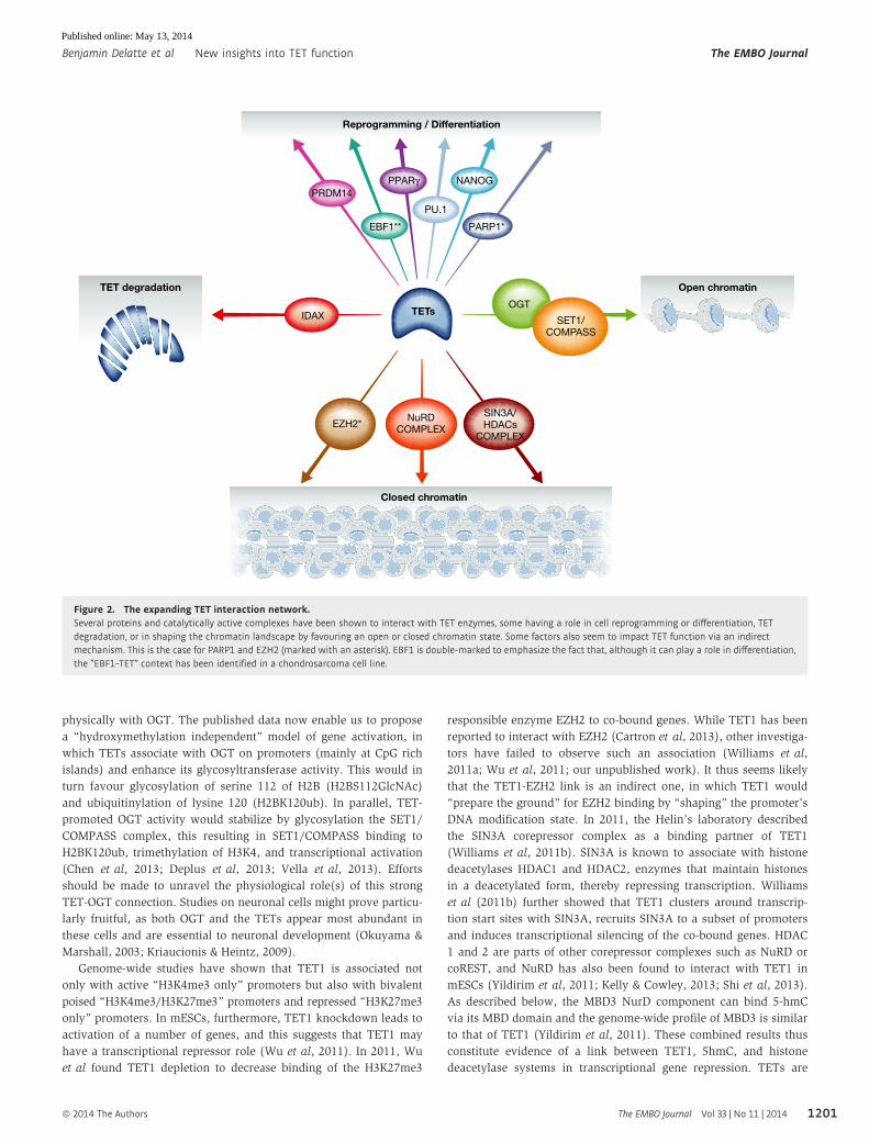
physically with OGT. The published data now enable us to propose
a “hydroxymethylation independent” model of gene activation, in
which TETs associate with OGT on promoters (mainly at CpG rich
islands) and enhance its glycosyltransferase activity. This would in
turn favour glycosylation of serine 112 of H2B (H2BS112GlcNAc)
and ubiquitinylation of lysine 120 (H2BK120ub). In parallel, TET-
promoted OGT activity would stabilize by glycosylation the SET1/
COMPASS complex, this resulting in SET1/COMPASS binding to
H2BK120ub, trimethylation of H3K4, and transcriptional activation
(Chen et al, 2013; Deplus et al, 2013; Vella et al, 2013). Efforts
should be made to unravel the physiological role(s) of this strong
TET-OGT connection. Studies on neuronal cells might prove particu-
larly fruitful, as both OGT and the TETs appear most abundant in
these cells and are essential to neuronal development (Okuyama &
Marshall, 2003; Kriaucionis & Heintz, 2009).
Genome-wide studies have shown that TET1 is associated not
only with active “H3K4me3 only” promoters but also with bivalent
poised “H3K4me3/H3K27me3” promoters and repressed “H3K27me3
only” promoters. In mESCs, furthermore, TET1 knockdown leads to
activation of a number of genes, and this suggests that TET1 may
have a transcriptional repressor role (Wu et al, 2011). In 2011, Wu
et al found TET1 depletion to decrease binding of the H3K27me3
responsible enzyme EZH2 to co-bound genes. While TET1 has been
reported to interact with EZH2 (Cartron et al, 2013), other investiga-
tors have failed to observe such an association (Williams et al,
2011a; Wu et al, 2011; our unpublished work). It thus seems likely
that the TET1-EZH2 link is an indirect one, in which TET1 would
“prepare the ground” for EZH2 binding by “shaping” the promoter’s
DNA modification state. In 2011, the Helin’s laboratory described
the SIN3A corepressor complex as a binding partner of TET1
(Williams et al, 2011b). SIN3A is known to associate with histone
deacetylases HDAC1 and HDAC2, enzymes that maintain histones
in a deacetylated form, thereby repressing transcription. Williams
et al (2011b) further showed that TET1 clusters around transcrip-
tion start sites with SIN3A, recruits SIN3A to a subset of promoters
and induces transcriptional silencing of the co-bound genes. HDAC
1 and 2 are parts of other corepressor complexes such as NuRD or
coREST, and NuRD has also been found to interact with TET1 in
mESCs (Yildirim et al, 2011; Kelly & Cowley, 2013; Shi et al, 2013).
As described below, the MBD3 NurD component can bind 5-hmC
via its MBD domain and the genome-wide profile of MBD3 is similar
to that of TET1 (Yildirim et al, 2011). These combined results thus
constitute evidence of a link between TET1, 5hmC, and histone
deacetylase systems in transcriptional gene repression. TETs are
Reprogramming / Differentiation
PPARγPRDM14
IDAX
EZH2*NuRD
COMPLEX
SIN3A/HDACs
COMPLEX
PARP1*EBF1**
PU.1
NANOG
TETsSET1/
COMPASS
OGT
Closed chromatin
TET degradation Open chromatin
Figure 2. The expanding TET interaction network.Several proteins and catalytically active complexes have been shown to interact with TET enzymes, some having a role in cell reprogramming or differentiation, TETdegradation, or in shaping the chromatin landscape by favouring an open or closed chromatin state. Some factors also seem to impact TET function via an indirectmechanism. This is the case for PARP1 and EZH2 (marked with an asterisk). EBF1 is double-marked to emphasize the fact that, although it can play a role in differentiation,the “EBF1-TET” context has been identified in a chondrosarcoma cell line.
1201ª 2014 The Authors The EMBO Journal Vol 33 | No 11 | 2014
Benjamin Delatte et al New insights into TET function The EMBO Journal
Published online: May 13, 2014

likely to be found to cooperate with yet other chromatin complexes
- both activating and repressive - in controlling gene expression.
TET1 and TET3 can bind DNA via their N-terminal CXXC and
C-terminal cys-rich domains, but TET2 has lost its CXXC domain
during evolution, which is now encoded by a separate gene called
CXXC4 or IDAX, a reported inhibitor of the Wnt/b-catenin pathway.
Interestingly, Anjana Rao’s laboratory has discovered that CXXC4
can associate with TET2 and recruit it to promoters. More surpris-
ingly, binding of CXXC4 to DNA stimulates caspase-dependent TET2
degradation. This is a unique case of epigenetic marking through
recruitment of a protein to DNA and its subsequent degradation (Ko
et al, 2013). Other transcription factors can recruit TET enzymes to
their genomic targets. This happens during cell reprogramming
to pluripotency: physical interaction between the NANOG and
TET1/TET2 proteins has been shown to facilitate reprogramming of
neural stem cells in a manner that depends on methylcytosine
hydroxylase activity (Costa et al, 2013). TET recruitment has also
been observed during differentiation. One example concerns the
differentiation of pre-adipocytes to adipocytes, triggered by the
nuclear receptor PPARc. During adipogenesis and in a ligand-
dependent manner, PPARc switches binding partners from a co-
repressor to a co-activator complex on the PPAR-response elements
(PPREs) of numerous genes expressed specifically in adipocytes.
PPARc poly(ADP-ribosyl)ation (PARylation), a post-translational
protein modification mediated by PARPs, is known to regulate
PPARc transcriptional activity, and this may involve DNA demethy-
lation (Fujiki et al, 2013). In a recent study, Fujiki et al showed that
during differentiation, TET1 interacts with PPARc after PARylation
of the nuclear receptor. They further revealed that TET1 and TET2
can bind to PAR polymers and propose a model in which active DNA
demethylation of key adipocyte-specific genes occurs via direct bind-
ing of TET1 and TET2 to the modified PPARc receptor (Fujiki et al,
2013). During osteoclast differentiation, likewise, the transcription
factor PU.1 interacts with TET2 and recruits it to genes that end up
hypomethylated (de la Rica et al, 2013). Interestingly, PU.1 is
required to generate most myeloid lineages (including macrophages,
neutrophils, and dendritic cells) and for B-cell differentiation; it can
be suppressed or mutated in leukaemia (Shereen & Rodney, 2011). It
can also interact with the spliceosome machinery and participate in
alternative splice site selection (Guillouf et al, 2006). These observa-
tions suggest an unanticipated involvement of the TET2-PU.1 associ-
ation in cancer and in essential processes such as mRNA
metabolism. On the basis of a search for consensus binding sites
surrounding hypermethylated positions in a meta-analysis of IDH-
mutant cancers, Guilhamon et al identified EBF1 as an interacting
partner of TET2 in a human chondrosarcoma cell line. Further exper-
iments showed that EBF1 and TET2 co-occupy certain genomic loci
(Guilhamon et al, 2013). It is known that EBF1 is required for
B lymphopoiesis and a significant fraction of genes, co-bound by
EBF1 and the transcription factor E2-alpha (E2A), correlates with
histones modifications of activated chromatin but also with poised
or repressed regions in pro-B cells (Hagman et al, 2012). Knowing
the interaction with EBF1 and that TET2 has a critical function in the
haematopoietic compartment, it will be of great interest to assess
its role in B-lymphocytes signaling and differentiation (Delatte &
Fuks, 2013).
TET1 and TET2 also interact with the transcriptional regulator
PRDM14, whose overexpression in mESCs leads to increased
hydroxymethylation, demethylation, and subsequent activation of a
set of pluripotency, germline, and imprinted genes (Okashita et al,
2013). As mentioned above, PGCs undergo global demethylation
during their maturation, as a result of global conversion of methyl-
cytosine to hydroxymethylcytosine and replication-dependent dilu-
tion. During this global wave of methylation resetting, imprinted
loci also become demethylated, and TET1 has been found to play a
role in erasing paternal and maternal genomic imprints, this being
particularly evident in a TET1 knockout mouse which also display a
defect in meiosis of developed gametes and some developmental
arrest between E16.5 and E18.5. As PRDM14 is highly expressed in
PGCs and interacts with TET1, it is tempting to speculate that these
two proteins might participate together in imprinting erasure during
PGC reprogramming (Okashita et al, 2013; Yamaguchi et al, 2013).
All in all, the functional diversity of the TET interaction network
(Fig 2) suggests that the TET proteins are more versatile than initi-
ally believed and participate in other processes besides demethyla-
tion of CpG dinucleotides. ChIP-seq experiments on histone marks
in relevant tissues will help to unravel the physiological roles of the
TETs beyond differentiation and reprogramming. Also, metabolic
and signalling pathways (such as those involved in OGT metabolism
or Wnt/b-catenin) are likely to modify the TET-related epigenome.
An important future task could thus be to compare maps of OGT or
CXXC4 with maps of TET2 and 5hmC, for example, under various
conditions affecting these pathways.
Readers of oxidized methylcytosines
In mammals, 5hmC levels vary according to the tissue and cell type.
The brain and spinal cord show the highest levels, and organs such
as the testes, spleen, and thymus display the lowest. An intermedi-
ate level is recorded in embryonic stem cells were approximately
0.1% of all bases in these cells are hydroxymethylcytosines
(Globisch et al, 2010; Ito et al, 2010; Szwagierczak et al, 2010). The
abundance of this oxidized methylcytosine, particularly in the brain,
has led the scientific community to seek proteins that can bind to
5hmC and interpret/translate the encoded information. Several stud-
ies have shown that methyl-binding proteins (MBDs) can to some
extend bind to 5hmC DNA, most often within a hemi-hydroxy-
methylated template and with lower affinity than to fully methylated
DNA. This is the case of MeCP2, MBD1, MBD2, and MBD4 (Jin
et al, 2010; Mellen et al, 2012; Otani et al, 2013; Hashimoto et al,
2014). MBD3 has been known for some time not to bind 5mC on its
own, but in 2011, Yildirim and coworkers reported that the repres-
sive NurD complex can bind both 5hmC-containing and non-modi-
fied DNA through its MBD3 subunit (Yildirim et al, 2011). The jury
is still out as to whether MBD3 is a true hmC reader, as it has not
been detected by others (Spruijt et al, 2013). The fact that MeCP2
can recognize 5hmC in vitro will force the community to re-evaluate
the MethylCap assay, which employs the MeCP2 MBD domain to
interrogate 5mC in genomic DNA. This applies particularly to tissues
such as brain tissue, where almost half of the methylcytosine is in
fact hydroxymethylcytosine (Brinkman et al, 2010). Further in silico
and in vitro studies have examined whether the DNMT1 partner
UHRF1 might also bind to hemi- or fully hydroxymethylated DNA
via its SRA domain. It appears that UHRF1 does bind 5hmC-contain-
ing DNA, but the function of this binding is not yet clear. The
protein is known to flip the base out of the DNA double helix. This
might enhance further oxidation of 5hmC to 5fC and 5caC by
The EMBO Journal Vol 33 | No 11 | 2014 ª 2014 The Authors
The EMBO Journal New insights into TET function Benjamin Delatte et al
1202
Published online: May 13, 2014
making the hydroxymethylated base more accessible to the TET
enzymes (Frauer et al, 2011). Accordingly, H293T cells overexpress-
ing the UHRF1 homologue UHRF2, which also binds hydroxymeth-
ylcytosine, display oxidation of 5mC to 5hmC, with a remarkable
increase in 5fC and 5caC (Spruijt et al, 2013).
The above-mentioned studies mostly describe in vitro approaches
where recombinant proteins are incubated with modified DNA frag-
ments and then used in electrophoretic mobility shift assays. Two
groups have reported identifying readers of 5hmC, 5fC, and 5caC by
quantitative mass spectrometry (Iurlaro et al, 2013; Spruijt et al,
2013). Interestingly, the first group found some readers to be
specific to mESCs, neural progenitor cells (NPCs), or adult mouse
brain tissue, while the second revealed proteins whose binding to
5hmC or 5fC depends on the CpG composition. This indicates (i)
that hydroxymethylcytosine probably behaves differently in differ-
ent cell types and (ii) that some presumptive readers bind oxidized
methylcytosines in a sequence-specific manner. The screens also
identified several specific 5fC and 5caC readers, among them TDG,
supporting the view that this protein can act as a DNA demethylase
(Iurlaro et al, 2013; Spruijt et al, 2013).
In summary, specific proteins are recruited to 5hmC, 5fC, or
5caC sites, and some of them are likely to regulate gene expression
or to drive active DNA demethylation. More in-depth characteriza-
tion by ChIP-seq and the use of knockout mice should help to show
whether the proteins identified so far are true “readers” that can
interpret 5hmC, 5fC, or 5caC modifications and translate them into
an activating or repressing signal, or merely “binders” with some
affinity for modified cytosines.
Emerging roles of TETs in normal physiology and disease
TETs/hmC in cell reprogramming and differentiation
The TET proteins participate in two waves of global DNA demethy-
lation that occur during reprogramming of primordial germ cells as
well as of paternal pronucleus in the zygote. Such reprogrammings
are needed to ensure resetting of the epigenomes for pluripotency
(Gu et al, 2011; Inoue et al, 2011; Iqbal et al, 2011; Wossidlo et al,
2011; Hackett et al, 2012; Nakamura et al, 2012). When PGCs
migrate to the future gonads, high levels of TET1 and TET2 allows
conversion of 5mC into 5hmC and this, not only globally but also
more locally (e.g. at genomic imprinted loci). This is followed by
subsequent passive “replication-coupled” dilution of 5hmC during
several round of cell divisions with a progressive loss of hydroxyme-
thylation (Hackett et al, 2012). A second wave of global DNA deme-
thylation appears when the spermatozoon fertilizes the oocyte. In
this case, TET3 hydroxylates the paternal DNA while PGC7 (also
called DPPA3/STELLA) maintains normal methylation levels on the
maternal genome as well as on imprinted genes by binding to the
repressive H3K9me2 histone mark and by repelling TET3 (Szabo &
Pfeifer, 2012). Worth mentioning, it has been observed that in the
paternal pronucleus, 5hmC is further oxidized into 5fC and 5caC
and that those modifications are also passively diluted through cell
divisions (Gu et al, 2011; Inoue & Zhang, 2011; Inoue et al, 2011;
Iqbal et al, 2011; Wossidlo et al, 2011; Nakamura et al, 2012).
The initial discovery of TET enzymes in ESCs has led many
groups to assess their importance in pluripotency maintenance and
cell differentiation. Although some reports suggest a role of TET1 in
maintenance of pluripotency, especially via direct binding and
demethylation of the NANOG promoter, other papers do not support
this view (Ito et al, 2010; Ficz et al, 2011; Koh et al, 2011; Williams
et al, 2011a; Wu et al, 2011; Xu et al, 2011b; Doege et al, 2012;
Freudenberg et al, 2012). Furthermore, in vivo results obtained with
TET1 knockout and TET1/TET2 double-knockout mice (under
conditions where TET3 is unlikely to compensate significantly for
the knockout) suggest that ESCs stemness can be maintained with-
out these methyldioxygenases (Dawlaty et al, 2011, 2013). In fact,
although a fraction of the double KO mice displayed midgestation
abnormalities and perinatal lethality, viable and fertile mice were
also obtained. Those however display a reduced size and weight as
compared to their wt counterparts and the females also show
smaller ovaries and reduced fertility. This indicates an overtly
normal development with variable phenotypes probably due to vari-
able penetrance of epigenetic defects. This double knockout is in
sharp contrast with the TET3 knockout embryos from Xu’s group
that show apparent lethality either around embryonic day 11.5 or
after birth, highlighting the crucial role of TET3 in reprogramming
the paternal pronucleus after fertilization (Gu et al, 2011). While
the jury is still out on whether TETs play a role in maintaining pluri-
potency in ESCs, another phenomenon involving TETs is coming to
light: reprogramming of developmentally committed cells such as
MEFs to produce induced pluripotent stem cells (iPSCs). iPSCs,
phenotypically comparable to ESCs, can be produced in vitro upon
overexpression of the Yamanaka “OSKM” factors (OCT4, SOX2,
KLF4 and C-MYC). During this process profound epigenetic repro-
gramming takes place, with loss of methylation on the promoters of
several pluripotency genes (Takahashi & Yamanaka, 2006). Several
studies have stressed the importance of TETs in iPSC reprogram-
ming. In the first of these studies, TET2 and PARP1 were identified
as key factors in this process: both are upregulated in MEFs upon
OSKM treatment, and both bind to ESRRB and NANOG to shape the
chromatin landscape essential for correct gene expression and iPSC
reprogramming (Doege et al, 2012). As mentioned earlier, another
report revealed that NANOG can interact with TET1 and TET2 and
facilitate reprogramming of neural stem cells in a TET-activity-
dependent manner (Costa et al, 2013). Remarkably, Gao et al
(2013) were able to induce iPSC formation in experiments where
they replaced OSKM with “TSKM” (TET1, SOX2, KLF4, and
C-MYC), probably because OCT4 is a direct binding target of TET1.
These results highlight the importance of TET1 and TET2 in the
demethylation and activation of key pluripotency genes such as
ESRRB, NANOG, and OCT4 during reprogramming of differentiated
cells, and thus suggest an important role for methyldioxygenases in
regenerative medicine studies.
Recent reports pinpoint a participation of TETs in terminal differ-
entiation as well. In 2013, Hahn et al found the 5hmC pattern to be
very dynamic during NPC differentiation to neurons. In gene bodies
they observed a global increase in 5hmC without any subsequent
change in DNA methylation. They further showed that TET2 and
TET3 are required for correct NPC differentiation and that their loss
leads to abnormal accumulation of cell clusters along the radial axis
in the intermediate and ventricular zones (Hahn et al, 2013). A
similar increase in hydroxymethylcytosine is seen during olfactory
neuron differentiation, and TET3 overexpression disrupts both the
olfactory receptor expression pattern and the targeting of axons to
the olfactory bulb (Colquitt et al, 2013). These two studies strongly
1203ª 2014 The Authors The EMBO Journal Vol 33 | No 11 | 2014
Benjamin Delatte et al New insights into TET function The EMBO Journal
Published online: May 13, 2014

suggest that the hydroxymethylation landscape is important for the
correct formation of brain tissues, as already reported by Shi’s
group, which found TET3 to be required for normal development of
the eye and brain in Xenopus laevis (Xu et al, 2012). Three recent
papers have shed further light on the involvement of TET enzymes
in brain development (Kaas et al, 2013; RudenKo et al, 2013; Zhang
et al, 2013b). Zhang et al found that TET1 is needed for NPC prolif-
eration and that in vivo depletion of TET1 affects neuron production
in the hippocampus. Further characterization of the mice in Morris
water maze experiments revealed that their short-term memory was
reduced (Zhang et al, 2013b). Rudenko et al (2013) reached differ-
ent conclusions: in fear conditioning and Morris water maze tests,
they found a normal neuronal density in the hippocampus of TET1-
knockout mice, with no short-term memory impairment but with a
significant decrease in memory extinction (which affects long-term
memory), possibly due to alteration of the long-term depression
pathway. As these last results have been partially reproduced in
mice overexpressing wild-type or mutant TET1 catalytic domains in
the hippocampus, TET1 appears to act as an “inhibitor” of long-term
memory, independently of its catalytic function (Kaas et al, 2013).
While a consensus is hard to reach at this stage, it would seem that
TETs affect both short- and long-term memory as well as NPC prolif-
eration and differentiation. The discrepancies between Zhang’s and
Rudenko’s results may be due to the use of different knockouts:
Zhang et al targeted exons 11–13 of the TET1 protein, whereas
Rudenko et al used knockout mice with a deleted exon 4 (previously
described by Jaenisch and colleagues Rudenko et al, 2013; Zhang
et al, 2013b). Further studies, including systematic conditional hippo-
campal knockout of TET1, TET2, and TET3 (and also double and
triple knockouts), should help to assess these divergences and to
distinguish the exact roles of the three TETs in neurogenesis.
Also beyond the neuronal context, reports show that the hydroxy-
methylcytosine pattern is highly dynamic during terminal cell
differentiation, as described for preadipocytes, germ cells, and
differentiating monocytes (Fujiki et al, 2013; Gan et al, 2013; Klug
et al, 2013; de la Rica et al, 2013). During adipogenesis, the genome
displays a global increase in 5hmC and local hydroxymethylation on
genes that are later expressed in mature adipocytes (Fujiki et al,
2013). A similar initial global increase in 5hmC, followed by a global
decrease, and the presence of differentially hydroxymethylated
regions (dhMRs) on certain coding and non-coding genes are also
observed during spermatogenesis (Gan et al, 2013). Finally, Klug
et al (2013) and de la Rica et al (2013) have provided evidence that
TETs, especially TET2, are essential to the proper differentiation of
monocytes to osteoclasts: during this process, DNA demethylation
preceded by 5hmC deposition is observed.
All in all, it thus seems that while 5hmC may decrease when
mESCs lose their pluripotency, a global increase in this epigenetic
mark can occur during terminal differentiation, sometimes
followed by a subsequent decrease. This highlights the dynamicity
of the hydroxymethylcytosine pattern and suggests a crucial role
for the TET enzymes in cell differentiation and organ (e.g. brain)
formation as well as a key role in cell reprogramming. With
proteins so essential and so versatile, it comes as no surprise that
their loss, or an alteration of the 5hmC pattern, can lead to
diseases such as cancer or neurological disorders. Roles of altered
TET functioning and hydroxymethylation in disease are discussed
below.
TETs/modified cytosines in cancer and non-cancer diseases
TETs are on the one hand recognized as tumour suppressor and a
hallmark of many cancers seems to be a significant decrease in
5hmC. This may be partly due to the well-known global hypomethy-
lation that takes place during cell transformation, but in breast
cancers, melanomas, and leukaemias without MLL rearrangements,
reports have additionally revealed either a mutation in the TET2
gene (although the level of one TET does not always correlate with
the global level of hydroxymethylcytosine) or substantial downregu-
lation of all three TETs, for example by micro-RNAs (Tefferi et al,
2009; Haffner et al, 2011; Nestor et al, 2011; Hsu et al, 2012; Lian
et al, 2012; Yang et al, 2012; Huang et al, 2013; Song et al, 2013a,
b). In 2012, Hsu et al (2012) reported TET1 to be downregulated in
prostate and breast cancer tissues, and found its loss to facilitate cell
invasion in xenograft mouse models, at least via deregulation of
“inhibitor of metalloproteinase” proteins (TIMPs). In human mela-
nomas, which show a marked genome-wide decrease in hydroxy-
methylcytosine and more subtle changes in methylation patterns,
the most decreased TET is TET2. When TET2 is overexpressed in
human melanoma cells xenografted onto immunodeficient NSG
mice, tumour growth is suppressed. This suggests a crucial involve-
ment of TET2 and 5hmC in melanoma development (Lian et al,
2012). Furthermore, TET2 is often mutated in myeloid malignan-
cies,. In 4–13% of myeloproliferative neoplasms and 20–25% of
myelodysplastic syndromes, somatic deletions and TET2-inactivat-
ing mutations are found, but do not correlate clearly with prognosis
(Tefferi et al, 2009; Bejar et al, 2011). TET2 mutations are also
found in 7–23% of AMLs, where they correlate with poor prognosis.
A major role of TET2 in leukaemia has been confirmed in TET2-
knockout mice, where loss of the enzyme appears to increase the
haematopoietic stem cell compartment and to skew cell differentia-
tion towards the myeloid compartment, causing symptoms
resembling those associated with TET2 mutations (Li et al, 2011;
Moran-Crusio et al, 2011; Quivoron et al, 2011).
In fascinating contrast to the above, there are contexts where
TETs appear to have an oncogenic action. They owe the designation
“Ten-Eleven translocation enzyme” to a rare translocation of the
histone methyl transferase gene MLL and of the TET1 coding
sequence in acute myeloid leukaemia (AML), yielding a 50
MLL-TET1 30 chimera (Lorsbach et al, 2003). The role of this trans-
location in leukaemia is unclear, and it seems that oncogenic trans-
formation driven by the rearranged MLL is observed with different
translocation partners (de Boer et al, 2013). Yet Huang et al
revealed in 2013 that TET1 plays an essential oncogenic role in
MLL-rearranged leukaemia. It is aberrantly overexpressed in MLL-
rearranged AML, while the TET2 and TET3 expression levels remain
unchanged. ChIP-qPCR and expression experiments have further
shown that the TET1 promoter is a direct target of MLL-fusion
proteins and that the increase in TET1 leads to a global genomic
increase of hydroxymethylcytosine. Furthermore, MLL fusions and
TET1 both co-regulate genes such as HOXA9 or MEIS1 that inhibit
apoptosis and enhance cell proliferation, causing transformation
and leukaemogenesis (Huang et al, 2013).
In summary, the hydroxymethylcytosine level is often deregulat-
ed in cancer, but not always in the same manner: globally there
may be an increase, as seen in MLL-rearranged leukaemia, or a
marked decrease, as observed in many other cancer types. Recently,
some elegant techniques have been created to precisely map 5hmC
The EMBO Journal Vol 33 | No 11 | 2014 ª 2014 The Authors
The EMBO Journal New insights into TET function Benjamin Delatte et al
1204
Published online: May 13, 2014

at single-nucleotide resolution (Booth et al, 2012; Yu et al, 2012).
This could make this epigenetic modification an interesting marker
for use in diagnosing cancers, especially blood cancers, for which
cell samples can easily be obtained.
Growing attention is also focusing on the involvement of TET
dysregulation in non-cancer diseases. There is increasing evidence
that epigenetic dysfunction and resulting changes in gene expression
may contribute at least partly to the aetiology of various patholo-
gies, especially neurological disorders (see Table 1) (Jakovcevski &
Akbarian, 2012). As mentioned above, the mammalian brain is very
rich in hydroxymethylcytosine. In Rett syndrome, mutations in
MeCP2 impair the binding of this protein to 5hmC, suggesting that
the altered deposition of this mark by TET methyldioxygenases may
also play a role in neurological diseases (Kriaucionis & Heintz,
2009; Mellen et al, 2012). Accordingly, several reports have revealed
altered TET expression and an abnormal 5hmC pattern in the brains
of patients suffering from psychosis, Huntington’s disease, Friedr-
eich’s ataxia, and fragile X syndrome (Campuzano et al, 1996;
Tassone et al, 2000; Saveliev et al, 2003; Sadri-Vakili & Cha, 2006;
Dong et al, 2012; Guidotti et al, 2012; Al-Mahdawi et al, 2013; Auta
et al, 2013; Chouliaras et al, 2013; Villar-Menendez et al, 2013;
Wang et al, 2013; Yao et al, 2013; Coppieters et al, 2014). Psychosis
is a broad category encompassing symptoms of schizophrenia and
bipolar disorder. Psychotic patients have been found to display
downregulated expression of several vulnerability genes, including
GAD67, reelin, NR2A, and GAT1, in parallel with hypermethylation
of the corresponding promoter regions (Dong et al, 2012). Recently,
two groups additionally reported hyper-hydroxymethylation of the
GAD67 promoter in the inferior parietal lobe, both in a psychotic
cohort and in mice born with psychosis-like disorders (Dong et al,
2012; Matrisciano et al, 2012). To evaluate the possible role of
5hmC in GAD67 repression, it would be interesting to see if varia-
tion of the 5hmC level in GAD67 might correlate with TET binding
to one or more corepressors such as SIN3A, and NurD. In addition,
Dong et al (2012) revealed local variations in hydroxymethylcyto-
sine content, a global increase in both TET1 and 5hmC in schizo-
phrenic and bipolar patients, and persons suffering from depression
displayed a similar, albeit insignificant trend. A TET1 increase has
likewise been observed in psychotic patients with a history of alco-
hol abuse and interestingly, in lymphocytes of schizophrenic
patients. The latter finding suggests that it might be possible to iden-
tify peripheral methylation/hydroxymethylation biomarkers for use
in diagnosis/prognosis when a biopsy is not an option (Guidotti
et al, 2012; Auta et al, 2013).
Huntington’s disease (HD) is a genetic disorder characterized by
expansion of glutamine residues in the protein huntingtin, due to
accumulation of the CAG triplet in the first exon of the correspond-
ing gene. The symptoms include difficulties in motor coordination
and also psychiatric disturbances, cognitive disorders, and weight
loss (Sadri-Vakili & Cha, 2006). In 2013, Villar-Menendez et al
(2013) showed that the promoter of the ADORA2A gene, encoding
the G-protein coupled receptor A2AR known to be downregulated in
HD and involved in the disease, displays a decreased 5hmC level
in a mouse model of HD and in the putamens of HD patients. In
contrast to the global increase in hydroxymethylcytosine found in
psychosis, dot-blot experiments and analysis of 5hmC patterns by
deep sequencing in an HD mouse model revealed a rather global
decrease in 5hmC, accompanied by a significant decrease in TET1
expression. Analysis of the differentially hydroxymethylated regions
further uncovered an alteration of canonical pathways involved in
neuronal development and differentiation pathways. Hence, the loss
of hydroxymethylcytosine may impair neuronal function in HD
brains and appears as a novel epigenetic marker of the disease
(Wang et al, 2013).
Friedreich’s ataxia (FRDA) is another neurodegenerative disease
due to expansion of a triplet (GAA). This expansion, here in the first
intron of the FXN gene, leads to heterochromatin formation and
gene silencing. Upstream from the repeats lay CpGs that appear
hypermethylated in patients suffering from the disease. In the stud-
ies concerned, however, the techniques used to map 5mC could not
distinguish this mark from 5hmC (Campuzano et al, 1996; Saveliev
et al, 2003; Al-Mahdawi et al, 2013). In 2013, Al-Mahdawi et al
(2013) addressed this issue, showing that nearly all the methylcyto-
sine is in fact hydroxymethylcytosine in both FRDA and normal
human cerebellar tissues, and that 5hmC is more abundant in the
former than in the latter. They further hypothesized that the
observed 5hmC increase might enhance production of FAST-1 anti-
sense RNA, which could in turn mediate heterochromatin formation
and FXN downregulation, causing the typical symptoms of the
disease. Lastly, the fragile X genetic syndrome, triggered by FMR1
gene repression, is a widespread inherited cause of autism and
mental retardation in boys (Tassone et al, 2000; Yao et al, 2013).
While the disorder is known to be associated with hypermethylation
of the FMR1 promoter and subsequent transcriptional gene silenc-
ing, possible global fragile-X-associated changes in DNA methyla-
tion/hydroxymethylation were not addressed until recently, when
Table 1. This table summarizes the involvement of TETs andmethylcytosine oxidation in neuronal diseases
TETs/hmC connection in neuronal diseases
Pathologies Observations References
Rettsyndrome
Decrease of MeCP2binding on 5hmC
Kriaucionis & Heintz(2009)Mellen et al (2012)
Psychosis GAD67 promoterhyper-hydroxymethylation
Dong et al (2012)Matrisciano et al(2012)
Global increase of 5hmCIncrease of TET1expression
Dong et al (2012)Guidotti et al (2012)Auta et al (2013)
Huntington’sdisease
Decrease of 5hmC onADORA2A promoter
Villar-Menendez et al(2013)
Global decrease of 5hmCDecrease of TET1expression
Wang et al (2013)
Friedreich’sataxia
Global increase of 5hmC Al-Mahdawi et al (2013)
Fragile Xsyndrome
Global decrease of 5hmC ongene bodies and promoters
Subtle increase of 5hmC onenhancers and repetitiveelements
Yao et al (2013)
Alzheimer’sdisease
Global increase of 5hmC inhippocampus
Global decrease of 5hmC intemporal tissues
Coppieters et al (2014)Chouliaras et al (2013)
1205ª 2014 The Authors The EMBO Journal Vol 33 | No 11 | 2014
Benjamin Delatte et al New insights into TET function The EMBO Journal
Published online: May 13, 2014

Yao et al reported a global decrease in 5hmC in gene bodies and
promoters and a more subtle increase in cerebellum-specific
enhancers and some repetitive elements. The same report also
presents dhMR analyses highlighting changes affecting functional
pathways in neuronal development. As these results were obtained
from a knockin mouse model, it will be important to assess the
global level of hydroxymethylcytosine in fragile X syndrome
patients (Yao et al, 2013).
In each of the above-mentioned neuronal diseases, the global
level of hydroxymethylation thus seems to be altered, either upward
or downward according to the disease. Also, some disorders may
involve several tissues in the brain, and both the global and specific
5hmC patterns may depend on the analysed tissue. This is exempli-
fied by two recent studies showing that in Alzheimer’s disease, the
most common form of dementia in humans, hippocampal regions
display a significant increase in 5hmC, while the middle frontal and
temporal gyri show a significant decrease (Chouliaras et al, 2013;
Coppieters et al, 2014).
In summary, the TET methyldioxygenase machinery seems
altered in various diseases, notably cancers and neudegenerative
disorders but also probably other important diseases such as oxida-
tive-stress related disorders (see modes of direct regulation of TET
enzymes). This strongly suggests that 5hmC patterns are important
in many pathologies. As DNA can be recovered easily from tissues
and is stable over time, 5hmC profiles might be used to find clini-
cally relevant epigenetic biomarkers. Yet questions remain to be
answered. It will notably be important to identify clearly the
pathways affecting 5hmC profile changes and those affected by
them. We also need to distinguish whether variations of the global
level of this mark are causes or consequences of the diseases
concerned. These important questions will surely keep a lot of labo-
ratories excited and busy for the next few years.
Concluding remarks
Since their discovery in 2009, the TET enzymes have sparked
tremendous interest in the epigenetic community. Key studies have
highlighted the importance of active or passive TET-primed deme-
thylation in both cell reprogramming and cell differentiation, and
the functional diversity of TET partner proteins suggests a more
multifaceted action than anticipated. TETs associate with diverse
chromatin-related machineries involved in transcriptional activation
or repression, and the marks they create may act as epigenetic
signals per se, i. e. as more than just intermediates in DNA demethy-
lation. TETs also seem to play roles that do not depend on their
catalytic activity.
Challenges abound in the world of TETs: untangling the intricate
web of TET interactions with the chromatin environment, answer-
ing burning questions such as: is the frequently observed positive
correlation of transcription with hydroxymethylcytosine in gene
bodies the cause or the consequence of gene expression? What
proteins act as true 5hmC, 5fC, and 5caC “readers” and what are
their precise biological roles?
Promoter
Oxidized DNA modifications
Transcriptionalregulators, e.g.:
• OGT • …
TETs TF, e.g.:
• CXXC4 • … fC hmC
Gene body
1
1
2 caC2 2
2
3
Small molecules, e.g.:
• Vit C • …
3
3
3 Binders/Readers, e.g.:
• UHRF1
• …
Figure 3. The evolving “TETris playground”.A picture is emerging in which TETs build up specific “DNAmodification blocks” (hmC, fC, caC) through their coordinated regulation at multiple levels. The following sequenceof events can therefore be suggested: (i) Transcription factors will result in (ii) 5hmC, 5fC, and 5caC formation with 5hmC being more abundant in gene bodies. (iii) Thesemodifications can be further bound by various “binders” or “readers” that will either participate in DNA demethylation or translate the epigenetic signal to trans-criptional activation or repression. Other active players are transcriptional regulators and small molecules such as vitamin C that directly affect TET activity and hencethe deposition of 5hmC, 5fC, and 5caC. These DNA modification blocks, TETs, and TET regulators can assemble in a gene- and/or a cell-state-dependent manner creatingprecise landscapes which will ultimately affect gene expression.
The EMBO Journal Vol 33 | No 11 | 2014 ª 2014 The Authors
The EMBO Journal New insights into TET function Benjamin Delatte et al
1206
Published online: May 13, 2014
It comes as no surprise that perturbed TET functioning leads to
disease, given the importance of TETs in so many key cellular
processes. In many cancers a global loss of hydroxymethylcytosine
is observed, but it remains uncertain whether altered hydroxy-
methylation patterns, like altered DNA methylation patterns, can be
viewed as true hallmarks of certain cancers. As various cancers
display altered TET-gene expression or TET mutations, and as TETs
play a role in generating fC and caC, the possible link between
cancers and changes in fC and/or caC patterns is certainly worth
investigating. This applies also to neuronal disorders, as several
show a perturbed 5hmC distribution. There remain crucial gaps in
our understanding of how TET dysfunctioning/dysregulation
contributes to disease aetiology, and filling them is likely to be
rewarding. Once we do understand the link between TET
functions and particular diseases, we can hope to develop
therapeutic tools. Just as known DNMT-modulating drugs
(e.g. selective non-nucleoside inhibitors) are commonly used to
treat myelodysplastic syndromes (Sekeres & Cutler, 2013), TET
modulators (inhibitors or activators) may prove clinically useful.
Small molecules and TET-microRNAs antagonists could be promis-
ing candidates. In fact, the therapeutic use of microRNAs antago-
nists might be just around the corner: a phase II study is in progress
on Miravirsen (Santaris Pharma), a miR-122 antisense Locked
Nucleic Acid that significantly reduces the level of viral RNA in
hepatitis-C-virus-infected patients (Janssen et al, 2013). Alterna-
tively, TET enzymes might be targeted to specific genomic loci to
restore the normal epigenetic landscape. Joung’s group has already
exploited this idea, successfully using TALE-TET fusion proteins to
target the TET1 catalytic domain to known methylated genes
(Maeder et al, 2013).
No wonder the world of TET is in ferment! Although still under
construction, a picture is emerging in which TETs both contribute to
building “DNA modification block” patterns through creation and
erasure of epigenetic marks and participate with diverse partners in
a more intricate scheme. As results accumulate with their share of
surprises, the simplistic view presented in Fig 3 is bound to become
much more elaborate. The challenge ahead will be to fully relate the
many levels in which TETs participate to the patterns they must
properly create.
Author contributionsBD and FF wrote the manuscript, RD designed the figures.
Conflict of interestThe authors declare that they have no conflict of interest.
References
Al-Mahdawi S, Sandi C, Mouro Pinto R, Pook MA (2013) Friedreich ataxia
patient tissues exhibit increased 5-hydroxymethylcytosine modification
and decreased CTCF binding at the FXN locus. PLoS ONE 8: e74956
Auta J, Smith RC, Dong E, Tueting P, Sershen H, Boules S, Lajtha A, Davis J,
Guidotti A (2013) DNA-methylation gene network dysregulation in
peripheral blood lymphocytes of schizophrenia patients. Schizophr Res 150:
312 – 318
Bejar R, Stevenson K, Abdel-Wahab O, Galili N, Nilsson B, Garcia-Manero G,
Kantarjian H, Raza A, Levine RL, Neuberg D, Ebert BL (2011) Clinical effect
of point mutations in myelodysplastic syndromes. N Engl J Med 364:
2496 – 2506
Blaschke K, Ebata KT, Karimi MM, Zepeda-Martinez JA, Goyal P, Mahapatra S,
Tam A, Laird DJ, Hirst M, Rao A, Lorincz MC, Ramalho-Santos M (2013)
Vitamin C induces Tet-dependent DNA demethylation and a
blastocyst-like state in ES cells. Nature 500: 222 – 226
de Boer J, Walf-Vorderwulbecke V, Williams O (2013) In focus:
MLL-rearranged leukemia. Leukemia 27: 1224 – 1228
Booth MJ, Branco MR, Ficz G, Oxley D, Krueger F, Reik W, Balasubramanian S
(2012) Quantitative sequencing of 5-methylcytosine and
5-hydroxymethylcytosine at single-base resolution. Science 336: 934 – 937
Brinkman AB, Simmer F, Ma K, Kaan A, Zhu J, Stunnenberg HG (2010)
Whole-genome DNA methylation profiling using MethylCap-seq. Methods
52: 232 – 236
Campuzano V, Montermini L, Molto MD, Pianese L, Cossee M, Cavalcanti F,
Monros E, Rodius F, Duclos F, Monticelli A, Zara F, Canizares J, Koutnikova H,
Bidichandani SI, Gellera C, Brice A, Trouillas P, De Michele G, Filla A, De
Frutos R et al (1996) Friedreich’s ataxia: autosomal recessive disease caused
by an intronic GAA triplet repeat expansion. Science 271: 1423 – 1427
Cartron PF, Nadaradjane A, Lepape F, Lalier L, Gardie B, Vallette FM (2013)
Identification of TET1 partners that control its DNA-demethylating
function. Genes Cancer 4: 235 – 241
Chen CC, Wang KY, Shen CK (2012) The mammalian de novo DNA
methyltransferases DNMT3A and DNMT3B are also DNA
5-hydroxymethylcytosine dehydroxymethylases. J Biol Chem 287:
33116 – 33121
Chen Q, Chen Y, Bian C, Fujiki R, Yu X (2013) TET2 promotes histone
O-GlcNAcylation during gene transcription. Nature 493: 561 – 564
Cheng J, Guo S, Chen S, Mastriano SJ, Liu C, D’Alessio AC, Hysolli E, Guo Y,
Yao H, Megyola CM, Li D, Liu J, Pan W, Roden CA, Zhou XL, Heydari K,
Chen J, Park IH, Ding Y, Zhang Y et al (2013) An extensive network of
TET2-targeting microRNAs regulates malignant hematopoiesis. Cell Rep 5:
471 – 481
Chojkier M, Spanheimer R, Peterkofsky B (1983) Specifically decreased
collagen biosynthesis in scurvy dissociated from an effect on proline
hydroxylation and correlated with body weight loss. In vitro studies in
guinea pig calvarial bones. J Clin Invest 72: 826 – 835
Chouliaras L, Mastroeni D, Delvaux E, Grover A, Kenis G, Hof PR, Steinbusch
HW, Coleman PD, Rutten BP, van den Hove DL (2013) Consistent decrease
in global DNA methylation and hydroxymethylation in the hippocampus
of Alzheimer’s disease patients. Neurobiol Aging 34: 2091 – 2099
Colquitt BM, Allen WE, Barnea G, Lomvardas S (2013) Alteration of genic
5-hydroxymethylcytosine patterning in olfactory neurons correlates with
changes in gene expression and cell identity. Proc Natl Acad Sci USA 110:
14682 – 14687
Coppieters N, Dieriks BV, Lill C, Faull RL, Curtis MA, Dragunow M (2014)
Global changes in DNA methylation and hydroxymethylation in
Alzheimer’s disease human brain. Neurobiol Aging 35: 1334 – 1344
Costa Y, Ding J, Theunissen TW, Faiola F, Hore TA, Shliaha PV, Fidalgo M,
Saunders A, Lawrence M, Dietmann S, Das S, Levasseur DN, Li Z, Xu M,
Reik W, Silva JC, Wang J (2013) NANOG-dependent function of TET1 and
TET2 in establishment of pluripotency. Nature 495: 370 – 374
Coulter JB, O’Driscoll CM, Bressler JP (2013) Hydroquinone increases
5-hydroxymethylcytosine formation through ten eleven
translocation 1 (TET1) 5-methylcytosine dioxygenase. J Biol Chem 288:
28792 – 28800
Dawlaty MM, Ganz K, Powell BE, Hu YC, Markoulaki S, Cheng AW, Gao Q, Kim
J, Choi SW, Page DC, Jaenisch R (2011) Tet1 is dispensable for maintaining
1207ª 2014 The Authors The EMBO Journal Vol 33 | No 11 | 2014
Benjamin Delatte et al New insights into TET function The EMBO Journal
Published online: May 13, 2014

pluripotency and its loss is compatible with embryonic and postnatal
development. Cell Stem Cell 9: 166 – 175
Dawlaty MM, Breiling A, Le T, Raddatz G, Barrasa MI, Cheng AW, Gao Q,
Powell BE, Li Z, Xu M, Faull KF, Lyko F, Jaenisch R (2013) Combined
deficiency of Tet1 and Tet2 causes epigenetic abnormalities but is
compatible with postnatal development. Dev Cell 24: 310 – 323
Delatte B, Fuks F (2013) TET proteins: on the frenetic hunt for new cytosine
modifications. Brief Funct Genomics 12: 191 – 204
Denis H, Ndlovu MN, Fuks F (2011) Regulation of mammalian DNA
methyltransferases: a route to new mechanisms. EMBO Rep 12: 647 – 656
Deplus R, Delatte B, Schwinn MK, Defrance M, Mendez J, Murphy N, Dawson
MA, Volkmar M, Putmans P, Calonne E, Shih AH, Levine RL, Bernard O,
Mercher T, Solary E, Urh M, Daniels DL, Fuks F (2013) TET2 and TET3
regulate GlcNAcylation and H3K4 methylation through OGT and SET1/
COMPASS. EMBO J 32: 645 – 655
Doege CA, Inoue K, Yamashita T, Rhee DB, Travis S, Fujita R, Guarnieri P,
Bhagat G, Vanti WB, Shih A, Levine RL, Nik S, Chen EI, Abeliovich A (2012)
Early-stage epigenetic modification during somatic cell reprogramming by
Parp1 and Tet2. Nature 488: 652 – 655
Dong E, Gavin DP, Chen Y, Davis J (2012) Upregulation of TET1 and
downregulation of APOBEC3A and APOBEC3C in the parietal cortex of
psychotic patients. Transl Psychiatry 2: e159
Ficz G, Branco MR, Seisenberger S, Santos F, Krueger F, Hore TA, Marques CJ,
Andrews S, Reik W (2011) Dynamic regulation of 5-hydroxymethylcytosine
in mouse ES cells and during differentiation. Nature 473: 398 – 402
Figueroa ME, Abdel-Wahab O, Lu C, Ward PS, Patel J, Shih A, Li Y, Bhagwat N,
Vasanthakumar A, Fernandez HF, Tallman MS, Sun Z, Wolniak K, Peeters
JK, Liu W, Choe SE, Fantin VR, Paietta E, Lowenberg B, Licht JD et al (2010)
Leukemic IDH1 and IDH2 mutations result in a hypermethylation
phenotype, disrupt TET2 function, and impair hematopoietic
differentiation. Cancer Cell 18: 553 – 567
Frauer C, Hoffmann T, Bultmann S, Casa V, Cardoso MC, Antes I, Leonhardt H
(2011) Recognition of 5-hydroxymethylcytosine by the Uhrf1 SRA domain.
PLoS ONE 6: e21306
Freudenberg JM, Ghosh S, Lackford BL, Yellaboina S, Zheng X, Li R,
Cuddapah S, Wade PA, Hu G, Jothi R (2012) Acute depletion of
Tet1-dependent 5-hydroxymethylcytosine levels impairs LIF/Stat3 signaling
and results in loss of embryonic stem cell identity. Nucleic Acids Res 40:
3364 – 3377
Fu X, Jin L, Wang X, Luo A, Hu J, Zheng X, Tsark WM, Riggs AD, Ku HT, Huang
W (2013) MicroRNA-26a targets ten eleven translocation enzymes and is
regulated during pancreatic cell differentiation. Proc Natl Acad Sci USA
110: 17892 – 17897
Fujiki R, Hashiba W, Sekine H, Yokoyama A, Chikanishi T, Ito S, Imai Y, Kim J,
He HH, Igarashi K, Kanno J, Ohtake F, Kitagawa H, Roeder RG, Brown M,
Kato S (2011) GlcNAcylation of histone H2B facilitates its
monoubiquitination. Nature 480: 557 – 560
Fujiki K, Shinoda A, Kano F, Sato R, Shirahige K, Murata M (2013)
PPARgamma-induced PARylation promotes local DNA demethylation by
production of 5-hydroxymethylcytosine. Nat Commun 4: 2262
Gan H, Wen L, Liao S, Lin X, Ma T, Liu J, Song CX, Wang M, He C, Han C, Tang
F (2013) Dynamics of 5-hydroxymethylcytosine during mouse
spermatogenesis. Nat Commun 4: 1995
Gao Y, Chen J, Li K, Wu T, Huang B, Liu W, Kou X, Zhang Y, Huang H, Jiang Y,
Yao C, Liu X, Lu Z, Xu Z, Kang L, Wang H, Cai T, Gao S (2013) Replacement
of Oct4 by Tet1 during iPSC induction reveals an important role of DNA
methylation and hydroxymethylation in reprogramming. Cell Stem Cell 12:
453 – 469
Globisch D, Munzel M, Muller M, Michalakis S, Wagner M, Koch S, Bruckl T,
Biel M, Carell T (2010) Tissue distribution of 5-hydroxymethylcytosine and
search for active demethylation intermediates. PLoS ONE 5: e15367
Gu TP, Guo F, Yang H, Wu HP, Xu GF, Liu W, Xie ZG, Shi L, He X, Jin SG, Iqbal
K, Shi YG, Deng Z, Szabo PE, Pfeifer GP, Li J, Xu GL (2011) The role of Tet3
DNA dioxygenase in epigenetic reprogramming by oocytes. Nature 477:
606 – 610
Guidotti A, Dong E, Gavin DP, Veldic M, Zhao W, Bhaumik DK, Pandey SC,
Grayson DR (2012) DNA methylation/demethylation network expression in
psychotic patients with a history of alcohol abuse. Alcohol Clin Exp Res 37:
417 – 424
Guilhamon P, Eskandarpour M, Halai D, Wilson GA, Feber A, Teschendorff AE,
Gomez V, Hergovich A, Tirabosco R, Fernanda Amary M, Baumhoer D,
Jundt G, Ross MT, Flanagan AM, Beck S (2013) Meta-analysis of
IDH-mutant cancers identifies EBF1 as an interaction partner for TET2.
Nat Commun 4: 2166
Guillouf C, Gallais I, Moreau-Gachelin F (2006) Spi-1/PU.1 oncoprotein affects
splicing decisions in a promoter binding-dependent manner. J Biol Chem
281: 19145 – 19155
Guo JU, Su Y, Zhong C, Ming GL, Song H (2011) Hydroxylation of
5-methylcytosine by TET1 promotes active DNA demethylation in the
adult brain. Cell 145: 423 – 434
Hackett JA, Sengupta R, Zylicz JJ, Murakami K, Lee C, Down TA, Surani MA
(2012) Germline DNA demethylation dynamics and imprint erasure
through 5-hydroxymethylcytosine. Science 339: 448 – 452
Haffner MC, Chaux A, Meeker AK, Esopi DM, Gerber J, Pellakuru LG, Toubaji A,
Argani P, Iacobuzio-Donahue C, Nelson WG, Netto GJ, De Marzo AM,
Yegnasubramanian S (2011) Global 5-hydroxymethylcytosine content is
significantly reduced in tissue stem/progenitor cell compartments and in
human cancers. Oncotarget 2: 627 – 637
Hagman J, Ramirez J, Lukin K (2011) B lymphocyte lineage specification,
commitment and epigenetic control of transcription by early B cell factor
1. Curr Top Microbiol Immunol 356: 17 – 38
Hahn MA, Qiu R, Wu X, Li AX, Zhang H, Wang J, Jui J, Jin SG, Jiang Y, Pfeifer
GP, Lu Q (2013) Dynamics of 5-hydroxymethylcytosine and chromatin
marks in Mammalian neurogenesis. Cell Rep 3: 291 – 300
Hashimoto H, Pais JE, Zhang X, Saleh L, Fu ZQ, Dai N, Correa IR, Zheng Y,
Cheng X (2014) Structure of a Naegleria Tet-like dioxygenase in complex
with 5-methylcytosine DNA. Nature 506: 391 – 395
He YF, Li BZ, Li Z, Liu P, Wang Y, Tang Q, Ding J, Jia Y, Chen Z, Li L, Sun Y, Li
X, Dai Q, Song CX, Zhang K, He C, Xu GL (2011) Tet-mediated formation of
5-carboxylcytosine and its excision by TDG in mammalian DNA. Science
333: 1303 – 1307
Hsu CH, Peng KL, Kang ML, Chen YR, Yang YC, Tsai CH, Chu CS, Jeng YM, Chen
YT, Lin FM, Huang HD, Lu YY, Teng YC, Lin ST, Lin RK, Tang FM, Lee SB, Hsu
HM, Yu JC, Hsiao PW et al (2012) TET1 suppresses cancer invasion by
activating the tissue inhibitors of metalloproteinases. Cell Rep 2: 568 – 579
Hu P, Shimoji S, Hart GW (2010) Site-specific interplay between
O-GlcNAcylation and phosphorylation in cellular regulation. FEBS Lett 584:
2526 – 2538
Hu L, Li Z, Cheng J, Rao Q, Gong W, Liu M, Shi YG, Zhu J, Wang P, Xu Y (2013)
Crystal structure of TET2-DNA complex: insight into TET-mediated 5mC
oxidation. Cell 155: 1545 – 1555
Huang H, Jiang X, Li Z, Li Y, Song CX, He C, Sun M, Chen P, Gurbuxani S,
Wang J, Hong GM, Elkahloun AG, Arnovitz S, Szulwach K, Lin L, Street C,
Wunderlich M, Dawlaty M, Neilly MB, Jaenisch R et al (2013) TET1 plays
an essential oncogenic role in MLL-rearranged leukemia. Proc Natl Acad
Sci USA 110: 11994 – 11999
The EMBO Journal Vol 33 | No 11 | 2014 ª 2014 The Authors
The EMBO Journal New insights into TET function Benjamin Delatte et al
1208
Published online: May 13, 2014
Inoue A, Zhang Y (2011) Replication-dependent loss of
5-hydroxymethylcytosine in mouse preimplantation embryos. Science 334:
194
Inoue A, Shen L, Dai Q, He C, Zhang Y (2011) Generation and
replication-dependent dilution of 5fC and 5caC during mouse
preimplantation development. Cell Res 21: 1670 – 1676
Iqbal K, Jin SG, Pfeifer GP, Szabo PE (2011) Reprogramming of the paternal
genome upon fertilization involves genome-wide oxidation of
5-methylcytosine. Proc Natl Acad Sci USA 108: 3642 – 3647
Ito S, D’Alessio AC, Taranova OV, Hong K, Sowers LC, Zhang Y (2010) Role of
Tet proteins in 5mC to 5hmC conversion, ES-cell self-renewal and inner
cell mass specification. Nature 466: 1129 – 1133
Ito S, Shen L, Dai Q, Wu SC, Collins LB, Swenberg JA, He C, Zhang Y (2011)
Tet proteins can convert 5-methylcytosine to 5-formylcytosine and
5-carboxylcytosine. Science 333: 1300 – 1303
Ito R, Katsura S, Shimada H, Tsuchiya H, Hada M, Okumura T,
Sugawara A, Yokoyama A (2014) TET3-OGT interaction increases
the stability and the presence of OGT in chromatin. Genes Cells 19:
52 – 65
Iurlaro M, Ficz G, Oxley D, Raiber EA, Bachman M, Booth MJ, Andrews S,
Balasubramanian S, Reik W (2013) A screen for hydroxymethylcytosine
and formylcytosine binding proteins suggests functions in transcription
and chromatin regulation. Genome Biol 14: R119
Jakovcevski M, Akbarian S (2012) Epigenetic mechanisms in neurological
disease. Nat Med 18: 1194 – 1204
Janssen HL, Kauppinen S, Hodges MR (2013) HCV infection and miravirsen. N
Engl J Med 369: 878
Jin SG, Kadam S, Pfeifer GP (2010) Examination of the specificity of DNA
methylation profiling techniques towards 5-methylcytosine and
5-hydroxymethylcytosine. Nucleic Acids Res 38: e125
Kaas GA, Zhong C, Eason DE, Ross DL, Vachhani RV, Ming GL, King JR, Song H,
Sweatt JD (2013) TET1 controls CNS 5-methylcytosine hydroxylation, active
DNA demethylation, gene transcription, and memory formation. Neuron
79: 1086 – 1093
Kelly RD, Cowley SM (2013) The physiological roles of histone deacetylase
(HDAC) 1 and 2: complex co-stars with multiple leading parts. Biochem
Soc Trans 41: 741 – 749
Klug M, Schmidhofer S, Gebhard C, Andreesen R, Rehli M (2013)
5-Hydroxymethylcytosine is an essential intermediate of active DNA
demethylation processes in primary human monocytes. Genome Biol 14:
R46
Ko M, An J, Bandukwala HS, Chavez L, Aijo T, Pastor WA, Segal MF, Li H, Koh
KP, Lahdesmaki H, Hogan PG, Aravind L, Rao A (2013) Modulation of TET2
expression and 5-methylcytosine oxidation by the CXXC domain protein
IDAX. Nature 497: 122 – 126
Koh KP, Yabuuchi A, Rao S, Huang Y, Cunniff K, Nardone J, Laiho A, Tahiliani
M, Sommer CA, Mostoslavsky G, Lahesmaa R, Orkin SH, Rodig SJ, Daley
GQ, Rao A (2011) Tet1 and Tet2 regulate 5-hydroxymethylcytosine
production and cell lineage specification in mouse embryonic stem cells.
Cell Stem Cell 8: 200 – 213
Kriaucionis S, Heintz N (2009) The nuclear DNA base 5-hydroxymethylcytosine
is present in Purkinje neurons and the brain. Science 324: 929 – 930
Li Z, Cai X, Cai CL, Wang J, Zhang W, Petersen BE, Yang FC, Xu M (2011)
Deletion of Tet2 in mice leads to dysregulated hematopoietic stem cells
and subsequent development of myeloid malignancies. Blood 118:
4509 – 4518
Lian CG, Xu Y, Ceol C, Wu F, Larson A, Dresser K, Xu W, Tan L, Hu Y, Zhan Q,
Lee CW, Hu D, Lian BQ, Kleffel S, Yang Y, Neiswender J, Khorasani AJ, Fang
R, Lezcano C, Duncan LM et al (2012) Loss of 5-hydroxymethylcytosine is
an epigenetic hallmark of melanoma. Cell 150: 1135 – 1146
Lorsbach RB, Moore J, Mathew S, Raimondi SC, Mukatira ST, Downing JR
(2003) TET1, a member of a novel protein family, is fused to MLL in
acute myeloid leukemia containing the t(10;11)(q22;q23). Leukemia 17:
637 – 641
Losman JA, Kaelin WG Jr (2013) What a difference a hydroxyl makes:
mutant IDH, (R)-2-hydroxyglutarate, and cancer. Genes Dev 27:
836 – 852
Maeder ML, Angstman JF, Richardson ME, Linder SJ, Cascio VM, Tsai SQ, Ho
QH, Sander JD, Reyon D, Bernstein BE, Costello JF, Wilkinson MF, Joung JK
(2013) Targeted DNA demethylation and activation of endogenous genes
using programmable TALE-TET1 fusion proteins. Nat Biotechnol 31:
1137 – 1142
Maiti A, Drohat AC (2011) Thymine DNA glycosylase can rapidly excise
5-formylcytosine and 5-carboxylcytosine: potential implications for active
demethylation of CpG sites. J Biol Chem 286: 35334 – 35338
Matrisciano F, Tueting P, Dalal I, Kadriu B, Grayson DR, Davis JM, Nicoletti F,
Guidotti A (2012) Epigenetic modifications of GABAergic interneurons are
associated with the schizophrenia-like phenotype induced by prenatal
stress in mice. Neuropharmacology 68: 184 – 194
Mellen M, Ayata P, Dewell S, Kriaucionis S, Heintz N (2012) MeCP2 binds to
5hmC enriched within active genes and accessible chromatin in the
nervous system. Cell 151: 1417 – 1430
Minor EA, Court BL, Young JI, Wang G (2013) Ascorbate induces ten-eleven
translocation (Tet) methylcytosine dioxygenase-mediated generation of
5-hydroxymethylcytosine. J Biol Chem 288: 13669 – 13674
Moran-Crusio K, Reavie L, Shih A, Abdel-Wahab O, Ndiaye-Lobry D, Lobry C,
Figueroa ME, Vasanthakumar A, Patel J, Zhao X, Perna F, Pandey S, Madzo
J, Song C, Dai Q, He C, Ibrahim S, Beran M, Zavadil J, Nimer SD et al
(2011) Tet2 loss leads to increased hematopoietic stem cell self-renewal
and myeloid transformation. Cancer Cell 20: 11 – 24
Morita S, Horii T, Kimura M, Ochiya T, Tajima S, Hatada I (2013) miR-29
represses the activities of DNA methyltransferases and DNA demethylases.
Int J Mol Sci 14: 14647 – 14658
Nakamura T, Liu YJ, Nakashima H, Umehara H, Inoue K, Matoba S, Tachibana
M, Ogura A, Shinkai Y, Nakano T (2012) PGC7 binds histone H3K9me2 to
protect against conversion of 5mC to 5hmC in early embryos. Nature 486:
415 – 419
Nestor CE, Ottaviano R, Reddington J, Sproul D, Reinhardt D, Dunican D, Katz
E, Dixon JM, Harrison DJ, Meehan RR (2011) Tissue type is a major
modifier of the 5-hydroxymethylcytosine content of human genes. Genome
Res 22: 467 – 477
Okashita N, Kumaki Y, Ebi K, Nishi M, Okamoto Y, Nakayama M, Hashimoto
S, Nakamura T, Sugasawa K, Kojima N, Takada T, Okano M, Seki Y (2013)
PRDM14 promotes active DNA demethylation through the Ten-eleven
translocation (TET)-mediated base excision repair pathway in embryonic
stem cells. Development 141: 269 – 280
Okuyama R, Marshall S (2003) UDP-N-acetylglucosaminyl transferase (OGT)
in brain tissue: temperature sensitivity and subcellular distribution of
cytosolic and nuclear enzyme. J Neurochem 86: 1271 – 1280
Otani J, Arita K, Kato T, Kinoshita M, Kimura H, Suetake I, Tajima S, Ariyoshi
M, Shirakawa M (2013) Structural basis of the versatile DNA recognition
ability of the methyl-CpG binding domain of methyl-CpG binding domain
protein 4. J Biol Chem 288: 6351 – 6362
Pastor WA, Aravind L, Rao A (2013) TETonic shift: biological roles of TET
proteins in DNA demethylation and transcription. Nat Rev Mol Cell Biol 14:
341 – 356
1209ª 2014 The Authors The EMBO Journal Vol 33 | No 11 | 2014
Benjamin Delatte et al New insights into TET function The EMBO Journal
Published online: May 13, 2014
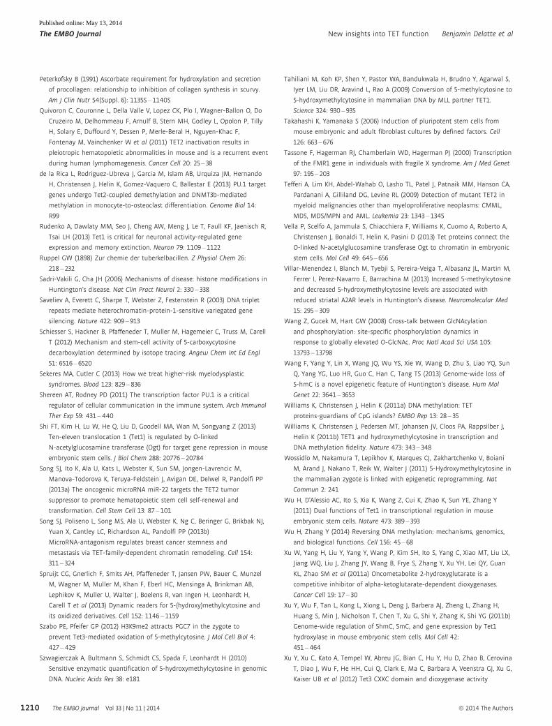
Peterkofsky B (1991) Ascorbate requirement for hydroxylation and secretion
of procollagen: relationship to inhibition of collagen synthesis in scurvy.
Am J Clin Nutr 54(Suppl. 6): 1135S – 1140S
Quivoron C, Couronne L, Della Valle V, Lopez CK, Plo I, Wagner-Ballon O, Do
Cruzeiro M, Delhommeau F, Arnulf B, Stern MH, Godley L, Opolon P, Tilly
H, Solary E, Duffourd Y, Dessen P, Merle-Beral H, Nguyen-Khac F,
Fontenay M, Vainchenker W et al (2011) TET2 inactivation results in
pleiotropic hematopoietic abnormalities in mouse and is a recurrent event
during human lymphomagenesis. Cancer Cell 20: 25 – 38
de la Rica L, Rodriguez-Ubreva J, Garcia M, Islam AB, Urquiza JM, Hernando
H, Christensen J, Helin K, Gomez-Vaquero C, Ballestar E (2013) PU.1 target
genes undergo Tet2-coupled demethylation and DNMT3b-mediated
methylation in monocyte-to-osteoclast differentiation. Genome Biol 14:
R99
Rudenko A, Dawlaty MM, Seo J, Cheng AW, Meng J, Le T, Faull KF, Jaenisch R,
Tsai LH (2013) Tet1 is critical for neuronal activity-regulated gene
expression and memory extinction. Neuron 79: 1109 – 1122
Ruppel GW (1898) Zur chemie der tuberkelbacillen. Z Physiol Chem 26:
218 – 232
Sadri-Vakili G, Cha JH (2006) Mechanisms of disease: histone modifications in
Huntington’s disease. Nat Clin Pract Neurol 2: 330 – 338
Saveliev A, Everett C, Sharpe T, Webster Z, Festenstein R (2003) DNA triplet
repeats mediate heterochromatin-protein-1-sensitive variegated gene
silencing. Nature 422: 909 – 913
Schiesser S, Hackner B, Pfaffeneder T, Muller M, Hagemeier C, Truss M, Carell
T (2012) Mechanism and stem-cell activity of 5-carboxycytosine
decarboxylation determined by isotope tracing. Angew Chem Int Ed Engl
51: 6516 – 6520
Sekeres MA, Cutler C (2013) How we treat higher-risk myelodysplastic
syndromes. Blood 123: 829 – 836
Shereen AT, Rodney PD (2011) The transcription factor PU.1 is a critical
regulator of cellular communication in the immune system. Arch Immunol
Ther Exp 59: 431 – 440
Shi FT, Kim H, Lu W, He Q, Liu D, Goodell MA, Wan M, Songyang Z (2013)
Ten-eleven translocation 1 (Tet1) is regulated by O-linked
N-acetylglucosamine transferase (Ogt) for target gene repression in mouse
embryonic stem cells. J Biol Chem 288: 20776 – 20784
Song SJ, Ito K, Ala U, Kats L, Webster K, Sun SM, Jongen-Lavrencic M,
Manova-Todorova K, Teruya-Feldstein J, Avigan DE, Delwel R, Pandolfi PP
(2013a) The oncogenic microRNA miR-22 targets the TET2 tumor
suppressor to promote hematopoietic stem cell self-renewal and
transformation. Cell Stem Cell 13: 87 – 101
Song SJ, Poliseno L, Song MS, Ala U, Webster K, Ng C, Beringer G, Brikbak NJ,
Yuan X, Cantley LC, Richardson AL, Pandolfi PP (2013b)
MicroRNA-antagonism regulates breast cancer stemness and
metastasis via TET-family-dependent chromatin remodeling. Cell 154:
311 – 324
Spruijt CG, Gnerlich F, Smits AH, Pfaffeneder T, Jansen PW, Bauer C, Munzel
M, Wagner M, Muller M, Khan F, Eberl HC, Mensinga A, Brinkman AB,
Lephikov K, Muller U, Walter J, Boelens R, van Ingen H, Leonhardt H,
Carell T et al (2013) Dynamic readers for 5-(hydroxy)methylcytosine and
its oxidized derivatives. Cell 152: 1146 – 1159
Szabo PE, Pfeifer GP (2012) H3K9me2 attracts PGC7 in the zygote to
prevent Tet3-mediated oxidation of 5-methylcytosine. J Mol Cell Biol 4:
427 – 429
Szwagierczak A, Bultmann S, Schmidt CS, Spada F, Leonhardt H (2010)
Sensitive enzymatic quantification of 5-hydroxymethylcytosine in genomic
DNA. Nucleic Acids Res 38: e181
Tahiliani M, Koh KP, Shen Y, Pastor WA, Bandukwala H, Brudno Y, Agarwal S,
Iyer LM, Liu DR, Aravind L, Rao A (2009) Conversion of 5-methylcytosine to
5-hydroxymethylcytosine in mammalian DNA by MLL partner TET1.
Science 324: 930 – 935
Takahashi K, Yamanaka S (2006) Induction of pluripotent stem cells from
mouse embryonic and adult fibroblast cultures by defined factors. Cell
126: 663 – 676
Tassone F, Hagerman RJ, Chamberlain WD, Hagerman PJ (2000) Transcription
of the FMR1 gene in individuals with fragile X syndrome. Am J Med Genet
97: 195 – 203
Tefferi A, Lim KH, Abdel-Wahab O, Lasho TL, Patel J, Patnaik MM, Hanson CA,
Pardanani A, Gilliland DG, Levine RL (2009) Detection of mutant TET2 in
myeloid malignancies other than myeloproliferative neoplasms: CMML,
MDS, MDS/MPN and AML. Leukemia 23: 1343 – 1345
Vella P, Scelfo A, Jammula S, Chiacchiera F, Williams K, Cuomo A, Roberto A,
Christensen J, Bonaldi T, Helin K, Pasini D (2013) Tet proteins connect the
O-linked N-acetylglucosamine transferase Ogt to chromatin in embryonic
stem cells. Mol Cell 49: 645 – 656
Villar-Menendez I, Blanch M, Tyebji S, Pereira-Veiga T, Albasanz JL, Martin M,
Ferrer I, Perez-Navarro E, Barrachina M (2013) Increased 5-methylcytosine
and decreased 5-hydroxymethylcytosine levels are associated with
reduced striatal A2AR levels in Huntington’s disease. Neuromolecular Med
15: 295 – 309
Wang Z, Gucek M, Hart GW (2008) Cross-talk between GlcNAcylation
and phosphorylation: site-specific phosphorylation dynamics in
response to globally elevated O-GlcNAc. Proc Natl Acad Sci USA 105:
13793 – 13798
Wang F, Yang Y, Lin X, Wang JQ, Wu YS, Xie W, Wang D, Zhu S, Liao YQ, Sun
Q, Yang YG, Luo HR, Guo C, Han C, Tang TS (2013) Genome-wide loss of
5-hmC is a novel epigenetic feature of Huntington’s disease. Hum Mol
Genet 22: 3641 – 3653
Williams K, Christensen J, Helin K (2011a) DNA methylation: TET
proteins-guardians of CpG islands? EMBO Rep 13: 28 – 35
Williams K, Christensen J, Pedersen MT, Johansen JV, Cloos PA, Rappsilber J,
Helin K (2011b) TET1 and hydroxymethylcytosine in transcription and
DNA methylation fidelity. Nature 473: 343 – 348
Wossidlo M, Nakamura T, Lepikhov K, Marques CJ, Zakhartchenko V, Boiani
M, Arand J, Nakano T, Reik W, Walter J (2011) 5-Hydroxymethylcytosine in
the mammalian zygote is linked with epigenetic reprogramming. Nat
Commun 2: 241
Wu H, D’Alessio AC, Ito S, Xia K, Wang Z, Cui K, Zhao K, Sun YE, Zhang Y
(2011) Dual functions of Tet1 in transcriptional regulation in mouse
embryonic stem cells. Nature 473: 389 – 393
Wu H, Zhang Y (2014) Reversing DNA methylation: mechanisms, genomics,
and biological functions. Cell 156: 45 – 68
Xu W, Yang H, Liu Y, Yang Y, Wang P, Kim SH, Ito S, Yang C, Xiao MT, Liu LX,
Jiang WQ, Liu J, Zhang JY, Wang B, Frye S, Zhang Y, Xu YH, Lei QY, Guan
KL, Zhao SM et al (2011a) Oncometabolite 2-hydroxyglutarate is a
competitive inhibitor of alpha-ketoglutarate-dependent dioxygenases.
Cancer Cell 19: 17 – 30
Xu Y, Wu F, Tan L, Kong L, Xiong L, Deng J, Barbera AJ, Zheng L, Zhang H,
Huang S, Min J, Nicholson T, Chen T, Xu G, Shi Y, Zhang K, Shi YG (2011b)
Genome-wide regulation of 5hmC, 5mC, and gene expression by Tet1
hydroxylase in mouse embryonic stem cells. Mol Cell 42:
451 – 464
Xu Y, Xu C, Kato A, Tempel W, Abreu JG, Bian C, Hu Y, Hu D, Zhao B, Cerovina
T, Diao J, Wu F, He HH, Cui Q, Clark E, Ma C, Barbara A, Veenstra GJ, Xu G,
Kaiser UB et al (2012) Tet3 CXXC domain and dioxygenase activity
The EMBO Journal Vol 33 | No 11 | 2014 ª 2014 The Authors
The EMBO Journal New insights into TET function Benjamin Delatte et al
1210
Published online: May 13, 2014

cooperatively regulate key genes for Xenopus eye and neural development.
Cell 151: 1200 – 1213
Yamaguchi S, Shen L, Liu Y, Sendler D, Zhang Y (2013) Role of Tet1 in erasure
of genomic imprinting. Nature 504: 460 – 464
Yang H, Liu Y, Bai F, Zhang JY, Ma SH, Liu J, Xu ZD, Zhu HG, Ling ZQ, Ye D,
Guan KL, Xiong Y (2012) Tumor development is associated with decrease
of TET gene expression and 5-methylcytosine hydroxylation. Oncogene 32:
663 – 669
Yao B, Lin L, Street RC, Zalewski ZA, Galloway JN, Wu H, Nelson DL, Jin P (2013)
Genome-wide alteration of 5-hydroxymethylcytosine in a mouse model of
fragile X-associated tremor/ataxia syndrome. Hum Mol Genet 23: 1095 –1107
Yildirim O, Li R, Hung JH, Chen PB, Dong X, Ee LS, Weng Z, Rando OJ, Fazzio
TG (2011) Mbd3/NURD complex regulates expression of
5-hydroxymethylcytosine marked genes in embryonic stem cells. Cell 147:
1498 – 1510
Yin R, Mao SQ, Zhao B, Chong Z, Yang Y, Zhao C, Zhang D, Huang H,
Gao J, Li Z, Jiao Y, Li C, Liu S, Wu D, Gu W, Yang YG, Xu GL, Wang H
(2013) Ascorbic acid enhances Tet-mediated 5-methylcytosine oxidation
and promotes DNA demethylation in mammals. J Am Chem Soc 135:
10396 – 10403
You JS, Jones PA (2012) Cancer genetics and epigenetics: two sides of the
same coin? Cancer Cell 22: 9 – 20
Yu M, Hon GC, Szulwach KE, Song CX, Zhang L, Kim A, Li X, Dai Q, Shen Y,
Park B, Min JH, Jin P, Ren B, He C (2012) Base-resolution analysis of
5-hydroxymethylcytosine in the mammalian genome. Cell 149: 1368 – 1380
Zhang L, Lu X, Lu J, Liang H, Dai Q, Xu GL, Luo C, Jiang H, He C (2012)
Thymine DNA glycosylase specifically recognizes
5-carboxylcytosine-modified DNA. Nat Chem Biol 8: 328 – 330
Zhang P, Huang B, Xu X, Sessa WC (2013a) Ten-eleven translocation (Tet) and
thymine DNA glycosylase (TDG), components of the demethylation
pathway, are direct targets of miRNA-29a. Biochem Biophys Res Commun
437: 368 – 373
Zhang RR, Cui QY, Murai K, Lim YC, Smith ZD, Jin S, Ye P, Rosa L, Lee YK, Wu
HP, Liu W, Xu ZM, Yang L, Ding YQ, Tang F, Meissner A, Ding C, Shi Y, Xu
GL (2013b) Tet1 regulates adult hippocampal neurogenesis and cognition.
Cell Stem Cell 13: 237 – 245
1211ª 2014 The Authors The EMBO Journal Vol 33 | No 11 | 2014
Benjamin Delatte et al New insights into TET function The EMBO Journal
Published online: May 13, 2014