pleiotropic age-dependent effects of mitochondrial ... · pleiotropic age-dependent effects of...
TRANSCRIPT
Pleiotropic age-dependent effects of mitochondrialdysfunction on epidermal stem cellsMichael C. Velardea, Marco Demariaa, Simon Melova, and Judith Campisia,b,1
aBuck Institute for Research on Aging, Novato, CA 94945 and bLawrence Berkeley National Laboratory, Berkeley, CA 94720
Edited by Philip C. Hanawalt, Stanford University, Stanford, CA, and approved July 15, 2015 (received for review March 21, 2015)
Tissue homeostasis declines with age partly because stem/pro-genitor cells fail to self-renew or differentiate. Because mitochon-drial damage can accelerate aging, we tested the hypothesis thatmitochondrial dysfunction impairs stem cell renewal or function. Wedeveloped a mouse model, Tg(KRT14-cre/Esr1)20Efu/J × Sod2tm1Smel,that generates mitochondrial oxidative stress in keratin 14-expressingepidermal stem/progenitor cells in a temporally controlled mannerowing to deletion of Sod2, a nuclear gene that encodes the mito-chondrial antioxidant enzyme superoxide dismutase 2 (Sod2). Epider-mal Sod2 loss induced cellular senescence, which irreversibly arrestedproliferation in a fraction of keratinocytes. Surprisingly, in youngmice, Sod2 deficiency accelerated wound closure, increasing epider-mal differentiation and reepithelialization, despite the reduced pro-liferation. In contrast, at older ages, Sod2 deficiency delayed woundclosure and reduced epidermal thickness, accompanied by epidermalstem cell exhaustion. In young mice, Sod2 deficiency accelerated epi-dermal thinning in response to the tumor promoter 12-O-tetradeca-noylphorbol-13-acetate, phenocopying the reduced regeneration ofolder Sod2-deficient skin. Our results show a surprising beneficialeffect of mitochondrial dysfunction at young ages, provide a poten-tial mechanism for the decline in epidermal regeneration at olderages, and identify a previously unidentified age-dependent role formitochondria in skin quality and wound closure.
cellular senescence | oxidative stress | skin aging | stem cell proliferation |superoxide dismutase 2
Stem and progenitor cells are crucial for tissue homeostasis,repair, and regeneration. In response to injury, they pro-
liferate and differentiate to replace damaged or dysfunctionalcells (1, 2). In the skin, epidermal basal cells differentiate to formdistinct epidermal layers: the stratum basale (SB), stratum spino-sum (SS), stratum granulosum (SG), and stratum corneum (SC)(3). In the SB layer, epidermal basal cells are identified by nucleithat stain strongly with hematoxylin and eosin (H&E). These cellsdifferentiate to form the SS layer, composed mainly of cells withlightly stained nuclei. SS cells differentiate into the SG layer,identified by cells with prominent cytoplasmic granules, whichterminally differentiate to form the SC layer containing acidophilicanucleated cells.Tissue homeostasis declines with age partly because stem/
progenitor cells fail to self-renew or differentiate (4). Oxidativedamage can contribute to this decline in compartments such asthe hematopoietic system (5, 6). Aging is caused by intrinsic andextrinsic factors that cooperate to drive aging phenotypes (7).Mitochondrial dysfunction has been suggested to play a majorrole in intrinsic aging (8). Furthermore, mitochondrial damage isassociated with extrinsic aging, particularly ultraviolet (UV) radia-tion-induced photoaging in the skin (9). Thus, mitochondrial dam-age may be a common link between intrinsic and extrinsic aging.Mitochondrial stress can decrease life span and health span.
Superoxide dismutase 2 (SOD2) scavenges mitochondrial super-oxide to protect against oxidative damage. Sod2 deficiency de-creases life span in several species. Mice with constitutive Sod2deficiency are neonatally lethal on multiple genetic backgrounds,presenting with neurodegeneration, spongiform encephalopathy,cardiomyopathy, hepatic fat accumulation, and failure to thrive;
cells from these mice exhibit impaired spare respiratory capacity,genomic instability, and mitochondrial functional defects (10–17). Sod2−/− cells also have a reduced proliferative capacity (17),consistent with the finding that constitutive Sod2 deficiency in-duces cellular senescence, a tumor-suppressive mechanism thatirreversibly arrests cell proliferation (18), in mouse skin (19).Conversely, overexpression of mitochondrial antioxidants canpartly rescue age-related pathologies (14, 20), increase organis-mal life span (21), and prolong stem cell replicative life span (22).Interestingly, some studies have suggested that mild mito-
chondrial stress can be beneficial (23). Here we show that mito-chondrial stress owing to Sod2 deficiency in epidermal cells canhave positive or negative effects on skin regeneration, and that theseeffects depend on age. We show that epidermal Sod2 deficiencyinduces cellular senescence, which reduces proliferative capacity inthe skin but stimulates the differentiation of epidermal stem/pro-genitor cells. This stimulation accelerates wound closure in youngmice, but the proliferative decline drains stem cell pools with agingand retards wound closure. Our findings extend the concept ofantagonistic pleiotropy, which stipulates that gene action can bebeneficial at young ages but deleterious at older ages, to mito-chondrial function in the skin.
ResultsMice with a Keratinocyte-Specific Mitochondrial Defect. Becauseconstitutive Sod2− deficiency is neonatal lethal (12), it is notpossible to study age-specific effects in these mice. Consequently,we constructed an inducible, temporally regulated, tissue-specificmouse model of Sod2 deficiency. We created mice carrying a Sod2gene into which we inserted LoxP sites (Sod2tm1Smel) into se-quences flanking a region required for catalytic activity. We thencrossed these animals to mice carrying a tamoxifen (TAM)-activated
Significance
Mitochondrial damage can accelerate features of aging, in-cluding impaired tissue regeneration, but little is known abouthow aging and this damage interact to impair tissue renewal.We show that mitochondrial oxidative stress in the epidermisalters wound healing depending on age. Epidermal mito-chondrial damage accelerated wound closure in young mice;however, in older mice, this damage limited epidermal cellproliferation and reduced epidermal stem cell numbers, leadingto delayed wound closure. Our findings uncover a surprisingbeneficial effect of mitochondrial dysfunction at young age(accelerated wound closure), and a potential mechanism forthe reduced epidermal regeneration at older ages (stem celldepletion).
Author contributions: M.C.V. and J.C. designed research; M.C.V. performed research; M.D.and S.M. contributed new reagents/analytic tools; M.C.V. analyzed data; and M.C.V.,M.D., S.M., and J.C. wrote the paper.
The authors declare no conflict of interest.
This article is a PNAS Direct Submission.1To whom correspondence should be addressed. Email: [email protected].
This article contains supporting information online at www.pnas.org/lookup/suppl/doi:10.1073/pnas.1505675112/-/DCSupplemental.
www.pnas.org/cgi/doi/10.1073/pnas.1505675112 PNAS | August 18, 2015 | vol. 112 | no. 33 | 10407–10412
CELL
BIOLO
GY
Dow
nloa
ded
by g
uest
on
Dec
embe
r 25
, 201
9
Cre recombinase (Cre-ERT) under control of the keratinocyte-specific keratin 14 promoter. This mouse, Tg(KRT14-cre/Esr1)20Efu/J x Sod2tm1Smel, herein designated K14S, allows deletionof critical Sod2 sequences in keratinocytes after TAM treatment.TAM, but not estrogen, causes nuclear translocation of Cre-ERT,a fusion protein comprised of Cre and a mutant estrogen receptorligand-binding domain, thus allowing Cre to excise sequencesbetween the LoxP sites (Fig. S1A). Because the K14 promoter isactive primarily in epidermal stem/progenitor cells (24, 25), TAMdeletes Sod2 mainly in epidermal, but not dermal, cells (Fig. S1 Band C). As expected, TAM deleted Sod2 in dorsal skin and skin-containing tails and toes, but not in the heart, liver, intestine, orlungs, of K14S mice (Fig. S1D). TAM did not delete Sod2 inSod2tm1Smel mice (designated S), which lack Cre recombinase(Fig. S1D).We measured the copy numbers of recombined and total Sod2
alleles by quantitative PCR (qPCR) (Fig. S1E). After TAMtreatment, >92% of Sod2 genes showed a deletion in the epi-dermis, but not the dermis, of K14S mice. No recombination oc-curred in the epidermis or dermis of Tg(KRT14-cre/Esr1)20Efu/J ×B6-Gt(ROSA)26Sortm1Sor/J mice (designated K14R), which lackLoxP sites. Accordingly, qPCR showed that Sod2 mRNA levelswere substantially lower in the epidermis (Fig. S1F), but not thedermis (Fig. S1G), of TAM-treated, but not vehicle-treated, K14Smice. As expected, Sod2 mRNA levels were comparably high inthe epidermis and dermis of vehicle- and TAM-treated K14Rmice. This finding indicates that TAM reduced Sod2 expression inepidermal cells of K14S mice, confirming successful generationof an inducible keratinocyte-specific Sod2-deficient mouse andovercoming the impediment of neonatal lethality caused by con-stitutive Sod2 deficiency.To determine the SOD2 expression pattern in skin, we used
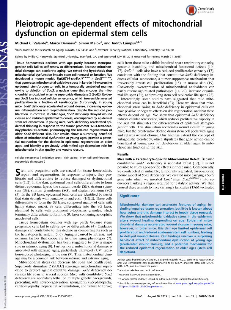
immunohistochemistry on whole mounts of tail epidermis fromvehicle- or TAM-treated K14S mice, counterstaining for CD49f,a keratinocyte marker to visualize epidermal compartments. SOD2was detectable in the sebaceous glands and epidermal layers, withhighest expression in the middle of the hair follicle or isthmus re-gion (Fig. 1A) in which stem/progenitor cells reside (26). Cells in thebulb area expressed lower SOD2 levels. TAM reduced epidermalSOD2 levels markedly, consistent with Sod2 deletion in thetargeted tissue.Constitutive Sod2 loss in mice severely reduces mitochondrial
complex II activity, but not complex IV activity, and decreasesmitochondrial spare respiratory capacity without altering the basalrespiration rate (12, 19). To determine whether keratinocyte-specific Sod2 loss has similar effects, we stained K14S skin for
succinate dehydrogenase (SDH) (complex II) and cytochrome coxidase (COX) (complex IV) activities. TAM-treated, but notvehicle-treated, K14S mice had significantly less SDH activity inthe hair follicles, but not in the underlying skeletal muscle (Fig. 1Band Fig. S1I). As expected (19), mitochondrial complex IV (COX)activity was similar in the TAM- and vehicle-treated K14S mice(Fig. S1 H and I). These results confirm that the mitochondrialdysfunction was confined to epidermal keratinocytes in TAM-treated K14S mice.Epidermal Sod2 deficiency did not significantly increase mor-
bidity in all cohorts tested up to ∼16 mo after TAM treatment of4-mo-old mice. Thus, epidermal deletion of Sod2 did not notice-ably compromise the health of mice up to 20 mo of age.
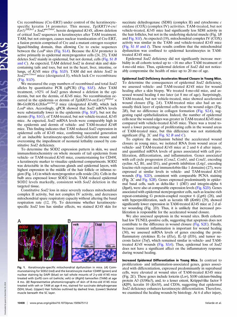
Epidermal Sod2 Deficiency Accelerates Wound Closure in Young Mice.To determine the consequences of epidermal Sod2 deficiency,we assessed vehicle- and TAM-treated K14S mice for woundhealing after a skin biopsy. We treated 4-mo-old mice, and as-sessed wound healing 4 mo later (at 8 mo of age). Surprisingly,TAM-treated, but not vehicle-treated, mice showed acceleratedwound closure (Fig. 2A). TAM-treated mice also had an un-usually thick layer of epidermal cells near the wound edges (Fig.2B), but no difference in collagen formation (Fig. S2A), sug-gesting rapid epithelialization. Indeed, the number of epidermalcells near the wound edges was greater in TAM-treated K14Smicecompared with vehicle-treated K14S mice. There was a trend to-ward a lower percentage of proliferating cells in the wound areasof TAM-treated mice, but this difference was not statisticallysignificant (Fig. 2C and Fig. S2 B and C).To explore the mechanism behind the accelerated wound
closure in young mice, we isolated RNA from wound areas ofvehicle- and TAM-treated K14S mice at 2 and 6 d after injury,and quantified mRNA levels of genes associated with cell pro-liferation, differentiation, and inflammation. Genes associatedwith cell cycle progression (Ccna2, Ccnb1, and Ccnd1, encodingcyclins A2, B1, and D1), and growth inhibition (Lrig1, encodingleucine-rich repeats and immunolobin-like 1 domains) (27), wereexpressed at similar levels in vehicle- and TAM-treated K14Swounds (Fig. S2D), consistent with comparable PCNA staining(Fig. 2C and Fig. S2B). Genes expressed predominantly in epider-mal basal cells, such as delta-like 1 (Dll1) and integrin-alpha 6(Itga6), were also at comparable expression levels (Fig. S2D). Genesassociated with epidermal stem/progenitor cells, such as leucine-richrepeat-containing G protein-coupled receptor 6 (Lgr6) (28), andwith hyperproliferation, such as keratin 6B (Krt6b) (29), showedsignificantly lower expression in TAM-treated K14S mice at 2 d af-ter wounding (Fig. 2D). Thus, it is unlikely that increased pro-liferation is responsible for the accelerated wound closure.We also assessed apoptosis in the wound sites. Both cohorts
had few TUNEL-positive cells, suggesting that apoptosis does notcontribute to the difference in wound closure (Fig. S2E). Finally,because transient inflammation is important for wound healing(30), we assessed mRNA levels of genes encoding the proin-flammatory cytokines IL-1α (Il1a), IL-1β (Il1b), and tumor ne-crosis factor (Tnf), which remained similar in vehicle- and TAM-treated K14S wounds (Fig. S3A). Thus, epidermal loss of Sod2does not have a significant affect on the inflammatory responseduring wound healing.
Increased Epidermal Differentiation in Young Mice. In contrast toproliferation- and inflammation-associated genes, genes associ-ated with differentiation, expressed predominantly in suprabasalcells, were elevated at wound sites of TAM-treated K14S mice(Fig. 2E). These genes include loricrin (Lor), S100 calcium-bindingprotein A3 (S100a3), and, to a lesser extent, Krüppel-like factor 9(Klf9), keratin 10 (Krt10), and CD36, suggesting that epidermalSod2 deficiency enhances keratinocyte differentiation. Therefore,we examined the healing wounds by histology. At 6 d after injury,
Fig. 1. Keratinocyte-specific mitochondrial dysfunction in mice. (A) Coim-munostaining for SOD2 (red) and the keratinocyte marker CD49f (green) andnuclear staining by DAPI (blue) on tail whole mounts of 2-y-old K14S micetreated with (Left) corn oil (vehicle; veh) or (Right) tamoxifen (TAM) at age4 mo. (B) Representative photomicrographs of skin of 8-mo-old K14S micetreated with veh or TAM at age 4 mo, stained for succinate dehydrogenase(SDH; blue). (Upper) Hair follicles outlined by dashed lines. (Lower) Skeletalmuscle beneath the SC layer.
10408 | www.pnas.org/cgi/doi/10.1073/pnas.1505675112 Velarde et al.
Dow
nloa
ded
by g
uest
on
Dec
embe
r 25
, 201
9

wounds in vehicle-treated mice had numerous cells in the SBlayer and a negligible SG layer (Fig. 2F and Fig. S3B). In contrast,wounds in TAM-treated mice had few cells in the SB layer and asubstantial SG layer (Fig. 2F and Fig. S3B). There were no ap-parent differences in the SC and SS layers.To confirm the prominent SG layer in TAM-treated K14S
mice, we immunostained wounds for loricrin (LOR), which wasmore prominent in the TAM-treated animals (Fig. 2G). Thesewounds also contained higher Lor mRNA levels (Fig. 2E). Be-cause SB cells differentiate into SS and SG layers, the increasedSG layer in wounds of TAM-treated K14S mice suggests thatSod2 deficiency accelerates epidermal differentiation during woundhealing. This acceleration was transient; at 10 d after wounding,epidermal stratification was similar in the TAM- and vehicle-treatedmice (Fig. S3C). Thus, increased differentiation, rather than cellproliferation, appears to drive the more rapid wound closure inyoung TAM-treated K14S mice.
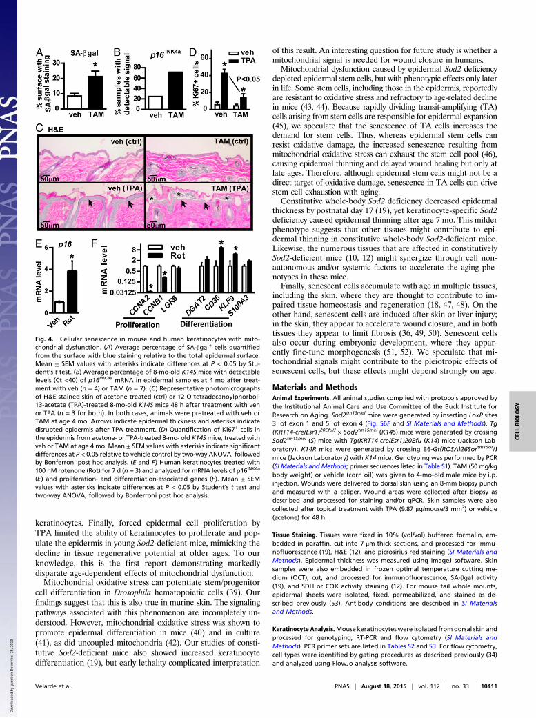
Delayed Wound Closure and Rapid Epidermal Thinning in Old Mice.The accelerated wound closure in young keratinocyte-specificSod2-deficient mice suggests that mitochondrial dysfunction im-proves skin repair, contradicting the free radical theory of aging.To explore this possibility, we monitored wound closure in older(age 11 and 14 mo) mice treated with TAM or vehicle at age 4 mo.Although keratinocyte-specific Sod2 deficiency accelerated woundclosure in young mice (Fig. 2A), this acceleration was lost in11-mo-old mice (Fig. S4A). Furthermore, Sod2 deficiency delayedwound closure in 14-mo-old mice (Fig. 3A). Thus, the Sod2 de-ficiency promoted epidermal differentiation and wound closure inyoung mice, but delayed wound closure in older mice. We alsomonitored epidermal thickness, which declines with age in humansand mice (31, 32). We treated K14S mice with vehicle or TAM atage 4 mo, and measured epidermal thickness 4, 7, and 10 mo later(at age 8, 11, and 14 mo). Vehicle-treated mice showed a
significant decline in epidermal thickness at 10 mo after treatment,whereas TAM-treated animals exhibited this decline at 7 mo aftertreatment (Fig. 3B and Fig. S4B). Thus, epidermal Sod2 deficiencyaccelerated age-associated thinning of the epidermis.
Depletion of Epidermal Stem Cells in Old Mice. One possible reasonfor the phenotypes of older TAM-treated K14Smice is a reducedcapacity of stem/progenitor cells to repopulate the tissue. Wequantified mRNA levels of genes expressed in K14-positive stemcells, involucrin (Ivl)-committed progenitor cells, total basal in-terfollicular epidermal (IFE) cells, and suprabasal IFE cells, asdescribed previously (33). Epidermal Sod2 deficiency did notsignificantly alter gene expression associated with K14-positivestem cells or Ivl-committed progenitor cells in young mice (Fig.3C and Fig. S4 C and D), but significantly decreased the ex-pression of genes associated with K14-positive stem cells in oldermice (Fig. 3C). These genes include kinesin family member 11(Kif11), Ccnb1, centromere protein E (Cenpe), cell division cycle20 (Cdc20), Cdca5, and Kif14. Because expression of these pro-proliferative genes declined in the TAM-treated, but not vehicle-treated, K14S epidermis at the older age but not the younger age,our data suggest that aging exacerbates the effects of theSod2 deficiency.mRNAs associated with Ivl-committed progenitor cells increased
with age, but only the ceramide synthase 4 (Lass4) mRNA sig-nificantly declined in the old Sod2-deficient epidermis (Fig. S4 Cand D). mRNA levels of genes expressed in basal and suprabasalcells showed no change (Fig. S4 C and D). Our data indicate thatthe Sod2 deficiency depletes K14-positive stem cells, but not Ivl-committed progenitor or total basal and suprabasal IFE cells, inaged mice.To verify these phenotypes, we analyzed epidermal stem cell
populations by flow cytometry. Using well-characterized markers(CD49f, CD34, and Sca1), as described previously (34), we found
Fig. 2. Wound closure in 8 mo old K14S mice treated with veh or TAM at age 4 mo. (A) Average wound size (mean ± SEM) after 8-mm punch biopsy in K14Smice (veh, n = 11; TAM, n = 8). Percent wound size refers to the wound area relative to the initial wound area × 100. Asterisks indicate differences at P < 0.05by Student’s t test. (B) Representative photomicrographs of H&E staining of wound areas in K14Smice at 2 d and 4 d after injury (n = 3 each). Layers above thedashed lines indicate the epidermis. White arrows show area of reepithelialization. (C) Quantification of PCNA immunofluorescence in wound areas of K14Smice at 2 d (n = 3 each) and 6 d (n = 5 each) after skin injury. (D and E) mRNA levels determined by qPCR of genes associated with proliferation (D), expressedpredominantly in epidermal basal cells, and differentiation (E), expressed predominantly in epidermal suprabasal cells, in wound areas of K14S mice at 2 d(veh and TAM, n = 3) or 6 d (veh, n = 5; TAM, n = 7) after injury. Mean ± SEM values with asterisks indicate differences at P < 0.05 by two-way ANOVAfollowed by Bonferroni post hoc analysis. (F) Representative photomicrographs of H&E staining of wound areas of K14Smice at 6 d after injury (veh and TAM,n = 5). Stratum basale (SB; arrows) cells are identified by strong nuclear staining (blue) at the wound bottom. Stratum spinosum (SS; black line) are cells withlow nuclear staining above the SB layer. Stratum granulosum (SG; asterisks) are cells with granules in the cytoplasm. Stratum corneum (SC; blue line) is theoutermost layer. (G) Representative photomicrographs of loricrin (LOR; red) staining by immunofluorescence and nuclear staining by DAPI (blue) in woundareas of K14S mice at 6 d after injury. Dashed lines indicate the wound site. Multiple photomicrographs of wound sections were taken under 10× magni-fication and tiled in Adobe Photoshop.
Velarde et al. PNAS | August 18, 2015 | vol. 112 | no. 33 | 10409
CELL
BIOLO
GY
Dow
nloa
ded
by g
uest
on
Dec
embe
r 25
, 201
9

a marked reduction in K14-positive stem cells in the junctionalzone and bulge area in 14-mo-old TAM-treated, but not vehicle-treated, K14S skin (Fig. 3D and Fig. S5). Because junctional zoneand bulge area stem cells are partly responsible for repopulatingthe epidermis after injury, their depletion in TAM-treated K14Smice suggests that mitochondrial dysfunction exhausts thesecells. This exhaustion takes time and manifests as delayed woundhealing only in older mice.
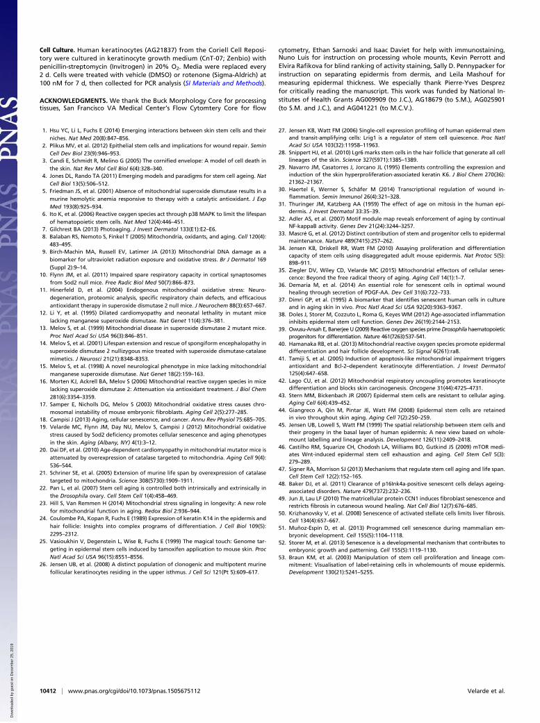
Cellular Senescence and Reduced Regeneration. Mitochondrialdysfunction can induce cellular senescence (19, 35). We haveshown that constitutive Sod2 deficiency causes the chronic pres-ence of senescent keratinocytes in the epidermis (19). We alsohave shown that cutaneous wounding transiently induces senes-cence in fibroblasts and endothelial cells, which promotes woundhealing (36). To determine whether senescent cells reside in theskin of keratinocyte-specific Sod2-deficient mice, we treated young(4 mo old) K14S mice with TAM or vehicle and stained the skinfor senescence-associated β-galactosidase (SA-βgal), an estab-lished senescence marker (37), 4 mo later. Relative to vehicle-treated mice, TAM-treated mice had ∼2.5-fold higher SA-βgalactivity in the epidermis (Fig. 4A and Fig. S6A). Interestingly, the
staining was most prominent in the stratum corneum or more dif-ferentiated layers; staining in hair follicles was nonspecific, as re-ported previously (37). We used qPCR to assess the expression ofp16INK4a, an additional senescence marker (36). Untreated 8-mo-old K14S mice have low to undetectable levels of epidermalp16INK4a mRNA. After TAM treatment, however, p16INK4a mRNAwas detectable in most (>70%) K14S skin samples (Fig. 4B).p16INK4a mRNA persisted even 10 mo after TAM treatment (Fig.S6B), confirming that Sod2 deficiency induced persistent senes-cence in the epidermis.Platelet-derived growth factor-A (Pdgfa) is secreted by senes-
cent cells during wound healing (36), but showed no differencein wounds of 8-mo-old vehicle- and TAM-treated K14S mice,treated at age 4 mo (Fig. S6C). We also assessed mRNA levels ofother factors that could contribute to wound healing, includingPdgfb, transforming growth factor-beta 1–3 (Tgfb1, Tgfb2, andTgfb3), fibroblast growth factors 2 and 7 (Fgf2 and Fgf7), andvascular endothelial growth factor (Vegf), none of which differedbetween the two groups (Fig. S6C). Thus, it is unlikely thatgrowth factors contribute to the altered wound healing in epi-dermal Sod2-deficient mice.The senescence growth arrest could reduce the ability of tissues to
regenerate. To determine the effect of Sod2 deficiency on epidermalproliferative capacity, we treated K14S mice with the propro-liferative agent 12-O-tetradecanoylphorbol-13-acetate (TPA). TPApromoted epidermal thickening (arrows) in vehicle-treated, but notin TAM-treated, K14S mice (Fig. 4C). Furthermore, it caused epi-dermal lesions and SC layer separation in TAM-treated K14S mice(Fig. 4C), which occurs when old mice are treated with TPA (38).Moreover, whereas TPA enhanced epidermal proliferation in vehicle-treated K14S mice, as determined by Ki67 staining, it induced lessproliferation in the TAM-treated epidermis (Fig. 4D and Fig. S6D).Thus, forced epidermal proliferation by TPA accelerated epider-mal thinning in young Sod2-deficient mice, phenocopying thereduced proliferative capacity observed at older ages.
Mitochondrial Dysfunction in Human Keratinocytes. To test the ideathat mitochondrial dysfunction reduces proliferation and in-creases differentiation in epidermal cells, we treated primaryhuman keratinocytes with the mitochondrial electron transportchain complex I inhibitor rotenone. We previously showed thatthese cells senesce in response to rotenone (19). Rotenone alsoincreased p16INK4a mRNA (Fig. 4E) and cell size (Fig. S6E), acharacteristic of senescent cells, and decreased mRNA levels ofthe proproliferation genes CCNA2 and CCNB1 (Fig. 4F), sup-porting the idea that persistent mitochondrial dysfunction haltskeratinocyte proliferation by inducing cellular senescence. Notably,rotenone increased the differentiation-associated mRNAs CD36and KLF9 and, to a lesser extent, S100A3 (Fig. 4F), consistent withmitochondrial dysfunction promoting keratinocyte differentiation.We conclude that mitochondrial dysfunction can accelerate woundclosure in young Sod2-deficient mice by increasing keratinocytedifferentiation, whereas persistent mitochondrial dysfunction dur-ing aging can deplete dividing keratinocytes.
DiscussionOur results demonstrate a previously unidentified age-dependentcontribution of mitochondria to stem cell and skin function.The use of K14S mice allowed us to study mitochondrialdysfunction in K14+ epidermal cells in young and old animals,leading to important and surprising conclusions. First, mitochon-drial dysfunction (owing to Sod2 deficiency) can acceleratewound closure in young mice. This unexpected response wasassociated with increased reepithelialization and epidermaldifferentiation. Second, mitochondrial dysfunction can depletethe stem cell reservoir in older mice, leading to decreased epi-dermal thickness and delayed wound closure. Third, mitochondrialdysfunction induces cellular senescence in many K14+ epidermal
Fig. 3. Wound closure in old K14S mice. (A) Average wound size (mean ±SEM) after injury by 8-mm punch biopsy in 14-mo-old K14Smice treated withveh or TAM (veh, n = 10; TAM, n = 11) at age 4 mo. Percent wound size refersto the wound area relative to the initial wound area × 100. (B) Quantificationof epidermal thickness (in μm) in H&E-stained K14S mouse skin at 4, 7, and10 mo after veh (Left) or TAM (Right) treatment (n = 5 each). (C) mRNA levelsof genes expressed predominantly in K14+ stem cells, analyzed by qPCR, in skinfrom K14S mice at 4 or 11–20 mo after treatment with veh or TAM. Mean ±SEM values with asterisks indicate differences at P < 0.05 relative to vehiclecontrol by two-way ANOVA, followed by Bonferroni post hoc analysis.(D) Quantification of flow cytometry histographs of epidermal cells stainedfor CD49f, CD34, and Sca1, isolated from the skin of 14-mo-old K14S micetreated with veh or TAM at age 4 mo (n = 5 for each). Populations of bulgestem cells (CD49fhi/CD34+), suprabasal bulge stem cells (CD49flo/CD34+), andjunctional zone stem cells (CD49fhi/CD34−/Sca1−) were quantified as theaverage percent cell population (mean ± SEM) in the epidermis. Asterisksindicate differences at P < 0.05 by Student’s t test.
10410 | www.pnas.org/cgi/doi/10.1073/pnas.1505675112 Velarde et al.
Dow
nloa
ded
by g
uest
on
Dec
embe
r 25
, 201
9
keratinocytes. Finally, forced epidermal cell proliferation byTPA limited the ability of keratinocytes to proliferate and pop-ulate the epidermis in young Sod2-deficient mice, mimicking thedecline in tissue regenerative potential at older ages. To ourknowledge, this is the first report demonstrating markedlydisparate age-dependent effects of mitochondrial dysfunction.Mitochondrial oxidative stress can potentiate stem/progenitor
cell differentiation in Drosophila hematopoietic cells (39). Ourfindings suggest that this is also true in murine skin. The signalingpathways associated with this phenomenon are incompletely un-derstood. However, mitochondrial oxidative stress was shown topromote epidermal differentiation in mice (40) and in culture(41), as did uncoupled mitochondria (42). Our studies of consti-tutive Sod2-deficient mice also showed increased keratinocytedifferentiation (19), but early lethality complicated interpretation
of this result. An interesting question for future study is whether amitochondrial signal is needed for wound closure in humans.Mitochondrial dysfunction caused by epidermal Sod2 deficiency
depleted epidermal stem cells, but with phenotypic effects only laterin life. Some stem cells, including those in the epidermis, reportedlyare resistant to oxidative stress and refractory to age-related declinein mice (43, 44). Because rapidly dividing transit-amplifying (TA)cells arising from stem cells are responsible for epidermal expansion(45), we speculate that the senescence of TA cells increases thedemand for stem cells. Thus, whereas epidermal stem cells canresist oxidative damage, the increased senescence resulting frommitochondrial oxidative stress can exhaust the stem cell pool (46),causing epidermal thinning and delayed wound healing but only atlate ages. Therefore, although epidermal stem cells might not be adirect target of oxidative damage, senescence in TA cells can drivestem cell exhaustion with aging.Constitutive whole-body Sod2 deficiency decreased epidermal
thickness by postnatal day 17 (19), yet keratinocyte-specific Sod2deficiency caused epidermal thinning after age 7 mo. This milderphenotype suggests that other tissues might contribute to epi-dermal thinning in constitutive whole-body Sod2-deficient mice.Likewise, the numerous tissues that are affected in constitutivelySod2-deficient mice (10, 12) might synergize through cell non-autonomous and/or systemic factors to accelerate the aging phe-notypes in these mice.Finally, senescent cells accumulate with age in multiple tissues,
including the skin, where they are thought to contribute to im-paired tissue homeostasis and regeneration (18, 47, 48). On theother hand, senescent cells are induced after skin or liver injury;in the skin, they appear to accelerate wound closure, and in bothtissues they appear to limit fibrosis (36, 49, 50). Senescent cellsalso occur during embryonic development, where they appar-ently fine-tune morphogenesis (51, 52). We speculate that mi-tochondrial signals might contribute to the pleiotropic effects ofsenescent cells, but these effects might depend strongly on age.
Materials and MethodsAnimal Experiments. All animal studies complied with protocols approved bythe Institutional Animal Care and Use Committee of the Buck Institute forResearch on Aging. Sod2tm1Smel mice were generated by inserting LoxP sites3′ of exon 1 and 5′ of exon 4 (Fig. S6F and SI Materials and Methods). Tg(KRT14-cre/Esr1)20Efu/J × Sod2tm1Smel (K14S) mice were generated by crossingSod2tm1Smel (S) mice with Tg(KRT14-cre/Esr1)20Efu (K14) mice (Jackson Lab-oratory). K14R mice were generated by crossing B6-Gt(ROSA)26Sortm1Sor/Jmice (Jackson Laboratory) with K14mice. Genotyping was performed by PCR(SI Materials and Methods; primer sequences listed in Table S1). TAM (50 mg/kgbody weight) or vehicle (corn oil) was given to 4-mo-old male mice by i.p.injection. Wounds were delivered to dorsal skin using an 8-mm biopsy punchand measured with a caliper. Wound areas were collected after biopsy asdescribed and processed for staining and/or qPCR. Skin samples were alsocollected after topical treatment with TPA (9.87 μg/mouse/3 mm2) or vehicle(acetone) for 48 h.
Tissue Staining. Tissues were fixed in 10% (vol/vol) buffered formalin, em-bedded in paraffin, cut into 7-μm-thick sections, and processed for immu-nofluorescence (19), H&E (12), and picrosirius red staining (SI Materials andMethods). Epidermal thickness was measured using ImageJ software. Skinsamples were also embedded in frozen optimal temperature cutting me-dium (OCT), cut, and processed for immunofluorescence, SA-βgal activity(19), and SDH or COX activity staining (12). For mouse tail whole mounts,epidermal sheets were isolated, fixed, permeabilized, and stained as de-scribed previously (53). Antibody conditions are described in SI Materialsand Methods.
Keratinocyte Analysis.Mouse keratinocytes were isolated from dorsal skin andprocessed for genotyping, RT-PCR and flow cytometry (SI Materials andMethods). PCR primer sets are listed in Tables S2 and S3. For flow cytometry,cell types were identified by gating procedures as described previously (34)and analyzed using FlowJo analysis software.
Fig. 4. Cellular senescence in mouse and human keratinocytes with mito-chondrial dysfunction. (A) Average percentage of SA-βgal+ cells quantifiedfrom the surface with blue staining relative to the total epidermal surface.Mean ± SEM values with asterisks indicate differences at P < 0.05 by Stu-dent’s t test. (B) Average percentage of 8-mo-old K14S mice with detectablelevels (Ct <40) of p16INK4a mRNA in epidermal samples at 4 mo after treat-ment with veh (n = 4) or TAM (n = 7). (C) Representative photomicrographsof H&E-stained skin of acetone-treated (ctrl) or 12-O-tetradecanoylphorbol-13-acetate (TPA)-treated 8-mo-old K14S mice 48 h after treatment with vehor TPA (n = 3 for both). In both cases, animals were pretreated with veh orTAM at age 4 mo. Arrows indicate epidermal thickness and asterisks indicatedisrupted epidermis after TPA treatment. (D) Quantification of Ki67+ cells inthe epidermis from acetone- or TPA-treated 8-mo- old K14Smice, treated withveh or TAM at age 4 mo. Mean ± SEM values with asterisks indicate significantdifferences at P < 0.05 relative to vehicle control by two-way ANOVA, followedby Bonferroni post hoc analysis. (E and F) Human keratinocytes treated with100 nM rotenone (Rot) for 7 d (n = 3) and analyzed for mRNA levels of p16INK4a
(E) and proliferation- and differentiation-associated genes (F). Mean ± SEMvalues with asterisks indicate differences at P < 0.05 by Student’s t test andtwo-way ANOVA, followed by Bonferroni post hoc analysis.
Velarde et al. PNAS | August 18, 2015 | vol. 112 | no. 33 | 10411
CELL
BIOLO
GY
Dow
nloa
ded
by g
uest
on
Dec
embe
r 25
, 201
9
Cell Culture. Human keratinocytes (AG21837) from the Coriell Cell Reposi-tory were cultured in keratinocyte growth medium (CnT-07; Zenbio) withpenicillin-streptomycin (Invitrogen) in 20% O2. Media were replaced every2 d. Cells were treated with vehicle (DMSO) or rotenone (Sigma-Aldrich) at100 nM for 7 d, then collected for PCR analysis (SI Materials and Methods).
ACKNOWLEDGMENTS. We thank the Buck Morphology Core for processingtissues, San Francisco VA Medical Center’s Flow Cytomtery Core for flow
cytometry, Ethan Sarnoski and Isaac Daviet for help with immunostaining,Nuno Luis for instruction on processing whole mounts, Kevin Perrott andElvira Rafikova for blind ranking of activity staining, Sally D. Pennypacker forinstruction on separating epidermis from dermis, and Leila Mashouf formeasuring epidermal thickness. We especially thank Pierre-Yves Desprezfor critically reading the manuscript. This work was funded by National In-stitutes of Health Grants AG009909 (to J.C.), AG18679 (to S.M.), AG025901(to S.M. and J.C.), and AG041221 (to M.C.V.).
1. Hsu YC, Li L, Fuchs E (2014) Emerging interactions between skin stem cells and theirniches. Nat Med 20(8):847–856.
2. Plikus MV, et al. (2012) Epithelial stem cells and implications for wound repair. SeminCell Dev Biol 23(9):946–953.
3. Candi E, Schmidt R, Melino G (2005) The cornified envelope: A model of cell death inthe skin. Nat Rev Mol Cell Biol 6(4):328–340.
4. Jones DL, Rando TA (2011) Emerging models and paradigms for stem cell ageing. NatCell Biol 13(5):506–512.
5. Friedman JS, et al. (2001) Absence of mitochondrial superoxide dismutase results in amurine hemolytic anemia responsive to therapy with a catalytic antioxidant. J ExpMed 193(8):925–934.
6. Ito K, et al. (2006) Reactive oxygen species act through p38 MAPK to limit the lifespanof hematopoietic stem cells. Nat Med 12(4):446–451.
7. Gilchrest BA (2013) Photoaging. J Invest Dermatol 133(E1):E2–E6.8. Balaban RS, Nemoto S, Finkel T (2005) Mitochondria, oxidants, and aging. Cell 120(4):
483–495.9. Birch-Machin MA, Russell EV, Latimer JA (2013) Mitochondrial DNA damage as a
biomarker for ultraviolet radiation exposure and oxidative stress. Br J Dermatol 169(Suppl 2):9–14.
10. Flynn JM, et al. (2011) Impaired spare respiratory capacity in cortical synaptosomesfrom Sod2 null mice. Free Radic Biol Med 50(7):866–873.
11. Hinerfeld D, et al. (2004) Endogenous mitochondrial oxidative stress: Neuro-degeneration, proteomic analysis, specific respiratory chain defects, and efficaciousantioxidant therapy in superoxide dismutase 2 null mice. J Neurochem 88(3):657–667.
12. Li Y, et al. (1995) Dilated cardiomyopathy and neonatal lethality in mutant micelacking manganese superoxide dismutase. Nat Genet 11(4):376–381.
13. Melov S, et al. (1999) Mitochondrial disease in superoxide dismutase 2 mutant mice.Proc Natl Acad Sci USA 96(3):846–851.
14. Melov S, et al. (2001) Lifespan extension and rescue of spongiform encephalopathy insuperoxide dismutase 2 nullizygous mice treated with superoxide dismutase-catalasemimetics. J Neurosci 21(21):8348–8353.
15. Melov S, et al. (1998) A novel neurological phenotype in mice lacking mitochondrialmanganese superoxide dismutase. Nat Genet 18(2):159–163.
16. Morten KJ, Ackrell BA, Melov S (2006) Mitochondrial reactive oxygen species in micelacking superoxide dismutase 2: Attenuation via antioxidant treatment. J Biol Chem281(6):3354–3359.
17. Samper E, Nicholls DG, Melov S (2003) Mitochondrial oxidative stress causes chro-mosomal instability of mouse embryonic fibroblasts. Aging Cell 2(5):277–285.
18. Campisi J (2013) Aging, cellular senescence, and cancer. Annu Rev Physiol 75:685–705.19. Velarde MC, Flynn JM, Day NU, Melov S, Campisi J (2012) Mitochondrial oxidative
stress caused by Sod2 deficiency promotes cellular senescence and aging phenotypesin the skin. Aging (Albany, NY) 4(1):3–12.
20. Dai DF, et al. (2010) Age-dependent cardiomyopathy in mitochondrial mutator mice isattenuated by overexpression of catalase targeted to mitochondria. Aging Cell 9(4):536–544.
21. Schriner SE, et al. (2005) Extension of murine life span by overexpression of catalasetargeted to mitochondria. Science 308(5730):1909–1911.
22. Pan L, et al. (2007) Stem cell aging is controlled both intrinsically and extrinsically inthe Drosophila ovary. Cell Stem Cell 1(4):458–469.
23. Hill S, Van Remmen H (2014) Mitochondrial stress signaling in longevity: A new rolefor mitochondrial function in aging. Redox Biol 2:936–944.
24. Coulombe PA, Kopan R, Fuchs E (1989) Expression of keratin K14 in the epidermis andhair follicle: Insights into complex programs of differentiation. J Cell Biol 109(5):2295–2312.
25. Vasioukhin V, Degenstein L, Wise B, Fuchs E (1999) The magical touch: Genome tar-geting in epidermal stem cells induced by tamoxifen application to mouse skin. ProcNatl Acad Sci USA 96(15):8551–8556.
26. Jensen UB, et al. (2008) A distinct population of clonogenic and multipotent murinefollicular keratinocytes residing in the upper isthmus. J Cell Sci 121(Pt 5):609–617.
27. Jensen KB, Watt FM (2006) Single-cell expression profiling of human epidermal stemand transit-amplifying cells: Lrig1 is a regulator of stem cell quiescence. Proc NatlAcad Sci USA 103(32):11958–11963.
28. Snippert HJ, et al. (2010) Lgr6 marks stem cells in the hair follicle that generate all celllineages of the skin. Science 327(5971):1385–1389.
29. Navarro JM, Casatorres J, Jorcano JL (1995) Elements controlling the expression andinduction of the skin hyperproliferation-associated keratin K6. J Biol Chem 270(36):21362–21367.
30. Haertel E, Werner S, Schäfer M (2014) Transcriptional regulation of wound in-flammation. Semin Immunol 26(4):321–328.
31. Thuringer JM, Katzberg AA (1959) The effect of age on mitosis in the human epi-dermis. J Invest Dermatol 33:35–39.
32. Adler AS, et al. (2007) Motif module map reveals enforcement of aging by continualNF-kappaB activity. Genes Dev 21(24):3244–3257.
33. Mascré G, et al. (2012) Distinct contribution of stem and progenitor cells to epidermalmaintenance. Nature 489(7415):257–262.
34. Jensen KB, Driskell RR, Watt FM (2010) Assaying proliferation and differentiationcapacity of stem cells using disaggregated adult mouse epidermis. Nat Protoc 5(5):898–911.
35. Ziegler DV, Wiley CD, Velarde MC (2015) Mitochondrial effectors of cellular senes-cence: Beyond the free radical theory of aging. Aging Cell 14(1):1–7.
36. Demaria M, et al. (2014) An essential role for senescent cells in optimal woundhealing through secretion of PDGF-AA. Dev Cell 31(6):722–733.
37. Dimri GP, et al. (1995) A biomarker that identifies senescent human cells in cultureand in aging skin in vivo. Proc Natl Acad Sci USA 92(20):9363–9367.
38. Doles J, Storer M, Cozzuto L, Roma G, Keyes WM (2012) Age-associated inflammationinhibits epidermal stem cell function. Genes Dev 26(19):2144–2153.
39. Owusu-Ansah E, Banerjee U (2009) Reactive oxygen species primeDrosophila haematopoieticprogenitors for differentiation. Nature 461(7263):537–541.
40. Hamanaka RB, et al. (2013) Mitochondrial reactive oxygen species promote epidermaldifferentiation and hair follicle development. Sci Signal 6(261):ra8.
41. Tamiji S, et al. (2005) Induction of apoptosis-like mitochondrial impairment triggersantioxidant and Bcl-2–dependent keratinocyte differentiation. J Invest Dermatol125(4):647–658.
42. Lago CU, et al. (2012) Mitochondrial respiratory uncoupling promotes keratinocytedifferentiation and blocks skin carcinogenesis. Oncogene 31(44):4725–4731.
43. Stern MM, Bickenbach JR (2007) Epidermal stem cells are resistant to cellular aging.Aging Cell 6(4):439–452.
44. Giangreco A, Qin M, Pintar JE, Watt FM (2008) Epidermal stem cells are retainedin vivo throughout skin aging. Aging Cell 7(2):250–259.
45. Jensen UB, Lowell S, Watt FM (1999) The spatial relationship between stem cells andtheir progeny in the basal layer of human epidermis: A new view based on whole-mount labelling and lineage analysis. Development 126(11):2409–2418.
46. Castilho RM, Squarize CH, Chodosh LA, Williams BO, Gutkind JS (2009) mTOR medi-ates Wnt-induced epidermal stem cell exhaustion and aging. Cell Stem Cell 5(3):279–289.
47. Signer RA, Morrison SJ (2013) Mechanisms that regulate stem cell aging and life span.Cell Stem Cell 12(2):152–165.
48. Baker DJ, et al. (2011) Clearance of p16Ink4a-positive senescent cells delays ageing-associated disorders. Nature 479(7372):232–236.
49. Jun JI, Lau LF (2010) The matricellular protein CCN1 induces fibroblast senescence andrestricts fibrosis in cutaneous wound healing. Nat Cell Biol 12(7):676–685.
50. Krizhanovsky V, et al. (2008) Senescence of activated stellate cells limits liver fibrosis.Cell 134(4):657–667.
51. Muñoz-Espín D, et al. (2013) Programmed cell senescence during mammalian em-bryonic development. Cell 155(5):1104–1118.
52. Storer M, et al. (2013) Senescence is a developmental mechanism that contributes toembryonic growth and patterning. Cell 155(5):1119–1130.
53. Braun KM, et al. (2003) Manipulation of stem cell proliferation and lineage com-mitment: Visualisation of label-retaining cells in wholemounts of mouse epidermis.Development 130(21):5241–5255.
10412 | www.pnas.org/cgi/doi/10.1073/pnas.1505675112 Velarde et al.
Dow
nloa
ded
by g
uest
on
Dec
embe
r 25
, 201
9