plgf, a placental marker of fetal brain defects after in ... a placental marker of fetal... ·...
TRANSCRIPT
RESEARCH Open Access
PLGF, a placental marker of fetal braindefects after in utero alcohol exposureMatthieu Lecuyer1, Annie Laquerrière1,3, Soumeya Bekri1,5, Céline Lesueur1,5, Yasmina Ramdani1, Sylvie Jégou1,Arnaud Uguen4, Pascale Marcorelles4, Stéphane Marret1,2 and Bruno J. Gonzalez1*
Abstract
Most children with in utero alcohol exposure do not exhibit all features of fetal alcohol syndrome (FAS), and achallenge for clinicians is to make an early diagnosis of fetal alcohol spectrum disorders (FASD) to avoid lostopportunities for care. In brain, correct neurodevelopment requires proper angiogenesis. Since alcohol altersbrain angiogenesis and the placenta is a major source of angiogenic factors, we hypothesized that it is involvedin alcohol-induced brain vascular defects. In mouse, using in vivo repression and overexpression of PLGF, weinvestigated the contribution of placenta on fetal brain angiogenesis. In human, we performed a comparativemolecular and morphological analysis of brain/placenta angiogenesis in alcohol-exposed fetuses. Results showedthat prenatal alcohol exposure impairs placental angiogenesis, reduces PLGF levels and consequently alters fetalbrain vasculature. Placental repression of PLGF altered brain VEGF-R1 expression and mimicked alcohol-inducedvascular defects in the cortex. Over-expression of placental PGF rescued alcohol effects on fetal brain vessels. Inhuman, alcohol exposure disrupted both placental and brain angiogenesis. PLGF expression was strongly decreasedand angiogenesis defects observed in the fetal brain markedly correlated with placental vascular impairments. PlacentalPGF disruption impairs brain angiogenesis and likely predicts brain disabilities after in utero alcohol exposure. PLGFassay at birth could contribute to the early diagnosis of FASD.
Keywords: Fetal alcohol exposure, Angiogenesis, Cortex, Placenta
Abbreviations: PLGF, placental growth factor; VEGF-R1, vascular endothelial growth factor receptor 1; PGF, placentalgrowth factor gene
IntroductionFetal alcohol exposure is one of the main causes of men-tal retardation worldwide and the primary cause of ac-quired mental retardation in industrialized countries [2].Fetal alcohol syndrome (FAS), which includes intrauter-ine growth retardation, characteristic cranio-facial dys-morphism, central nervous system malformations, andneurobehavioral neurocognitive deficits with seizures, isthe most severe expression of fetal alcohol spectrum dis-orders (FASD) [41, 42]. Prenatal alcohol exposure causesa continuum of disabilities and most children with inutero exposure do not exhibit the characteristic physicalfeatures of FAS [32]. Nevertheless, these infants have
neurobehavioral disabilities (attention deficits, hyper-activity), which may remain undetected until they areschool age [7]. Diagnosing FASD as early as possible isimportant for the most appropriate interventions.Based on research in mice and humans, fetal alcohol
exposure affects the brain vasculature by impairing cor-tical microvessel organization [22]. Preclinical modelsalso showed impacts on the expression of receptors forpro-angiogenic factors belonging to the vascular endo-thelium growth factor (VEGF) family [22]. In particular,VEGF-R1 is the unique receptor of placental growth fac-tor (PLGF), a member of the VEGF family that is nor-mally weakly expressed in the brain [3], although it canbe detected in pathologic conditions [12].The placenta transfers oxygen and nutrients from the
mother to the fetus and removes waste products releasedby the fetus. With regard to pro-angiogenic factors,trophoblast cells express both VEGF and PLGF [14, 47].
* Correspondence: [email protected], Inserm U1245 and Rouen University Hospital, Normandy Centrefor Genomic and Personalized Medicine, Normandie University, Rouen,FranceFull list of author information is available at the end of the article
© The Author(s). 2017 Open Access This article is distributed under the terms of the Creative Commons Attribution 4.0International License (http://creativecommons.org/licenses/by/4.0/), which permits unrestricted use, distribution, andreproduction in any medium, provided you give appropriate credit to the original author(s) and the source, provide a link tothe Creative Commons license, and indicate if changes were made. The Creative Commons Public Domain Dedication waiver(http://creativecommons.org/publicdomain/zero/1.0/) applies to the data made available in this article, unless otherwise stated.
Lecuyer et al. Acta Neuropathologica Communications (2017) 5:44 DOI 10.1186/s40478-017-0444-6
The placenta represents the major source of PLGF duringfetal growth [3], and because significant amounts of PLGFappear in the fetal blood [35], it is conceivable that it canreach the fetal brain. Several reports indicate that disrup-tion of VEGF/PLGF balance during pregnancy could bepathogenic [8, 15]. For example, pre-eclampsia featureslimited endovascular trophoblast invasion and impairedexpression of angiogenic factors, which could have a prog-nostic value in early-onset preeclampsia [15]. Altogether,these data support the hypothesis that altered PLGF ex-pression in the placenta could predict placental vascularpathologies. However, vascular consequences in the fetalbrain remain unexplored.Numerous reports describe the impact of alcohol con-
sumption during pregnancy on placental growth [19, 30].In addition, up-regulation of VEGF, a permeability inducerand a pro-angiogenic factor, was found in the mouseplacenta after acute alcohol exposure [19], and a transcrip-tomic approach revealed reduced PLGF expression aftermoderate alcohol exposure [43]. Surprisingly, whereasseveral animal studies suggest an impact of alcohol onplacental angiogenesis [19, 43], characteristic effects of al-cohol on the vasculature and the VEGF/PLGF system inhuman placentae have never been reported.In summary, alcohol during pregnancy impairs the
development of the placenta, which is the main sourceof PLGF. In addition, high levels of VEGF-R1 areexpressed by brain microvessels during development,with angiogenesis in the fetal brain being impaired byalcohol. Hence, we hypothesized that the effects of al-cohol on placental pro-angiogenic factors may be asso-ciated with vascular defects in the fetal brain. Wetherefore conducted a preclinical and clinical study tocharacterize the effects of prenatal alcohol exposure onthe brain and placental vasculatures. We also intendedto shed light on the effects of prenatal alcohol exposureon the expression of members of the VEGF/PLGF path-way in both the placenta and the brain. Additionalgoals were to demonstrate that PLGF can reach thefetal brain, to show that PLGF repression in placentaimpacts VEGF-R1 expression and vasculature in fetalbrains, to determine the impact of placental PLGF over-expression on alcohol-induced vascular defects in thefetal brain and to establish a statistical correlation be-tween placental and brain vascular defects in alcohol-exposed human neonates.
Materials and methodsChemicalsFast green, Hoechst 33,258, povidone iodine, proteaseinhibitor cocktail and reagents for electron microscopywere obtained from Sigma Aldrich (Saint-QuentinFallavier, France). The characteristics of the antibodiesraised against CD31, PLGF, VEGFA, VEGF-R1, VEGF-
R2, ZO-1, Glut-1, MCT-1 and β-actin (Additional file 1:Table S1). The goat anti-rabbit IgG-HRP (sc-2030) forWestern blot experiments, the lentiviral shRNA and theCRISPR-dCas9 plasmids targeting PLGF used for inutero electroporation were obtained from Santa CruzBiotechnology (Santa Cruz, CA, USA). Alexa Fluor® 488donkey anti-rabbit IgG (A-21206) and Alexa Fluor® 594donkey anti-goat IgG (A-11058) used for immunohisto-chemistry were from Invitrogen. The recombinant hu-man PLGF was obtained from RayBiotech (Norcross,GA, USA) and the human PLGF Elisa kit by CohesionBiosciences (London, UK). Isoflurane was from Baxter(Maurepas, France). The lysis buffer was from Cell Sig-naling Technology (Danvers, MA).
In vivo treatment of pregnant miceNMRI (National Marine Research Institute) mice (JanvierLabs, Le Genest-Saint-Isle, France) were used according tothe recommendations of the French Ethical Committeeand the European Directive EC/86/609 (Council Directive86/609/EEC, license no. 21CAE035), and experimentswere carried out under the supervision of authorizedinvestigators (B.J.G., authorization n° 7687 from the Min-istère de l’Agriculture et de la Pêche). Modalities of admin-istration, dose of alcohol used for in vivo treatments andthe follow-up of blood alcohol levels (BALs) in pregnantmice was defined from a previous study [22]. In particular,injections were performed from GD15 to GD20. After-wards placentae and brains of the fetuses were collected atGD20 for histological and biochemical studies.
Visualization and quantification of the corticalmicrovascular network in GD20 mouse embryosAn immunohistochemical study targeting the endothelialcell marker CD31 was carried out to visualize the brainmicrovascular network on histological sections fromcontrol and alcohol-exposed animals. Immunolabelingswere analyzed under a DMI 6000 fluorescence micro-scope (Leica) equipped with a CCD camera (RoperScientific, Lisses, France). For vascular network mea-surements, a morphometric approach was employedusing the software Metamorph (Roper Scientific) [22].In particular, quantification of the angular orientationwas performed in the fronto-parietal cortex on twoslices per animal and five to seven mice from four dif-ferent litters per group.
Immunohistochemistry in mouse brains and placentaeSections previously fixed with 4% PFA in PBS were in-cubated overnight at 4 °C with various primary anti-bodies (CD31, ZO-1, Glut-1, MCT-1, VEGF-R1, VEGF-R2) diluted in incubation buffer (PBS containing 1%bovine serum albumin [BSA] and 3% Triton X-100).Then, the slices were rinsed twice with PBS for 20 min
Lecuyer et al. Acta Neuropathologica Communications (2017) 5:44 Page 2 of 20
and incubated with the same incubation buffer con-taining the appropriate secondary antibody. Cell nucleiwere visualized by incubating the slices for 5 min with1 μg/mL Hoechst 33,258 in PBS. Control for nonspe-cific binding of the secondary antibody was done byomitting the primary antibodies.
Visualization and histomorphometric quantification ofvascular criteria in mouse placentaAnonymized Cresyl violet stained slices from control andalcohol-exposed placentae were used for blind quantifica-tion of the protrusion number and the protrusion length.Practically, z-series of images were acquired and saved inTIFF format with a confocal laser scanning microscope(Leica DMI 6000B microscope and a Leica TCS SP2AOBS confocal laser scanning imaging system (LeicaMicrosystems AG). Afterwards, acquired images weredeconvoluted using AutoQuant X3 software (Media Cy-bernetics Inc., Rockville, MD, USA) and loaded intoIMARIS imaging software (Bitplane, Zurich, Switzerland).Image segmentation was used to discriminate protrusionswithin the labyrinth zone for 3D reconstruction and quan-tification of protrusion density and protrusion length. Forquantification of the Reichert’s membrane thichness andthe density of round shape giant trophoblasts, anonymizedtoluidin blue stained semithin sections were used. Ac-quired TIFF format images were opened in the computer-assisted image analysis station Metamorph (Roper Scien-tific, Evry, France). After calibration of the objective usedfor acquisitions, thickness and cell density were quantifiedusing the integrated morphometric analysis tools. Becauseof anatomic specificities between mouse and human pla-centae (hemotrichorial versus hemomonochorial), mor-phometric criteria quantified were different between thetwo species.
Electron microscopyPregnant mice (GD 20) were anesthetized with isoflur-ane and fixed by intracardiac perfusion of glutaralde-hyde 2% in a Sorensen phosphate buffer solution(0.2 M NaH2PO4, 0.2 M Na2HPO4, pH 7.3). Placentaewere removed and post-fixed 1 h at 4 °C under agita-tion in the same solution of glutaraldehyde and rinsedat 22 °C in Sorensen phosphate buffer. Tissues werepost-fixed with an osmium tetroxide solution 1% andferricyanide potassium 1.5% in Sorensen phosphate buf-fer in the dark at 4 °C for 1 h. After three washes of10 min with Sorensen phosphate buffer, placentae weredehydrated under agitation by successive baths of acet-one anhydrous 50–70–90-100%. Samples were embed-ded in resin epoxy and placed at 60 °C for 48 h forresin polymerization. Semithin and ultrathin sectionswere cut using an ultra-microtome (Ultracut S, Leica).Semithin sections were stained with toluidine blue and
observed under a conventional optic microscope (LeicaDMI 6000B) Ultrathin sections were contrasted withuranyl acetate and lead citrate and examined under aTecnai Biotwin (Hillsboro, OR).
Visualization and quantification of placenta/fetal brainperfusion by transUV-illuminationTo characterize blood perfusion from the placenta to thefetal brain, 3 μL of Evans blue (2% in PBS) were injectedinto the placenta of GD15 pregnant mice following sur-gical procedures similar to the in utero transfectionprotocol. Placentae and their corresponding fetal brainswere collected at different times ranging from 10 to40 min. The fluorescence properties of Evans blue at ex-citation wavelengths of 530–550 nm were used tovisualize and quantify the placenta/brain fluorescence ra-tio by transUV-illumination using a Bio-Rad Imager(Bio-Rad Laboratories, Marne la Coquette, France).
Quantification of mouse PLGF levels by ELISAPlacentae from control and alcohol exposed mice werecollected at E20 and the labyrinth zone (materno-fetalexchange zone) microdissected. Tissue homogenateswere rinced in ice-cold PBS and weighted beforehomogenization in PBS with a glass homogenizer on iceand ultrasonic cell disruption. ELISA was then per-formed using the instructions provided in the commer-cial kit (RayBiotech, Norcross, GA).
Quantification of human PLGF placenta/fetal brainperfusion by ELISAThree microliters of human recombinant PLGF (2 ng/μL)were injected in placentae of pregnant mice at GD15using a surgery protocol similar to the in utero trans-fection protocol. Thirty minutes after placental injection,whole brains were rapidly collected and a human-specificELISA kit (Cohesion Biosciences, London, UK) was usedto quantify PLGF levels in brain extracts following theinstructions provided in the commercial kit.
In utero placental transfection of lentiviral vectorsencoding PLGF shRNAPregnant mice timed at GD13 were anesthetized withisoflurane using an anesthetic vaporizer for a maximumof 40 min (Datex-Ohmeda, GE Healthcare, Aulnay sousbois, France). A laparotomy was realized to allow theaccess to uterine horns. The abdominal cavity, especiallythe exposed uterine horn, was kept moist with warmedphysiological solution. During surgery, the bodytemperature of the mouse was maintained using a hot-plate (Lab-Line Instruments, Melrose Park, IL). Injectionof lentiviral plasmids encoding PLGF shRNA was doneusing micropipettes made of glass capillaries (0.58 mminner diameter, 1.0 mm outer diameter, Harvard
Lecuyer et al. Acta Neuropathologica Communications (2017) 5:44 Page 3 of 20
Apparatus, UK) with a P-97 flaming/brown micropipettepuller (Sutter Instrument Company; Novato, CA). PLGFshRNA plasmids consisted in a pool of three target-specific lentiviral vectors each encoding 19–25 nt (plushairpin) shRNAs designed to knock down gene expres-sion (#sc-39,836-SH, Santa Cruz Biotechnology). To fol-low the injection process, the DNA solution was coloredby adding Fast Green solution (1 μg/μL in 0.1 M PBSpH 7.2). The injection depth within the placenta was0.5 mm and 2 μL of the solution mix injected. For elec-troporation, the appropriate voltage was applied via spe-cialized platinum electrodes Nepagene CUY 650P(Nepagene Co., Ichikawa, Japan). The voltage conditionswere controlled on the NEPA21 type II Electroporator(Nepagene Co., Ichikawa, Japan). After electroporationwas done, the uterine horn was carefully replaced in theabdominal cavity and the abdominal walls sutured withsterile Silk Suture Prolene 6–0, MPP2832H (ETHICON,Lidingö, Sweden). Fetal brains corresponding to in uterotransfected placentae were collected four days after elec-troporation for Western blot experiments and vascularmorphometric analysis.
Placental overexpression of PLGF by in utero transfectionof PGF CRISPR-dCas9 activation plasmidsPGF CRISPR-dCas9 activation plasmids (sc-422,211-ACT) constituting the synergistic activation mediator(SAM) complex were designed and provided by SantaCruz Biotechnology. PGF CRISPR-dCas9 activation plas-mids were transfected by in utero electroporation atGD13. Surgical procedure was similar to that alreadydescribed for shRNA plasmid transfection. Alcohol expos-ure was done from GD15 to GD20 as previously describedin the paragraph “In vivo treatment of pregnant mice”.The gap of two days between in utero transfection of PGFCRISPR-dCas9 activation plasmids and alcohol exposurewas required to allow plasmid expression and PLGF over-expression. For a given pregnant mice, 3 placentae weretransfected with PGF CRISPR-dCas9 activation plasmids,3 placentae were transfected with negative controlCRISPR-Cas9 plasmids (sc-418,922) targeting a non-specific 20 nt guide RNA while other placentae were nottransfected and used as internal controls.
Control and alcohol-exposed human brainsFetal human cortices were obtained from a collection ofarchival tissues as previously reported [22]. Sixteen fetalbrains ranging from gestational week (GW) 19 to GW38were subdivided into two groups. Seven brains belongingto the control group were obtained from fetuses whosebrains were macroscopically and microscopically free ofdetectable abnormalities (Additional file 2: Table S2) andwhose biometric and maturation data were normal ac-cording to Guihard-Costa and Larroche [17] and Fess
Higgins and Larroche [11]. Eleven brains were obtainedafter spontaneous in utero death or after medical ter-mination of the pregnancy for in utero alcohol exposure(Additional file 3: Table S3). For both groups, a completeautopsy had been performed in each case with the in-formed consent of the parents. Medical termination ofthe pregnancies had been accepted by the local ethicalcommittee of the Prenatal Diagnosis MultidisciplinaryCenter according to the French law. Neuropathologicaldata of alcohol-exposed fetuses and neonates are de-tailed in the Additional file 3: Table S3. In each case,brain growth was evaluated according to the histomor-phometric criteria of Guihard-Costa and Larroche [17].Macroscopic evaluation of brain maturation, in particu-lar gyration, was performed using the atlas of Fess-Higgins and Larroche [11]. Seven-micrometer paraffin-embedded sections were stained using hematoxylin-eosin and cresyl violet, which enabled confirming theabsence of cerebral lesions or evaluating the existence oflesions due to prenatal ethanol exposure. The morph-ology of the brain structures was compared with the ageof the patients, which was evaluated by using skeletalmeasurements, ossification points and the maturationalstages of different viscera.
Control and alcohol-exposed human placentaeEighty-three placentae from 21 to 42 WG were selectedthrough a collaborative study involving two French cen-ters over a 12-year period (from 2002 to 2013). These pla-centae were divided in two main groups: a control group(41 placentae) and a group in which maternal alcohol in-take sometimes associated with other drug addictionswere well documented. Both groups were then subdividedinto three subgroups according to the term, i.e. 21 to <25WG, 25 to <35 WG and 35 to 42 WG. For all subgroups,data from maternal and fetal or neonatal medical history,fetal or neonatal outcome, placental macroscopic andhistological examination were provided whenever possibleand are summarized in Additional file 4: Table S4 andAdditional file 5: Table S5. Fetal biometry was performedaccording to Guihard-Costa and co-workers [18] andPinar and co-workers [37].
Identification of alcohol consuming pregnant women andspecific casesAlcohol consuming pregnant women were identified byobstetricians from the Brest and Rouen University Hos-pitals after obvious alcohol consumption (drunkenness)either during visit or emergency admission. In somecases, identification of alcohol consumption was associ-ated with blood assays (gamma GT and MGV whenavailable). Two cases presented genetic abnormalities:trisomy 21 (case 1/Additional file 2: Table S2) and tri-somy 18 (case 1/Additional file 4: Table S4). Based on a
Lecuyer et al. Acta Neuropathologica Communications (2017) 5:44 Page 4 of 20
previous report which showed that the separation ofmaternal serum PLGF levels was small in unaffectedand affected (fetal trisomy 18 and trisomy 21) pregnan-cies, we included these two cases in their respectivegroups [44]. Concerning twins, only dichorionic dia-mniotic pregnancies were included.
Human placental immunohistochemical studiesSix-μm paraffin-embedded sections from central (nearcord insertion) were mounted on coated slides (SuperfrostSlides, Thermo Scientific, France) and dried overnight in aconvection oven (37 °C). Induced epitope retrieval wasperformed by immersion in a citrate buffer solution pH 6at 95 °C to 99 °C for 1 h. Incubations were performed for1 h at room temperature using the Benschmark Ultra sys-tem (Ventana Medical Systems, Tucson, AZ), the primaryantibody being diluted in an antibody diluant reagent so-lution (Invitrogen). Primary antibodies used were CD31,VEGF-A, VEGF-R1, VEGF-R2 and PLGF, whose charac-teristics and working dilutions are described in Additionalfile 1: Table S1. Peroxidase was visualized using the DABdetection Kit (Ventana Medical Systems). Slides wererinsed in tap water, counterstained with hematoxylin andmounted in mounting medium. Negative controls wereobtained by omission of the primary antibody or the useof other antibodies of known reactivity. The distributionof immunoreactive placental components was semi-quantitatively evaluated using the following scale: UD,undetected; +, weak immunoreactivity; ++, moderate im-munoreactivity; +++, strong immunoreactivity (Additionalfile 6: Table S6).
Western blot analysis of cortical and placental extractsfrom mouse or humanPlacentae and/or brain extracts were prepared from con-trol and alcohol-exposed mice and from control and al-cohol consuming women. Tissues were homogenized in300 μL of lysis buffer (Cell Signaling Technology). Onehundred micrograms of protein extracts prepared fromcortical and placental samples were suspended inLaemmli buffer (100 mM Hepes; pH 6.8; 10% β-mercaptoethanol; 20% SDS), boiled for 5 min, and loadedonto a 10% SDS-polyacrylamide gel. After separation, pro-teins were electrically transferred onto a nitrocellulosemembrane. The membrane was incubated with blockingsolution at room temperature for 1 h and incubated over-night with primary antibodies (Additional file 1: Table S1).After incubation with the corresponding secondary anti-bodies coupled to peroxidase (Santa Cruz Biotechnology,Santa Cruz, CA), proteins were visualized using an en-hanced chemiluminescence ECL Plus immunoblotting de-tection system (Amersham Biosciences Europe GmbH,Freiburg, Germany). The intensity of the immunoreac-tive bands was quantified using a blot analysis system
(Bio-Rad Laboratories, Marne la coquette, France) andβ-actin was used as a loading control. Commercialmarkers (Seeblue prestained standard, Invitrogen) wereused as molecular weight standards.
Visualization and histomorphometric quantification ofvascular criteria in human placentaVessels were studied by means of CD31 immunohisto-chemistry. For each placenta, histomorphometric ana-lysis was carried out on sections from the central regionusing the Metamorph software (Roper Scientific). Ana-lyses consisted of i) the quantification and the classifica-tion of placental villi according to their size, ii) thedistribution of vessels per class of villous sizes and theluminal vascular area per class of villous sizes. Analyseswere performed on the three groups of ages previouslydefined. Images (×20 magnification) were acquired usinga conventional transmission microscope (Leica DMI6000B), saved in TIFF format and subsequently openedunder the Mercator software (Explora Nova, La Rochelle,France). A grid was affixed on images, allowing for the de-termination of three regions of interest (ROIs). For eachROI, two parameters were quantified by using the “countobjects” application of the software (i.e. number of villiand number of vessels). Moreover, two additional parame-ters were quantified using the “area” application (i.e. vesseland villous areas).
Statistical analysisStatistical analyses were performed using the biostatisticPrism software. Tests used for each experiments, thenumber of independent experiments and p values weresummarized in Additional file 7: Table S7.
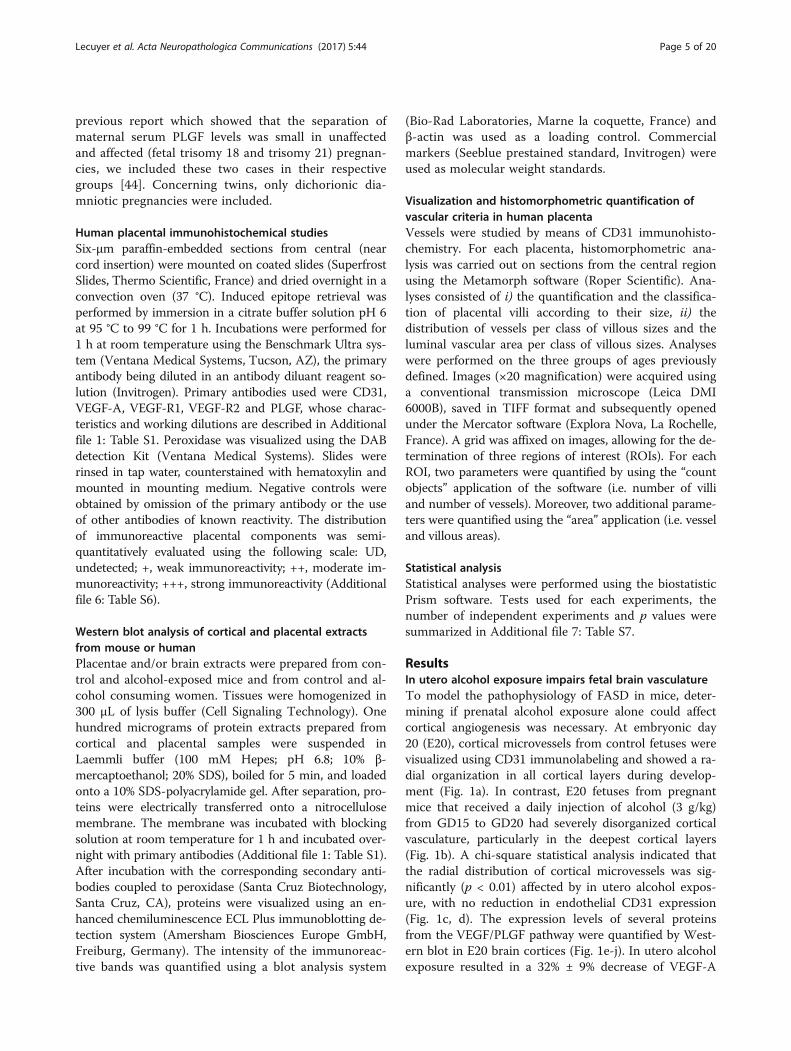
ResultsIn utero alcohol exposure impairs fetal brain vasculatureTo model the pathophysiology of FASD in mice, deter-mining if prenatal alcohol exposure alone could affectcortical angiogenesis was necessary. At embryonic day20 (E20), cortical microvessels from control fetuses werevisualized using CD31 immunolabeling and showed a ra-dial organization in all cortical layers during develop-ment (Fig. 1a). In contrast, E20 fetuses from pregnantmice that received a daily injection of alcohol (3 g/kg)from GD15 to GD20 had severely disorganized corticalvasculature, particularly in the deepest cortical layers(Fig. 1b). A chi-square statistical analysis indicated thatthe radial distribution of cortical microvessels was sig-nificantly (p < 0.01) affected by in utero alcohol expos-ure, with no reduction in endothelial CD31 expression(Fig. 1c, d). The expression levels of several proteinsfrom the VEGF/PLGF pathway were quantified by West-ern blot in E20 brain cortices (Fig. 1e-j). In utero alcoholexposure resulted in a 32% ± 9% decrease of VEGF-A
Lecuyer et al. Acta Neuropathologica Communications (2017) 5:44 Page 5 of 20
Fig. 1 (See legend on next page.)
Lecuyer et al. Acta Neuropathologica Communications (2017) 5:44 Page 6 of 20
(p < 0.05; Fig. 1e), whereas PLGF was undetectable byWestern blot (Fig. 1f ). With regard to VEGF-A andPLGF receptors, both soluble and membrane forms ofVEGF-R1 were decreased (p < 0.05; Fig. 1g-h), whereasVEGF-R2 had no significant variations (Fig. 1i). To valid-ate the Western blot conditions for PLGF detection, acontrol experiment compared PLGF protein levels in thefetal cortex at E20 and in the placenta at GD20(p < 0.001; Fig. 1j). These results indicate that alcoholexposure restricted to the fetal life alters cortical angio-genesis in the mouse brain.
Alcohol exposure impairs the placental integrity and theVEGF/PLGF systemAlthough alcohol has long been known to impair fetalgrowth [23], studies have only recently focused on meta-bolic dysfunctions of the placenta [30] and very few re-ports have targeted the VEGF/PLGF system [19]. Inutero alcohol exposure from GD15 to GD20 in mice re-sulted in abnormal lamination of the placenta with a sig-nificant increase of both number and length ofprotrusions of the junctional zone within the labyrinthzone (p < 0.05; p < 0.01; Additional file 8: Figure S1a, b,i and j). Reichert’s membrane thickness was significantlyreduced (p < 0.01; Additional file 8: Figure S1c, d and k),and the morphology of giant trophoblasts was altered(Additional file 8: Figure S1c, d). In the control group,giant trophoblasts possessed a typical rectangular shape(Additional file 8: Figure S1c, l; arrows). In contrast, inthe alcohol-exposed group, cell shape was markedlymodified and alcohol exposure induced a significant in-crease of the proportion of “round shape” giant tropho-blasts (p < 0.0001; Additional file 8: Figure S1d, l;arrows). Electronic microscopy revealed that giant tro-phoblasts were cohesive in the control group but not inthe alcohol-exposed group, in which tight junctions werenearly absent from the placentae (Additional file 8:Figure S1e-h). We also investigated the effect of in uteroalcohol exposure on the expression of the tight junctionprotein ZO-1, the monocarboxylate transporter MCT-1(Additional file 9: Figure S2a-d), and on placental angio-genic factors from the VEGF/PLGF family (Fig. 2a-f ). Inalcohol-exposed placentae, PLGF protein expression wasreduced (p < 0.05; Fig. 2b). Soluble and membrane forms
of VEGF-R1 as well as VEGF-R2 protein expressionwere also significantly reduced (p < 0.05; Fig. 2c-e)whereas CD31 expression was not modified (Fig. 2f ).VEGF-R1 and VEGF-R2 immunohistochemistry revealeda typical dot-like pattern (Fig. 2g, h and Additional file 9:Figure S2e, f ). PLGF levels in the microdissected labyrinthzone was reduced by −28.5% in alcohol-exposed placentae(p < 0.01; Fig. 2i and Additional file 9: Figure S2 g). Theseresults indicate that alcohol exposure during pregnancyimpairs placenta integrity at the ultrastructural level andthe expression of proteins involved in angiogenesis.
PLGF originating from placenta reaches the fetal brain,affects VEGF-R1 expression and impairs angiogenesisWhereas VEGF-R1 is expressed in the fetal brain (Fig. 1g,h), PLGF is massively expressed by the placenta (Fig. 1f, j)[3], suggesting that some alcohol-induced brain vascu-lar defects may result from placental angiogenic factors.To confirm this hypothesis, we performed transUV-illumination experiments after in utero placental injec-tions in mice (Fig. 3a-d). In time-course studies, Evansblue fluorescence was immediately detectable in theplacenta after in utero injection (Fig. 3b, e). Fluores-cence reached a maximum at 10 min and then progres-sively decreased (Fig. 3e). Evans blue fluorescence wasalso detected in the matched fetal brains 20 to 30 minafter placental injection (Fig. 3d, f ). Through the sameprotocol, human recombinant PLGF was injected intothe placenta of pregnant mice at GD15. A specifichPLGF ELISA detected recombinant hPLGF in the fetalbrain 30 min after the injection (p < 0.05; Fig. 3g). More-over, PLGF was detected by Western blot in the cephalicblood of E20 fetuses (Additional file 9: Figure S2 h).Altogether these data indicate that pro-angiogenic factorsreleased by the placenta can reach the fetal brain.Daily injection of pregnant mice with alcohol from
GD15 to GD20 resulted in decreased VEGF-R1 proteinlevels in the fetal brain (Fig. 1g, h). To determine ifPLGF is involved in this effect, a shRNA strategycoupled with in utero placenta transfection wasconducted (Fig. 3h-n). Electroporation of an eGFP-expressing vector revealed that the syncytiotrophoblastlayer cells expressed eGFP 48 h post transfection (Fig. 3h).Triple fluorescent labeling indicated that fetal
(See figure on previous page.)Fig. 1 Effects of in utero alcohol exposure on brain angiogenesis and expression of members of the VEGF/PLGF family from E20 embryos. a, b Effectsof fetal alcohol exposure from GD15 to GD20 on the organization of cortical microvessels in control and alcohol-exposed animals. Brain microvesselswere visualized by immunohistochemistry against CD31. Arrows indicate brain microvessels presenting a radial orientation in the control group. Note aloss of the radial organization in the alcohol-exposed group. I-VI: Cortical layers; CC: Corpus callosum. c Distribution of the orientation (angle classes) ofcortical microvessels in the immature cortex from GD20 fetuses. Statistical analysis was performed using the χ2 test. d Quantification by Western blot ofthe effects of fetal alcohol exposure during the last gestational week on the cortical expression of CD31 at GD20. e-i Quantification by Western blot ofVEGFA, PLGF, sVEGF-R1, mVEGF-R1 and VEGF-R2 protein levels in the cortex from control and alcohol-exposed groups. *p < 0.05 vs the control groupusing the unpaired t test. j Comparison by Western blot of the PLGF protein levels in the cortex and the placenta of E20 embryos from the controlgroup. ***p < 0.001 vs the control group using the unpaired t test
Lecuyer et al. Acta Neuropathologica Communications (2017) 5:44 Page 7 of 20
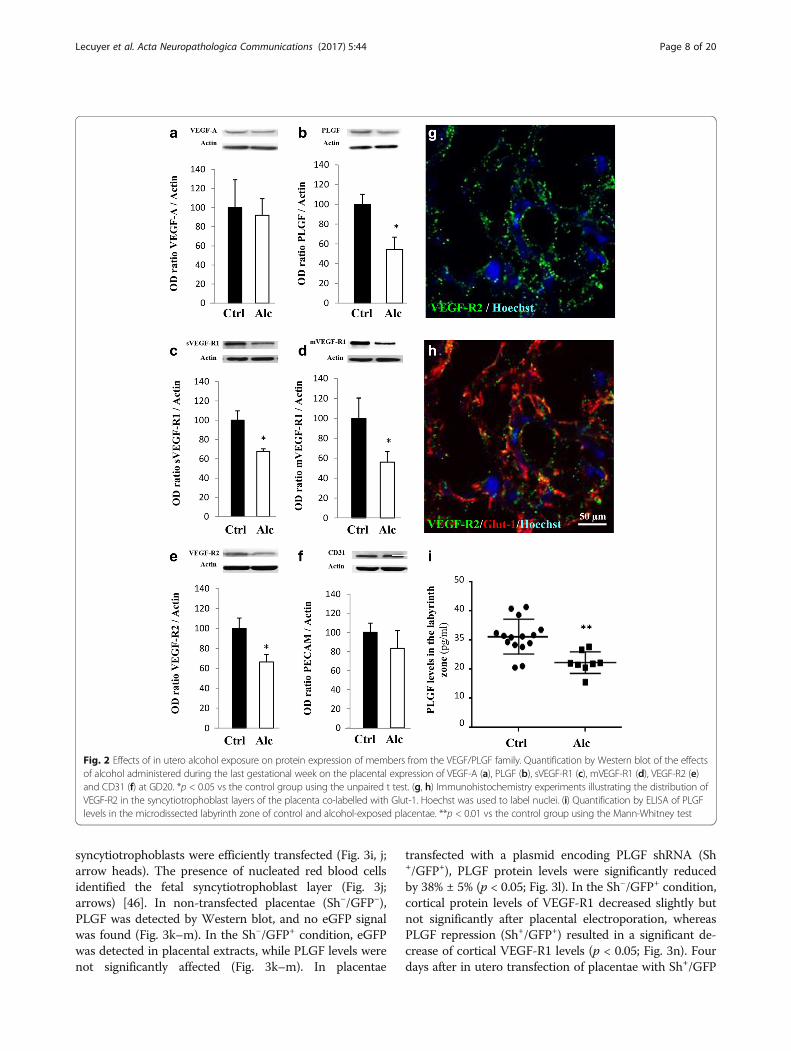
syncytiotrophoblasts were efficiently transfected (Fig. 3i, j;arrow heads). The presence of nucleated red blood cellsidentified the fetal syncytiotrophoblast layer (Fig. 3j;arrows) [46]. In non-transfected placentae (Sh−/GFP−),PLGF was detected by Western blot, and no eGFP signalwas found (Fig. 3k–m). In the Sh−/GFP+ condition, eGFPwas detected in placental extracts, while PLGF levels werenot significantly affected (Fig. 3k–m). In placentae
transfected with a plasmid encoding PLGF shRNA (Sh+/GFP+), PLGF protein levels were significantly reducedby 38% ± 5% (p < 0.05; Fig. 3l). In the Sh−/GFP+ condition,cortical protein levels of VEGF-R1 decreased slightly butnot significantly after placental electroporation, whereasPLGF repression (Sh+/GFP+) resulted in a significant de-crease of cortical VEGF-R1 levels (p < 0.05; Fig. 3n). Fourdays after in utero transfection of placentae with Sh+/GFP
Fig. 2 Effects of in utero alcohol exposure on protein expression of members from the VEGF/PLGF family. Quantification by Western blot of the effectsof alcohol administered during the last gestational week on the placental expression of VEGF-A (a), PLGF (b), sVEGF-R1 (c), mVEGF-R1 (d), VEGF-R2 (e)and CD31 (f) at GD20. *p < 0.05 vs the control group using the unpaired t test. (g, h) Immunohistochemistry experiments illustrating the distribution ofVEGF-R2 in the syncytiotrophoblast layers of the placenta co-labelled with Glut-1. Hoechst was used to label nuclei. (i) Quantification by ELISA of PLGFlevels in the microdissected labyrinth zone of control and alcohol-exposed placentae. **p < 0.01 vs the control group using the Mann-Whitney test
Lecuyer et al. Acta Neuropathologica Communications (2017) 5:44 Page 8 of 20
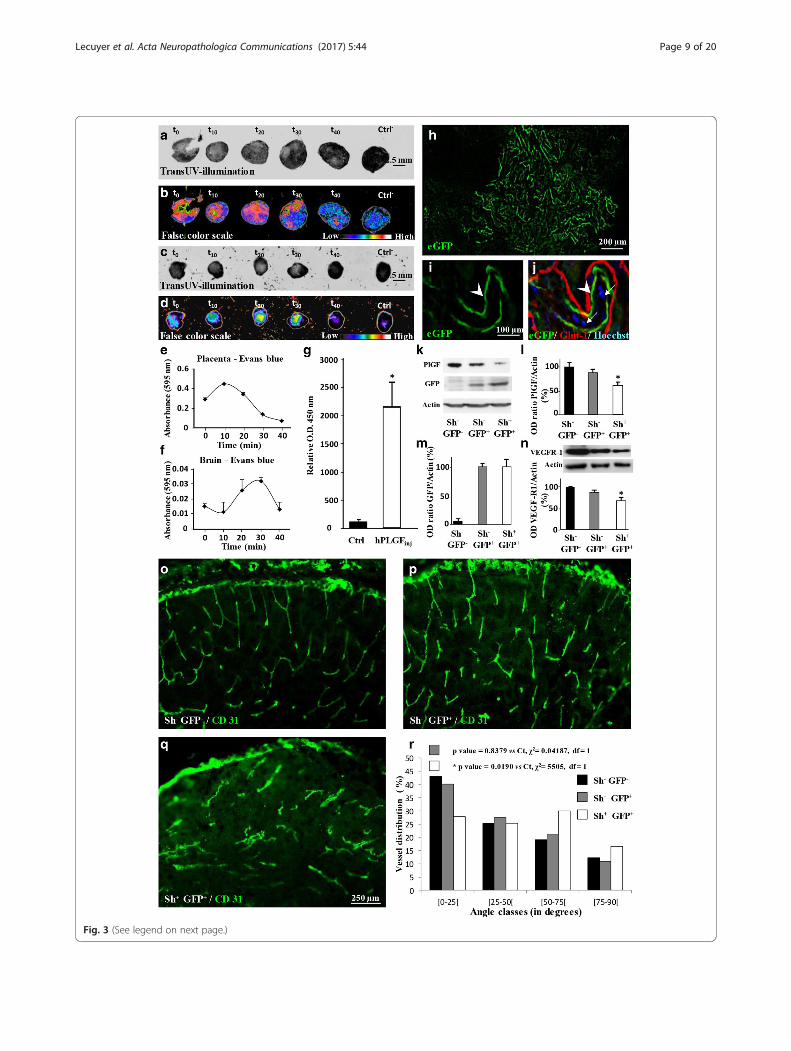
Fig. 3 (See legend on next page.)
Lecuyer et al. Acta Neuropathologica Communications (2017) 5:44 Page 9 of 20
+ plasmids (Fig. 3o-r), PLGF repression induced a markedimpairment of the vasculature in the fetal brain (Fig. 3q,r). No effect on the brain vasculature was found in the Sh−/GFP+ group (Fig. 3p). These data indicate that repres-sion of placental PLGF alters brain VEGF-R1 expressionand impairs cortical angiogenesis of the fetus, supportingthe idea of placental contribution to alcohol-induced brainvascular defects.
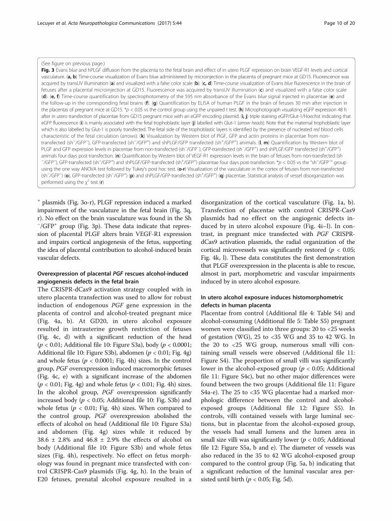
Overexpression of placental PGF rescues alcohol-inducedangiogenesis defects in the fetal brainThe CRISPR-dCas9 activation strategy coupled with inutero placenta transfection was used to allow for robustinduction of endogenous PGF gene expression in theplacenta of control and alcohol-treated pregnant mice(Fig. 4a, b). At GD20, in utero alcohol exposureresulted in intrauterine growth restriction of fetuses(Fig. 4c, d) with a significant reduction of the head(p < 0.01; Additional file 10: Figure S3a), body (p < 0.0001;Additional file 10: Figure S3b), abdomen (p < 0.01; Fig. 4g)and whole fetus (p < 0.0001; Fig. 4h) sizes. In the controlgroup, PGF overexpression induced macromorphic fetuses(Fig. 4c, e) with a significant increase of the abdomen(p < 0.01; Fig. 4g) and whole fetus (p < 0.01; Fig. 4h) sizes.In the alcohol group, PGF overexpression significantlyincreased body (p < 0.05; Additional file 10: Fig. S3b) andwhole fetus (p < 0.01; Fig. 4h) sizes. When compared tothe control group, PGF overexpression abolished theeffects of alcohol on head (Additional file 10: Figure S3a)and abdomen (Fig. 4g) sizes while it reduced by38.6 ± 2.8% and 46.8 ± 2.9% the effects of alcohol onbody (Additional file 10: Figure S3b) and whole fetussizes (Fig. 4h), respectively. No effect on fetus morph-ology was found in pregnant mice transfected with con-trol CRISPR-Cas9 plasmids (Fig. 4g, h). In the brain ofE20 fetuses, prenatal alcohol exposure resulted in a
disorganization of the cortical vasculature (Fig. 1a, b).Transfection of placentae with control CRISPR-Cas9plasmids had no effect on the angiogenic defects in-duced by in utero alcohol exposure (Fig. 4i–l). In con-trast, in pregnant mice transfected with PGF CRISPR-dCas9 activation plasmids, the radial organization of thecortical microvessels was significantly restored (p < 0.05;Fig. 4k, l). These data constitutes the first demonstrationthat PLGF overexpression in the placenta is able to rescue,almost in part, morphometric and vascular impairmentsinduced by in utero alcohol exposure.
In utero alcohol exposure induces histomorphometricdefects in human placentaPlacentae from control (Additional file 4: Table S4) andalcohol-consuming (Additional file 5: Table S5) pregnantwomen were classified into three groups: 20 to <25 weeksof gestation (WG), 25 to <35 WG and 35 to 42 WG. Inthe 20 to <25 WG group, numerous small villi con-taining small vessels were observed (Additional file 11:Figure S4). The proportion of small villi was significantlylower in the alcohol-exposed group (p < 0.05; Additionalfile 11: Figure S4c), but no other major differences werefound between the two groups (Additional file 11: FigureS4a-e). The 25 to <35 WG placentae had a marked mor-phologic difference between the control and alcohol-exposed groups (Additional file 12: Figure S5). Incontrols, villi contained vessels with large luminal sec-tions, but in placentae from the alcohol-exposed group,the vessels had small lumens and the lumen area insmall size villi was significantly lower (p < 0.05; Additionalfile 12: Figure S5a, b and e). The diameter of vessels wasalso reduced in the 35 to 42 WG alcohol-exposed groupcompared to the control group (Fig. 5a, b) indicating thata significant reduction of the luminal vascular area per-sisted until birth (p < 0.05; Fig. 5d).
(See figure on previous page.)Fig. 3 Evans blue and hPLGF diffusion from the placenta to the fetal brain and effect of in utero PLGF repression on brain VEGF-R1 levels and corticalvasculature. (a, b) Time-course visualization of Evans blue administered by microinjection in the placenta of pregnant mice at GD15. Fluorescence wasacquired by transUV illumination (a) and visualized with a false color scale (b). (c, d) Time-course visualization of Evans blue fluorescence in the brain offetuses after a placental microinjection at GD15. Fluorescence was acquired by transUV illumination (c) and visualized with a false color scale(d). (e, f) Time-course quantification by spectrophotometry of the 595 nm absorbance of the Evans blue signal injected in placentae (e) andthe follow-up in the corresponding fetal brains (f). (g) Quantification by ELISA of human PLGF in the brain of fetuses 30 min after injection inthe placentas of pregnant mice at GD15. *p < 0.05 vs the control group using the unpaired t test. (h) Microphotograph visualizing eGFP expression 48 hafter in utero transfection of placentae from GD15 pregnant mice with an eGFP encoding plasmid. (i, j) triple staining eGFP/Glut-1/Hoechst indicating thateGFP fluorescence (i) is mainly associated with the fetal trophoblastic layer (j) labelled with Glut-1 (arrow heads). Note that the maternal trophoblastic layerwhich is also labelled by Glut-1 is poorly transfected. The fetal side of the trophoblastic layers is identified by the presence of nucleated red blood cellscharacteristic of the fetal circulation (arrows). (k) Visualization by Western blot of PlGF, GFP and actin proteins in placentae from non-transfected (sh−/GFP−), GFP-transfected (sh−/GFP+) and shPLGF/GFP transfected (sh+/GFP+) animals. (l, m) Quantification by Western blot ofPLGF and GFP expression levels in placentae from non-transfected (sh−/GFP−), GFP-transfected (sh−/GFP+) and shPLGF/GFP transfected (sh+/GFP+)animals four days post-transfection. (n) Quantification by Western blot of VEGF-R1 expression levels in the brain of fetuses from non-transfected (sh−/GFP−), GFP-transfected (sh−/GFP+) and shPLGF/GFP-transfected (sh+/GFP+) placentae four days post-transfection. *p < 0.05 vs the “sh−/GFP−” groupusing the one way ANOVA test followed by Tukey’s post hoc test. (o-r) Visualization of the vasculature in the cortex of fetuses from non-transfected(sh−/GFP−) (o), GFP-transfected (sh−/GFP+) (p) and shPLGF/GFP-transfected (sh+/GFP+) (q) placentae. Statistical analysis of vessel disorganization wasperformed using the χ2 test (r)
Lecuyer et al. Acta Neuropathologica Communications (2017) 5:44 Page 10 of 20
Fig. 4 (See legend on next page.)
Lecuyer et al. Acta Neuropathologica Communications (2017) 5:44 Page 11 of 20
We also performed a time-course study of villous andvessel densities (Fig. 5e, f ). In the control group, villousdensity was similar between 20 to 35 WG (Fig. 5e) thenincreased from 35 to 42 WG (p < 0.0001; Fig. 5e). In thealcohol-exposed group, villous density was similar tothe control group from 20 to 35 WG (Fig. 5e), but themassive increase of the villous density observed in thecontrol group during the last 2 month of gestation didnot occur in the alcohol-exposed group (p < 0.0001;Fig. 5e). Moreover, in the control group, the vesselintravillous area regularly increased during the gesta-tion (p < 0.05; Fig. 5f ) in contrast to the alcohol-exposed group, in which vessel area tended to decrease(p < 0.05; Fig. 5f ). These data constitute the first dem-onstration that alcohol has deleterious effects on hu-man placental vasculature.
In utero alcohol exposure impairs the VEGF/PLGF systemin human placentaSince histomorphometric data showed major differencesfrom 35 to 42 WG (Fig. 5a–f ), further analyses were per-formed at these stages by Western blot. A significant de-crease in the expression of ZO-1 was found in placentaefrom women who consumed alcohol (p < 0.05; Fig. 5g).Levels of MCT-1 tended to increase, but statistical ana-lysis showed no significance (Fig. 5h). PLGF and VEGF-A levels were also quantified, and significant decreaseswere found regarding PLGF in the alcohol-exposedgroup (p < 0.05; Fig. 5i, j). Quantification of VEGF-R1and VEGF-R2 indicated no differences between controland alcohol-exposed groups (Fig. 5k, l). We then per-formed immunohistochemical studies using VEGF,PLGF, VEGF-R1 and VEGF-R2 antibodies in both con-trol and alcohol-exposed human placentae (Additionalfile 6: Table S6 and Additional file 13: Figure S6). Thetwo groups had some differences in PLGF immunoreac-tivity, which was lower in the alcohol-exposed groupthan in the control group in extravillous and intravilloustrophoblasts, as well as in decidual and intravillous ves-sel endothelial cells. In particular, after 34 WG, the
alcohol-exposed group showed very low immunoreac-tivity, whereas it was apparent in villous vessel lumensof controls (Additional file 6: Table S6 and Additionalfile 13: Figure S6). VEGF-R1 and R2 immunoreactivitydid not differ among groups and cell types except forvillous trophoblasts and endothelial cells in whichVEGF-R1 and R2 immunoreactivity was low from 35WG in the alcohol group (Additional file 6: Table S6).These data indicate that in utero alcohol exposure in-duced major differences in the expression profile ofproteins involved in angiogenesis in human placentaduring the third trimester of pregnancy.
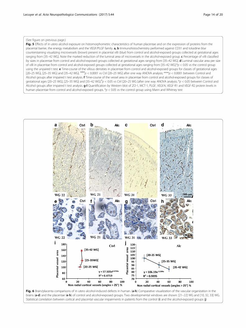
Placental impairments correlate with brain vasculardefects in humanSince preclinical data showed that prenatal alcohol ex-posure induced disorganized orientation of corticalmicrovessels (Fig. 1), impaired expression of placentalangiogenic factors (Fig. 2), that targeted repression ofPLGF in the placenta mimicked the effects of in uteroalcohol exposure on both VEGF-R1 expression and ves-sel organization in the fetal brain (Fig. 3) and that pla-cental over-expression of PLGF rescued alcohol-inducedvascular defects (Fig. 4), we researched in human an as-sociation between vascular defects found in the placen-tae and in the brain after in utero alcohol exposure.Immunohistological studies showed that the corticalorientation of brain microvessels was similar betweencontrol and alcohol-exposed groups from 20 to 25 WG(Fig. 6a, b), with most vessels being radially oriented. Inaddition, placental vasculature did not differ (Fig. 6e, f ).In contrast, from 35 to 42 WG, cortical microvesselorganization was markedly impaired in the alcohol-exposed group (Fig. 6c, d) along with vessel luminal areain the placentae (Fig. 6g, h). In the control group, nocorrelation existed between cortical vessel organizationand placental vessel area; the radial organization of cor-tical microvessels remained unchanged between gesta-tional groups, while placental vessel area strongly
(See figure on previous page.)Fig. 4 Effects of in utero PGF overexpression on fetal growth and cortical vasculature during prenatal alcohol exposure. (a, b) PGF CRISPR-dCas 9 activationapproach coupled with in utero electroporation of the placenta was done at GD13 (a) and overexpression of PLGF controlled at GD20 (b). In the alcoholgroup, in utero exposure occurred from GD15 to GD20. (c, d) Visualization of E20 fetuses from pregnant mice exposed to NaCl (c) or alcohol (d). Note thesmall size of alcohol-exposed fetuses. Green bars indicate morphometric measures that have been done (head size (a); body size (b); abdomen size (c) andwhole fetus size (a + b). (e, f) Visualization of E20 fetuses after in utero electroporation of PGF CRISPR-dCas9 plasmids in placentae from control (e) oralcohol-exposed pregnant mice (f). (g, h) Quantification of abdomen (g) and whole fetus (h) sizes in control (NaCl) and alcohol groups. In a same uterinehorn some placentae were not electroporated (black bars), electroporated with control CRISPR-Cas9 plasmids (grey bars) or electroporated with PGFCRISPR-dCas9 plasmids (white bars). ##p < 0.01; ###p < 0.001; ####p < 0.0001 vs the control group and *p < 0.05; **p < 0.01; ****p < 0.0001 as indicatedusing the two way ANOVA test followed by Tukey’s post hoc test. (i-k) Visualization of the vasculature in the cortex of E20 fetuses from control (NaCl)/non-transfected (i), alcohol/control CRISPR-Cas9 transfected (j) and alcohol/ PGF CRISPR-dCas9 transfected (k) placentae. (l) Quantification of thepercentage of radial vessels in the cortex of E20 fetuses from not electroporated (black bars), electroporated with control CRISPR-Cas9 plasmids (grey bars)and electroporated with PGF CRISPR-dCas9 plasmids (white bars) placentae. #p < 0.05 vs the control group and *p < 0.05 as indicated using the two wayANOVA test followed by Tukey’s post hoc test
Lecuyer et al. Acta Neuropathologica Communications (2017) 5:44 Page 12 of 20
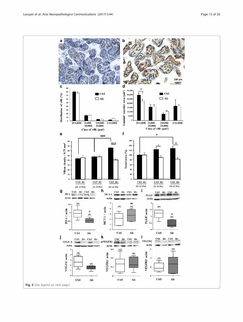
Fig. 5 (See legend on next page.)
Lecuyer et al. Acta Neuropathologica Communications (2017) 5:44 Page 13 of 20
(See figure on previous page.)Fig. 5 Effects of in utero alcohol exposure on histomorphometric characteristics of human placentae and on the expression of proteins from theplacental barrier, the energy metabolism and the VEGF/PLGF family. a, b Immunohistochemistry performed against CD31 and toluidine bluecounterstaining visualizing microvessels (brown) present in placental villi (blue) from control and alcohol-exposed groups collected at gestational agesranging from [35–42 WG]. Note the marked reduction of the luminal area of microvessels in the alcohol-exposed group. c Percentage of villi classifiedby sizes in placentae from control and alcohol-exposed groups collected at gestational ages ranging from [35–42 WG]. d Luminal vascular area per sizeof villi in placentae from control and alcohol-exposed groups collected at gestational ages ranging from [35–42 WG].*p < 0.05 vs the control groupusing the unpaired t test. e Time-course of the villous densities in placentae from control and alcohol-exposed groups for classes of gestational ages[20–25 WG], [25–35 WG] and [35–42 WG]. ####p < 0.0001 vs Ctrl [20–25 WG] after one way ANOVA analysis; ****p < 0.0001 between Control andAlcohol groups after impaired t test analysis. f Time-course of the vessel area in placentae from control and alcohol-exposed groups for classes ofgestational ages [20–25 WG], [25–35 WG] and [35–42 WG].#p < 0.05 vs Ctrl [20–25 WG [after one way ANOVA analysis; *p < 0.05 between Control andAlcohol groups after impaired t test analysis. g-l Quantification by Western blot of ZO-1, MCT-1, PLGF, VEGFA, VEGF-R1 and VEGF-R2 protein levels inhuman placentae from control and alcohol-exposed groups. *p < 0.05 vs the control group using Mann and Whitney test
Fig. 6 Brain/placenta comparisons of in utero alcohol-induced defects in human. (a-h) Comparative visualization of the vascular organization in thebrains (a-d) and the placentae (e-h) of control and alcohol-exposed groups. Two developmental windows are shown: [21–22] WG and [10, 32, 33] WG.Statistical correlation between cortical and placental vascular impairments in patients from the control (i) and the alcohol-exposed groups (j)
Lecuyer et al. Acta Neuropathologica Communications (2017) 5:44 Page 14 of 20
increased during gestation (Fig. 6i). In contrast, in thealcohol-exposed group, the disorganized orientation ofthe cortical microvessels massively increased with preg-nancy duration (Fig. 6j) and was markedly correlatedwith the lack of increase of placental vessel area (Fig. 6j).
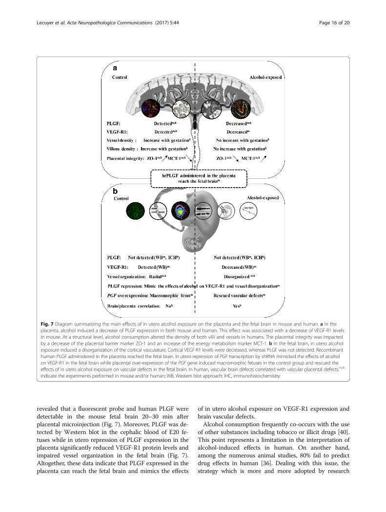
DiscussionUsing preclinical and clinical approaches, we investigatedthe effects of prenatal alcohol exposure on both brain andplacental vasculatures. We demonstrated that the angio-genesis and the expression of VEGF/PLGF proteins are al-tered in both placentae and fetal brains. We also showedthat PLGF can reach the fetal brain and that targeted inutero repression of PLGF in the mouse placenta mimicsthe effect of prenatal alcohol exposure on both VEGF-R1expression and vasculature impairments in the fetal brain.In addition, PLGF overexpression by PGF CRISPR-dCas9activation rescues brain vascular defects induced by inutero alcohol exposure. Our results in mice are similar tothose observed in humans, with placental and brain vascu-lar defects being strongly correlated in alcohol-exposedhuman fetuses. Since decreased PLGF levels in the pla-centa after in utero alcohol exposure are associated tobrain angiogenesis defects, the levels may serve as a pre-dictive marker for subsequent neurodevelopmental out-comes of exposed fetuses. Compared with the knownexposition markers of maternal alcohol intake, this newgeneration of “effect” biomarkers could facilitate earlydiagnosis of FASD.Data on the effect of alcohol on the fetal brain vascula-
ture during pregnancy are scarce [22], but several adultstudies indicate that alcohol interacts with angiogenesis[39, 50]. We showed that a transient exposure of thefetus to alcohol during a developmental window inwhich cranio-facial dysmorphism is not induced [29] caninterfere with brain angiogenesis. The critical role ofangiogenesis in neurodevelopment is evident [5, 51]. Notonly are the guidance molecules used by neurons andendothelial cells to migrate to their final destination simi-lar [5], but the migrating cells closely interact [28, 48].Regarding in utero alcohol exposure, our data revealed amarked decrease in VEGF-R1 levels in cortical extractsfrom E20 fetuses (Fig. 7). In addition, PLGF, which bindsexclusively to VEGF-R1, is poorly expressed in the fetalbrain but is massively synthesized by the placenta [3].At a mechanistic level, the way in which placental PLGF
could act to regulate fetal brain angiogenesis have to befully investigated. However, several data from the litera-ture would plaid in favor of a direct effect. Indeed, consist-ent with the present data, it has been recently showed thatPLGF is detected in the fetal blood of human neonates[35]. In addition, VEGF-R1 is expressed by tip cells andrelative levels of VEGF-R1 and VEGF-R2 contribute in tipcell position [21]. In particular, it has been shown that
PLGF may regulate angiogenesis by competing withVEGFA for VEGF-R1 [4]. Alternatively, PLGF might alsocontribute to the control of angiogenesis by modulatingintracellular signals through VEGF-R1 [1].At a neurodevelopmental level, multiple aspects of
central nervous system development can be affected byalcohol exposure, but it has been clearly described ab-normalities of neuronal migration as well as corpus cal-losum defects [41]. Recently, it has been demonstratedthat some nervous cell types such as oligodendrocytes[48] and GABA interneurons [51] require a vascular-dependent interaction to migrate. Taken together, thesedata suggest that abnormal brain vascular developmentresulting from PGF dysfunction may contribute in somealterations described in FASD children such as impairedcell migration.Maternal ethanol consumption from the beginning of
pregnancy is well known to impair placental develop-ment. However, while previous transcriptomic studiesshowed that alcohol altered the expression of angiogenicfactors in the placenta of pregnant rats, [43] its impacton the placental vasculature and the VEGF/PLGF systemwas little investigated in animal models [19] and not de-scribed in humans. The placenta appears to be a forgot-ten organ, although it is surely a promising source ofbiomarkers [34]. In our series, the analysis of at least 40placentae per group indicated that, in the control group,a strong increase of vessel density occurred during thelast trimester of gestation, consistent with the massiveincrease of energetic needs of the developing fetal brain(Fig. 7) [9]. In contrast, in the alcohol-exposed group,this increase did not occur and the total lumen area ofvessels was markedly reduced. Western blot experimentsshowed that alcohol induced a marked decrease of PLGFlevels in mouse and human placentae. In human, PLGFimmunolabeling and protein levels were reduced frommidgestation compared with control placentae in whichPLGF was present at term, suggesting a role of PLGF inplacenta development and maintenance until term. Con-sistent with this hypothesis, adult knock-out mice invali-dated for PLGF have been shown to have a markedreduction of the vessel lumen diameter [4]. Altogether,these data demonstrate for the first time that during hu-man pregnancy alcohol impairs the protein expressionof angiogenic markers in the placenta and these molecu-lar defects are associated with marked histomorpho-metric abnormalities affecting the placental vasculature.During pregnancy the umbilical cord blood contains
PLGF [6, 35]. Our data show that in utero alcohol ex-posure impairs both the expression of brain VEGF-R1and the radial organization of cortical microvessels, lead-ing to speculation that PLGF can reach the fetal brainand contributes to alcohol-induced angiogenic impair-ments. Both trans-UV illumination and ELISA approaches
Lecuyer et al. Acta Neuropathologica Communications (2017) 5:44 Page 15 of 20
revealed that a fluorescent probe and human PLGF weredetectable in the mouse fetal brain 20–30 min afterplacental microinjection (Fig. 7). Moreover, PLGF was de-tected by Western blot in the cephalic blood of E20 fe-tuses while in utero repression of PLGF expression in theplacenta significantly reduced VEGF-R1 protein levels andimpaired vessel organization in the fetal brain (Fig. 7).Altogether, these data indicate that PLGF expressed in theplacenta can reach the fetal brain and mimics the effects
of in utero alcohol exposure on VEGF-R1 expression andbrain vascular defects.Alcohol consumption frequently co-occurs with the use
of other substances including tobacco or illicit drugs [40].This point represents a limitation in the interpretation ofalcohol-induced effects in human. On another hand,among the numerous animal studies, 80% fail to predictdrug effects in human [36]. Dealing with this issue, thestrategy which is more and more adopted by research
Fig. 7 Diagram summarizing the main effects of in utero alcohol exposure on the placenta and the fetal brain in mouse and human. a In theplacenta, alcohol induced a decrease of PLGF expression in both mouse and human. This effect was associated with a decrease of VEGF-R1 levelsin mouse. At a structural level, alcohol consumption altered the density of both villi and vessels in humans. The placental integrity was impactedby a decrease of the placental barrier marker ZO-1 and an increase of the energy metabolism marker MCT-1. b In the fetal brain, in utero alcoholexposure induced a disorganization of the cortical vasculature. Cortical VEGF-R1 levels were decreased, whereas PLGF was not detected. Recombinanthuman PLGF administered in the placenta reached the fetal brain. In utero repression of PGF transcription by shRNA mimicked the effects of alcoholon VEGF-R1 in the fetal brain while placental over-expression of the PGF gene induced macromorphic fetuses in the control group and rescued theeffects of in utero alcohol exposure on vascular defects in the fetal brain. In human, vascular brain defects correlated with vascular placental defects.m,h
indicate the experiments performed in mouse and/or human; WB, Western blot approach; IHC, immunohistochemistry
Lecuyer et al. Acta Neuropathologica Communications (2017) 5:44 Page 16 of 20

groups is translational medicine [13]. In the present study,most data found in human have been confirmed in ouranimal model of mono-intoxication with alcohol (Fig. 7)supporting a robust link between alcohol, placental PLGFand brain vascular defects. In addition, overexpressionPLGF experiments revealed effects on somatic growth ofthe fetus opening new research avenues regarding PLGFand in utero growth retardation (IURG) [24].Most infants with FASD are not diagnosed at birth. A
recent cohort study revealed that 86.5% of children oradolescents (4 to 18 years old) with FASD had neverbeen previously diagnosed or had been misdiagnosed[7]. This high rate of missed diagnosis significantlyaffects therapeutic care, the social integration of the in-fants and economic costs [38]. In addition, as demon-strated for other pathologies such as autism spectrumdisorders, the earlier medical care is started, the better theoutcomes probably because of the high plasticity of thenervous system in the first years following birth [27]. Themajor limitation of the existing biomarkers of alcohol con-sumption during pregnancy can be summed up in onequestion: “Was the fetus exposed to alcohol?” [20]. Deter-mining prenatal alcohol exposure is crucial to identify thechildren/population at risk, but it is not realistic to assessall infants with prenatal alcohol exposure. First, a “safe”dose of alcohol is controversial and highly debated [16,33]; second, patterns of alcohol consumption differ(chronic/acute) and their effect on the fetus is not thesame [10]; and third, the developing brain has windowsof vulnerability during which potential harm is greater[25, 49]. These limits also contribute to the discrepan-cies between different cohort studies on the impact ofalcohol consumption on the infant [26, 31, 45]. Thus, theidentification of biomarkers of alcohol-induced brain ef-fects after fetal exposure is required. The present study re-vealed a strong correlation between placental and brainvascular defects in the context of prenatal alcohol expos-ure. The PLGF levels (<40%) in placentae from womenwho consumed alcohol during pregnancy appeared tohave a predictive value for vascular brain defects. Inaddition, the demonstration that PGF CRISPR-dCas9 acti-vation is able to restore a correct cortical angiogenesisopens new avenues of research regarding a possible pre-vention of alcohol-induced behavioral troubles. Indeed, asobserved in human, several preclinical studies reportedneonatal behavioral troubles and long-term deficits in ani-mals exposed in utero to alcohol such as increased motoractivity [22, 42]. PLGF assay could help identify infantswith brain damage associated with in utero alcohol expos-ure, thus contributing to an early diagnosis of FASD andprompt intervention. In addition, the present study high-lights the necessity to plan a clinical protocol consisting infollowing both placental PLGF levels at birth and longterm behavioral troubles in infants exposed in utero to
alcohol. This work was patented (FR1555727 / PCT/EP2016/064480) and (FR1661813).
ConclusionThe present study provides the first mechanistic andclinical evidence that decreased PLGF levels in the pla-centa after in utero alcohol exposure are associated tobrain angiogenesis defects. Measurement of PLGF levelsat birth in the placenta or the fetal blood may serve as apredictive marker for subsequent neurodevelopmentaloutcomes of exposed fetuses. Compared with the knownexposition markers of maternal alcohol intake, this newgeneration of “effect” biomarkers could facilitate earlydiagnosis of FASD.
Additional files
Additional file 1: Table S1. Origin and characteristics of the primaryantibodies used for the immunohistochemical and Western blot studiesperformed in mouse and human tissues. (DOCX 26 kb)
Additional file 2: Table S2. Main clinical and morphological characteristicsof human control group for brain studies. (DOCX 17 kb)
Additional file 3: Table S3. Main clinical and morphological characteristicsof the alcohol-exposed group of patients for brain studies. (DOCX 21 kb)
Additional file 4: Table S4. Main clinical and morphological characteristicsof human placentae from the control group. (DOC 89 kb)
Additional file 5: Table S5. Main clinical and morphological characteristicsof human placentae from the alcohol-exposed group. (DOC 131 kb)
Additional file 6: Table S6. Immunohistochemical characteristics ofmembers of the VEGF-PLGF family in human placentae from the “Control”and “Alcohol” groups. (DOCX 25 kb)
Additional file 7: Table S7. Statistical analysis. (DOCX 23 kb)
Additional file 8: Figure S1. Effects of in utero alcohol exposure onmorphometric and ultrastructural characteristics of the placenta from GD20mice. (a) Visualization by Cresyl violet staining of the effect of alcoholexposure on the laminar structuration of the placenta. The placenta isoriented with its maternal side at the top. Note that alcohol inducedprotrusions of the junctional zone within the labyrinth zone (dotted lines).(b) Visualization of a typical 3D reconstruction of placental protrusions usedfor morphometric analysis. (c-d) Visualization at low magnification of thegiant trophoblast layer from control (c) and alcohol-exposed (d) groups.Arrows indicate giant trophoblasts. Note the typical rectangular shape ofthis cell type in placentae from the control group whereas in the alcohol-ex-posed group the trophoblasts present a round shape. (e-h) Imagesacquired by electron microscopy at moderate (e and f) and high (g andh) magnifications visualizing the morphology of giant trophoblasts andthe presence of zonula occludens (arrows) from control (e and g) andalcohol-exposed (f and h) groups. Note a loss of zonula occludens (stars) inalcohol-treated animals. Inserts present in e and f indicate the area visualizedat high magnification in g and h, respectively. d: maternal decidua;j: junctional zone; l: labyrinth zone; tg: trophoblast giant layer. (i-l)Quantification by morphometric analysis of the effect of alcohol on thenumber of placental protrusions (i), the length of protrusions (j), thethickness of the Reichert’s membrane (k) and the proportion of round-shape giant trophoblasts in control and alcohol-exposed placentae (l).*p < 0.05; **p < 0.01; ****p < 0.0001 vs the control group using the unpairedt test. (TIFF 15056 kb)
Additional file 9: Figure S2. Effects of in utero alcohol exposure on ZO-1 and MCT-1 expression and visualization of VEGF-R1 in the mouse placenta.(a, b) Visualization by immunohistochemistry of the ZO-1 protein in thelabyrinth zone of the mouse placenta from the control (a) and thealcohol-exposed (b) groups. Note that ZO-1 immunolabeling is dotted
Lecuyer et al. Acta Neuropathologica Communications (2017) 5:44 Page 17 of 20
and clustered (arrows) in the control group whereas it is diffuse in thealcohol-exposed group. Immunoreactivity against the glucose transporterGlut-1 was done to visualize the trophoblast layers. Hoechst was used tolabel nuclei. (c) Double immunolabeling experiment performed withthe monocarboxylate and the glucose transporters MCT-1 and Glut-1,respectively in the labyrinth zone of a control placenta. Note that, contrastingwith Glut-1, MCT-1 expression is associated with one syncytiotrophoblast layer(maternal side). Hoechst was used to label nuclei. (d) Quantification byWestern blot of the expression levels of the proteins ZO-1 and MCT-1in the placentae from control and alcohol-exposed groups. Westernblot experiments showed that placentae from alcohol-exposed animalshad significantly decreased ZO-1 levels while MCT-1 protein levels weresignificantly increased. *p < 0.05, **p < 0.01 vs the control group usingthe unpaired t test. (e, f) Immunohistochemistry experiments illustratingthe distribution of VEGF-R1 (e) and Glut-1 (f) in the syncytiotrophoblastlayers of the mouse placenta. Hoechst was used to visualize nuclei.(g) Immunohistochemistry experiments visualizing Glut-1 and PLGFimmunoreactivity in the syncytiotrophoblast layers of the mouseplacenta. Hoechst was used to visualize nuclei. (h) Visualization byWestern blot of PLGF in 100 μg protein extracts from GD20 placenta andE20 brain and in 4 μl plasma from E20 cephalic blood. (TIFF 23009 kb)
Additional file 10: Figure S3. Effects of placental in utero PGFoverexpression on head and body sizes of E20 fetuses in control andalcohol groups. (a, b) Quantification of head (a) and body (b) sizes in control(NaCl) et alcohol groups. In a same uterine horn some placentaewere not electroporated (black bars), electroporated with controlCRISPR-Cas9 plasmids (grey bars) or electroporated with PGF CRISPR-dCas9plasmids (white bars). ##p < 0.01; ###p < 0.001; ####p < 0.0001 vs thecontrol group and *p < 0.05; **p < 0.01 as indicated using the two wayANOVA test followed by Tukey’s post hoc test. (TIFF 7601 kb)
Additional file 11: Figure S4. Histomorphometric characterization ofthe effects of in utero alcohol exposure on human placentae from WG20to WG25. (a, b) Immunohistochemistry performed against CD31 andtoluidine blue counterstaining visualizing microvessels (brown) present inplacental villi (blue) from control and alcohol-exposed groups collected atgestational ages ranging from [20–25 WG]. (c) Percentage of villi classifiedby sizes in placentae from control and alcohol-exposed groups collected atgestational ages ranging from [20–25 WG].*p < 0.05 vs the controlgroup using the unpaired t test (d) Repartition of vessels per size of villi inplacentae from control and alcohol-exposed groups collected at gestationalages ranging from [20–25 WG]. (e) Luminal vascular area per size of villi inplacentae from control and alcohol-exposed groups collected at gestationalages ranging from [20–25 WG]. (TIFF 19794 kb)
Additional file 12: Figure S5. Histomorphometric characterization ofthe effects of in utero alcohol exposure on human placentae from WG25to WG35. (a, b) Immunohistochemistry performed against CD31 andtoluidine blue counterstaining visualizing microvessels (brown) present inplacental villi (blue) from control and alcohol-exposed groups collected atgestational ages ranging from [25–35 WG]. (c) Percentage of villi classified bysizes in placentae from control and alcohol-exposed groups collected atgestational ages ranging from [25–35 WG]. *p < 0.05 vs the control groupusing the unpaired t test. (d) Repartition of vessels per size of villi in placentaefrom control and alcohol-exposed groups collected at gestational agesranging from [25–35 WG]. *p < 0.05 vs the control group using the unpaired ttest. (e) Luminal vascular area per size of villi in placentae from control andalcohol-exposed groups collected at gestational ages ranging from [25–35WG]. *p < 0.05 vs the control group using the unpaired t test. (TIFF 19271 kb)
Additional file 13: Figure S6. PlGF immunoreactivity of the mainplacental compartments in control and prenatally alcohol-exposed neonatesfor gestational ages [35–42 WG]. (a, b) Strongly immunoreactive decidualcells in a control placental maternal floor (arrow), conversely to those of aprenatally alcohol-exposed neonate, where decidual cells exhibit a weakimmunoreactivity (arrow). (c, d) Circulating PlGF in the villous capillaries(arrow) in a normal placenta at term, contrasting with absent intra-luminalPlGF immunoreactivity in the villous vessels of a prenatally ethanol-exposedneonate (arrow). (e, f) Strong PlGF immunoreactivity of the villoussyncytiotrophoblasts in a control placenta at term (arrow), contrastingwith weak and irregular PlGF immunoreactivity in the villous trophoblasts ina prenatally ethanol-exposed neonate (arrow). (TIFF 25948 kb)
AcknowledgementsThis work was supported by the Normandy University, INSERM, the ConseilRégional de Normandie, Normandie Valorisation, the Fédération de Rechercheen Alcoologie (FRA), the LARC-Neuroscience network and the Regional Platformfor Cell Imaging (PRIMACEN). M.L. is a recipient of a fellowship from the RégionHaute-Normandie.
Authors’ contributionsML, CL YR and AU performed experiments; AL, PM and SM contributed tothe acquisition and analysis of data; PM, SB, SJ, AL, SM and BJG designed theexperiments and conceived the project; SM and BJG directed the work. Allauthors read and approved the final manuscript.
Competing interestsThe authors declare that they have no competing of interests.
Ethics approval and consent to publishPreclinical experiments were done according to the recommendations of theFrench Ethical Committee and the European Directive EC/86/609 (CouncilDirective 86/609/EEC, license no. 21CAE035), and were carried out under thesupervision of authorized investigators (B.J.G., authorization n° 7687).For experiments performed in post-mortem tissues and placentae, tissueswere collected in each case with the informed consent of the parents.Medical termination of the pregnancies had been accepted by the localethical committee of the Prenatal Diagnosis Multidisciplinary Centeraccording to the French law.
Publisher’s NoteSpringer Nature remains neutral with regard to jurisdictional claims inpublished maps and institutional affiliations.
Author details1UNIROUEN, Inserm U1245 and Rouen University Hospital, Normandy Centrefor Genomic and Personalized Medicine, Normandie University, Rouen,France. 2Department of Neonatal Paediatrics and Intensive Care, RouenUniversity Hospital, Rouen, France. 3Department of Pathology, RouenUniversity Hospital, Rouen, France. 4Department of Pathology, BrestUniversity Hospital, Rouen, France. 5Department of Molecular Biochemistry,Rouen University Hospital, Rouen, France.
Received: 25 March 2017 Accepted: 20 May 2017
References1. Autiero M, Waltenberger J, Communi D, Kranz A, Moons L, Lambrechts D,
Kroll J, Plaisance S, De Mol M, Bono F et al (2003) Role of PlGF in the intra-and intermolecular cross talk between the VEGF receptors Flt1 and Flk1. NatMed 9:936–943. doi:10.1038/nm884
2. Bakoyiannis I, Gkioka E, Pergialiotis V, Mastroleon I, Prodromidou A, VlachosGD, Perrea D (2014) Fetal alcohol spectrum disorders and cognitive functionsof young children. Rev Neurosci 25:631–639. doi:10.1515/revneuro-2014-0029
3. Cao Y, Ji WR, Qi P, Rosin A, Cao Y (1997) Placenta growth factor: identificationand characterization of a novel isoform generated by RNA alternative splicing.Biochem Biophys Res Commun 235:493–498. doi:10.1006/bbrc.1997.6813
4. Carmeliet P, Moons L, Luttun A, Vincenti V, Compernolle V, De Mol M, Wu Y,Bono F, Devy L, Beck H et al (2001) Synergism between vascular endothelialgrowth factor and placental growth factor contributes to angiogenesis andplasma extravasation in pathological conditions. Nat Med 7:575–583
5. Carmeliet P, Tessier-Lavigne M (2005) Common mechanisms of nerve andblood vessel wiring. Nature 436:193–200. doi:10.1038/nature03875
6. Catarino C, Rebelo I, Belo L, Rocha S, Castro EB, Patrício B, Quintanilha A,Santos-Silva A (2009) Fetal and maternal angiogenic/anti-angiogenic factors innormal and preeclamptic pregnancy. Growth Factors Chur Switz 27:345–351.doi:10.3109/08977190903184670
7. Chasnoff IJ, Wells AM, King L (2015) Misdiagnosis and missed diagnoses infoster and adopted children with prenatal alcohol exposure. Pediatrics 135:264–270. doi:10.1542/peds.2014-2171
8. Conti E, Zezza L, Ralli E, Caserta D, Musumeci MB, Moscarini M, Autore C,Volpe M (2013) Growth factors in preeclampsia: a vascular disease model. Afailed vasodilation and angiogenic challenge from pregnancy onwards?Cytokine Growth Factor Rev 24:411–425. doi:10.1016/j.cytogfr.2013.05.008
Lecuyer et al. Acta Neuropathologica Communications (2017) 5:44 Page 18 of 20
9. Cunnane SC, Crawford MA (2014) Energetic and nutritional constraints oninfant brain development: implications for brain expansion during humanevolution. J Hum Evol 77:88–98. doi:10.1016/j.jhevol.2014.05.001
10. de la Monte SM, Kril JJ (2014) Human alcohol-related neuropathology.Acta Neuropathol (Berl) 127:71–90. doi:10.1007/s00401-013-1233-3
11. Feess-Higgins A, Larroche J.C (1987) Development of the human foetal brain.Masson Paris
12. Freitas-Andrade M, Carmeliet P, Charlebois C, Stanimirovic DB, Moreno MJ(2012) PlGF knockout delays brain vessel growth and maturation uponsystemic hypoxic challenge. J Cereb Blood Flow Metab 32:663–675.doi:10.1038/jcbfm.2011.167
13. Fudge N, Sadler E, Fisher HR, Maher J, Wolfe CDA, McKevitt C (2016) Optimisingtranslational research opportunities: a systematic review and narrative synthesis ofbasic and clinician scientists’ perspectives of factors which enable or hindertranslational research. PLoS One 11:e0160475. doi:10.1371/journal.pone.0160475
14. Ghosh D, Sharkey AM, Charnock-Jones DS, Dhawan L, Dhara S, Smith SK,Sengupta J (2000) Expression of vascular endothelial growth factor (VEGF)and placental growth factor (PlGF) in conceptus and endometrium duringimplantation in the rhesus monkey. Mol Hum Reprod 6:935–941
15. Gómez-Arriaga PI, Herraiz I, López-Jiménez EA, Escribano D, Denk B, GalindoA (2014) Uterine artery doppler and sFlt-1/PlGF ratio: prognostic value inearly-onset pre-eclampsia. Ultrasound Obstet Gynecol 43:525–532. doi:10.1002/uog.13224
16. Gray R, Mukherjee RAS, Rutter M (2009) Alcohol consumption duringpregnancy and its effects on neurodevelopment: what is known and whatremains uncertain. Addict Abingdon Engl 104:1270–1273. doi:10.1111/j.1360-0443.2008.02441.x
17. Guihard-Costa AM, Larroche JC (1990) Differential growth between the fetalbrain and its infratentorial part. Early Hum Dev 23(1):27–40
18. Guihard-Costa A-M, Ménez F, Delezoide A-L (2002) Organ weights in humanfetuses after formalin fixation: standards by gestational age and body weight.Pediatr Dev Pathol 5:559–578. doi:10.1007/s10024-002-0036-7
19. Haghighi Poodeh S, Salonurmi T, Nagy I, Koivunen P, Vuoristo J, Räsänen J,Sormunen R, Vainio S, Savolainen MJ (2012) Alcohol-induced prematurepermeability in mouse placenta-yolk sac barriers in vivo. Placenta 33:866–873.doi:10.1016/j.placenta.2012.07.008
20. Himes SK, Dukes KA, Tripp T, Petersen JM, Raffo C, Burd L, Odendaal H,Elliott AJ, Hereld D, Signore C, Prenatal Alcohol in SIDS and Stillbirth (PASS)Network et al (2015) Clinical sensitivity and specificity of meconium fatty acidethyl ester, ethyl glucuronide, and ethyl sulfate for detecting maternal drinkingduring pregnancy. Clin Chem 61:523–532. doi:10.1373/clinchem.2014.233718
21. Jakobsson L, Franco CA, Bentley K, Collins RT, Ponsioen B, Aspalter IM,Rosewell I, Busse M, Thurston G, Medvinsky A, Schulte-Merker S, Gerhardt H(2010) Endothelial cells dynamically compete for the tip cell position duringangiogenic sprouting. Nat Cell Biol 12:943–953. doi:10.1038/ncb2103
22. Jégou S, El Ghazi F, de Lendeu PK, Marret S, Laudenbach V, Uguen A, MarcorellesP, Roy V, Laquerrière A, Gonzalez BJ (2012) Prenatal alcohol exposure affectsvasculature development in the neonatal brain. Ann Neurol 72:952–960.doi:10.1002/ana.23699
23. Jones KL, Smith DW (1973) Recognition of the fetal alcohol syndrome in earlyinfancy. Lancet Lond Engl 302:999–1001
24. Joó JG, Rigó J, Börzsönyi B, Demendi C, Kornya L (2016) Placental geneexpression of the placental growth factor (PlGF) in intrauterine growth restriction.J Matern Fetal Neonatal Med. 30:1471-75. doi:10.1080/14767058.2016.1219993
25. Karaçay B, Li S, Bonthius DJ (2008) Maturation-dependent alcohol resistancein the developing mouse: cerebellar neuronal loss and gene expressionduring alcohol-vulnerable and -resistant periods. Alcohol Clin Exp Res 32:1439–1450. doi:10.1111/j.1530-0277.2008.00720.x
26. Landgren M, Svensson L, Strömland K, Andersson Grönlund M (2010) Prenatalalcohol exposure and neurodevelopmental disorders in children adopted fromeastern Europe. Pediatrics 125:e1178–e1185. doi:10.1542/peds.2009-0712
27. Lerna A, Esposito D, Conson M, Massagli A (2014) Long-term effects of PECS onsocial-communicative skills of children with autism spectrum disorders: a follow-up study. Int J Lang Commun Disord 49:478–485. doi:10.1111/1460-6984.12079
28. Li S, Haigh K, Haigh JJ, Vasudevan A (2013) Endothelial VEGF sculptscortical cytoarchitecture. J Neurosci 33:14809–14815. doi:10.1523/JNEUROSCI.1368-13.2013
29. Lipinski RJ, Hammond P, O’Leary-Moore SK, Ament JJ, Pecevich SJ, Jiang Y,Budin F, Parnell SE, Suttie M, Godin EA et al (2012) Ethanol-induced face-brain dysmorphology patterns are correlative and exposure-stagedependent. PLoS One 7:e43067. doi:10.1371/journal.pone.0043067
30. Lui S, Jones RL, Robinson NJ, Greenwood SL, Aplin JD, Tower CL (2014)Detrimental effects of ethanol and its metabolite acetaldehyde, on firsttrimester human placental cell turnover and function. PLoS One 9:e87328. doi:10.1371/journal.pone.0087328
31. Lundsberg LS, Illuzzi JL, Belanger K, Triche EW, Bracken MB (2015) Low-to-moderate prenatal alcohol consumption and the risk of selectedbirth outcomes: a prospective cohort study. Ann Epidemiol 25:46–54.e3.doi:10.1016/j.annepidem.2014.10.011
32. Memo L, Gnoato E, Caminiti S, Pichini S, Tarani L (2013) Fetal alcoholspectrum disorders and fetal alcohol syndrome: the state of the art andnew diagnostic tools. Early Hum Dev 89(Suppl 1):S40–S43. doi:10.1016/S0378-3782(13)70013-6
33. Mills JL, Graubard BI, Harley EE, Rhoads GG, Berendes HW (1984)Maternal alcohol consumption and birth weight. How much drinking duringpregnancy is safe? JAMA 252:1875–1879
34. O’Keeffe GW, Kenny LC (2014) Predicting infant neurodevelopmentaloutcomes using the placenta? Trends Mol Med 20:303–305. doi:10.1016/j.molmed.2014.04.005
35. Paredes V, Espinoza-Caicedo JA, Salazar-Pousada D, Escobar GS, Pérez-López FR, Chedraui P (2017) Lower placental growth factor and higherfree β-hCG and PAPP-A levels in the fetal circulation of near-termpregnancies complicated with severe preeclampsia. Gynecol Endocrinol33:79–81. doi:10.1080/09513590.2016.1241228
36. Perrin S (2014) Preclinical research: make mouse studies work. Nature 507:423–425. doi:10.1038/507423a
37. Pinar H, Sung CJ, Oyer CE, Singer DB (1996) Reference values for singletonand twin placental weights. Pediatr Pathol Lab Med 16:901–907
38. Popova S, Lange S, Burd L, Rehm J (2015) Cost attributable to fetal alcoholspectrum disorder in the Canadian correctional system. Int J Law Psychiatry.doi:10.1016/j.ijlp.2015.03.010
39. Radek KA, Matthies AM, Burns AL, Heinrich SA, Kovacs EJ, Dipietro LA(2005) Acute ethanol exposure impairs angiogenesis and theproliferative phase of wound healing. Am J Physiol Heart Circ Physiol289:H1084–H1090. doi:10.1152/ajpheart.00080.2005
40. Richter L, Pugh BS, Smith PH, Ball SA (2016) The co-occurrence ofnicotine and other substance use and addiction among youth andadults in the United States: implications for research, practice, andpolicy. Am J Drug Alcohol Abuse:1–14. doi:10.1080/00952990.2016.1193511
41. Riley EP, Infante MA, Warren KR (2011) Fetal alcohol spectrum disorders: anoverview. Neuropsychol Rev 21:73–80. doi:10.1007/s11065-011-9166-x
42. Riley EP, Mattson SN, Li T-K, Jacobson SW, Coles CD, Kodituwakku PW,Adnams CM, Korkman MI (2003) Neurobehavioral consequences ofprenatal alcohol exposure: an international perspective. Alcohol Clin ExpRes 27:362–373. doi:10.1097/01.ALC.0000052703.38558.B2
43. Rosenberg MJ, Wolff CR, El-Emawy A, Staples MC, Perrone-Bizzozero NI,Savage DD (2010) Effects of moderate drinking during pregnancy onplacental gene expression. Alcohol Fayettev N 44:673–690. doi:10.1016/j.alcohol.2009.10.002
44. Spencer K, Liao AW, Ong CY, Geerts L, Nicolaides KH (2001) Firsttrimester maternal serum placenta growth factor (PIGF)concentrationsin pregnancies with fetal trisomy 21 or trisomy 18. Prenat Diagn 21:718–722
45. Srikartika VM, O’Leary CM (2015) Pregnancy outcomes of mothers withan alcohol-related diagnosis: a population-based cohort study for theperiod 1983-2007. BJOG 122:795–804. doi:10.1111/1471-0528.12983
46. Steiner R (1973) On the kinetics of erythroid cell differentiation in fetalmice. II. DNA and hemoglobin measurements of individual erythroblastsduring gestation. J Cell Physiol 82:219–230. doi:10.1002/jcp.1040820210
47. Tayade C, Hilchie D, He H, Fang Y, Moons L, Carmeliet P, Foster RA,Croy BA (2007) Genetic deletion of placenta growth factor in micealters uterine NK cells. J Immunol 178:4267–4275. doi:10.4049/jimmunol.178.7.4267
48. Tsai H-H, Niu J, Munji R, Davalos D, Chang J, Zhang H, Tien A-C, Kuo CJ,Chan JR, Daneman R, Fancy SPJ (2016) Oligodendrocyte precursors migratealong vasculature in the developing nervous system. Science 351:379–384.doi:10.1126/science.aad3839
49. Washburn SE, Ramadoss J, Chen W-JA, Cudd TA (2015) Effects of all threetrimester moderate binge alcohol exposure on the foetal hippocampalformation and olfactory bulb. Brain Inj 29:104–109. doi:10.3109/02699052.2014.947629
Lecuyer et al. Acta Neuropathologica Communications (2017) 5:44 Page 19 of 20
50. Williams JK, Baptista PM, Daunais JB, Szeliga KT, Friedman DP, Soker S (2008)The effects of ethanol consumption on vasculogenesis potential innonhuman primates. Alcohol Clin Exp Res 32:155–161. doi:10.1111/j.1530-0277.2007.00558.x
51. Won C, Lin Z, Kumar TP, Li S, Ding L, Elkhal A, Szabó G, Vasudevan A (2013)Autonomous vascular networks synchronize GABA neuron migration in theembryonic forebrain. Nat Commun. doi:10.1038/ncomms3149
• We accept pre-submission inquiries
• Our selector tool helps you to find the most relevant journal
• We provide round the clock customer support
• Convenient online submission
• Thorough peer review
• Inclusion in PubMed and all major indexing services
• Maximum visibility for your research
Submit your manuscript atwww.biomedcentral.com/submit
Submit your next manuscript to BioMed Central and we will help you at every step:
Lecuyer et al. Acta Neuropathologica Communications (2017) 5:44 Page 20 of 20