plus - american red cross...2 plus | spring/summer 2012in the news 3 xmrv - 2 4 “you can’t...
TRANSCRIPT
PLUSAN EDUCATIONAL RESOURCE PUBLISHED BY AMERICAN RED CROSS BLOOD SERVICES | SPRING/SUMMER 2012

2 PLUS | Spring/Summer 2012
In the News
3 XMRV - 2
4 “You can’t always get what you want...sometimes you get what you need.” (with apologies to the Rolling Stones)
5 Blood money? Or, Give us your iron...
6 Dulling one edge of a double-edged sword
7 Positive experiences with bacterial detection
8 A thoughtful look at stem cell engineering for therapy
9 Stem cell cultures for RBCs for transfusion?
10 The doomsday virus?
11 Young blood
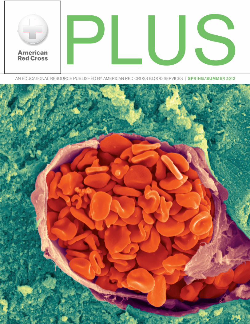
Cover photo: Arteriole in the human cerebellum containing red blood cells.
In his play of 1939, “Galileo,” the German writer Bertolt Brecht has a wonderful line for the scientist, Galileo Galilei: “The aim of science,” he says, “is not to open the door to infinite wisdom, but to set a limit to infinite error.” These are words well worth remembering.
XMRV is the abbreviation for xenotropic murine leukemia virus-related virus, a mouse virus that purportedly can grow in another species, in this case humans, and is related to or derived from the known mouse leukemia virus. It is a retrovirus, but unrelated to the HIV or HTLV retroviruses we know. In a study in 2006, it was associated with 40% of men with prostate cancer. Then in 2009, in a paper in Science, it was said to be present in the lymphocytes of patients with chronic fatigue syndrome (CFS), a rare, devastating illness of unknown cause. A year later, in another study, gene sequences of the virus were reported to be present in the blood of both CFS patients and healthy blood donors. Subsequent to that, the AABB, the American Red Cross and other blood banks asked donors with CFS to defer themselves. The evidence wasn’t all there, but the concern that CFS might be transmitted by blood transfusion posed an ominous possibility that required action.
Other researchers, however, have not been able to confirm these data from the two original reports. Some studies found no XMRV evidence in prostate cancer or CSF patients, whereas the original studies reported 80% or more prevalence in CSF patients and 7% in healthy blood donors. Some studies used the exact same detailed procedures of the original CFS paper (in some cases actually using samples from the same
patients) and could not replicate the findings. Other workers identified the fact that XMRV was actually a recombinant event involving two precursor viruses found only in strains of laboratory mice, and that XMRV does not exist in wild strains of mice, nor in mice from other laboratories unrelated to the original studies.
Since no lab has been able to reproduce the findings, now including also the two original reporting labs, the editors of Science have called for, and recently received, a partial retraction of the first CSF manuscript and a full retraction of the second one. It seems likely that the cautions concerning CFS and blood donation will be considered unnecessary, especially given the summary of findings of very recently published clinical studies (see next article).
The authors of this commentary in Transfusion (see citation below) cite in their paper the development of the “scientific method,” based on the teachings of another eminent scientist of the 16th century, Sir Francis Bacon of England, born within a few years of Galileo. They note that Sir Bacon would be proud to see work that “clearly demonstrates the potency of a 400-year-old method.” It is compelling that these two men, born in the mid-1500s, have so much of importance to say to us still. There is no evidence that Galileo actually said the words given him by the playwright, but he is known as the father of inductive logic, and it is known that the two men corresponded through translating intermediaries.
The scientific method at work: xenotropic murine leukemia virus-related virus is neither a cause of chronic fatigue syndrome nor a threat to the blood supply. Karafin MS, Stramer SL. Transfusion 2012; 52:222–225.
XMRV: Say it isn’t so!

Spring/Summer 2012 | PLUS 3
The same February issue of Transfusion that published the commentary reviewed here presented several additional papers on XMRV, the cumulative impact of which is very powerful.
Dodd (American Red Cross) and colleagues looked at about 15,000 routine blood donor samples, as well as 3,741 samples of units transfused to a group of frequently transfused patients. 830 pre- and post-transfusion samples from 109 of those frequently transfused patients receiving components derived from the donations of the 3,741 units in the study were also evaluated. They were tested for antibodies to the recombinant XMRV and related antigens. 1,435 of the regular donor samples were tested for the presence of XMRV RNA, using a transcription-mediated amplification (TMA) assay. There was no evidence of XMRV infection in any of the samples tested, including none of the nearly 100 samples known to be positive for HTLV antibodies.
Another report, from Qiu (Abbott Labs, USA) and co-workers, examined plasma from 1,000 U.S donors, 100 HIV-infected Cameroonian donors, 642 HTLV-1 infected Japanese donors and 311 STD diagnostic samples, using a chemo-luminescent immunoassay (CMIA), followed by additional CMIA, Western blot and reverse transcriptase PCR for positive results from the screening test. HTLV-1 infected donors had a low (5%) level of seroreactivity, but none had any detectable XMRV sequences. These few reactive samples were based on a single XMRV protein, thought to be due to cross-reactivity with HTLV-1. Thus, they found no evidence of XMRV in normal or retrovirus infected (HIV and HTLV) blood donors.
It is known that, in addition to being vectors/carriers of Lyme disease, hantaviruses and other organisms, “wild” mice can infect humans with lymphocytic choriomeningitis virus. Brooks, et al, examined the blood of 43 animal workers in Ottawa, Canada, with long (6 years) and high levels (50,000 mice) of exposure to laboratory mice. They found no evidence of infection with XMRV or to related mouse leukemia virus, suggesting that cross-species transmission of such viruses from mice to people must be really quite low, especially given the level of exposure.
All of these, and other more limited data, would seem to augment what the commentary of Karafin and Stramer so clearly illustrates. The findings of XMRV in patients with CFS and in small numbers of blood donors were related to laboratory contamination, not human infection. The work could not be reproduced by others, the original lab or those looking at some of the same CFS patients. The scientific community seems to now agree that the hypothesis linking XMRV in blood donors and CFS in recipients has no substance and is rejected.
1) Xenotropic murine leukemia virus-related virus does not pose a risk to blood recipient safety. Dodd RY, Hackett J Jr., Linnen JM. et al. Transfusion 2012; 52:298–306.
2) Seroprevalence of xenotropic murine leukemia virus-related virus in normal and retrovirus-infected blood donors. Qiu X, Swanson P, Tang N, et al. Transfusion 2012; 52:307–316.
3) No evidence of cross-species transmission of mouse tetroviruses to animal workers exposed to mice. Brooks J, Lycett-Lambert K, Caminiti K, et al. Transfusion 2012; 52:317–325.
Xenotropic Murine Leukemia Virus
XMRV - 2

4 PLUS | Spring/Summer 2012
Discussions about appropriate “transfusion triggers” in various settings and circumstances continues. Although it is a medical pleasure to prescribe a treatment for someone that makes them feel or perform better in specified circumstances, it is sometimes hard to document that improvement objectively. Some take satisfaction in seeing that withdrawal of a treatment can often make a patient better, such as when taking too much opioid or digitalis or blood pressure medication. And it’s good not to prescribe a treatment that doesn’t really make any objective difference, especially if it has measurable risks associated with it.
With such thoughts in mind, staff from the Notre Dame Hospital in Montreal and the University of Montreal Department of Mathematics and Statistics carried out a study in 305 patients, 60 years or older, who had a total hip or knee replacement. Looking at the question of whether anemia is associated with a decrease in postoperative vigor and function, they hypothesized that there is a threshold hemoglobin (Hgb) level below which both function and quality of life were adversely affected. Their results rejected this hypothesis.
In a prospective but unblinded observational cohort study, they categorized the 305 patients according to their 1 day postoperative Hgb level, 80 gm/L (8.0 gm/dl) or less, 81–90, 91–100 and >100gm/L. The study patients were 64% women, the average age 72, and the patients were generally obese with a mean BMI (body mass index) of 31, +/- 6, a BMI of 25 being the upper limit of normal. The measures evaluated were: distance walked in 6 minutes (6MWT); scores on the Borg
Scale (a recognized standard) for the patient’s perception of effort; maximal dominant hand strength; and a quality-of-life assessment. Measurements were done preoperatively and in the week following surgery. The 6MWT was done on Day 5. The preoperative Hgb levels ranged from 120–140 (12–14 gm/dl) in all groups.
There were no significant differences (p<0.05) noted in functional recovery (6MWT), hand strength measurements or quality of life assessments within each group, comparing pre- and postoperative measurements, even though Hgb levels in some cases dropped dramatically. Similarly, comparisons between the groups showed no appreciable differences. Approximately 50%, 30%, 30% and 20%, respectively, of the groups, starting with the lowest Hgb group first, were transfused perioperatively, an average of 1 unit of red cells for the lowest group and about 0.5 units for the rest. (Obviously half or more of the patients got no transfusions at all; a few got several).
The authors believe their data support the use of a restrictive transfusion strategy in the immediate postoperative period. They did not examine these parameters weeks after surgery, but since no functional problems were seen immediately postoperatively, and since Hgb levels recover quickly, it is unlikely that any effects of an initially low Hgb would be found.
Postoperative anemia does not impede functional outcome and quality of life early after hip and knee arthroplasties. Vuille-Lessard E, Boudreault D, Girad F et al. Transfusion 2012; 52:261–270.
PLUS has a new writer, Robert (Bob) G. Westphal, MD, MPH, FACP. Dr. Westphal has been a university and medical school professor at the University of Vermont College of Medicine and at the School of Public Health, University at Albany, SUNY. He has clinical, classroom, international and online curriculum experience. Dr. Westphal is Board Certified in Internal Medicine and Hematology and Board Qualified in Preventive Medicine. The American Red Cross is delighted to share Dr. Westphal’s engaging, expert writing with our readers of PLUS.
“ You can’t always get what you want...sometimes you get what you need.” (with apologies to the Rolling Stones)
Dr. Robert G. Westphal
To view previous issues of PLUS, go to redcrossblood.org/hospitals/plus-quarterly
PLUSAN EDUCATIONAL RESOURCE PUBLISHED BY AMERICAN RED CROSS BLOOD SERVICES | WINTER 2012

Spring/Summer 2012 | PLUS 5
Donors appear at blood centers and mobile drawings and casually comment that their doctor says they probably have a high iron level. Or, their doctor finds that they do have a high iron level, or high Hgb level, and sends them to donate blood. Maybe their Hgb level is high because they have lung disease. Generally, they will be told that the collector cannot take their blood, since it is considered a therapeutic phlebotomy. In order to transfuse blood to a patient that is removed for treatment of the donor, one must label the unit with the name of the disease process necessitating the phlebotomy, and most large-scale blood collectors are reluctant to do that, for a variety of reasons.
Hemachromatosis, iron storage disease, is not a common condition, but it is not rare. There are several types, the most common of which is due to the presence of the HFE gene mutation (which has many polymorphs), with about 5% of the northern European population being homozygous for this gene. Not all so affected manifest with disease, however. Men are more frequently seen than women, since childbearing and regular menstrual flow diminish the amount of iron stored. Iron is quite toxic to animal tissues, as evidenced by its highly protected place in the folds of globin chains, the very small amount absorbed from the upper small intestine and the complex system of carrier proteins used to transport it in its journeys through human metabolic and storage processes. Too much iron, whether from excessive transfusion requirements or faulty genes, results in a “bronze” skin pigmentation, diabetes, liver disease and high levels of cardiac dysfunction due to an iron-overload effect on those respective tissues.
Blood bank staff at a small, rural hospital in Massachusetts (142 beds) have offered therapeutic phlebotomy as a community service and recently described the need for it in their community, the nature of the illnesses so treated and the positive financial impact this program provided for their hospital. (See article below.) They obtained a variance from the Food and Drug Administration in order to develop their program for people with hemachromatosis. Overall, the program is small, but traditionally they have collected the major portion of the blood they use, maintaining, on average, an inventory of about 300–400 units over a month’s time. Over the course of a year, their therapeutic phlebotomy contribution to inventory was 4.5%. Over a 13
month period, they provided 390 therapeutic phlebotomies of which 207 were eligible, allogeneic hemachromatosis units. Other conditions necessitating therapeutic phlebotomy included polycythemia vera, erythrocytosis and secondary polycythemia.
Units that were discarded from hemachromatosis donors numbered 17 and had positive viral markers or other reasons for non-use. Of the 84 patients in the program, 62 had hemachromatosis, 43 of these met eligibility requirements and 207 units were collected for the blood bank inventory for a net savings of $21,000 in blood costs. In addition, the 22 non-hemachromatosis therapeutic phlebotomy patients underwent 183 procedures, the fees for which generated additional revenue in excess of $15,000.
Not every hospital will wish to engage in this service, but clearly blood banks can do well by doing good!
Therapeutic phlebotomy procedures and their impact on a rural hospital’s red blood cell inventory and fiscal stature. Flynn RC, Bryant BJ. Transfusion 2011; 51, issue 12 part 2:2761–2766. Special issue: J of Blood Services Management.
Blood money? Or, Give us your iron...

6 PLUS | Spring/Summer 2012
Many of today’s blood bankers and clinicians remember not too long ago when having platelets for transfusion was a near miracle. They were a precious commodity that was severely supply-constrained. As improved methods of collection and storage came along, the result of many dedicated laboratories and individuals, we took platelet transfusion availability for granted (at least most of the time) and began to focus on the problems that inevitably follow broader implementation of a new therapeutic modality. Chief among these problems was, and is, bacterial contamination, primarily because in order to maintain function, platelet concentrates must be stored at room temperature. An unfortunate fact is that those who need platelet transfusions most generally are also maximally depleted of phagocytic and other white blood cells and are quite immunologically stressed, often already infected. One of the important precepts of medicine, Primum non nocere—“First, do no harm,” was often put to the test in the early days of platelet transfusions, miraculous though they were.
In an editorial in Transfusion last December (see reference below) Drs. Peter Tomasulo and Leon Su review the various incremental steps we have used to reduce this problem, and they evaluate three reports from that same issue that docu-ment the continued risk of bacterial contamination after early testing. Skin disinfection procedures, in-line diversion pouches for isolating the first ounce or two of blood from the collection and cultures with bacterial detection identifiers have been
chief among the applied “solutions,” yet some contaminated products continue to evade detection. Almost all of these efforts have been developed for use in blood collection and storage centers, usually after 24–36 hours of storage. One of the three papers in that issue of Transfusion presents results from a study of testing of platelet concentrates on the day of issue, almost “at the bedside,” using a test with a rapidly available result.
All of these tests have attendant problems with implementa-tion and timeliness, which the authors discuss. They also point out that in some countries, notably France, pathogen-reduc-tion activity has been very effective in reducing contamination. Canada is evaluating such a system, but the authors state that this intervention will not be feasible in the U.S. “in the near future.” They don’t say why, but one can guess it relates to the difficulties of proving the efficacy and safety of such a process in a highly regulated environment with rather severe political and economic restraints. And although the risk of sepsis from platelet transfusion is substantially less than it was 10 years ago, it is still several orders of magnitude greater than the risks of transfusion-transmitted HBV, HBC and HIV. This is clearly a situation worthy of continued and serious effort.
Is it time for new initiatives in the blood center and/or the hospital to reduce bacte-rial risk of platelets? Tomasulo, P and Su, L. Transfusion 2011; 51:2527–2533.
Dulling one edge of a double-edged sword

Spring/Summer 2012 | PLUS 7
As noted in the accompanying article, bacterial contamination of platelet concentrates remains a problem in search of a more effective solution. A collection of all the publications on this issue over the last 3–4 decades would likely fill a small library. Here we look briefly at three articles, two from the Canadian Blood Services, one from a multi-center U.S. study group.
In the first paper, the authors gathered data from the last 6 full years (2004–2010) from the 12 Canadian Blood Services sites. They compared bacterial contamination rates in platelet concentrates (PCs) prepared from apheresis collections, buffy coats and platelet-rich plasma (PRP). The PRP was the earliest method in use in many places; in Canada and most of Europe it has been phased out in favor of pooled buffy coats from four-to-six (in Europe) units. Samples for culture were obtained from the platelet “tails” for PRP PCs and from integral sample pouches in the other two types. They used the BacT/Alert BPA culture system in all three sets of measurements. Their protocols were changed twice, once to a 2-step arm prep method, which made no difference, and also by increasing the sample volume incubated. The latter method greatly increased the rate of confirmed positive tests. Initially positive rates differed between PC groups, with apheresis PCs having the highest; however, confirmed positives were similar between groups. During the period of analysis, there were 5 false negative tests with resultant transfusion reactions. Three additional such PCs were found during in-house sterility testing begun in 2009. Thus, although risk was reduced it was not eliminated.
The second Canadian article studied the rate of detection failures during initial screening using the aerobic BacT/Alert system and then added anaerobic incubation/testing and an immunoassay (Verax Pan Genera Detection test, or PGD, see next paragraph) to evaluate longer-term storage. Their PCs were all of the BC variety. They tested 4,000 outdated (7 days storage) BC PCPs and found one true positive (same sample) in the BacT and PGD tests. Fifty-four false positive BacT anaerobic cultures were found, mostly due to problems with the anaerobic instrumentation. Eleven Gram-stain positive false-positive PGD tests were noted. Correctible problems were noted in the PGD testing due to the need for a humidity chamber. They concluded that introducing anaerobic cultures would waste many platelets unnecessarily, and that (initial
testing is known to miss certain contaminated products) extended platelet storage might be feasible using a rapid screening method, such as PGD, right before transfusion.
Finally, a look at the large multi-center evaluation of the PGD test. Briefly, the test itself is a dual-sandwich immunoassay designed to detect very common antigens found on Gram-negative and on Gram-positive bacteria, a window on the disposable testing device for each class. Samples of up to 6 concentrates can be combined. As the sample (0.5 ml) moves along the two windows, it is exposed to gold-conjugated antibodies to the common Gram-negative and Gram-positive antigens in the respective window. These bound indicators adhere in a detectable line at opposite ends of the device, indicating a Gram-negative or -positive result. Controls built into the same device must also indicate positivity in every case. The assay takes about 30 minutes. Of the nearly 28,000 apheresis PCs tested, all of which had been released after negative BacT/Alert testing, 9 were repeatedly reactive and verified by culture. There was a false-positive rate of 0.5% (142/27,629). At hospitals where the PCs were cultured as well as tested with the PGD method, 2 contaminated units and one transfusion reactive specimen were also found to be culture positive. The authors state that day of transfusion testing using this method will prevent many, if not most, septic reactions to PC transfusion.
Cost was not evaluated in any of these studies, and although pathogen reduction techniques, as noted by Tomasulo and Su, might be extremely effective at low cost, such methods are not on the immediate horizon in the U.S. Whether or not the cost-benefit ratios of this two-method approach to testing PCs would bear up to close scrutiny remains to be determined, especially since such a large proportion of recipients of PCs have such devastating, life-shortening illness.
1) Bacterial contamination in platelets: Incremental improvements drive down but do not eliminate risk. Jenkins C, Ramirez-Arcos S, Goldman M, and Devine D. Transfusion 2011; 51:2555–2565.
2) Bacterial screening of outdated buffy coat platelet pools using a culture system and rapid immunoassay. Ramirez-Arcos S, Kou Y, Mastronardi C, Perkins H, and Goldman M. Transfusion 2011; 51:2566–2572.
3) Detection of bacterial contamination in prestorage culture-negative apheresis platelets on day of issue with the Pan Genera Detection test. Jacobs MR, Smith D, Heaton WA and the PGD Study Group. Transfusion 2011; 51:2573–2582.
Positive experiences with bacterial detection

8 PLUS | Spring/Summer 2012
A thoughtful look at stem cell engineering for therapy
In ages past (long past, actually) alchemists strove mightily to devise ways of turning lead into gold. A more modern search for miraculous results has involved the idea of growing blood cells from bone marrow or other-source stem cells. Some recent work has focused on the potential of growing red cells for transfusion in vitro.
However, the use of engineered blood stem cells for the treatment of some hereditary and even acquired diseases involving hematopoietic cells has already begun, but occasionally with a disastrous result. Cases of myelodys-plasia and leukemia have resulted from a few of these efforts, often related to the cell systems chosen as vectors, or deliverers, of the altered genes. Much of the work in this area was begun and carried out at the National Institutes of Health (NIH), in Bethesda. The paper cited below, which gives an in-depth review of where things stand today, also comes from the NIH and from the Memorial Sloan-Kettering Center in New York. Since the work of the early 1990s, nearly 100 patients have been treated with genetically modified CD34+ hematopoietic progenitors worldwide.
Some unwanted, tragic events occurred earlier on, with 5 cases of leukemic transformation out of 20 patients treated for severe, combined immune deficiency (SCID), and other similar occurrences in patients with chronic granulomatous disease (CGD) and Wiskott-Aldrich syndrome (WAS). But these tragic events have led to more refined, improved techniques. These new trends in HSC engineering fall into 3 broad categories: improvements in the design of retroviral vectors (it was problems in the use of these that caused such heartbreaking events in the past); development of technolo-gies for targeted gene delivery; and novel approaches made possible by the advent of the use of patient-specific pluripo-tent stem cells, that is, stem cells collected from the patients themselves. It is interesting to note that one of the promising new vectors used to transmit the therapeutically altered gene
is the murine leukemia virus, which appears in another article in this issue of PLUS. Researchers have devised a way to turn off the self-replicating gene in these vectors that was the cause of previous disastrous events in earlier trials.
Most of the diseases that have been studied have been in patients suffering from severe immune deficiency disorders such as SCID, WAD, CGD and some others. Obvious afflic-tions that might be targeted with a new assault using these improved techniques include thalassemia, sickle cell disease and other hemoglobinopathies. There are already two patients treated with beta thalassemia, one with combined Hgb E/thalassemia.
For those fellow humans who suffer from these difficult, often tragic but not rare diseases (and to some extent for those of us who care for them), positive developments along these lines would indeed seem like turning lead into gold. Stay in touch, King Midas!
Hematopoietic stem cell engineering at a crossroads. Riviere I, Dunbar C, and Sadelain M. Blood 2012; 119:1107–1116.
A stylised view of a stem cell surrounded by red blood cells

Spring/Summer 2012 | PLUS 9
In the accompanying article of this issue of PLUS, we talked of the potential role of hematopoietic stem cells (HSCs) as modulators of gene therapy for the treatment of a number of hereditary and acquired disorders affecting elements of the blood and immune systems. As blood bankers, most of us feel there are constantly recurring shortages of blood, especially during certain times of the year, and under certain circumstances. The authors of the paper discussed here (see below) state, without reference to year or source, that over 93 million donations are made worldwide and that the number of units outdated in the U.S. represented 13% of total donations in 2008. An older estimate in 1995 from the World health Organization (WHO) was of “… more than 90 million…” In 2002, also using WHO sources, the figure was about “…75 million donations, of which 45 million were collected and tested in more developed countries and of the other 30 million roughly only half were completely tested for critical infectious disease markers.” [The author’s paraphrase.]*
The point is not necessarily which figures are the most accurate, but that in much of the world we really don’t have good numbers about blood collection and transfusion. There are still very highly risky blood transfusions going on these days, and for some people, even in developed countries, suitably compatible and safe blood is not always a “given.” So, wouldn’t it be great to be able to grow safe blood in cultures? Or red cells with the antigens stripped off their outer shell? Or provide Hgb without those pesky red cells’ membranes altogether? Or find chemicals that could deliver oxygen and carry off carbon dioxide? Sure! However, none of these approaches has yet borne fruit. Even Hgb solutions, from cattle or even humans, even polymerized and packaged, have been associated with too much risk of myocardial infarction and death.
Enter the idea of using HSCs to grow RBCs for transfusion. Enough of them have been grown and shown to have normal RBC functions, and to have normal life spans as measured with radioactive chromium standards. One might use them autologously to temporarily support bone marrow ablation/suppression during cancer therapy, or for targeting gene therapy for sickle cell disease, or perhaps to grow RBCs from donors with special rare types of blood for more general clinical use. Advances in the area have generated a lot of
excitement, but the authors of this article spend considerable time discussing the challenges and hurdles associated with such goals.
The challenges and hurdles have been numerous, but the article separates them into three categories: growth capacity, cellular differentiation and in vitro enucleation.
With regard to growth capacity, the generated red cells seem unable to increase in concentrations past about 106 per ml; thus one would need 2.5x10³ (2,500) liters in order to contain a dose of 2x10¹² RBCs. There are about 2.5x10¹² cells in one pint of human blood. So, the engineering problems would be virtually insurmountable, especially considering sterility and cell fragility. In culture systems, HSCs have poor erythroid expansion potential, making final differentiation into RBCs problematic. In addition, the final enucleation that turns RBCs into a flexible biconcave disc with no cellular nucleus, which occurs naturally in human marrow, cannot be done in culture except with stromal cells of animal origin. Chemicals that might promote the task have yet to be developed.
Although great strides have been made in our understanding of how to grow RBCs from HSCs in cultures, current knowledge would have us focus on lesser goals, say the authors. Reagent red cells could be used for targeted drug delivery, say chemotherapy to vascular tumors. Certain blood types might be propagated for reagent use in antibody-screening scenarios. A compelling case is made for the use of cultured erythroblasts as a way to assay for promotion of Hgb F production for use in sickle cell disease; or for intracellular methods to inhibit the growth of malarial parasites or to investigate erythropoietic stimulants for use in cases of myelodysplasia, or aplasia.
A lot of work remains to see even these limited goals come to fruition, but this paper opens a window to an exciting part of our current world of scientific exploration.
The potential of stem cells as an in vitro source of red blood cells for transfusion. Migliaccio AR, Whitsett C, Papayannopoulou T and Sadelain M. Cell Stem Cell 2012; 10:115–119.
* International aspects of blood services. Bianco, C. Rossi’s Principles of Transfusion Medicine, 3rd edition, 2002: 953. And, International aspects of blood services. Westphal RG. Principles of Transfusion Medicine, 2nd edition, 1996: 917.
Stem cell cultures for RBCs for transfusion?
A stylised view of a stem cell surrounded by red blood cells

10 PLUS | Spring/Summer 2012
The doomsday virus?This past December, there were two manuscripts submitted for publication in two of the more eminent scientific journals, Science and Nature. One came from a virology lab in Rotterdam, the Netherlands, the other from the University of Wisconsin-Madison. Both labs are funded for this work by the U.S. National Institutes of Health (NIH). Both articles were to be published in March of 2012, but considerable controversy and discussion have developed since the nature of the manuscripts has become known. Both groups of scientists were working on “bird flu,” and appeared to have altered the genes of the virus so that it could be transmitted between ferrets, and thus perhaps humans, since the immune system of the ferret closely resembles that of humans.
You remember bird flu, don’t you? The H5N1 influenza virus filled our minds and newspapers a few years ago with great concern about the risks of a lethal pandemic. This avian virus was first identified as the cause of a particularly lethal form of influenza in Hong Kong in 1997, and public health organizations, hospitals, bioterrorism preparedness officials, schools and newspapers all began discussing the potential risks if a large-scale outbreak were to occur. The epidemic was linked to poultry and classified as avian influenza A (H5N1). Migrating wildfowl could carry it great distances, but early on it was clear that all of the human cases that were reported, primarily from South East Asia, seemed to be due to direct contact with infected fowl, and person-to-person spread had not been seen. The reported fatality rate was around 60%, but no one knew the actual denominator, how many cases
there were that were undetected, as well as those known. Nonetheless, the concept was frightening, and we continued to worry about it up to and through the SARS outbreak of 2003.
The avian flu virus (H5N1) has been shown to survive in the environment for long periods of time. Infection may be spread simply by touching contaminated surfaces. Birds who were infected with this flu continue to release the virus in their feces and saliva for as long as 10 days after initial infection. The ongoing emergence of this and other infectious diseases has lingered in the public consciousness, probably ever since the AIDS epidemic was recognized in 1981, and certainly after September 11, 2001. However, the fact that virtually no human-to-human transmission has been documented was somewhat reassuring. Now things seem about to change and fears of accidental or purposeful release of this altered virus have led to calls to suppress publication of these data.
The scientists involved have said the work would allow for health organizations to improve their surveillance of influenza in birds and other animals so as to have an early warning of mutations occurring that might put humans at greater risk. Critics, of which there are many, argue that our current surveillance systems are not equipped to detect these sorts of mutated flu viruses. The Dutch scientist now says his findings have been misconstrued and exaggerated, and that the only ferrets that died had received very large inoculae into their trachea. Despite immunological similarities, several people point out that people are not ferrets, and we don’t know what might occur. The National Science Advisory Board for Biosecurity (U.S.) recommended that a limited version of the papers be published; however, an international panel of experts convened by the World Health Organization (WHO) met in February and recommended complete publication. Critics contend the panel was heavily weighted by participants with a clear stake in publication, and many very highly respected and influential people in both public health (Dr. Osterholm and Dr. Henderson) and biosecurity (Dr. Inglesby and Dr. Henderson) urge restricting dissemination of the information and allowing it only in BSL-4 (the highest level of biosecurity laboratory facilities) equipped institutions to continue the work. Dr. Henderson is credited with eliminating the scourge of smallpox from the world.
Given the charges of censorship and intellectual hindrance on the one hand, and concerns about the risks of bioterrorism
Influenza A virus subtype H5N1 or Avian Flu Virus.

Spring/Summer 2012 | PLUS 11
Young bloodMany years ago at a blood center in New England, a young high school girl, who weighed 116 pounds soaking wet and had previously donated 2–3 times, brought a couple of friends to share the experience. One was a strapping 190–200 pound halfback on the football team who fainted dead away when the young woman had her finger pricked to measure her Hgb level. There is just no accounting for how some people will react to the blood-donation process. But it is very clear that younger donors, on whom we rely more and more, and especially first-time and high school donors, are far more likely to have an untoward reaction to donation. Which is not good when one considers the changing demographics of longevity and blood utilization.
There have been a number of studies evaluating the donation-process in young people; the January 2012, issue of Transfusion Medicine Reviews contains a thorough summary of these problems and makes some recommendations for future interventions to mitigate them. Dr. Anne Eder, from the American Red Cross Biomedical Services, reviews the recent articles on the subject, many of which were written by her and other colleagues, and looks at the measures taken by several institutions to reduce the rate of reactions in young and first-time donors. In 2005, 15 states allowed donation by teens as young as 16, with parental permission. As of 2010, 34 states allowed blood donations by young donors, and others were deliberating the issue.
Almost every study notes that donor reactions are related more to young age than any other factor. In addition to being, on average, smaller than more mature donors, younger donors, especially young women, do not always have a total circulating blood volume that is large enough (>3500ml) to allow for a safe donation of a 525 ml blood donation. Blood
volume is calculated based on the donor weight, gender and height. One report, using this formula, noted that 9% of donors less than 23 years old had an estimated blood volume of less than the 3,500 ml projected for a minimum weight of 110 lbs (50 kg). Half to two-thirds of first-time donors with these low blood volumes have a reaction, especially young women. The American Red Cross has now begun to limit donations from these low blood volume donors at high school drives.
In addition to sometimes marginal blood volumes, the iron stores of younger donors lag behind those of their elders, which presents a more long-term concern. But even minor reactions and temporary deferrals have a negative impact on future blood donations. Age, blood volume and first-time donation status all contribute independently to the risk of syncopal reactions. Young, first-time donors are more likely to be apprehensive and susceptible to the anxiety seen in those around them. Interventions that diminish both anxiety and volume loss, such as muscle flexing, mental distraction and extra hydration pre- and immediately post-donation have been shown to be useful.
As the population ages and medical interventions follow us into our waning years (not always for good, some feel), the number of older donors is declining while the need for blood increases. Thus, it is important, indeed imperative, that blood collection agencies explore and implement new practices to reduce the anxiety surrounding the donation process; foster enthusiasm and fulfillment in young donors; and carefully orchestrate collections so that the principle of “First do no harm” is borne out through the entire experience.
Improving safety for young blood donors. Eder, AF. Trans. Med. Rev. 2012; 26: 14–26.
or accidents on the other, this discussion likely will not be over soon. The U.S. NIH’s Dr. Anthony Fauci has asked the biosecurity advisory board (U.S.) to meet again and reconsider its call to redact/omit some of the details prior to publication, and the WHO is planning further advisory meetings with a broader range of participants. Many of us have not forgotten the fact that the only deaths from a bioterrorism incident in the entire history of the U.S. (Lord Jeffrey Amherst’s smallpox-contaminated blankets during the French and Indian wars not counted) occurred in the fall of 2001 when a lethal, inhaled form of Bacillus anthracis (anthrax) killed seven innocent people, sickened 17 others and was perpetrated
using an agent from a highly protected bioterrorism laboratory of the U.S. Army, and probably by an employee of same, though we may never know who was really responsible.
1) Life sciences at a crossroads: respiratory transmissible H5N1. Osterholm MT, Henderson DA. Science 2012; 335:801–802.
2) Engineered H5N1: a rare time for restraint in science. Inglesby TV. Annals of Internal Medicine, www.annals.org 2012.
3) Despite safety worries, work on deadly flu to be released. Grady D. New York Times, February 17, 2012.
4) Genetically altered bird flu virus not as dangerous as believed, its maker asserts. Grady D. New York Times, February 29, 2012.
5) The truth about the doomsday virus? Editorial. New York Times. March 3, 2012.

Reimbursement resourcesReimbursement of hospitals by the Centers for Medicare and Medicaid is a complicated affair. The American Red Cross provides various forms assistance to its valued hospital partners. These materials can be found at redcrossblood.org/reimbursement:
• Updates on reimbursement in the hospital outpatient and inpatient settings are published at least four times a year.
• Educational materials, including a summary of blood billing guidelines and FAQs regarding coding, coverage and reimbursement.
• Technical support through the email address [email protected].
The Red Cross also sponsors a limited number of on-site live learning opportunities. Attendance at one of these events will normally earn participants continuing education credits.
Remember these WebsitesImmunohematology Journal redcross.org/immunohematology
Reimbursement redcrossblood.org/reimbursement
SUCCESS success.redcross.org
PLUSSpring/Summer 2012, Volume Six, Issue Two
H21239
Publications CornerRecent publications by American Red Cross scientists and physicians:
Exploring nursing students' level of preparedness for disaster response. Schmidt CK, Davis JM, Sanders JL, Chapman LA, Cisco MC, Hady AR. Nurs Educ Perspect. 2011; 32:380–3.
Syncope after whole blood donation: factors associated with increased donor injury. Newman B, Siegfried B. Transfusion 2012; 52:210–1.
Prevalence of anti-dengue immunoglobulin G antibodies among American Red Cross blood donors in Puerto Rico, 2006. Mohammed H, Tomashek KM, Stramer SL, Hunsperger E. Transfusion 2012. Epub ahead of print doi: 10.1111/j.1537–2995.2011.03492.x.
New SUCCESS course “Group AB Plasma and the Risk of TRALI”This presentation describes two case reports of fatal transfusion reactions occurring in 2010 and 2011 that were ascribed to transfusion-related acute lung injury (TRALI) following transfusion of group AB plasma. The diagnosis and etiology of TRALI are described and hemovigilance data implicating high-plasma-volume components are reviewed, as is the impact of the AABB recommendation that transfusion services minimize patient exposure to high-volume plasma products from alloimmunized or potentially alloimmunized donors. The persistent demand for group AB plasma in excess of blood center production capacity is detailed. Hospital use of group AB plasma is reviewed and recommendations made to restrict group AB plasma use to those patients who require this group, despite its consideration as “universal plasma.”