pneumonia
DESCRIPTION
Pneumonia. Andriy Lepyavko, MD, PhD Department of Internal Medicine № 2. Definition. Pneumonia – this is an inflammation in the lung parenchyma caused by bacteria, viruses or fungi which is characterized by intraalveolar exudation. Morphology. Classification. Etiology (if it is known) - PowerPoint PPT PresentationTRANSCRIPT
Andriy Lepyavko, MD, PhDDepartment of Internal Medicine
№ 2

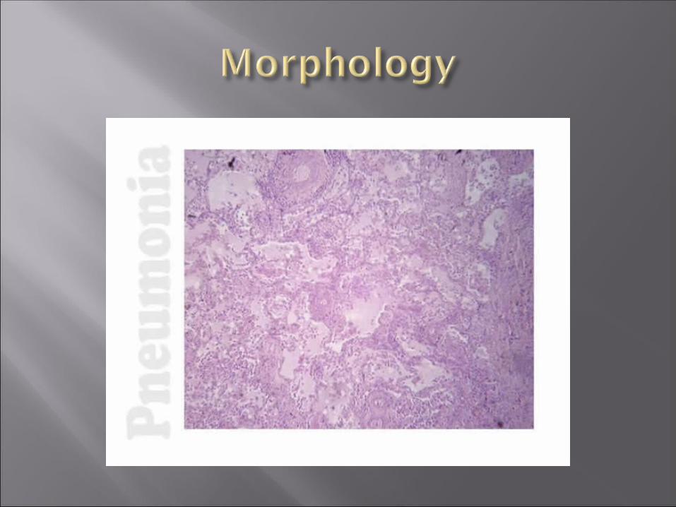
Pneumonia – this is an inflammation in the lung parenchyma caused by bacteria, viruses or fungi which is characterized by intraalveolar exudation


I. Etiology (if it is known)II. Variants: Community-acquired pneumonia Nosocomial pneumonia – when patient
was hospitalized with any another diagnosis, and after 48 hours in the hospital (not earlier!) pneumonia was diagnosed, or pneumonia after artificial lung ventilation
Pneumonia due to aspiration. It results from the aspiration of gastric contents in addition to aspiration of upper respiratory flora in secretions.
Pneumonia in immunocompromised host – patients with AIDS or immunodeficit of other origin. Causes of pneumonia – viruses, fungi of saprofites (E.coli etc.)

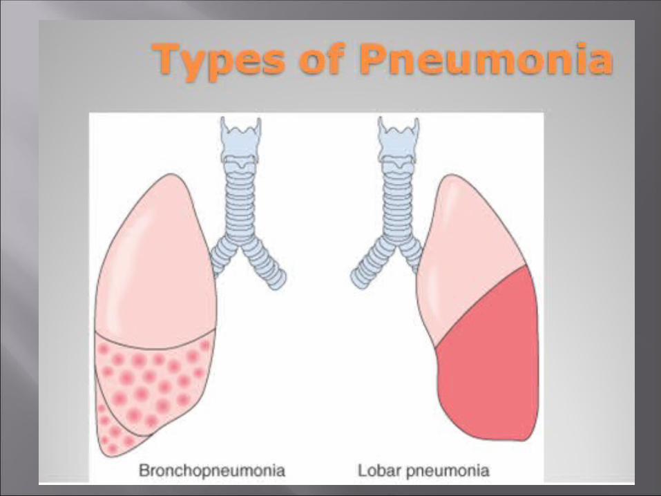
III. Localization (side, lobe, segment)IV. Stages of severity: Mild stage –conciousness is clear, t less than 38,
heart rate less than 90, BP normal, dyspnea mild in case of physical activity, CXR – small infiltration
Moderate – conciousness is clear, sweating, general weakness, t 38-39, heart rate 90-100, moderate dccreased BP, dyspnea, large size of infiltration
Severe – t 39-40, conciousness is not clear, heart rate more than 100, low BP, severe dyspnea, cyanosis, large size of infiltration and presence of complications
V. Complications.

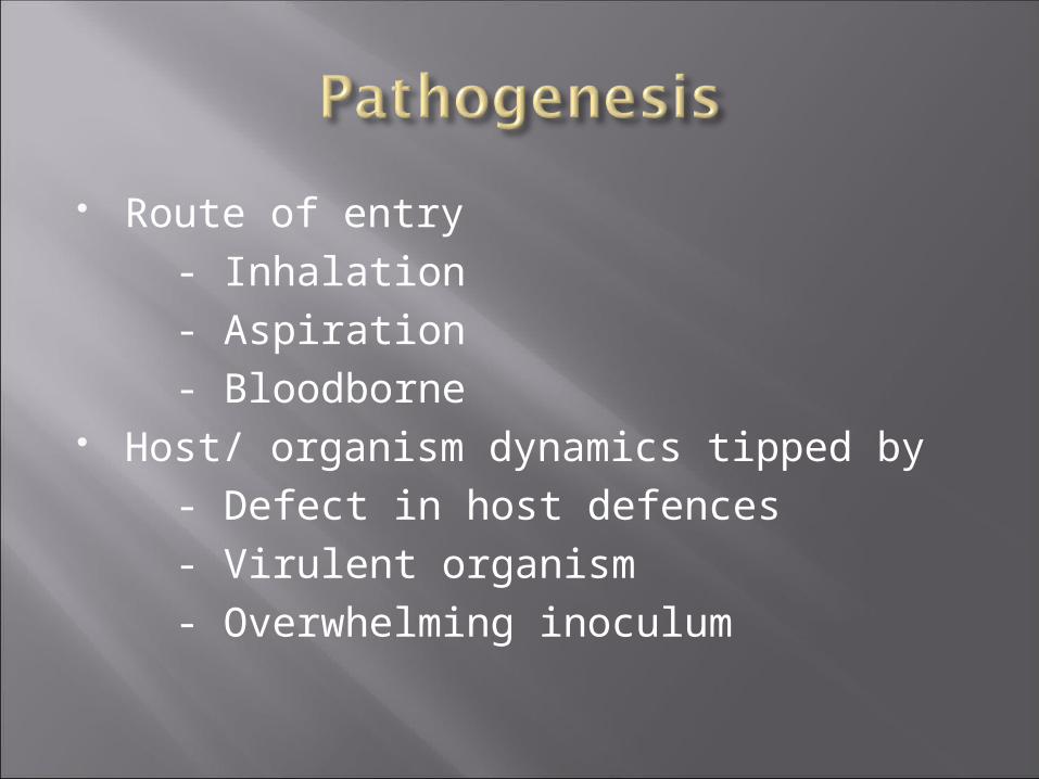
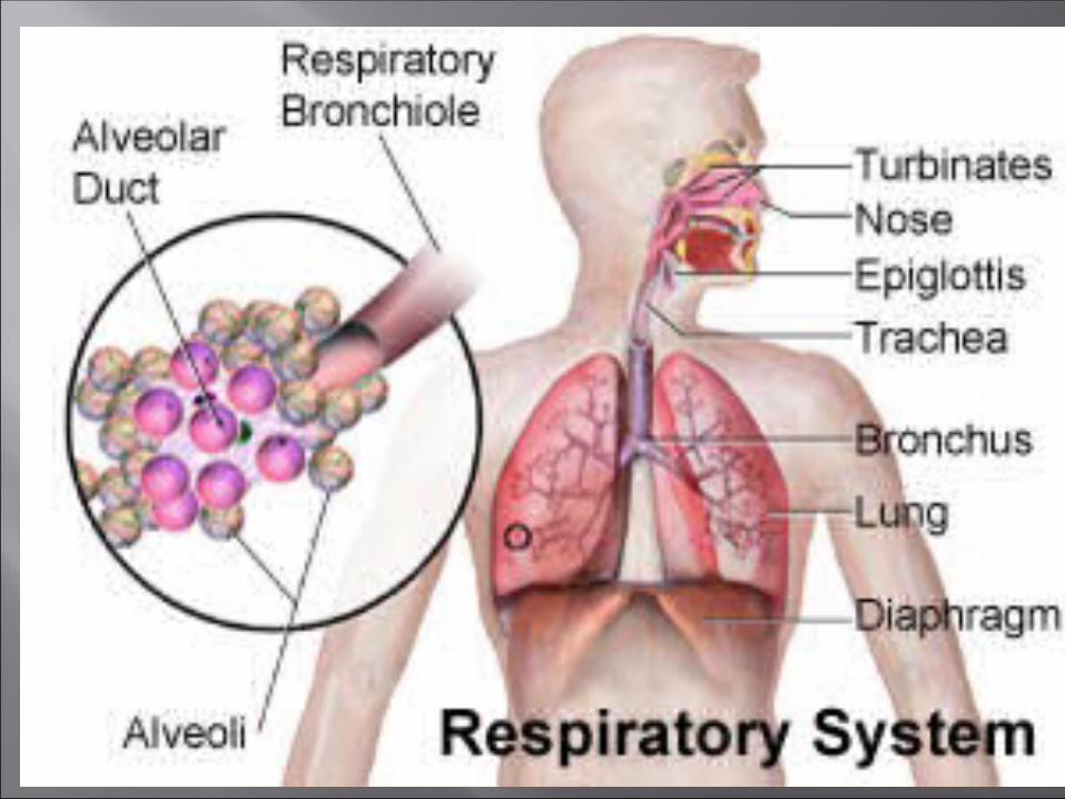
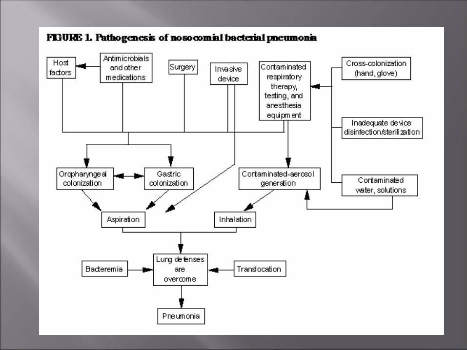
Route of entry - Inhalation - Aspiration - Bloodborne Host/ organism dynamics tipped by - Defect in host defences - Virulent organism - Overwhelming inoculum

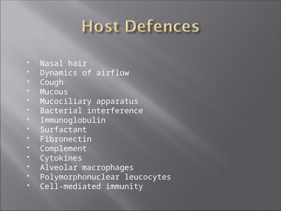
Nasal hair Dynamics of airflow Cough Mucous Mucociliary apparatus Bacterial interference Immunoglobulin Surfactant Fibronectin Complement Cytokines Alveolar macrophages Polymorphonuclear leucocytes Cell-mediated immunity


Predisposition – CHF, diabetes, alcoholism, COPD
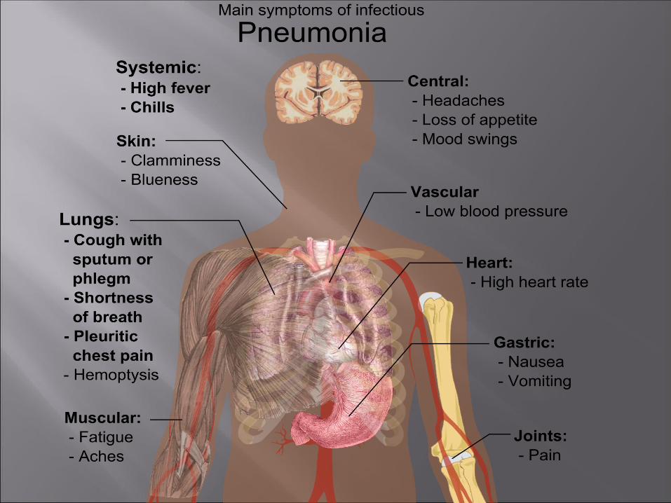
Classic symptoms – cough, fever, sputum production, dyspnea
Clinical syndrome – fever, pleuritic chest pain, productive cough with mucopurulent sputum
Focal pulmonary findings (rales, crapitation or signs of consolidation) – less sensitive than CXR
General blood analysis – increased ESR, leucocytosis, shift to the left
Sputum analysis – causative microorganism and its sencitivity to antibiotics may be found

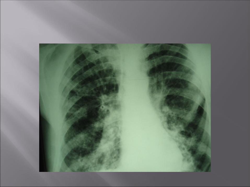
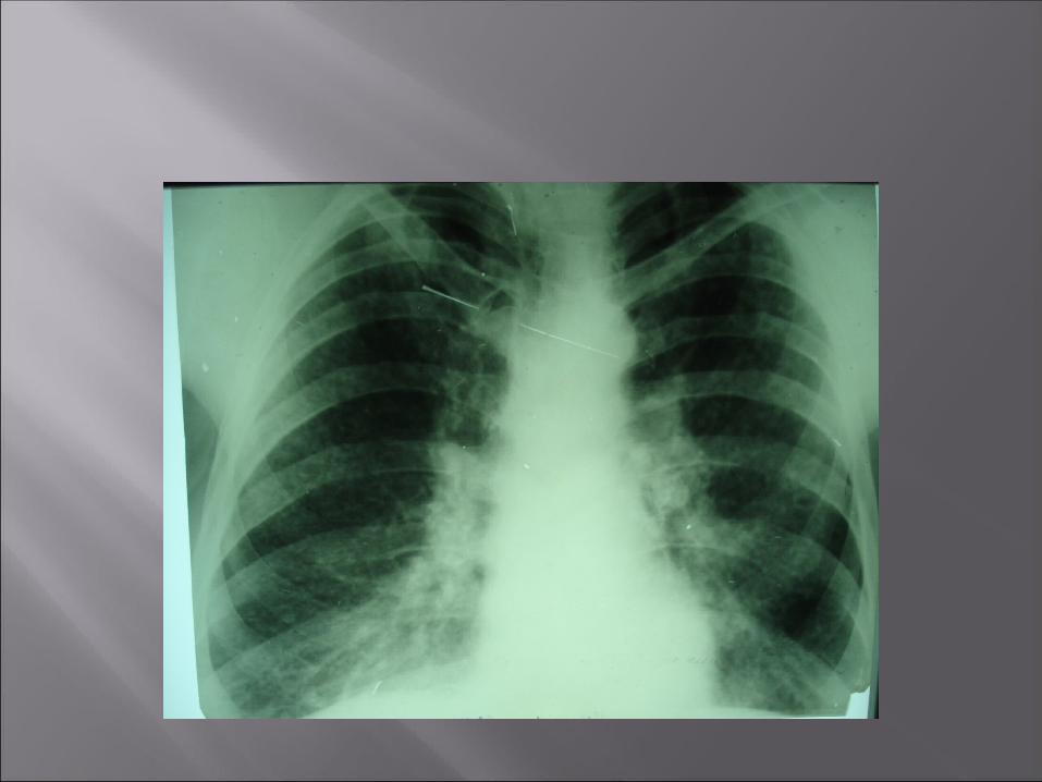
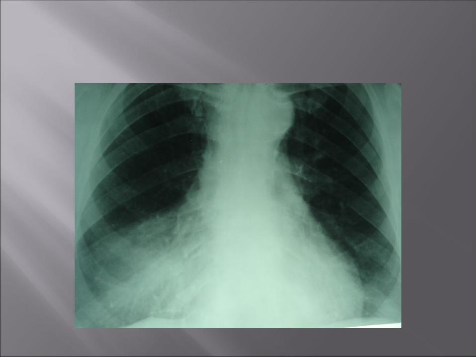
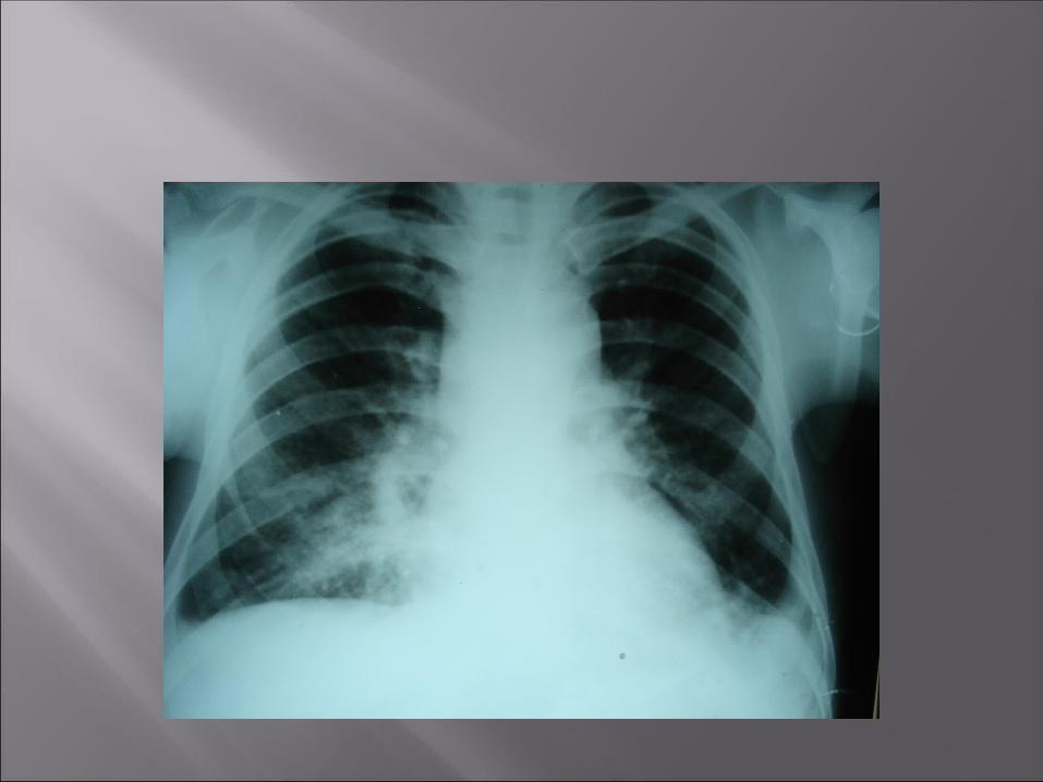
CXR with infiltrates – diagnosis “pneumonia” is invalid without it


Most common pathogens: Streptococcus pneumoniae (9% to 75%; mean,
33%), Haemophilus influenzae (0 to 50%; mean, 10%), Legionella species (0 to 50%; mean, 7%), Chlamydia pneumoniae (0 to 20%; mean, 5%). Mycoplasma pneumoniae
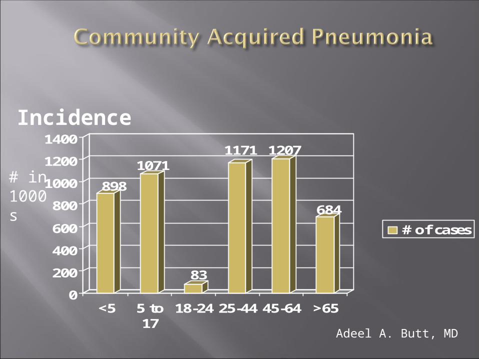
Adeel A. Butt, MD
898
1071
83
1171 1207
684
0
200
400
600
800
1000
1200
1400
<5 5 to17
18-24 25-44 45-64 >65
# of cases
# in 1000s
Incidence

Adeel A. Butt, MD
25,7
74,9
0
10
20
30
40
50
60
70
80
<4 5 to 14 15-24 25-44 45-64 >65
# of deaths# in 1000s
Mortality


At least 5 days Until afebrile for 48-72 hours Stable vital signs Longer course needed if Initial antibiotic choice did not cover the
pathogen Extrapulmonary infection (meningitis) Lung abscess, cavitation or empyema Gram negative pathogen or S.aureus
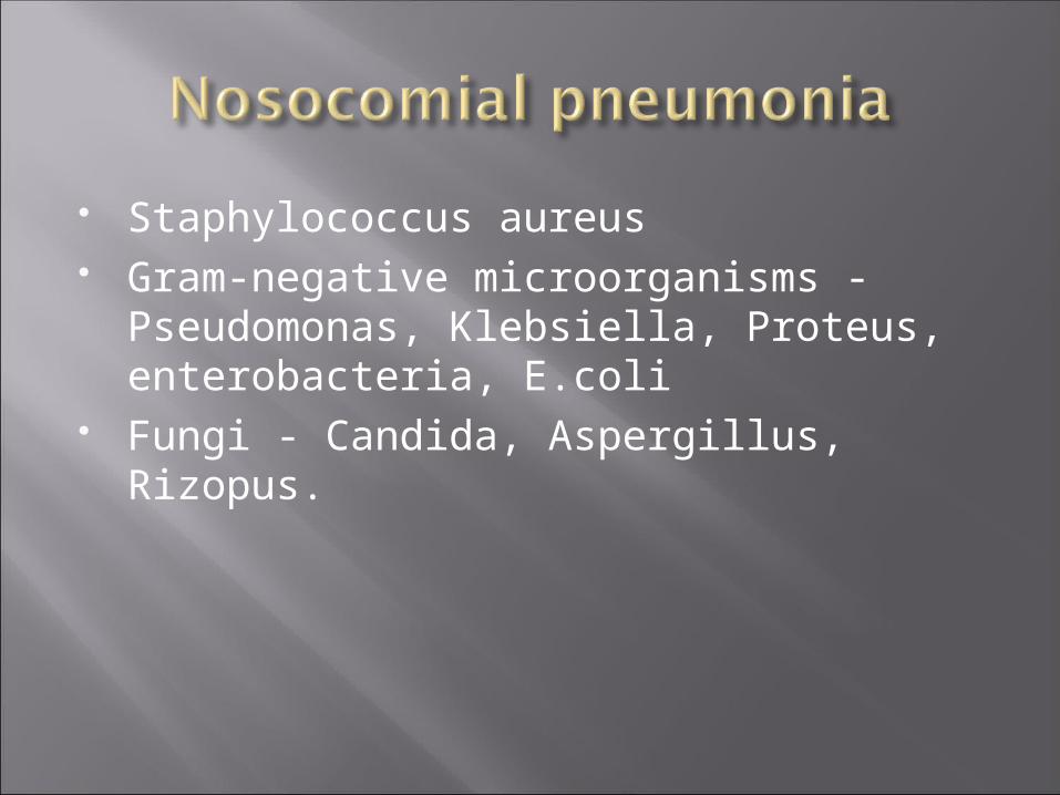
Staphylococcus aureus Gram-negative microorganisms -
Pseudomonas, Klebsiella, Proteus, enterobacteria, E.coli
Fungi - Candida, Aspergillus, Rizopus.

Most effective are: Aminoglycozyde (tobramycin, sizomycin)
+ Metronidazol Cephalosporini III-IV
generation+Metronidazol