polycationic calixarene ptx013, a potent cytotoxic agent against tumors and drug resistant cancer
TRANSCRIPT

PRECLINICAL STUDIES
Polycationic calixarene PTX013, a potent cytotoxic agentagainst tumors and drug resistant cancer
Ruud P. M. Dings & Joseph I. Levine & Susan G. Brown &
Lucile Astorgues-Xerri & John R. MacDonald &
Thomas R. Hoye & Eric Raymond & Kevin H. Mayo
Received: 29 November 2012 /Accepted: 24 January 2013# Springer Science+Business Media New York 2013
Summary Previously, we reported on the anti-tumor activ-ities of two designed calix[4]arene-based topomimetics(PTX008 and PTX009) of the amphipathic, angiostatic pep-tide Anginex. Here, we chemically modified the hydropho-bic and hydrophilic faces of PTX008 and PTX009, anddiscovered new calixarene compounds that are more potent,cytotoxic anti-tumor agents. One of them, PTX013, is par-ticularly effective at inhibiting the growth of several humancancer cell lines, as well as drug resistant cancer cells.Mechanistically, PTX013 induces cell cycle arrest in sub-G1 and G0/G1 phases of e.g. SQ20B cells, a radio-resistanthuman head and neck carcinoma model. In the syngeneicB16F10 melanoma tumor mouse model, PTX013(0.5 mg/Kg) inhibits tumor growth by about 50-fold better
than parent PTX008. A preliminary pharmacodynamicsstudy strongly suggests that PTX013 exhibits good in vivoexposure and a relatively long half-life. Overall, this re-search contributes to the discovery of novel therapeutics aspotentially useful agents against cancer in the clinic.
Keywords Calixarenes . Galectin-1 . Structure-activityrelationships . Therapeutics
AbbreviationsEC Endothelial cellHUVEC Human umbilical vein ECSAR Structural activity relationshipsPBS Phosphate buffered saline
Introduction
FDA approval of new anti-cancer agents has decreased overthe past decade, and many of those approved therapeuticsare usually antibodies or compounds that target molecularentities within the same pathway(s) of previously approvedagents, e.g. growth factors and related kinase inhibitors. Ingeneral, this mind-set has hindered the development of newclasses of compounds for treating cancer [1]. Moreover,targeting the same component(s) within the same pathway(s) has contributed to limited therapeutic effectiveness, part-ly due to cancer cell selection of alternative pathways anddeveloped drug resistance. Clearly, there is a need for alter-native approaches and paradigms to drug discovery anddevelopment.
There are two general strategies to therapeutic drug dis-covery: target-based and activity-based. Target-based drugdiscovery involves first identifying compounds that bind
RPM Dings and JI Levine contributed equally to this work.
Electronic supplementary material The online version of this article(doi:10.1007/s10637-013-9932-0) contains supplementary material,which is available to authorized users.
R. P. M. Dings : J. I. Levine :K. H. Mayo (*)Department of Biochemistry, Molecular Biology & Biophysics,University of Minnesota,Minneapolis, MN 55455, USAe-mail: [email protected]
J. I. Levine : S. G. Brown : T. R. HoyeDepartment of Chemistry, University of Minnesota, Minneapolis,MN 55455, USA
L. Astorgues-Xerri : E. RaymondINSERM U728 and Department of Medical Oncology, BeaujonUniversity Hospital, (AP-HP – PRES Paris 7 Diderot), 100 bd duGénéral Leclerc,92110 Paris-Clichy, France
J. R. MacDonaldPepTx, Inc., Excelsior, MN, USA
Invest New DrugsDOI 10.1007/s10637-013-9932-0

specifically to a therapeutically validated molecular target.Recent developments in genomics (e.g. the human genomeproject) and the emergence of increasing proteomicsinsights have encouraged and promoted the target-basedapproach [1, 2]. The activity- or phenotypic-based approachdominated drug discovery prior to the molecular age, andwas largely based on testing a diverse set of compounds(e.g. natural products, substrate analogues, combinatorialsyntheses) using in vitro cell-based assays followed by invivo assessment of toxicity and efficacy [1–3]. Ideally,identification of the drug target and mechanism of actionwould follow during later stages of the drug developmentprocess [2].
Although in recent years the activity-based approachhad become less favored in the pharmaceutical industry,it is undergoing a renaissance because of shortcomingswith the molecular-based approach that cannot, e.g., pre-dict issues related to in vivo toxicity and in vivo effica-cy. It is at this level that most drugs fail in development.Because of this, the current paradigm to drug discoverymore often employs a combination of these approaches.Initial identification of an effective compound in cell-based and initial animal studies results in efforts toidentify the molecular target and to elucidate structure–activity relationships (SAR) that contribute to furtherlead optimization [2].
Previously, we used the activity-based approach to dis-cover that the amphipathic ß-sheet peptide 33mer Anginexis an angiostatic and anti-tumor agent [4–8] and then iden-tified galectin-1 (gal-1) as its molecular target [5–8]. Sub-sequently, we designed topomimetics of Anginex using thecalix[4]arene scaffold, which is particularly well-suited forface-selective functionalization in order to impart facialamphiphilicity [9, 10], and identified two compounds(PTX008 and PTX009, Fig. 1) as potent anti-tumor agents[11]. Recently, we reported that PTX008, like Anginex, alsotargets gal-1, and it does so at a site different from the gal-1canonical carbohydrate binding site [12]. In this regard,PTX008 functions as a non-competitive, allosteric inhibitorof galectin-1 function. PTX008 is currently in a humanPhase I clinical trial.
Here, we report on four new calixarene compounds(Fig. 1) that we have also observed to have cytotoxicand anti-tumor properties. Of these, PTX013, at sub-micromolar concentrations, was most effective at inhib-iting cancer cell proliferation, in particular that of drugresistant tumor cells. In the syngeneic B16F10 melano-ma mouse model, PTX013 inhibited tumor growth byup to 80 % at a dose of 0.5 mg/Kg. Overall, thisresearch contributes to our ability to design therapeuticagents that inhibit tumor growth, potentially useful inthe clinic.
Experimental
Chemicals and chemistry
Calix[4]arene PTX008 (PepTx, Excelsior MN; USA) isalso known as OTX-008 (OncoEthix, Lausanne; Switzer-land) and was synthesized as previously described,wherein it was referred to as 0118 [11]. PTX009 wassynthesized as previously described, wherein it was re-ferred to as calixarene compound 1097 [11]. The fournew calixarene compounds (PTX012–PTX015) discussedhere were synthesized using similar approaces. Thedetails of these syntheses as well as the spectroscopiccharacterization of all new compounds are provided asSupplemental Material.
Cell lines
Human umbilical vein derived EC (HUVEC), fibroblastsand MA148, a human epithelial ovarian carcinoma cellline were kindly provided by Prof. Dr. S. Ramakrishnan(University of Minnesota) and cultured as described ear-lier [5, 7, 8, 11, 13]. HUVEC and fibroblasts werecultured in gelatin-coated tissue-culture flasks (0.2 %)in culture medium [RPMI 1640 with 20 % (v/v) humanserum, supplemented with 2 mM glutamine, 100units/mL penicillin and 0.1 mg/mL streptomycin]. Mu-rine cell lines: endothelial (2H11), melanoma (B16F10),
Fig. 1 Structural representations of calix[4]arene diguanidine compound PTX009 and tetra-amine compounds PTX008, PTX012, PTX013,PTX014, and PTX015
Invest New Drugs

mammary carcinoma (SCK), and fibrosarcoma (FSAII)were kindly provided by Prof. Dr. R. Griffin [5, 13, 14]and human cell lines: lung carcinoma (A549), head andneck carcinoma (SQ20B), breast adenocarcinoma (MCF-7), and colon adenocarcinoma (Colo205 and DLD-1)were obtained from ATCC (Rockville, MD, USA) or
t h e
National Cancer Institute collection (Bethesda, MD,USA). The resistant SQ20B-R cell line was developedfrom the head and neck SQ20B cancer cell line byinducing acquired resistance to PTX008. The resistantMCF7-R cell line was developed by knocking downWISP-2/CCN5 by RNA interference in estrogen receptor
0.1 1 10 1000
20
40
60
80
100
120B16F10H
PTX013
PTX015
PTX009
PTX014PTX012
PTX008
0.1 1 10 1000
20
40
60
80
100
120
Cel
l Su
rviv
al (
%)
MA148E
PTX013 PTX015
PTX009
PTX014
PTX008
0.1 1 10 1000
20
40
60
80
100
120FSAIID
PTX013
PTX015
PTX009PTX014
PTX012
PTX008
0.1 1 10 1000
20
40
60
80
100
120A549F
PTX013 PTX015
PTX009
PTX014
PTX012
PTX008
0.1 1 10 1000
20
40
60
80
100
1202H11C
PTX013PTX015
PTX009PTX014
PTX008
0.1 1 10 1000
20
40
60
80
100
120FibroblastsB
PTX013
PTX015
PTX009
PTX014
PTX012
PTX008
0.1 1 10 1000
20
40
60
80
100
120HUVECA
PTX013PTX015
PTX009
PTX014
PTX012
PTX008
0.1 1 10 1000
20
40
60
80
100
120
Concentration (µM)
SCK
PTX013
PTX012
PTX009 PTX008
PTX014PTX015
G
Fig. 2 Cytotoxicity effects ofcalix[4]arenes. Cell viability ofHUVEC, fibroblasts, andhuman ovarian carcinoma(MA148) and lung carcinoma(A549) cells, as well as murineendothelial (2H11),fibrosarcoma (FSAII),mammary carcinoma (SCK),and melanoma (B16F10) cells,was measured in the absence orpresence of variousconcentrations of calixarenes.Cell viability and survival wasassessed by colorimetricanalysis as described in the“Experimental”
Invest New Drugs

alpha positive MCF-7 breast cancer cells [15]. The resis-tant Colo205-R cell line was developed from the colonColo205 cancer cell line by inducing acquired resistanceto ingenol-3-angelate (a protein kinase C modulator); thisline additionally displayed cross-resistance to Enzastaurin(Eli Lilly) [12, 16]. The resistant DLD-R cell line wasdeveloped from the colon DLD-1 cell line by inducingconditional expression of the human transcriptional re-pressor Snail [17]. Both murine and human cell lineswere cultured on non-coated flasks using 10 % fetalbovine serum and 1 % penicillin/streptomycin in RPMI1640 as described before [5, 13, 18] and regularlychecked to confirm absence of Mycoplasma.
Cytotoxicity assay
All cell types were seeded at a concentration of 3,000cells per well and allowed to adhere for at least 3 h at37 °C in 5 % CO2/95 % air before treatments wereinitiated. The cells were then exposed to various con-centrations of calixarenes for 72 h. Colorimetric assays[CCK-8 (Dojindo; Gaithersburg, MD) and MTT (Sigma;Saint-Quentin Fallavier, France] were used to assess cellviability relative to untreated cells, as described earlier[6, 12, 19, 20]. All measurements were done in tripli-cate, and the experiments were done at least three times.
Cell cycle analysis
Cell cycle analysis was assessed by flow cytometry. In brief,cells were seeded onto 25 cm3 flasks and treated with variousconcentrations of PTX013. At various time-points adherentand non-adherent cells were recovered, washed with PBS,fixed in 70 % ethanol and stored at 4 °C until use. Cells wererehydrated in PBS, incubated for 20 min at room temperaturewith 250 μg/mL RNAse A, and for 20 min at 4 °C with50 μg/mL propidium iodide in the dark. The cell cycle distri-bution and percentage of apoptotic cells were determined witha flow cytometer [FACSCalibur and Cell Quest Pro software(BD, Le-Pont-de-Claix, France)].
Tumor mouse model
Female C57/BL6 mice were purchased from the JacksonLaboratories and allowed to acclimate to local conditions forat least 1 week. Animals were given water and standardchow ad libitum and were kept on a 12-h light/dark cycle.Experiments were approved by the University of MinnesotaResearch Animal Resources ethical committee. Exponen-tially growing B16/F10 cells were cultured, harvested, sus-pended in serum free RPMI (2.0 x 10⁶ cells/mL), and 100μL was inoculated subcutaneously (s.c.) into the right flankof the mice (n=10 each group), as described previously [5,8, 19]. Studies were carried out in a therapeutic interventionmodel with established tumors to test the capacity of treat-ment to inhibit tumor growth [6]. Tumors were allowed togrow to the size of approximately 75 mm3 prior to random-ization and initiation of treatment. Compounds were admin-istered via intraperitoneal (IP) injection twice daily (BID) inthe concentrations indicated. Tumor volume was determinedby measuring the size of the tumors on the flanks of themice. The diameters of tumors were measured using cal-lipers (Scienceware; Pequannock, NJ), and the volume wascalculated using the equation to determine the volume of aspheroid: (a2 x b x π)/6, where a is the width of the tumorand b is the length of the tumor. As an indirect measurementof general toxicity, body weights of mice were monitoredtwice weekly using a digital balance (Ohaus; Florham, NJ).
Results and discussion
Synthesis of calixarenes
Given the relatively good therapeutic potential of calixar-enes PTX008 and PTX009 [11], we performed this focusedSAR study by modifying the hydrophobic and hydrophilicfaces of these compounds as shown in Fig. 1. To test theeffect of having a different molecular footprint to the calix-arene scaffold, we synthesized calix[6]arene PTX012 thatmaintains the chemical character of both hydrophobic and
Table 1 IC50 values (μM) ofcalixarene-based compounds oncell viability
“nd” indicates not done
PTX008 PTX009 PTX012 PTX013 PTX014 PTX015
HUVEC 3 3 30 2 8 2
2H11 >100 80 nd 0.7 15 4
Fibroblasts 35 5 >20 0.2 6 1
FSAII >20 10 >20 0.7 10 2
MA148 0.5 40 nd 1.5 15 3
A549 2 8 100 0.8 6 1
SCK 100 4 100 0.7 9 0.7
B16F10 80 10 70 1 8 2
Invest New Drugs

hydrophilic faces of PTX008, but with a larger footprint dueto addition of two aromatic moieties within the calixarenescaffold. PTX013 has the same footprint and hydrophobicface as PTX008, but has shorter 2-dimethylaminoethyoxygroups on its hydrophilic face vis-à-vis the larger and some-what more polar N-(2-dimethylamino)ethyl acetamidogroups. PTX014 and PTX015 were designed as hybrids.
PTX014 has the hydrophobic face of PTX009 and thehydrophilic face of PTX008, whereas PTX015 has the hy-drophobic face of PTX008 and the hydrophilic face ofPTX013. We then assessed the functional efficacy of thesecompounds for their ability to inhibit the proliferation ofcancer cells, including drug resistant cells, and the growth oftumors in murine models.
Sub-G1 G0/G1 S G2/M0
10
20
30
40
50
60
To
tal c
ells
(%
)
ControlPTX013
A
B
1E-3 0.01 0.1 1 10 1000
20
40
60
80
100
Colo205Colo205-R
1E-3 0.01 0.1 1 10 1000
20
40
60
80
100C
ell S
urv
ival
(%
)
SQ20BSQ20B-R
1E-3 0.01 0.1 1 10 1000
20
40
60
80
100
Concentration [µM]
MCF7MCF7-R
1E-3 0.01 0.1 1 10 1000
20
40
60
80
100
DLDDLD-R
Fig. 3 Cytotoxicity effects ofPTX013 on human cancer celllines and correspondingresistant counterparts. a Humanhead and neck carcinoma(SQ20B), colonadenocarcinoma (Colo-205),breast adenocarcinoma (MCF-7), and colon adenocarcinoma(DLD-1) and their respectiveresistant variations (please see“Material and Methods” fordetails) were exposed to variousconcentrations of PTX013 for72 h. A dose dependentinhibition of cancer cellsurvival was observed (with anoverall approximate IC50
~3 μM), as assessed bycolorimetric analysis (“Materialand Methods”). b Cell cycleanalysis was performed inSQ20B cells using PTX013 at3 μM. Exposure to PTX013increased the number of cells insub-G1 and GO/G1 phases ofthe cell cycle and wasassociated with decreasednumbers of cells in S and G2/Mphases
Invest New Drugs
Cytotoxicity studies
We first assessed the ability of these calixarenes (Fig. 1) toinduce cytotoxicity in eight different cell types: two normal,primary cells [human umbilical vein endothelial cells(HUVEC) and fibroblasts]; immortalized mouse endothelialcells (2H11), and five cancer cell lines (the human MA148ovarian carcinoma and A549 lung adenocarcinoma and themurine SCH breast carcinoma, FSaII fibrosarcoma, andB16F10 melanoma). Dose response curves are shown inFig. 2 as the percentage of cell survival relative to that fromcultures treated only with vehicle (100 %). Data pointsrepresent the means obtained in at least three independentexperiments, each using triplicate cell cultures. ApparentIC50 values (i.e., the concentration required for 50 % cellviability) derived from these curves are given in Table 1 andcompared with those from PTX008 and PTX009.
PTX013 and PTX015 were the most effective at inducingcytotoxicity in a variety of cell types tested. For example,PTX013 and PTX015 had an IC50 of 2 μM against HUVEC.Whereas PTX015 revealed a similar single digit micromolaractivity (~2 μM) against most cell types, PTX013 generallyachieved IC50 values ranging from 0.2 μM to 2 μM. Over-all, PTX013 demonstrated the greatest cytotoxic effects.Results from PTX013 and PTX015 showed markedly in-creased potency over the cytotoxicity profiles of PTX008and PTX009, which on average exhibited mid-single-digitmicromolar IC50 values (Table 1).
The absence of significant activity from PTX012 allowsus to conclude that enlarging the calixarene molecular foot-print greatly attenuates activity. This is interesting, becausewe recently reported that PTX008 targets galectin-1 as anon-competitive, allosteric inhibitor [12]; therefore, increas-ing the calixarene footprint suggests that there would beweaker interactions between the calixarene scaffold andthe lectin. Moreover, increased activity with PTX013 andPTX015 vis-à-vis PTX014 indicates that reducing the lengthof the hydrophilic, tertiary amine substituents and/or de-creasing their polarity by removing the amide linkage groupsignificantly improves function. These data also suggest thatPTX013 and PTX015 probably do not target galectin-1,because the cell proliferation inhibitory profile of PTX008against these cell types is quite different from those ofPTX013 and PTX015 (see Fig. 2 and Table 1).
Because PTX013 was overall the most active compound,we performed further cell cytotoxicity studies with it usingfour drug resistant, human cancer cell lines (Colo205-R,SQ20B-R, MCF7-R, and DLD-R) that all demonstrate anepithelial to mesenchymal transition (EMT) phenotype. TheColo205-R cell line was derived from Colo205 cells with anacquired resistance to protein kinase C modulators likeEnzastaurin (LY317615-HCI, Eli Lilly) [16, 21]. SQ20B-Rcells were derived from inherent radio-resistant SQ20B head
& neck squamous cancer cells with acquired resistance toPTX008 that developed after about 6 months of treatment.MCF7-R is derived from the breast cancer cell line MCF-7by knocking down WISP-2/CCN5 [15]. The DLD-R cellline was generated from the human DLD-1 epithelial coloncell line by inducing conditional expression of the humantranscriptional repressor Snail [17].
PTX013 exhibited a strong cytotoxic effect with IC50 val-ues of about 3 μM or less against all four parental cell types(Fig. 3a). More importantly, PTX013 shows the same activityagainst their drug resistant, EMTanalogs. In general, PTX013functions more like a cytotoxic drug than a cytostatic agent,like PTX008 and various kinase inhibitors. As exemplifiedwith SQ20B cells in Fig. 3b, exposure to PTX013 inducessub-G1 DNA fragmentation that can be either due to cellnecrosis or to apoptosis. Furthermore, cell cycle analysisreveals that exposure of these cells to PTX013 induces accu-mulation in G0/G1 phase, suggesting a reduction in DNAsynthesis. Besides the differential activity profile of PTX008
2 4 6 8 10
0
200
400
600
800
1000
1200
um
or
Vo
lum
eT (
mm
³) ControlB16F10
A
PTX008PTX009
PTX015
PTX013
2 4 6 8 100
80
90
100
110
Bo
dy
Wei
gh
ts (
%)
Time (days)
B
ControlPTX008PTX009
PTX015
PTX013
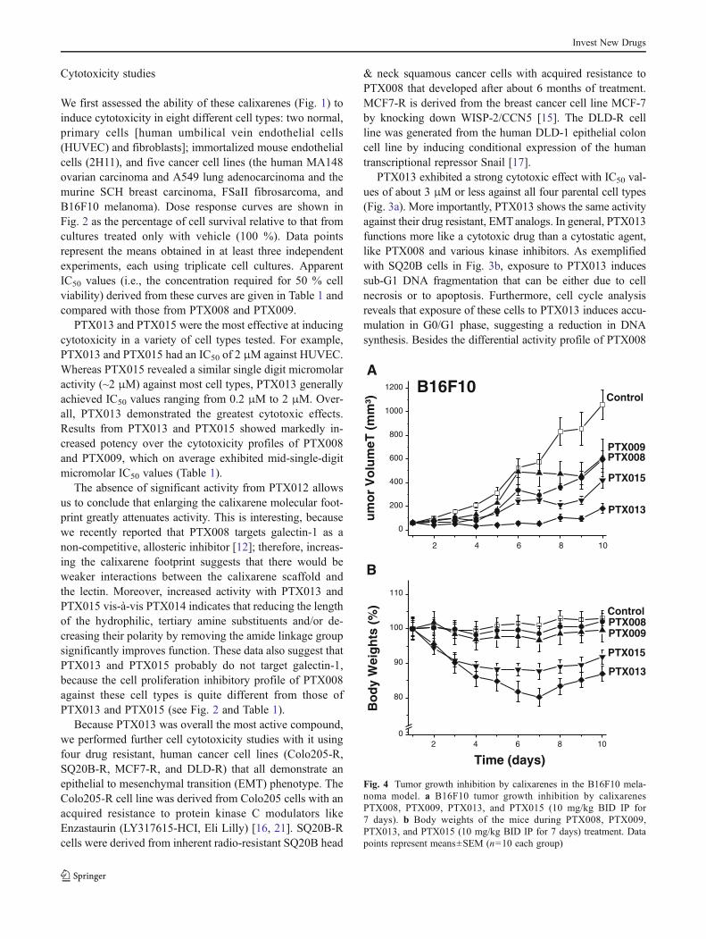
Fig. 4 Tumor growth inhibition by calixarenes in the B16F10 mela-noma model. a B16F10 tumor growth inhibition by calixarenesPTX008, PTX009, PTX013, and PTX015 (10 mg/kg BID IP for7 days). b Body weights of the mice during PTX008, PTX009,PTX013, and PTX015 (10 mg/kg BID IP for 7 days) treatment. Datapoints represent means±SEM (n=10 each group)
Invest New Drugs
and PTX013, since SQ20B and Colo205 cells express rela-tively low levels of galectin-1, SQ20B-R cells show a loss ofgal-1 expression (data not shown), and MCF-7 cells expresshigh levels of galectin-1, these cytotoxicity data further sup-port the proposal that the cellular mechanisms of action ofPTX008 and PTX013 are quite different.
Tumor growth inhibition studies in mice
We assessed the efficacy of the most active of thesecalixarenes PTX013 and PTX015 (along with PTX008,PTX009 and phosphate buffered saline vehicle) in theB16F10 tumor mouse model. These compounds wereadministered (IP BID) in a total dose of 10 mg/kg/mouse)for 8 days (q1dx8), with treatment being initiated whentumors were approximately 75 mm3 in size. Compared toPBS controls, PTX013 was observed to be the mosteffective of these calixarenes at inhibiting B16F10 tumorgrowth, and even appeared to promote tumor growthregression at this dose (Fig. 4a). In addition, althoughtumors in all groups generally grew more rapidly posttreatment, tumor growth in mice treated with PTX013occurred more slowly than in all other groups.
As an indirect measure of toxicity, Fig. 4b plots animalbody weights during the course of this study. Whereas bodyweights of mice treated with PTX008 and PTX009remained constant during the course of treatment, the bodyweights of mice treated with PTX015, and especially withPTX013, fell significantly. With PTX013, treatment washalted on day 6 because 1 out of 10 of these mice diedand the others showed significant reductions (~20 %) inbody weights. In all instances, however, animal bodyweights regained normally post-treatment.
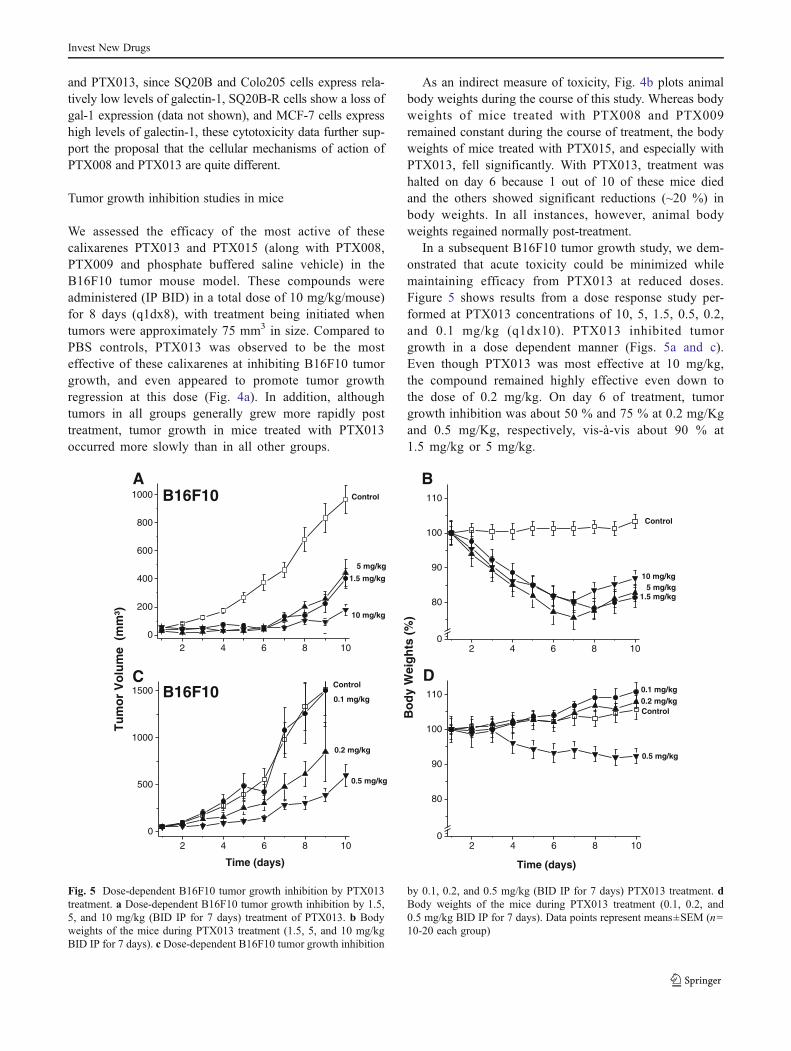
In a subsequent B16F10 tumor growth study, we dem-onstrated that acute toxicity could be minimized whilemaintaining efficacy from PTX013 at reduced doses.Figure 5 shows results from a dose response study per-formed at PTX013 concentrations of 10, 5, 1.5, 0.5, 0.2,and 0.1 mg/kg (q1dx10). PTX013 inhibited tumorgrowth in a dose dependent manner (Figs. 5a and c).Even though PTX013 was most effective at 10 mg/kg,the compound remained highly effective even down tothe dose of 0.2 mg/kg. On day 6 of treatment, tumorgrowth inhibition was about 50 % and 75 % at 0.2 mg/Kgand 0.5 mg/Kg, respectively, vis-à-vis about 90 % at1.5 mg/kg or 5 mg/kg.
2 4 6 8 100
200
400
600
800
1000A
B16F10 Control
1.5 mg/kg
5 mg/kg
10 mg/kg
2 4 6 8 100
80
90
100
110
Bo
dy
Wei
gh
ts (
%)
Time (days)
D
Control
0.1 mg/kg0.2 mg/kg
0.5 mg/kg
2 4 6 8 100
80
90
100
110
B
Control
5 mg/kg1.5 mg/kg
10 mg/kg
2 4 6 8 10
0
500
1000
1500
Time (days)
Tu
mo
r V
olu
me
(m
m³)
CControl
0.1 mg/kg
0.2 mg/kg
0.5 mg/kg
B16F10
Fig. 5 Dose-dependent B16F10 tumor growth inhibition by PTX013treatment. a Dose-dependent B16F10 tumor growth inhibition by 1.5,5, and 10 mg/kg (BID IP for 7 days) treatment of PTX013. b Bodyweights of the mice during PTX013 treatment (1.5, 5, and 10 mg/kgBID IP for 7 days). c Dose-dependent B16F10 tumor growth inhibition
by 0.1, 0.2, and 0.5 mg/kg (BID IP for 7 days) PTX013 treatment. dBody weights of the mice during PTX013 treatment (0.1, 0.2, and0.5 mg/kg BID IP for 7 days). Data points represent means±SEM (n=10-20 each group)
Invest New Drugs

Trends in body weights shown in Figs. 5b and cindicate that apparent toxicity from PTX013 is consid-erably less at these lower doses. Still, some acute tox-icity was noted at the higher doses (10 mg/kg and5 mg/kg), where again treatment was halted on day 6when 1/10 mice died in either group, and the othersshowed that body weights on average dropped by about20 %. Local rashes were also observed at the site ofinjection. In all instances, however, animal bodyweights increased normally and the rashes resolvedpost-treatment. At 0.5 mg/kg, body weights were ratherconstant throughout the entire study, whereas at 0.1 and0.2 mg/kg, mice gained weight normally during treat-ment. However, because post-treatment tumor growthoccurred at a slower rate in mice treated at the0.5 mg/kg dose, the optimal IP dose of PTX013 appearsto be between 0.2 and 0.5 mg/kg/day, which is 20- to50-fold lower than that of PTX008 at 10 mg/kg/day.
Preliminary pharmacodynamics
As an initial read-out on PTX013 pharmacodynamics, wetreated mice using different schedules, while maintainingthe same cumulative dose of 20 mg/kg throughout thetreatment span of 10 days. In this regard, each group ofmice (n = 10) rece ived ei ther 10 in jec t ions of2 mg/kg/mouse daily (q1dx10), 4 injections of5 mg/kg/mouse once every 3 days (q3dx4), or 2 injectionsof 10 mg/kg/mouse once every 5 days (q5dx2). Unexpect-edly, the q5dx2 group that received two injections of10 mg/kg each showed the greatest tumor growth inhibi-tion of about 90 % on day 10 (Fig. 5a), although thesecond injection was accompanied by a transient bodyweight loss of about 8 % (Fig. 6b). The lower and morefrequently administered doses of 2 mg/kg (10 injections,1qdx10) and 5 mg/kg (4 injections, q3dx4) suppressedtumor growth to a lesser extent (Fig. 6a) by about 80 %and 70 %, respectively, but without any apparent loss inaverage body weight (Fig. 5b). Overall, these data strong-ly suggest that PTX013 has good in vivo exposure and arelatively long half-life.
Calixarene-based compounds PTX008 and PTX009 havebeen shown previously to be highly effective therapeuticagents against cancer. By chemically modifying the hydro-phobic and hydrophilic faces of PTX008 and PTX009, wediscovered that two other calix[4]arenes, PTX013 andPTX015, are highly effective as general cytotoxic agents.The most promising of these, PTX013, is considerably moreeffective in vitro at inhibiting proliferation of human cancercells, including various drug resistant cells that have anepithelial to mesenchymal transition (EMT) phenotype.Moreover, PTX013 is highly effective at inhibiting tumorgrowth in mice. Even at a dose of 0.2 mg/kg, PTX013
inhibits tumor growth with about 20-fold greater potencythan PTX008, and preliminary pharmocodynamic data sug-gest that PTX013 exhibits good in vivo exposure and arelatively long half-life. Future studies are needed to identifythe molecular target(s) of PTX013 and PTX015, whichappear to be different from the PTX008 target, galectin-1.Overall, this research contributes to the discovery of thera-peutics as potentially useful agents against cancer and, es-pecially, against drug resistant cancer in the clinic.
Acknowledgements This work was supported by research grantsfrom the National institute of Health - National Cancer Institute (CA-096090 and CA-76497) and OncoEthix Inc. to KHM. ER and LA-Xwere supported by OncoEthix Inc., the Foundation Nelia & AmadeoBarleta (FNAB), and the Association pour la Recherche & l’Enseigne-ment en Cancérologie (AAREC).
2 4 6 8 10 12 14
0
500
1000
1500
2000
q5dx2
q1dx10
Tu
mo
r V
olu
me
(mm
³)
B16F10A
Control
q3dx4
2 4 6 8 10 12 140
80
90
100
110
Bo
dy
Wei
gh
ts (
%)
Time (days)
B
Control
q3dx4q5dx2
q1dx10
Fig. 6 Tumor growth inhibition by PTX013 in the B16F10 melanomamodel using different treatment schedules. a The effect of differentscheduling of PTX013 while maintaining the same accumulative totaltreatment dose. All groups received a total of 20 mg/kg PTX013 at theend of the treatment span of 10 days. However, the groups weredivided to receive either 10 injections of 2 mg/kg every day(q1dx10), 4 injections of 5 mg/kg every 3 days (q3dx4), or 2 injectionsof 10 mg/kg every 5 days (q5dx2). b Body weights of the mice duringdifferent treatment schedules of PTX013. Data points represent means±SEM (n=10 each group)
Invest New Drugs

Conflict of interest Co-authors K.H. Mayo and J. R. MacDonaldhave a financial interest in PepTx, a pharmaceutical company thatholds license to commercialize the PTX compounds
References
1. Hambley TW, Hait WN (2009) Is anticancer drug develop-ment heading in the right direction? Cancer Res 69(4):1259–1262
2. Dings RP, Mayo KH (2007) A journey in structure-based drugdiscovery: from designed peptides to protein surface topomimeticsas antibiotic and antiangiogenic agents. Acc Chem Res 40(10):1057–1065
3. ChenX,Dings RP, Nesmelova I, Debbert S, Haseman JR,Maxwell J,Hoye TR,Mayo KH (2006) Topomimetics of amphipathic beta-sheetand helix-forming bactericidal peptides neutralize lipopolysaccharideendotoxins. J Med Chem 49(26):7754–7765
4. Griffioen AW, van der Schaft DW, Barendsz-Janson AF, Cox A,Struijker Boudier HA, Hillen HF, Mayo KH (2001) Anginex, adesigned peptide that inhibits angiogenesis. Biochem J 354(Pt2):233–242
5. Dings RP, Loren M, Heun H, McNiel E, Griffioen AW, Mayo KH,Griffin RJ (2007) Scheduling of radiation with angiogenesis inhib-itors Anginex and Avastin improves therapeutic outcome via ves-sel normalization. Clin Cancer Res 13(11):3395–3402
6. Dings RP, Van Laar ES, Loren M, Webber J, Zhang Y, Waters SJ,Macdonald JR, Mayo KH (2010) Inhibiting tumor growth by target-ing tumor vasculature with galectin-1 antagonist anginex conjugatedto the cytotoxic acylfulvene, 6-hydroxylpropylacylfulvene.Bioconjug Chem 21(1):20–27
7. Dings RP, van der Schaft DW, Hargittai B, Haseman J, GriffioenAW, Mayo KH (2003) Anti-tumor activity of the novel angiogen-esis inhibitor anginex. Cancer Lett 194(1):55–66
8. Dings RP, Yokoyama Y, Ramakrishnan S, Griffioen AW, MayoKH (2003) The designed angiostatic peptide anginex synergisti-cally improves chemotherapy and antiangiogenesis therapy withangiostatin. Cancer Res 63(2):382–385
9. Ortiz Mellet C, Benito JM, Garcia Fernandez JM (2010) Preorganized,macromolecular, gene-delivery systems. Chemistry 16(23):6728–6742
10. Bagnacani V, Franceschi V, Fantuzzi L, Casnati A, Donofrio G,Sansone F, Ungaro R (2012) Lower Rim Guanidinocalix[4]arenes:Macrocyclic Nonviral Vectors for Cell Transfection. BioconjugChem 23:993-1002
11. Dings RP, Chen X, Hellebrekers DM, van Eijk LI, Zhang Y, HoyeTR, Griffioen AW, Mayo KH (2006) Design of nonpeptidic top-omimetics of antiangiogenic proteins with antitumor activities. JNatl Cancer Inst 98(13):932–936
12. Dings RP, Miller MC, Nesmelova I, Astorgues-Xerri L, Kumar N,Serova M, Chen X, Raymond E, Hoye TR, Mayo KH (2012)Antitumor agent calixarene 0118 targets human galectin-1 as anallosteric inhibitor of carbohydrate binding. J Med Chem 55(11):5121–5129
13. Dings RP, Williams BW, Song CW, Griffioen AW, Mayo KH,Griffin RJ (2005) Anginex synergizes with radiation therapy toinhibit tumor growth by radiosensitizing endothelial cells. Int JCancer 115(2):312–319
14. Dings RP, Loren ML, Zhang Y, Mikkelson S, Mayo KH, Corry P,Griffin RJ (2011) Tumour thermotolerance, a physiological phenom-enon involving vessel normalisation. Int J Hyperth 27(1):42–52
15. Fritah A, Saucier C, De Wever O, Bracke M, Bieche I, Lidereau R,Gespach C, Drouot S, Redeuilh G, Sabbah M (2008) Role ofWISP-2/CCN5 in the maintenance of a differentiated and nonin-vasive phenotype in human breast cancer cells. Mol Cell Biol 28(3):1114–1123
16. Ghoul A, SerovaM, Astorgues-Xerri L, Bieche I, Bousquet G, VarnaM, VidaudM, Phillips E, Weill S, Benhadji KA, Lokiec F, CvitkovicE, Faivre S, Raymond E (2009) Epithelial-to-mesenchymal transitionand resistance to ingenol 3-angelate, a novel protein kinase C mod-ulator, in colon cancer cells. Cancer Res 69(10):4260–4269
17. De Craene B, Gilbert B, Stove C, Bruyneel E, van Roy F, Berx G(2005) The transcription factor snail induces tumor cell invasionthrough modulation of the epithelial cell differentiation program.Cancer Res 65(14):6237–6244
18. Griffin RJ, Dings RP, Jamshidi-Parsian A, Song CW (2010) Mildtemperature hyperthermia and radiation therapy: role of tumourvascular thermotolerance and relevant physiological factors. Int JHyperth 26(3):256–263
19. Mayo KH, Dings RP, Flader C, Nesmelova I, Hargittai B, van derSchaft DW, van Eijk LI, Walek D, Haseman J, Hoye TR, GriffioenAW (2003) Design of a partial peptide mimetic of anginex withantiangiogenic and anticancer activity. J Biol Chem 278(46):45746–45752
20. Dings RP, Van Laar ES, Webber J, Zhang Y, Griffin RJ, Waters SJ,MacDonald JR, Mayo KH (2008) Ovarian tumor growth regres-sion using a combination of vascular targeting agents anginex ortopomimetic 0118 and the chemotherapeutic irofulven. Cancer Lett265(2):270–280
21. Serova M, Astorgues-Xerri L, Bieche I, Albert S, Vidaud M,Benhadji KA, Emami S, Vidaud D, Hammel P, Theou-Anton N,Gespach C, Faivre S, Raymond E (2010) Epithelial-to-mesenchymal transition and oncogenic Ras expression in resis-tance to the protein kinase Cbeta inhibitor enzastaurin in coloncancer cells. Mol Cancer Ther 9(5):1308–1317
Author contribution
Participated in research design:Ruud P.M. Dings, John R. MacDonald, Thomas R. Hoye, Eric
Raymond, and Kevin H. Mayo.Conducted experiments:Ruud P.M. Dings, Joseph I. Levine, Susan G. Brown, Lucile
Astorgues-Xerri.Performed data analysis and wrote the paper:All authors.
Invest New Drugs