polydnavirus dna parasitoid - pnas maxd. summers, august9, 1991 abstract...
TRANSCRIPT

Proc. Nati. Acad. Sci. USAVol. 88, pp. 9770-9774, November 1991Microbiology
Polydnavirus DNA is integrated in the DNA of its parasitoidwasp host
(insect vius/icemod/Campokis sonorensis/parstic hyMenoptera)
Jo-ANN G. W. FLEMING* AND MAX D. SUMMERS*t*Department of Entomology and the tCenter for Advanced Invertebrate Molecular Sciences, Texas A&M University, College Station, TX 77843
Contributed by Max D. Summers, August 9, 1991
ABSTRACT The polydnavirus Campolekis sonorensis virus(CsV) is present in the oviducts of all adult C. sonorensis femalewasps and appears to be required for these wasps to parasitizehosts successfully. Physical mapping, Southern blot analysis,and nucleotide sequence analysis demonstrate that the viralDNA B-specific sequences in cloned wasp DNA are colinearwith viral genomic segmentDNA B from nucleocapsids and arecovalently linked to nonviral wasp sequences. Integrated DNAB terminates in 59-nucleotide imperfect direct repeats, but asingle repeat exists in the extrachromosomal superhelical viralDNA B. Sequences near each junction form imperfect invertedrepeats with sequences near the ends of an internal viral540-base-pair repeat element gene. CsV appears to be the frstdocumented integrated, nonretroviral DNA virus of insects andprobably is vertically transmitted as a provirus.
Polydnaviruses are distinguished from other viruses by theirlarge genomes composed of multiple superhelical DNAs.These viruses appear to be mutualistically associated withcertain wasp species that appear to require the viruses forsuccessful parasitization of host larvae. The polydnavirusCampoletis sonorensis virus (CsV) replicates in the calyxepithelia of the oviducts of all adult female C. sonorensiswasps (1). During oviposition the wasp injects eggs and virusfrom the lumen of the oviducts into lepidopteran host larvaewhere viral genes are expressed (2, 3). Several pronouncedimmunological, physiological, and developmental changesoccur in parasitized hosts concomitant with viral geneexpression and appear to be essential for normal endopara-sitic development of the wasp in the host (4, 5).
All adult females in several species of ichneumonid orbraconid wasps contain polydnaviruses that appear to bespecies specific (6-10). Genetic studies suggested that thepolydnavirus of Cotesia melanoscela braconid wasps is ver-tically transmitted, but physical evidence for viral integrationhas not been reported (11). In contrast, CsV-specific DNA isdetectable as offsize restriction fragments in nonoviductsomatic tissues of both male and female C. sonorensisichneumonid wasps, suggesting that CsV DNA is integratedin the wasp genome (12). However, an alternative interpre-tation that the CsV offsize restriction fragments representedpolymorphic or rearranged viral DNAs could not be excludeddue to the incompletely understood, complex patterns ofintragenomic cross-hybridization (3, 6, 12-15).
In this study, we examine CsVDNA segment B and relatedcloned C. sonorensis sequences by Southern blot hybridiza-tion and nucleotide sequence analysis to determine whetherthe offsize restriction fragments detected in wasp DNAindicate the integration of polydnaviral DNA or rearrangedsequences in the viral genome A Our data show that unrear-ranged viral DNA B is covalently linked to wasp sequences.
Polydnaviruses like CsV likely are maintained in parasitichymenoptera as integrated proviruses.
MATERIALS AND METHODSInsects, Virus, Genomic Libraries, and Plasmid DNA. Meth-
ods of rearing an inbred population of C. sonorensis waspsand virus and viral DNA purification have been published (2,7, 12). Total cellularDNA was extracted from male wasps bystandard methods because they lack oviducts (16). Thepredominant form of viral DNA in males is detectable asoffsize restriction fragments (12). A C. sonorensis genomiclibrary was constructed by ligating BamHI-cleaved EMBL3A DNA arms (Promega) with size-selected Mbo I partialdigestion products of male wasp DNA (16). Methods ofplaque purification, A DNA isolation, plasmid extraction andpurification, and generation of overlapping deletions withexonucleases Exo III and Exo VII or exonuclease BAL-31have been described (16-20). Extrachromosomal CsV DNAB was cloned in its entirety as independent 6.7-kilobase-pair(kbp) BamHI (pBB6700) or EcoRI (pBE6700) fragments byD. A. Theilmann, who provided the clones (21). The BamHIclone (pBB6700) was used routinely.
Southern Blots. Southern blotting, high-stringency hybrid-ization and washing conditions, autoradiography, and nick-repair labeling of gel-purified, vector-free DNAs with[32P]dATP to high specific activity (>108 cpm/Ixg) wereperformed as described (12). For lower-stringency conditionhybridizations, the percentage of formamide in the hybrid-ization buffer was decreased to 30%, and the washing con-ditions were altered by lowering the temperature of the 0.1 xstandard saline citrate/0.1% SDS washes to 370C.DNA Sequence Analysis. The nucleotide sequence of both
strands was determined with single-stranded DNA by thedideoxynucleotide chain-termination method (22). Sequencedata were analyzed with the University of Wisconsin Genet-ics Computer Group (UWGCG) software forVAX computers(release 60) (23).
RESULTSCharacterization of Wasp DNAs Related to Viral DNA B.
Previous Southern blot analyses suggested that a 1.25-kbpXho I fragment ofCsV DNA B (6.7 kbp) is detectable in maleC. sonorensis cellular DNA as offsize restriction fragments(12). Additional Southern blots of male wasp DNA and CsVDNA digested with several other restriction endonucleasesand hybridized with radiolabeled cloned CsV DNA B(pBB6700) or subcloned fragments of it indicated that otherDNA B sequences are not detectably altered in wasp DNA
Abbreviations: CsV, Campoletis sonorensis virus; nt, nucleotide(s);DRL, direct repeat, left; DRR, direct repeat, right; IR, invertedrepeat; ORF, open reading frame.MThe sequences reported in this paper have been deposited in theGenBank data base (accession nos. M80621, M80622, and M80623).
9770
The publication costs of this article were defrayed in part by page chargepayment. This article must therefore be hereby marked "advertisement"in accordance with 18 U.S.C. §1734 solely to indicate this fact.
Proc. Natl. Acad. Sci. USA 88 (1991) 9771
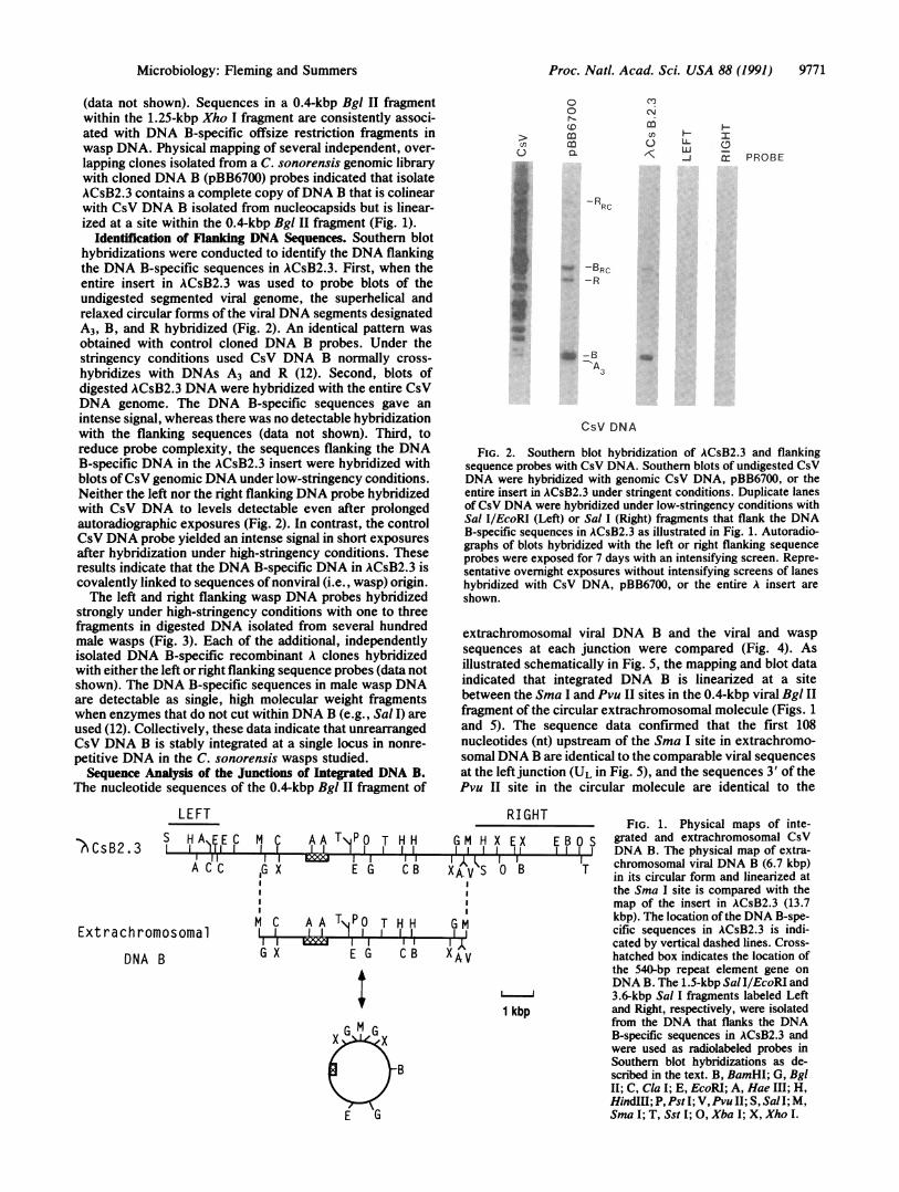
(data not shown). Sequences in a 0.4-kbp Bgl II fragmentwithin the 1.25-kbp Xho I fragment are consistently associ-ated with DNA B-specific offsize restriction fragments inwasp DNA. Physical mapping of several independent, over-lapping clones isolated from a C. sonorensis genomic librarywith cloned DNA B (pBB6700) probes indicated that isolateACsB2.3 contains a complete copy ofDNA B that is colinearwith CsV DNA B isolated from nucleocapsids but is linear-ized at a site within the 0.4-kbp Bgl II fragment (Fig. 1).
Identification of Flanking DNA Sequences. Southern blothybridizations were conducted to identify the DNA flankingthe DNA B-specific sequences in ACsB2.3. First, when theentire insert in ACsB2.3 was used to probe blots of theundigested segmented viral genome, the superhelical andrelaxed circular forms of the viral DNA segments designatedA3, B, and R hybridized (Fig. 2). An identical pattern wasobtained with control cloned DNA B probes. Under thestringency conditions used CsV DNA B normally cross-hybridizes with DNAs A3 and R (12). Second, blots ofdigested ACsB2.3 DNA were hybridized with the entire CsVDNA genome. The DNA B-specific sequences gave anintense signal, whereas there was no detectable hybridizationwith the flanking sequences (data not shown). Third, toreduce probe complexity, the sequences flanking the DNAB-specific DNA in the ACsB2.3 insert were hybridized withblots ofCsV genomic DNA under low-stringency conditions.Neither the left nor the right flanking DNA probe hybridizedwith CsV DNA to levels detectable even after prolongedautoradiographic exposures (Fig. 2). In contrast, the controlCsV DNA probe yielded an intense signal in short exposuresafter hybridization under high-stringency conditions. Theseresults indicate that the DNA B-specific DNA in ACsB2.3 iscovalently linked to sequences of nonviral (i.e., wasp) origin.The left and right flanking wasp DNA probes hybridized
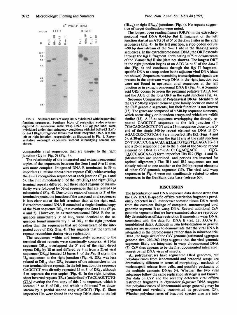
strongly under high-stringency conditions with one to threefragments in digested DNA isolated from several hundredmale wasps (Fig. 3). Each of the additional, independentlyisolated DNA B-specific recombinant A clones hybridizedwith either the left or right flanking sequence probes (data notshown). The DNA B-specific sequences in male wasp DNAare detectable as single, high molecular weight fragmentswhen enzymes that do not cut within DNA B (e.g., Sal I) areused (12). Collectively, these data indicate that unrearrangedCsV DNA B is stably integrated at a single locus in nonre-petitive DNA in the C. sonorensis wasps studied.Sequence Analysis of the Junctions of Integrated DNA B.
The nucleotide sequences of the 0.4-kbp Bgl II fragment of
LEFT
)BCSB2.3 S H EC M C AA T PO T H H
A C C lb X
lll
Extrachromosomal
DNA B
baw I IE G CB
MCa AAT T HHI I ) II II
I I
B
en0
S
a,
00
(DmmcL
m
An
c) LA ELU 0
-4 x PROBE
-RRC
_W -BRCf _R
3
CSV DNA
FIG. 2. Southern blot hybridization of ACsB2.3 and flankingsequence probes with CsV DNA. Southern blots of undigested CsVDNA were hybridized with genomic CsV DNA, pBB6700, or theentire insert in ACsB2.3 under stringent conditions. Duplicate lanesof CsV DNA were hybridized under low-stringency conditions withSal IlEcoRI (Left) or Sal I (Right) fragments that flank the DNAB-specific sequences in ACsB2.3 as illustrated in Fig. 1. Autoradio-graphs of blots hybridized with the left or right flanking sequenceprobes were exposed for 7 days with an intensifying screen. Repre-sentative overnight exposures without intensifying screens of laneshybridized with CsV DNA, pBB6700, or the entire A insert areshown.
extrachromosomal viral DNA B and the viral and waspsequences at each junction were compared (Fig. 4). Asillustrated schematically in Fig. 5, the mapping and blot dataindicated that integrated DNA B is linearized at a sitebetween the Sma I and Pvu II sites in the 0.4-kbp viral Bgl IIfragment of the circular extrachromosomal molecule (Figs. 1and 5). The sequence data confirmed that the first 108nucleotides (nt) upstream of the Sma I site in extrachromo-somalDNA B are identical to the comparable viral sequencesat the left junction (UL in Fig. 5), and the sequences 3' of thePvu II site in the circular molecule are identical to the
RI GHTFIG. 1. Physical maps of inte-
G M H X E X E B o S grated and extrachromosomal CsV..,L,4l..l il.Jl... JrJ DNA B. The physical map of extra-XA.V S IO'B IT chromosomal viral DNA B (6.7 kbp)XA. V S 0 B T in its circular form and linearized at
the Sma I site is compared with themap of the insert in ACsB2.3 (13.7kbp). The location of the DNA B-spe-cific sequences in ACsB2.3 is indi-cated by vertical dashed lines. Cross-
X A V hatched box indicates the location ofthe 540-bp repeat element gene onDNA B. The 1.5-kbp Sal I/EcoRI and3.6-kbp Sal I fragments labeled Left
1 kbp and Right, respectively, were isolatedfrom the DNA that flanks the DNAB-specific sequences in ACsB2.3 andwere used as radiolabeled probes inSouthern blot hybridizations as de-scribed in the text. B, BamHI; G, BglII; C, Cla I; E, EcoRI; A, Hae III; H,HindIII; P, Pst I; V, Pvu II; S, Sal I; M,Sma I; T, Sst I; 0, Xba I; X, Xho I.
II IG X E G
Microbiology: Fleming and Summers
9772 Microbiology: Fleming and Summers
dWASP DNA
RIGHT
-r -V
C a- X u X
(V (
0.CL1 LUI
I.
Aw
0 6-
FIG. 3. Southern blots ofwasp DNA hybridized with the nonviralflanking sequences. Southern blots of restriction endonuclease-digested C. sonorensis male wasp DNA (10 ,Ag per lane) were
hybridized under high-stringency conditions with Sal I/EcoRI (Left)or Sal I (Right) fragment DNAs that flank integrated DNA B at theleft or right junction, respectively, as illustrated in Fig. 1. Repre-sentative overnight exposures without intensifying screens are
shown.
comparable viral sequences that are unique to the rightjunction (UR in Fig. 5) (Fig. 4).The relationship of the integrated and extrachromosomal
copies of the sequences between the Sma I and Pvu II siteswas more complex. Integrated DNA B terminated in 59-ntimperfect (11 mismatches) direct repeats (DR), which overlapthe Sma I recognition sequences at each junction (Figs. 4 and5). The 7 nt immediately 3' of the left (DRL) and right (DRR)terminal repeats differed, but these short regions of dissim-ilarity were followed by 53-nt sequences that are related (14mismatches) (Fig. 4). Due to this region of similarity 3' of theterminal repeats, the junction of the wasp and viral sequencesis less clear-cut at the left terminus than at the right end.Extrachromosomal DNA B contained a single identical copyof the 59-nt sequence DRL that overlaps the Sma I site (Figs.4 and 5). However, in extrachromosomal DNA B the se-
quences immediately 3' of DRL were identical to the se-
quences found immediately 3' of DRR at the right junctionrather than the sequences that are downstream of the inte-grated copy of DRL (Fig. 4). This suggests that the terminalrepeats recombine during virus replication.The sequences within and immediately adjacent to the
terminal direct repeats were structurally complex. A 21-bpsequence DR3A overlapped the 3' end of the right directrepeat DRR by 18 nt and differed by 4 nt from a 21-nt viralsequence (DR3B) located 25 bases 3' of the Pvu II site in theUR sequences at the right junction (Fig. 4). DRL was lessrelated to DR3B than DRR because of the mismatches in thetwo terminal direct repeats. In the left junction, the sequenceCAGCTCT was directly repeated 15 nt 3' of DRL, although5 nt separate the two copies (Fig. 4). In the right junction,short inverted repeats (IRv) (underlined) (TACCAGCTC(Ti-GTIA) overlap the first copy of this 7-nt sequence, which islocated 15 nt 3' of DRR and which is followed 7 nt down-stream by a partial second copy (CAGCT) (Fig. 4). Shortimperfect IRs were found in the wasp DNA close to the left
(IRWL) or right (IRWR) junctions (Fig. 4). No repeats sugges-tive of target duplications were noted.The longest open reading frames (ORFs) in the extrachro-
mosomal viral DNA 0.4-kbp Bgl II fragment or the leftjunction start at an ATG 31 nt 5' of the Sma I sites in the viralsequences (Fig. 4). In the left junction, a stop codon occurs':90 bp downstream of the Sma I site in the flanking waspsequences. In the extrachromosomal DNA, the ORF extendsthrough the Bgl II fragment, terminating -75 nt downstreamof the 3'-most Bgl II site (data not shown). The longest ORFin the right junction begins at an ATG 34 nt 5' of the Sma Isite (Fig. 4) and continues through the Bgl II fragment-specific DNA to a stop codon in the adjacent viral DNA (datanot shown). Sequences resembling transcriptional signals arepresent in the upstream wasp DNA in the right junction butwere not found in upstream viral sequences at the leftjunction or in extrachromosomal DNA B (Fig. 4). A 5-aminoacid ORF occurs between the proximal putative TATA boxand the AUG of the long ORF in the right junction (Fig. 4).Sequence Comparison of Polydnaviral DNAs. Members of
the CsV 540-bp repeat element gene family occur on most ofthe CsV genomic segments, but their function is not known(15). The genes are composed of -540-bp sequence elements,which occur singly or in tandem arrays and which are -60%similar (15). A 13-nt sequence overlapping the directly re-peated CAGCTCT sequence at the left junction (5'-TGACAGCAGCTCT-3') and a 13-nt sequence close to the 5'end of the single 540-bp repeat element on DNA B (5'-AGAGCGGCTGTCA-3') are imperfect IRs IR1 (Figs. 4 and5). A 30-nt sequence near the Bgl II site at the right junction(5'-TTGCTCGGAACAGATGACGTiGTGiCAGATG-3')and a 26-nt sequence close to the 3' end of the 540-bp repeatelement on DNA B (5'-CATCTGjACGACG .... CCG-TACCGAGCAA-3') form imperfect IRs IR2 (Figs. 4 and 5).(Mismatches are underlined, and periods are inserted foroptimal alignment.) The IR1 and IR2 sequences are notclosely related to one another or the 540-bp repeat elementsof other CsV genomic segments (15). The viral and waspsequences in Fig. 4 were not significantly related to othersequences in the GenBank data base (release 60).
DISCUSSIONThe hybridization and DNA sequence data demonstrate thatthe CsV DNA B-specific offsize restriction fragments previ-ously detected in C. sonorensis somatic tissue DNA resultfrom the covalent linkage of complete, unrearranged viralgenomic segment B to wasp DNA. Several additional CsVgenomic segments that we have examined also are reproduc-ibly detectable as offsize restriction fragments in wasp DNA,consistent with the data for DNA B (ref. 12; J.G.W.F.,unpublished data). Although in situ hybridization or geneticanalyses are necessary to demonstrate that the viral DNA isintegrated in the chromosomes rather than in mitochondrialDNA, the large size of the CsV genome (estimated aggregategenome size, 210-260 kbp) suggests that the viral genomicsegments likely are integrated in wasp chromosomal DNA(7). CsV thus appears to be the first documented integrated,nonretroviral DNA virus of insects.
All polydnaviruses have segmented DNA genomes, butpolydnaviruses from ichneumonid and braconid wasps aredramatically different in terms of morphology, methods ofnucleocapsid release from cells, and possibly packaging ofthe multiple genomic DNAs (4). Whether the two viralsubgroups follow the same replication strategy is not known.Our data on CsV and the recently detected viral offsizerestriction fragments in Hyposoter fugitivus DNA suggestthat polydnaviruses of ichneumonid wasps generally may beintegrated and vertically transmitted as proviruses (24).Whether polydnaviruses of braconid species also are inte-
Probe LEFT
_0M C
23 1-94-66- _
4 4-
23-20-
Proc. Natl. Acad Sci. USA 88 (1991)

Microbiology: Fleming and Summers Proc. Natl. Acad. Sci. USA 88 (1991) 9773
AGATCTCGGT CAGAGAGAGA TTCCAGAAAT AGACTCGTCT TTTAGTTGTT TCGTTGTTAT TGCCTAGTTT CTCTCGTTAG 80BglII-
LDRL
TGGCACAATT GCAG¶6ZC TAAGAGTGAC TGCATCAACT GGGTGGCCCG GGTGCTTATC TGTTCCCGCA GGAAGCGTAC 160-SmaI-
* *ATCGTGTCAA CTACTCACGT ACCAGCTCTG GTATAGCAGC TGCCGAACGG TAGCGAGTCT GGCCTTCATC AAGCGTCTAC 240
-IRV- -IRv -PVUII- DR3B
CTTGTCAAAA AAGGCTATGT TTCAAATGTC TAGCTGCCGT AATATCATCC ATAGCGACCT GTGCCAGGCA TCAGCCGTAC 320
CAATCGTACC TTGCGCTAAT TATGACTCAT AGCAATTATA TTATCTACGA CAAAAATGTT TGCTCGGAAC AGATGACGTG 400
GTGCAGATGC AGATCT 416BglII-
R
AGATCTCGGT CAGAGAGAGA TTCCAGAAAT AGACTCGTCT TTTAGTTGTT TCGTTGTTAT TGCCTAGTTT CTCTCGTTAG 80BglII-
LDRL
TGGCACAATT GCAG C TAAGAGTGAC TGCATCAACT GGGTGGCCCG GGTGCTTATC TGTTCCCGCA GGAAGCGTAC 160viraa * * -SmaI-
ATCGTGTTGG TCTATCACGA ATCAGCTCTG ACAGCAGCTC TTAAACCGTA GCAAGTCTAA TCTTCGTGTA ACTCCCAACA 240IR1
TATCCAAAGT CAGCTCATCT TCAGAATGAC TACCGGCTCG ATTGTTACAG TTGATGTTTC AACCAACAAA AAACGCCGTA 320
CTCGCGAGGC AGAACAACGG TAATCTCCCT ACTTTAAGTC TAAAAAATCA
GGGACCGCGA AAACGTCaTG GAAAATTGTC CATATCAGGC GCAACTCTAC
-IRWL--IRW-
CATTTTCGGG TATGTAAACC TGGGTTTTTC 400
450
T 7 J[CTI
AATCGAATTA ACCCTAGAAC TCAATGCATC
ATTTGGAGGT TGACATATTT TCTI ¶A
ATCC A AAAACTCACC ACTTAACATAIRWR-
ATCGTCGACC TCTGCACGTG AAGTTCCCTC
GTTTATTATT TCATAACAGT GACTAGTGAC
CTGTCG7I TTCTATCATC GTTACTGCAT~~~~~~~*GTCAACTACT CACGTACCAG CTCTGGTATA
-IRv- -IRv-
CAAAAAAGGC TATGTTTCAA ATGTCTAGCT
GTACCTTGCG CTAATTATGA CTCATAGCAA
ATTTTTTTTA
CTTAAATTCT
TCGACTACGA
AAAAAGCACT
AAAAAGAATA AAACTAAGTA
GACC*TGGT GGTTAGGCTT|
AAAGACTAGT TTTTTCGTTG
DRR-CGGCAGGCTG TCCCGGGAGA
-SmaI-
GCAGCTGCCG AACGGTAGCG
-PvuI I-
GCCGTAATAT CATCCATAGC
TTATATTATC TACGACAAAA
TTTTATCTP GTACAG GATGTCTAAC 80
GCTTACGCCA AAATCAAAAA AAGAATCAGC 160
CAATTGCATT TTGACTTGCG ATAGGTTTGC 240
TGAATATATT AAATACGTGA AATTTCTCGC 320
TGTCGTGAGG CTTCTTTGAT CCGTGGCACC 400IRW1
TCATCTGTTC CCGAAGCAAG CGTATATCGT 480DR3A
AGTCTGGCCT TCATCAAGCG TCTACCTTGT 560
DR3B -
GACCTGTGCC AGGCATCAGC CGTACCAATC 640
ATGTTTGCTC GGAACAGATG ACGTGGTGCA 720IR2
GATGCAGATC T 731-BglII-
R
FIG. 4. Nucleotide sequences of the viral and wasp sequences at the junctions of integrated DNA B or the extrachromosomal viralhomologue. Nucleotide sequences of the 0.4-kbp Bgl II fragment of extrachromosomal DNA B, the comparable viral sequences at the left orright junctions of integrated DNA B, and the flanking wasp DNA are shown in standard 5' -+ 3' orientation. The terminal direct repeats DRLor DRR are indicated by lines above the sequences. Other sequence pairs forming direct (DR) or inverted (IR) repeats are underlined and areidentified by similar subscripts. Sequences related to the 540-bp repeat element gene (IR1 and IR2) are noted. A 7-nt sequence motif discussedin the text is indicated by an asterisk. Possible CCAAT and TATA sequences, the ATGs of the longest ORFs detected, and a 5-amino acid ORFlocated upstream of the ATG of the long ORF in the right junction are boxed.
grated is not clear from conflicting published data. Only regated among wasp progeny in ratios consistent with pos-episomal viral DNAs were detected in male C. melanoscela sible chromosomal inheritance of the virus (24):braconid wasps, and viral DNA probes did not hybridize to The replication strategy of polydnaviruses is not known.the chromosomal DNA (11). In a genetic analysis of the same Unlike plant multipartite mitochoidrial genomes, no evi-species, however, two polymorphic forms of the virus seg- dence for a large, "master" circular DNA from which the
9774 Microbiology: Fleming and Summers
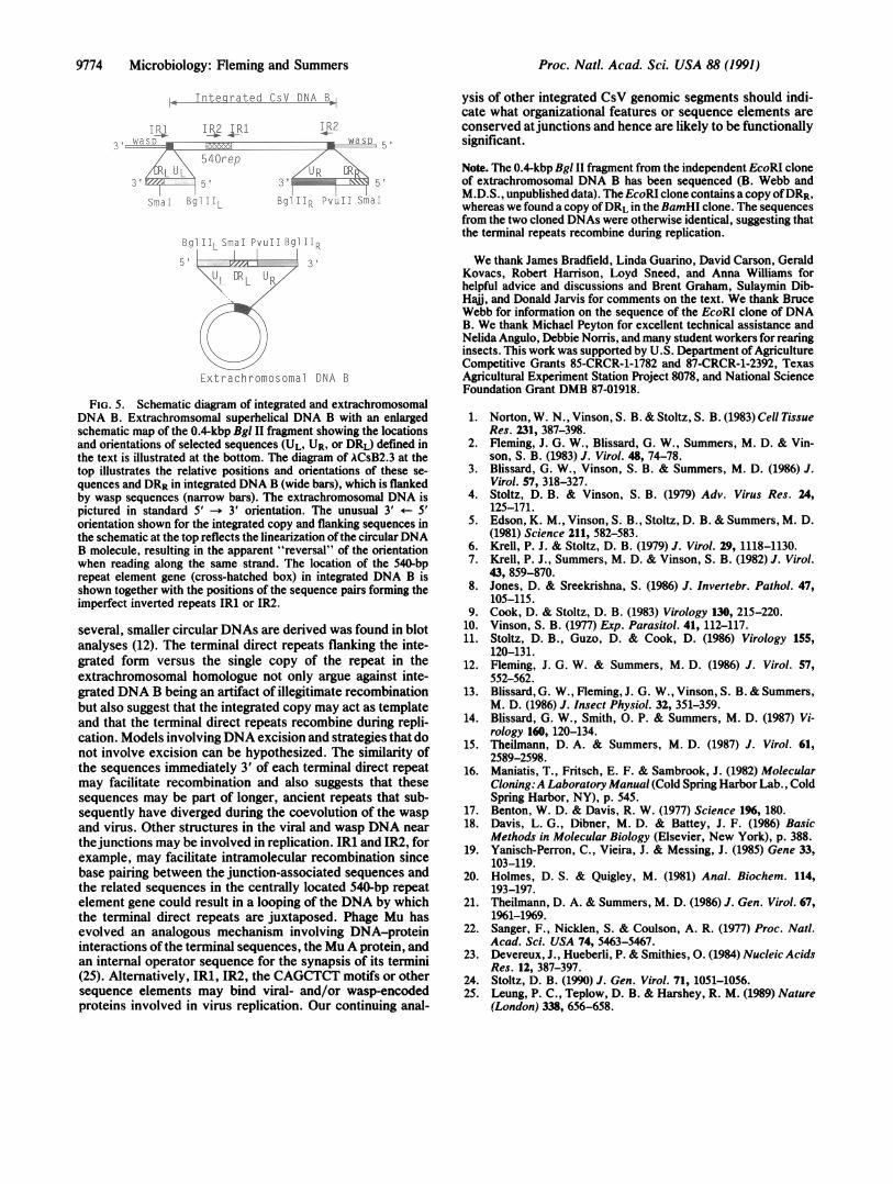
I1ntegrated CsV DNA B
r' IRT2 I RI!.N -E 'f L"_,
540repLRL
t . _ b-
.2 a I
I R?
LI r ; I I
ER t b
iR
Lxtrachromosomal DNA B
FIG. 5. Schematic diagram of integrated and extrachromosomalDNA B. Extrachromsomal superhelical DNA B with an enlargedschematic map of the 0.4-kbp Bgl II fragment showing the locationsand orientations of selected sequences (UL, UR, or DRL) defined inthe text is illustrated at the bottom. The diagram of ACsB2.3 at thetop illustrates the relative positions and orientations of these se-quences and DRR in integrated DNA B (wide bars), which is flankedby wasp sequences (narrow bars). The extrachromosomal DNA ispictured in standard 5' -* 3' orientation. The unusual 3' +- 5'
orientation shown for the integrated copy and flanking sequences inthe schematic at the top reflects the linearization ofthe circularDNAB molecule, resulting in the apparent "reversal" of the orientationwhen reading along the same strand. The location of the 540-bprepeat element gene (cross-hatched box) in integrated DNA B isshown together with the positions of the sequence pairs forming theimperfect inverted repeats IR1 or IR2.
several, smaller circular DNAs are derived was found in blotanalyses (12). The terminal direct repeats flanking the inte-grated form versus the single copy of the repeat in theextrachromosomal homologue not only argue against inte-grated DNA B being an artifact of illegitimate recombinationbut also suggest that the integrated copy may act as templateand that the terminal direct repeats recombine during repli-cation. Models involvingDNA excision and strategies that donot involve excision can be hypothesized. The similarity ofthe sequences immediately 3' of each terminal direct repeatmay facilitate recombination and also suggests that thesesequences may be part of longer, ancient repeats that sub-sequently have diverged during the coevolution of the waspand virus. Other structures in the viral and wasp DNA nearthejunctions may be involved in replication. IR1 and IR2, forexample, may facilitate intramolecular recombination sincebase pairing between the junction-associated sequences andthe related sequences in the centrally located 540-bp repeatelement gene could result in a looping of the DNA by whichthe terminal direct repeats are juxtaposed. Phage Mu hasevolved an analogous mechanism involving DNA-proteininteractions of the terminal sequences, the MuA protein, andan internal operator sequence for the synapsis of its termini(25). Alternatively, IR1, IR2, the CAGCTCT motifs or othersequence elements may bind viral- and/or wasp-encodedproteins involved in virus replication. Our continuing anal-
ysis of other integrated CsV genomic segments should indi-cate what organizational features or sequence elements areconserved atjunctions and hence are likely to be functionallysignificant.
Note. The 0.4-kbp Bgl II fragment from the independent EcoRI cloneof extrachromosomal DNA B has been sequenced (B. Webb andM.D.S., unpublished data). The EcoRI clone contains a copy ofDRR,whereas we found a copy ofDRL in theBamHI clone. The sequencesfrom the two cloned DNAs were otherwise identical, suggesting thatthe terminal repeats recombine during replication.
We thank James Bradfield, Linda Guarino, David Carson, GeraldKovacs, Robert Harrison, Loyd Sneed, and Anna Williams forhelpful advice and discussions and Brent Graham, Sulaymin Dib-Haii, and Donald Jarvis for comments on the text. We thank BruceWebb for information on the sequence of the EcoRI clone of DNAB. We thank Michael Peyton for excellent technical assistance andNelida Angulo, Debbie Norris, and many student workers for rearinginsects. This work was supported by U.S. Department ofAgricultureCompetitive Grants 85-CRCR-1-1782 and 87-CRCR-1-2392, TexasAgricultural Experiment Station Project 8078, and National ScienceFoundation Grant DMB 87-01918.
1. Norton, W. N., Vinson, S. B. & Stoltz, S. B. (1983) Cell TissueRes. 231, 387-398.
2. Fleming, J. G. W., Blissard, G. W., Summers, M. D. & Vin-son, S. B. (1983) J. Virol. 48, 74-78.
3. Blissard, G. W., Vinson, S. B. & Summers, M. D. (1986) J.Virol. 57, 318-327.
4. Stoltz, D. B. & Vinson, S. B. (1979) Adv. Virus Res. 24,125-171.
5. Edson, K. M., Vinson, S. B., Stoltz, D. B. & Summers, M. D.(1981) Science 211, 582-583.
6. Krell, P. J. & Stoltz, D. B. (1979) J. Virol. 29, 1118-1130.7. Krell, P. J., Summers, M. D. & Vinson, S. B. (1982) J. Virol.
43, 859-870.8. Jones, D. & Sreekrishna, S. (1986) J. Invertebr. Pathol. 47,
105-115.9. Cook, D. & Stoltz, D. B. (1983) Virology 130, 215-220.
10. Vinson, S. B. (1977) Exp. Parasitol. 41, 112-117.11. Stoltz, D. B., Guzo, D. & Cook, D. (1986) Virology 155,
120-131.12. Fleming, J. G. W. & Summers, M. D. (1986) J. Virol. 57,
552-562.13. Blissard, G. W., Fleming, J. G. W., Vinson, S. B. & Summers,
M. D. (1986) J. Insect Physiol. 32, 351-359.14. Blissard, G. W., Smith, 0. P. & Summers, M. D. (1987) Vi-
rology 160, 120-134.15. Theilmann, D. A. & Summers, M. D. (1987) J. Virol. 61,
2589-2598.16. Maniatis, T., Fritsch, E. F. & Sambrook, J. (1982) Molecular
Cloning:A Laboratory Manual (Cold Spring Harbor Lab., ColdSpring Harbor, NY), p. 545.
17. Benton, W. D. & Davis, R. W. (1977) Science 196, 180.18. Davis, L. G., Dibner, M. D. & Battey, J. F. (1986) Basic
Methods in Molecular Biology (Elsevier, New York), p. 388.19. Yanisch-Perron, C., Vieira, J. & Messing, J. (1985) Gene 33,
103-119.20. Holmes, D. S. & Quigley, M. (1981) Anal. Biochem. 114,
193-197.21. Theilmann, D. A. & Summers, M. D. (1986) J. Gen. Virol. 67,
1%1-1969.22. Sanger, F., Nicklen, S. & Coulson, A. R. (1977) Proc. Natl.
Acad. Sci. USA 74, 5463-5467.23. Devereux, J., Hueberli, P. & Smithies, 0. (1984) Nucleic Acids
Res. 12, 387-397.24. Stoltz, D. B. (1990) J. Gen. Virol. 71, 1051-1056.25. Leung, P. C., Teplow, D. B. & Harshey, R. M. (1989) Nature
(London) 338, 656-658.
Proc. Natl. Acad Sci. USA 88 (1991)