polyethylene glycol-coated biocompatible surfaces - diyhpl.usdiyhpl.us/~nmz787/pdf/polyethylene...
TRANSCRIPT

Polyethylene glycol–coated biocompatible surfaces
Norma A. Alcantar, Eray S. Aydil, Jacob N. IsraelachviliChemical Engineering Department and Materials Department, University of California, Santa Barbara, California 93106
Received 17 August 1999; accepted 10 January 2000
Abstract: Surfaces covered with polyethylene glycol (PEG;HO–(CH2–CH2–O)n–H) have been shown to be biocompat-ible because PEG’s properties yield nonimmunogenicity,nonantigenicity, and protein rejection. To produce a biocom-patible surface coating, we have developed a method forgrafting PEG onto activated silica films. We first depositedan amorphous silica film by plasma-enhanced chemical va-por deposition from SiH4 and O2 gases, which provided theflexibility to coat diverse materials with different chemistriesand shapes. The silica films were activated by exposure towater plasma, increasing the number of silanol groups (Si–OH) on their surface. The surface silanol groups were thenchemically reacted with the hydroxyl end of PEG to form anester bond, Si–O–C, and to cover the surface with PEG. Thesurface reactions were monitored using attenuated total re-flection Fourier transform infrared spectroscopy. The vibra-tional absorption bands of the C–O and –CH2 bonds in-creased with time and saturated, indicating that PEG wasadsorbed to saturation coverage on the surface. Simulta-
neously, the Si–OH absorption band decreased, showingthat the surface silanols reacted with PEG and were de-pleted. The PEG-covered surfaces were physically character-ized by atomic force microscopy, Auger electron spectros-copy, ellipsometry, and contact angle measurements. Thesecharacterization techniques provided additional evidencefor the existence of chemically bonded PEG on the surfaces.Efficacy of protein rejection on PEG-covered surfaces wasstudied through measurements of the fluorescence intensityof Texas red–labeled bovine serum albumin brought in con-tact with such surfaces in solution. Significantly less proteinadsorption was observed on surfaces covered with PEGcompared to uncovered surfaces. © 2000 John Wiley & Sons,Inc. J Biomed Mater Res, 51, 343–351, 2000.
Key words: polyethylene glycol; silica, silicon oxide; chemi-cal grafting of PEG; biocompatible surface; biocompatiblecoating
INTRODUCTION
The ability to design, fabricate, and optimize sur-faces tailored for a particular biological application isof crucial importance to the synthesis of biocompatiblematerials.1 For example, one of the more difficultproblems in bioengineering science is finding versatilematerials to be used in constructing artificial parts thatare able to reproduce the properties and functionalityof human tissues and organs.2 Biomaterials that are indemand include materials for temporary and long-term therapeutic devices, prostheses for bone, skin,joints, cartilage, teeth, heart valves, blood vessels, andcontact and intraocular lenses, and encapsulation for
drug delivery systems.3–5 Although efforts devoted inthe past to developing biomaterials have resulted inrecovery of specific functions or prolongation of lifefor many patients, successful applications are limitedin scope, and there are many problems yet to besolved. One of the major problems is the undesiredinteractions of foreign materials introduced into thebody with the immune system (e.g., leukocytes,phagocytes) and biomolecules such as lipids, proteins,fats, and enzymes. This problem is generally referredas bioincompatibility.6,7
The two most desirable characteristics of prostheticdevices are precise medicofunctionality and excellentbiocompatibility. The former characteristic defines theperformance or function of the device needed to real-ize a medical aim efficiently. The second characteristicis essential for any prosthesis that comes into contactwith the human body and specifically with the im-mune system. When any foreign material is intro-duced into an organism, disturbances are generated atthe contacting interfaces. Examples of these distur-bances are blood coagulation, cytotoxic effects, im-mune response deficiency, injuries of biological mem-branes, and tissue damage. Furthermore, the device
Correspondence to: J. N. Israelachvili; e-mail: [email protected]
Contract grant sponsor: NSF; Contract grant numbers:DMR-9123048, ECS-9457758
Contract grant sponsor: U. C. Biostar Program; Contractgrant number: 97-21
Contract grant sponsor: NIH; Contract grant number: PHSGM 47334
© 2000 John Wiley & Sons, Inc.
itself can suffer deterioration such as chemical attack,physical deformation, and mechanical dysfunction.8
One of the most successful approaches to producingsurfaces that are able to resist protein adhesion andbiological attack has been to use polyethylene glycol(PEG) as a surface protector. For example, it has beenfound that grafting PEG to solid surfaces reduces pro-tein adsorption and cell adhesion.9–13 Furthermore, itwas demonstrated both in vitro and in vivo that PEGcoatings suppress platelet adhesion, leading to re-duced risk of thrombus formation, tissue damage, andother cytotoxic effects.14 Others have found that somesubstances covered with a PEG coating do not showantigenic activity.11–13 PEG is unusually effective atexcluding other polymers from its presence in aque-ous solution. This property is thought to be directlyrelated to its ability to repel proteins.15 PEG formstwo-phase systems with other polymers, it is nontoxic,it exhibits immunogenicity and nonantigenicity, and itdoes not harm active proteins or cells even when itinteracts with them directly.9 In addition, it attaches tosurfaces with very little effect on their chemistry; it issoluble in water and most organic solvents such astoluene and chloroform, and it increases the solubilityof large molecules irrespective of their size. Further-more, molecules and surfaces coupled or coated withPEG often gain the favorable characteristics men-tioned above. For example, PEG-coated surfaces be-come hydrophilic and protein rejecting.16
The inert character of PEG is based on its molecularconformation in aqueous solution, where PEG exposesuncharged hydrophilic groups and shows very highsurface mobility (steric exclusion).10,12 The solubilityof PEG in water and some other polar compounds isdue to the fact that it has a similar molecular structureto water and can participate in strong hydrogen bond-ing with the oxygen in ethers and hydrogens in wa-ter.15 The ability of a PEG-coated surface to preventproteins and other biomolecules from approaching thesurface is thought to be further enhanced by a stericstabilization force.8,17 There are two main contribu-tions to this repulsive force: an excluded volume com-ponent and a mixing interaction component.18,19 Theformer is an elastic response from the loss of con-formation entropy. When a protein gets close to aPEG-covered surface, the available volume for eachpolymer segment is reduced, and consequently, a re-pulsive force is developed owing to loss of conforma-tional freedom of the PEG chains. The second is theosmotic interaction between the protein and the PEG-covered surface. In this case, the number of availableconformations of the PEG segments is reduced owingto either compression or interpenetration of the pro-tein chains generating an osmotic repulsive force.8,18
Whether the dominant effect is compression or inter-penetration depends on the grafting density of PEG onthe surface.10 Compression is preferred for dense
grafting.19 On the other hand, interpenetration islikely to dominate at low grafting densities. Therefore,the functionality of PEG as a surface protector is in-terpreted as biocompatibility and PEG can be used for“camouflaging” drugs, implants, artificial medical de-vices, medical instruments, etc.9
The term “biocompatible surfaces” is used to definesurfaces that are introduced in the human body with-out causing any allergic or rejective reactions. To pro-duce such surfaces, it is necessary to form a permanentchemical bond between a material surface (substrate)and PEG. If PEG is only physically absorbed, it willeventually be removed by biofluids because it issoluble in water and in a great variety of organic sol-vents.9
Previous attempts at grafting PEG on surfaces ofmaterials involved two or more reaction steps. One ofthe earliest works in this field was presented by Buck-mann et al.,20 who prepared PEG ligands with bro-mide, amine sulfonate and N-hydroxysuccinimide.Zalipsky et al.21 described the synthesis of amino-, iso-cyanato-, and carboxylated PEG for attachment todrugs. Gombotz et al.22 reported the covalent immo-bilization of high-molecular-weight polyethylene gly-col on polyethylene terephthalate (PET) films. ThePET films were produced by using a radiofrequencyglow discharge polymer deposition process, and sub-sequently modified by introducing amino or hydroxylgroups with plasma-chemical coupling of allyl alcoholor allyl amine compounds. Finally, those surfaceswere activated with cyanuric chloride and reactedwith bisaminopolyethylene glycol. As a final example,Lea et al.23 tried to react and coat aldehyde terminatedmonomethoxy-PEG on silicon nitride films. In thatcase, they used a silicon nitride surface that had beenactivated with 3-aminopropyltriethoxisilane. All of theabove schemes used to attach PEG to surfaces involveeither the functionalized derivatives of PEG (e.g., cy-anuric chloride-activated PEG or PEG-silyl reagent) orfunctionalized substrate surfaces (e.g., silica sol withan amino reagent or an alkyl silylation reagent) intwo-step reactions or more.12,13,18,20,24 Moreover, thesemethods have a considerable disadvantage in thatthey work for only specific substrate materials andreactive schemes.
Our goal was to develop a general surface coatingprocedure that could be used with a large class ofmaterials to produce biocompatible interfaces be-tween body fluids and materials such as polymers,ceramics, and metals which may be used in artificialimplants. In this article we demonstrate a versatileapproach that allows the grafting of PEG on water-plasma “activated” silica films through an Si–O–C es-ter linkage. Silica films are produced by plasma-enhanced chemical vapor deposition (PECVD), whichprovides flexibility, because silica can be homoge-neously deposited on many different materials (e.g.,
344 ALCANTAR, AYDIL, AND ISRAELACHVILI

ceramics, metals, polymers) with complex shapes. Theability to coat arbitrary shapes and common materialsthat may be used in constructing functional parts isthe most important advantage of our synthesis ap-proach. Activation with water plasma produces alarge number of hydroxyl groups on the silica surface,which can react with the alcohol end group of PEG.Silica was selected because its surface can be readilymodified to create hydroxyl groups by exposing it towater plasma; it is nontoxic at low concentrations, andit is easy to deposit.25–28 The surfaces produced by thismethod were characterized using several techniquesto understand the effect of deposition parameters andwater plasma conditions on the final surface proper-ties.
MATERIALS AND METHODS
Experimental method
Our aim was to be able to graft PEG chemically to anydesired substrate. We did this by initially coating the desiredsubstrate with an amorphous silicon dioxide (i.e., silica) filmdeposited by PECVD. This silica film was then made reac-tive by exposure to water plasma which was used to hy-droxylate the silica surface layer fully. That is, a saturatedsurface of hydroxyl or silanol (SiOH) groups was produced[Fig. 1(a)]. The silanol-covered silica surface was exposed toPEG 400 molecular weight (Mw) vapor in vacuum. The SiOHgroups on the silica surface subsequently reacted with theend alcohol group of PEG, and a PEG film was createdthrough an Si–O–C ester linkage on the silica layer [Fig.1(b)]. During the reaction, the temperature of the substratewas maintained constant at about 100°C.
Materials
Polyethylene glycol
Polyethylene glycol is a linear neutral polyether. Itschemical structure representation is HO–(CH2CH2O)n–CH2CH2OH. PEG is also known as polyethylene oxide(PEO), polyoxyethylene (POE), and polyoxirane. We usedPEG of low molecular weight (400 g/mol) from Sigma (P-3265). PEG 400 is a viscous liquid which is transported asvapor using a turbomolecular pump to react with the silicasurface.
Silica
Silica is used as an abbreviation for silicon dioxide in all itsforms, such as crystalline, amorphous, hydrated, and hy-droxylated, with the general formula SiO2 or SiO2 × H2O.The silica structure consists of interlinked SiO4 tetrahedralarrays. At the surface, the silica structure terminates either ina siloxane group (Si–O–Si) with the oxygen at the surface orin one of several forms of silanol groups (Si–OH). The silanolgroups can be divided into isolated groups, vicinal groups,or geminal silanols.26 If the surface silicon atom has threebonds pointing into the bulk structure and the fourth isattached to a single OH group, it is defined as an isolatedgroup or as a free silanol group. The vicinal silanols, whichare also called bridged or associated silanols, occur whentwo silanol groups attached to different silicon atoms areclose enough together to form a hydrogen bond.
The silica films used in these experiments are depositedby reacting SiH4 and O2 gases in a PECVD reactor (Fig. 2).The reactor is a six-way stainless-steel cross-vacuum cham-ber with a 16-in.-long, 2-in.-diameter Pyrex cylinder con-nected to the feedthrough port at the top. The plasma isproduced by a helical resonator discharge source using a
Figure 1. Schematic of the method used to create a chemi-sorbed PEG coating. (a) A silica film, deposited fromPECVD, is treated with water plasma. This treatment pro-duces a high density of silanol groups on the surface. (b) Thewater plasma–treated surface is exposed to low-molecular-weight PEG vapor under vacuum. The silanol groups on thesilica surface react with the end alcohol group of the PEG toform an Si–O–C linkage. A uniform, dense, and stable PEGsurface is thereby produced.
Figure 2. Schematic representation of the plasma enhancedchemical vapor deposition reactor with a plasma source, andin situ attenuated total reflection Fourier transform infraredspectroscopy apparatus used to detect PEG adsorbed on thesilica surfaces and monitor the progress of the grafting re-action.
345PEG-COATED SURFACES

radiofrequency (rf)-powered copper coil at 13.56 MHz,which is surrounded by a grounded cylindrical coppershield enclosing the Pyrex tube. The rf power, provided byan RF Plasma Products Model RF5S power supply, main-tains the plasma in the tube.29
This reactor also has an in situ attenuated total reflectionFourier transform infrared (ATR-FTIR) apparatus, which isused to monitor and analyze the deposition of the silica, theactivation of silica with water plasma, and the reaction be-tween PEG and silanol-saturated silica. The substrates usedin this study were rectangular (50 × 10 × 0.71 mm) galliumarsenide (GaAs) wafers with 45° bevels at each of the shortsides (i.e., trapezoidal ATR crystals).30,31 GaAs crystal wasused as the substrate because it is transparent above 770cm−1 and allows monitoring of the C–O stretching vibrationof PEG. Infrared radiation from an infrared spectrometer(Nicolet Magna 550) was focused on one of the bevelededges of the ATR crystal with a lens (KBr), traversedthrough the GaAs substrate, and exited from the oppositebeveled edge undergoing multiple total internal reflections.The transmitted IR beam was collected by another KBr lensand focused on an HgCdTe detector by an off-axis parabolicmirror. Detailed descriptions of this reactor and ATR-FTIRapparatus were previously published by Han et al.27 andDeshmukh et al.29
The oxygen gas was fed from the top of the Pyrex tubeand flowed through the plasma where it was dissociated.The silane gas (SiH4), diluted to 0.93% in argon (Ar), wasintroduced into the reactor through a gas injection ring sur-rounding the substrate on the electrode stage. The flow rateof gases were controlled by flow controllers (EdwardsModel 825 or needle valves). A 300-L/s turbomolecularpump (Edwards, Model EXT351) backed with a two-stagemechanical pump (Edwards, Model E2M40) provided a basepressure of about 10−6 Torr and evacuated the reactor. Thepressure in the reactor was controlled independently fromthe flow rates by a throttle valve (Edwards, Model 1850).The temperature of the substrate was held constant duringthe deposition by circulating heated oil through the sub-strate platter.
The deposition parameters used for producing silicon di-oxide films of variable thickness were as follows. The systempressure for all depositions was 25 mTorr. The total flowrate was maintained constant at 50 sccm by varying the flowrates of SiH4 and O2. The rate of deposition was controlledbetween 14 and 100 Å/min by adjusting the SiH4/O2 flowrate ratio and the film thickness was controlled by adjustingthe deposition time. The substrate temperature and rf powerduring each deposition were held constant at 250°C and 100W, respectively. The mechanism of the deposition reactionswas described in detail by Han et al.27,28,32 In summary,PECVD of silica from SiH4 and O2 discharges produced oxy-gen excited molecules, atomic oxygen, and a mixture of si-lane molecular fragments, SiHX (0 # x # 3), forming anoxide film. The surface of the as-deposited film was coveredby SiOH or SiH species whose coverage depended on theSiH4-to-O2 flow rate ratio. Films used in this study weredeposited under oxygen-rich conditions, which left the sur-face largely OH terminated. Full hydroxylation was ensuredby following the deposition by water plasma treatment.
Water plasma treatment
After deposition of the silica layer, the sample was sub-jected to water plasma treatment to increase the hydroxylgroup concentration on the surface controllably and repro-ducibly. The water plasma treatments were done in thePECVD reactor described above and in a capacitive parallelplate plasma reactor (not shown). Repeated experimentsshowed that both reactors produce similar hydroxylatedsilica surfaces. Deionized water vapor entered the reactorfrom the top of the plasma source and was dissociated bythe plasma electrons as it flowed through the tube. Most ofthese treatments were done at low water vapor pressure (20mTorr) and low rf power (10 W for 15 min). Such a gentletreatment was sufficient to saturate the surface with hy-droxyl groups completely, without roughening the surface.We found that higher pressure or power resulted in roughersurfaces, as determined by AFM. In fact, water plasma treat-ment reduced surface roughness.33
RESULTS AND DISCUSSION
Silica surfaces
The PECVD silica films were deposited at differentrates. The deposition rate depended on the ratio ofSiH4/O2 gas flow rates.27 The SiH4/O2 ratio rangedfrom 0.002 to 0.2 during deposition, which led to de-position rates between 14 and 100 Å/min. Severalsilica films were produced at different deposition ratesto analyze the effect of the above parameters on thesurface properties of the resulting silica films.
Silica films approximately 5000 Å thick were depos-ited at different deposition rates in five sets of threesamples each. Table I shows thickness and root meansquare (RMS) roughness values for these samplesobtained from ellipsometry and AFM, respectively.The RMS roughness increased slightly with the depo-sition rate, although this dependence was weak. Therougher surface of sample 3 (14.8 nm) compared toother samples was most likely due to an initially dam-aged surface. From these data, we conclude that de-
TABLE IThickness and RMS Roughness Values for Different
Deposition Rate Silica Films
SampleNo.
FilmThickness
(Å)
DepositionTime(min)
DepositionRate
(Å/min)
RMSRoughness
(nm)
1 3860 ± 85 277 14 4.5 ± 1.22 5030 ± 20 152 33 6.6 ± 0.83 6370 ± 10 119 54 14.8 ± 2.14 4520 ± 30 62 73 6.9 ± 0.35 5070 ± 25 61 83 9.4 ± 2.5
346 ALCANTAR, AYDIL, AND ISRAELACHVILI

position of bare silica at a low deposition rate is pref-erable for producing smooth surfaces.33
We also discovered that the water plasma treat-ment, originally planned solely to increase the amountof hydroxyl groups on the silica surface, had an addi-tional beneficial effect. Water plasma treatment dra-matically decreased the surface roughness of the silicafilms. A set of five samples of varying roughness(Table I) was exposed to the same water plasma treat-ment. As shown in Table II, the roughness of all fivesamples decreased to between 2 and 3 nm. Note thatsample 3, which originally had a higher roughnessthan the other samples, also had a low roughness afterthe plasma treatment. The thickness of the films beforeand after the plasma treatment were the same withinthe experimental error (5–10%), indicating that thesmoothing was not accompanied by significant etch-ing of the film. The effects of water plasma treatmenton silica film surfaces and mechanism of smoothingwere discussed in a previous publication33 and areoutside of the scope of this article.
Grafting of PEG to activated silica
After depositing and hydroxylating the silica, wegrafted PEG onto the activated surface. In situ ATR-FTIR (Fig. 2) was used to follow the species adsorbedon the activated silica surfaces, and thus the progressof the PEG reaction as a function of time. For thesemeasurements, a thin amorphous film of silica (285 Å)was deposited at approximately 50 Å/min and ex-posed to water plasma until hydroxyl saturation wasobtained at 100°C. Following this, the hydroxylatedsilica surface was exposed to PEG 400 for 43 h at100°C.
The reaction between a surface silanol group andthe end alcohol group on a PEG chain is a simplecondensation reaction that produces an ester bond (Si–O–C). The mechanism of this reaction resembles theFicher esterification mechanism, where a carboxylicacid is directly converted to an ester by heating it withan alcohol in the presence of a mineral acid. Thismechanism is an acid-catalyzed nucleophilic acyl sub-
stitution which needs to be acid catalyzed because thecarbonyl group of a carboxylic acid is not sufficientlyelectrophilic to be attacked by an alcohol. An acidcatalyst protonates the carbonyl group and activates ittoward nucleophilic attack, where loss of a protongives the hydrate of an ester and loss of water from thehydrate of the ester yields ester and water as finalproducts. In the same manner that the acid is the pro-moter of the Ficher esterification reaction, waterplasma treatment was the promoter of this PEG-grafting reaction. Water plasma protonated the silicondioxide surface forming silanol groups. The reactivityof the silanols can be compared with the reactivity ofcarboxylic acids, because both compounds had an acidend and the alpha species were strongly nucleophilic.The end alcohol group of the PEG molecule bonded tothis protonated oxygen giving a metastable hydrate ofan ester. Finally, loss of water occurred, and stabiliza-tion of the ester bond attached the PEG onto the silicasurface covalently. The esterification reaction mayhave been driven to the ester side by using an excessof one of the reactants, heating the substrate, or re-moving one of the products. In our procedure, weused these three options simultaneously: Water wasconstantly removed because the reaction chamber wasunder vacuum, the substrate was maintained at 100°C,and PEG was continuously brought to the substratesurface. Consequently, saturation coverage of PEG onthe surface was attainable.
The surface reaction was monitored using in situand in real-time ATR-FTIR spectroscopy. A back-ground spectrum was taken after the water plasmatreatment and used as reference for spectra taken dur-ing the PEG exposure and grafting reaction as a func-tion of time. Figure 3(a) shows the infrared spectrumof the species adsorbed on the silica surface after 43 hof PEG 400 exposure. For comparison, Figure 3(b)shows the infrared spectrum of liquid PEG 400. Thevibration bands in Figure 3(a) were assigned by com-paring the absorption frequencies with values re-ported in the literature and the vibrational spectrumof liquid PEG [Fig. 3(b)]. The largest peak, at 1100cm−1, and the vibration band at 1300 cm−1 (antisym-metric stretch) in Figure 3(a,b) correspond to theC–O–C ether stretch. In the same manner, the bandspeaking at 2960 and 2869 cm−1 corresponded to –CH2stretching vibrations. The area of the C–O–C and–CH2 bands increased with time during exposure ofthe surface to PEG until saturation after 43 h at 100°C[Fig. 4(a,b)]. The absorption at 3740 cm−1 was assignedto the stretching vibration of isolated Si–OH on thesilica surface26 [Fig. 4(c)]. Similarly, the –OH peak de-creased with time, corresponding to its reaction withthe PEG chains, until a constant value was reachedcoinciding with the saturation of the C–O–C and –CH2peaks. This temporal evolution of the C–O–C, –CH2and –OH peaks was strong evidence that PEG was
TABLE IIThickness and RMS Roughness Values for Water
Plasma–Treated Silica Samples
SampleNo.
Thickness beforePlasma Treatment
(Å)
Thickness afterPlasma Treatment
(Å)
RMSRoughness
(nm)
1 3860 ± 85 3870 ± 25 2.3 ± 0.42 5030 ± 20 4625 ± 20 2.4 ± 0.33 6370 ± 10 6080 ± 40 2.9 ± 0.24 4520 ± 30 4290 ± 40 2.5 ± 0.35 5070 ± 25 5090 ± 10 3.3 ± 0.3
347PEG-COATED SURFACES
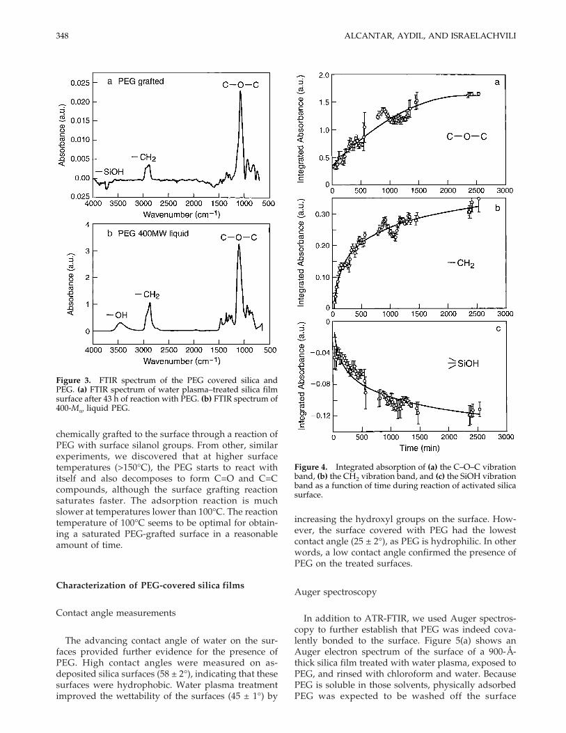
chemically grafted to the surface through a reaction ofPEG with surface silanol groups. From other, similarexperiments, we discovered that at higher surfacetemperatures (>150°C), the PEG starts to react withitself and also decomposes to form C=O and C=Ccompounds, although the surface grafting reactionsaturates faster. The adsorption reaction is muchslower at temperatures lower than 100°C. The reactiontemperature of 100°C seems to be optimal for obtain-ing a saturated PEG-grafted surface in a reasonableamount of time.
Characterization of PEG-covered silica films
Contact angle measurements
The advancing contact angle of water on the sur-faces provided further evidence for the presence ofPEG. High contact angles were measured on as-deposited silica surfaces (58 ± 2°), indicating that thesesurfaces were hydrophobic. Water plasma treatmentimproved the wettability of the surfaces (45 ± 1°) by
increasing the hydroxyl groups on the surface. How-ever, the surface covered with PEG had the lowestcontact angle (25 ± 2°), as PEG is hydrophilic. In otherwords, a low contact angle confirmed the presence ofPEG on the treated surfaces.
Auger spectroscopy
In addition to ATR-FTIR, we used Auger spectros-copy to further establish that PEG was indeed cova-lently bonded to the surface. Figure 5(a) shows anAuger electron spectrum of the surface of a 900-Å-thick silica film treated with water plasma, exposed toPEG, and rinsed with chloroform and water. BecausePEG is soluble in those solvents, physically adsorbedPEG was expected to be washed off the surface
Figure 3. FTIR spectrum of the PEG covered silica andPEG. (a) FTIR spectrum of water plasma–treated silica filmsurface after 43 h of reaction with PEG. (b) FTIR spectrum of400-Mw liquid PEG.
Figure 4. Integrated absorption of (a) the C–O–C vibrationband, (b) the CH2 vibration band, and (c) the SiOH vibrationband as a function of time during reaction of activated silicasurface.
348 ALCANTAR, AYDIL, AND ISRAELACHVILI

whereas covalently bonded PEG would remain on thesurface even after vigorous rinsing in these solvents.The peak at 503 eV represented the characteristic en-ergy of the Auger electron (KLL transition) of oxygen.The peak with low electron energy at 76 eV was char-acteristic of atomic silicon in an SiO2 compound (LMMtransition), and the peak at 271 eV was characteristicof atomic carbon (KLL transition). Therefore, we ob-served all the atomic species of the PEG molecule. Toascertain whether the carbon peak was due to con-tamination, characterization of a silica surface after thesame treatment without the PEG exposure was alsoperformed. Figure 5(b) shows the Auger electron spec-trum of this control sample; whereas the same char-acteristic peaks for oxygen and silicon were detectedowing to the silica, there was no carbon on the surface.Therefore, nonremovable carbon due to the PEG graft-ing (CH2–CH2–O)n was detected only in the spectrumof the PEG-covered silica surfaces.
AFM
The surface topology and smoothness of the baresilica and PEG-covered silica surfaces were deter-
mined with tapping mode AFM. The silica surfaces as-deposited showed an RMS roughness of 5.1 ± 0.2 nm[Fig. 6(a)]. After water plasma activation, the modifiedsurface had an RMS roughness of 2.3 ± 0.2 nm [Fig.6(b)]. However, after PEG was chemically bonded tothe surface, the RMS roughness decreased further to0.6 ± 0.2 nm and the surfaces appeared to be uniform[Fig. 6(c)]. We believe that PEG was filling the asym-metries on the silica, carpeting the surface completely.Consequently, PEG not only chemically modified thesilica films, but also appeared to smooth the surfacetopography.
Fluorescence microscopy
We also investigated the biocompatibility of thePEG coating by measuring its ability to resist proteinadsorption. PEG-coated silica surfaces, water plasma–treated silica surfaces, and bare silica films were allexposed to a 2-mg/mL solution of bovine serum al-bumin (BSA) labeled with a Texas red probe for 120min at 37°C and pH 7.4 (imitating physiological con-ditions). Protein adsorption onto these surfaces was
Figure 5. (a) PEG-covered silica surface Auger spectrum.This surface was treated with water plasma and exposed toPEG until saturation. It was then rinsed with chloroform(CHCl3) and water. Oxygen (503 eV), silicon (92 eV), andcarbon (272 eV) were detected on the surface, which formthe PEG chains. (b) Auger electron spectrum of a silica sur-face. This surface was subjected to water plasma and rinsedwith CHCl3 and water, but it was not exposed to PEG. Oxy-gen and silicon were on the surface because they are theatomic species for silica; however, carbon was not found.
Figure 6. Three-dimensional AFM images (1 × 1 mm) of (a)as-deposited silica, (b) silica after water plasma treatment,and (c) PEG-covered silica. The root mean square roughnessof the three films are 5, 2, and 0.5 nm, respectively.
349PEG-COATED SURFACES

detected with a CCD camera mounted on a fluores-cence microscope. Figure 7(a–c) shows the fluores-cence-intensity illumination difference from one sam-ple of each of these three surface chemistries. Thesurface that was covered with PEG had very little fluo-rescence activity owing to low or absent protein ad-sorption [Fig. 7(c)]. Conversely, the bare silica surfacewas much brighter owing to relatively high amountsof adsorbed protein [Fig. 7(a)]. The surface treated
with water plasma adsorbed some proteins, falling be-tween the bare silica and PEG coated surfaces [Fig.7(b)]. Quantitative analysis of protein adsorption wasdone by comparing the normalized adsorption inten-sity (NAI) of scans for the three different surfaces(scan size: 350 × 450 mm). The NAI is the percent ratioof the emission intensity of the surface and the emis-sion intensity measured for the background. The back-ground is taken on the surface that has not been ex-
Figure 7. Fluorescence detectionof adsorbed bovine serum albu-min labeled with a Texas redprobe. Incubation time was 120min at 37°C and pH 7.4 (physi-ological conditions). (a) Bare silicawith normalized adsorption in-tensity (NAI) of 71%. (b) Waterplasma–treated silica with NAI of62%. (c) PEG-covered silica sur-face with NAI of 7%.
350 ALCANTAR, AYDIL, AND ISRAELACHVILI

posed to the protein. Bare silica exhibited the greatestNAI: 71%. The NAI for water plasma–treated silicawas 62%. The silica surface that was reacted with PEGshowed an NAI of only 7%. The adsorption of otherproteins is currently under investigation.
SUMMARY AND CONCLUSIONS
A new PEG grafting method was optimized for pro-ducing biocompatible coatings. The coatings wereproduced by grafting PEG on a film of activated amor-phous silica. Our approach is simple and flexible be-cause silica can be easily deposited by PECVD on vari-ous materials, including metals and plastics. The pro-posed synthesis route is also efficient because PEG isgrafted onto the surface by a direct condensation of itsalcohol end group with the silanol groups on the silicasurface. Surface characterization of the resulting filmsusing infrared spectroscopy, Auger electron spectros-copy, contact angle measurements, and AFM corrobo-rated that PEG was chemisorbed on the surface. ThePEG films were smoother and more hydrophilic thaneither bare or activated silica surfaces. The measure-ments of fluorescence intensity showed that PEG-covered silica films prevented protein adsorption.Hence, PEG binding modifies the resulting surface to-pology, chemistry, and interactions, rendering sur-faces coated with our protective layers biocompatible.
The authors would like to thank the General Office ofAcademic Affairs, UNAM, for its support.
References
1. Agrawal CM. Reconstructing the human body using biomate-rials. JOM-J Min Met Mat S 1998;50:31–35.
2. Caruana CM. Biomaterials boost body repair. Chem Eng Prog1999;95:9–14.
3. Dunn SE, Brindley A, Davis SS, Davies MC, Illum L. Polysty-rene-poly(ethylene glycol) (PS-PEG2000) particles as modelsystems for site specific drug delivery. 2. The effect of PEGsurface density on the in vitro cell interaction and in vivo bio-distribution. Pharm Res 1994;11:1016–1022.
4. Beduaddo FK, Huang L. Interaction of PEG-phospholipid con-jugates with phospholipid: Implications in liposomal drug de-livery. Adv Drug Deliver Rev 1995;16:235–247.
5. Kuhl TL, Leckband DE, Lasic DD, Israelachvili JN. Modulationof interaction forces between bilayers exposing short-chainedethylene oxide headgroups. Biophys J 1994;66:1479–1488.
6. Service RF. Designer tissues take hold. Science 1995;270:230–232.
7. Ratner BD. New ideas in biomaterials science, a path to engi-neering biomaterials. J Biomed Mater Res 1993;27:837–850.
8. Churaev NV, Sergeeva IP, Sonolev VD. Hydrodynamic, thick-ness and deformation of adsorbed layers of polyethylene ox-ides. J Colloid Interface Sci 1995;169:300–305.
9. Harris JM, editor. Poly(ethylene glycol) chemistry, biotechnicaland biomedical applications. New York: Plenum Press, 1992.
10. Andrade JD, Hlady V, Jeon SI. Poly(ethylene oxide) and pro-tein resistance: Principles, problems, and possibilities. AdvChem Ser 1996;248:51–59.
11. Lee JH, Lee HB, Andrade JD. Blood compatibility of polyeth-ylene oxide surfaces. Prog Poly Sci 1995;20:1043–1079.
12. Golander C-G, Herron JN, Lim K, Claesson P, Stenius P, An-drade JD. Properties of immobilizated PEG films and the in-teraction with proteins. In: Harris JM, editor. Poly(ethyleneglycol) chemistry, biotechnical and biomedical applications.New York: Plenum Press, 1992. p 221–245.
13. Holmberg K, Bergstrom K, Stark MB. Immobilization of pro-teins via PEG chains. In: Harris JM, editor. Poly(ethylene gly-col) chemistry, biotechnical and biomedical applications. NewYork: Plenum Press, 1992. p 303–324.
14. Nagaoka S, Nakao A. Clinical application of antithrombogenichydrogel with long poly(ethylene oxide) chains. Biomaterials1990;11:119–121.
15. Jeon SI, Lee JH, Andrade JD, Gennes PG. Protein surface in-teractions in the presence of polyethylene oxide. 1. Simplifiedtheory. J Colloid Interface Sci 1991;142:149–166.
16. Harris JM, Dust JM, McGill RA, Harris PA, Edwell MJ,Sedaghatherati RM, Karr LJ, Donnelly DL. New polyethyleneglycols for biomedical applications. ACS Sym Ser 1991;467:418–429.
17. Sheth SR, Leckband D. Measurements of attractive forces be-tween proteins and end-grafted poly(ethylene glycol) chains.Proc Natl Acad Sci USA 1997;94:8378–8379.
18. Atha DH, Ingham KC. Mechanism of precipitation of proteinsby polyethylene glycols: Analysis in terms of excluded volume.J Biol Chem 1981;256:2108–2117.
19. Israelachvili JN. The different faces of poly(ethylene glycol).Proc Natl Acad Sci USA 1997;94:8399–8404.
20. Buckmann MM, Johansson G. Functionalization of poly(ethyl-ene glycol) and monomethoxy-poly(ethylene glycol). Makro-mol Chem 1981;182:1379–1384.
21. Zalipsky S, Gilon C, Zilkha A. Attachment of drugs to poly-ethylene glycols. Eur Polym J 1983;19:1177–1183.
22. Gombotz WR, Guanghui W, Horbett TA, Hoffman AS. Proteinadsorption to poly(ethylene oxide) surfaces. J Biomed MaterRes 1991;25:1547–1562.
23. Lea AS, Andrade JD, Hlady V. Compression of polyethyleneglycol chains grafted onto silicon nitride as measured by scan-ning force microscopy. Colloid Surf A 1994;93:349–357.
24. Yoshinaga K, Kito T, Yamate M. Effective immobilization ofprotein linked with polyethylene glycol on silica via hydrogelsusing silica sol. J Appl Polymer Sci 1990;41:1443–1450.
25. Iler RK. The chemistry of silica. New York: John Wiley & Sons;1979. p. 622–783.
26. Vansant EF, Van Der Voort P, Vrancken KC. Characterizationand chemical modification of the silica surfaces. Amsterdam,The Netherlands: Elsevier, 1995.
27. Han M, Aydil ES. Study of surface reactions during plasmaenhanced chemical vapor deposition of SiO2 from SiH4, O2
−,and Ar plasma. J Vac Sci Technol A 1996;14:2062–2070.
28. Han M, Aydil ES. Plasma and surface diagnostics duringplasma-enhanced chemical vapor deposition of SiO2 fromSiH4/O2
−/Ar discharges. Thin Solid Films 1996;291:427–434.29. Deshmukh SC, Aydil ES. Investigation of low temperature
SiO2 plasma enhanced chemical vapor deposition. J Vac SciTechnol A 1996;14:2355–2367.
30. Harrick NJ. Internal reflection spectroscopy. New York: Inter-science, 1967.
31. Aydil ES, Gottscho RA. Probing plasma/surface interactions.Solid State Technol 1997;40:181.
32. Han SM, Aydil ES. Silanol concentration depth profiling dur-ing plasma deposition of SiO2 using multiple internal reflec-tion infrared spectroscopy. Appl Phys Lett 1997;70:3269–3271.
33. Alcantar NA, Aydil ES, Israelachvili JN. Effect of water plasmaon silica surfaces. In: Blitz J, Little CB, editors. Fundamentaland applied aspects of chemically modified surfaces. Cam-bridge, UK: Royal Society of Chemistry, 1999. p 212–222.
351PEG-COATED SURFACES