polymer chemistry creative commons attribution
TRANSCRIPT

PolymerChemistry
COMMUNICATION
Cite this: Polym. Chem., 2016, 7,6293
Received 30th August 2016,Accepted 30th September 2016
DOI: 10.1039/c6py01523a
www.rsc.org/polymers
Glyconanoparticles with controlled morphologiesand their interactions with a dendritic cell lectin†
Gokhan Yilmaz,a,b,c Lea Messager,d Anne S. Gleinich,e Daniel A. Mitchell,e
Giuseppe Battaglia*d and C. Remzi Becer*c
Well-defined amphiphilic block glycopolymers with equal
mannose content have been self-assembled in aqueous solution to
form glyconanoparticles with different morphologies. The size and
shape of nanoparticles have significant effects on the interactions
with the dendritic cell-specific intercellular adhesion molecule-3-
grabbing non-integrin (DC-SIGN; CD209), characterized using a
surface plasmon resonance spectrometer (SPR).
Protein–carbohydrate interactions play an important role inmany biological processes including cell interactions withimmune systems, tumor metastasis, adhesion of infectiousagents to host cells and many more.1 Proteins involved inthese signaling and processing pathways are known aslectins.2 These carbohydrate binding proteins are wide-spreadin nature and they differ from antibodies or enzymes entities.3
For example, it has been shown that some viruses such as HIVexpress many carbohydrate entities at their surface, whichenable them to bind to these lectins on the cell surface in theimmune system. Therefore, one of the promising strategies forfighting against infectious diseases would be to design com-peting systems with higher lectin affinity than pathogens, thuspreventing their adhesion.
Recent and elegant synthetic routes have allowed polymerchemists to prepare a wide range of glyco-polymers/particlesthat provide strong and selective recognition propertiestowards lectins.4–8 In particular, amphiphilic block glycopoly-mers (GPs) have attracted a great attention in terms of theirability to form various types of glyconanoparticles. These
amphiphilic GPs are generally composed of biocompatible,biodegradable hydrophobic polymer blocks covalently bondedto a biocompatible hydrophilic block.9–11 Some recent studieshave shown that amphiphilic block glycopolymers withdifferent carbohydrate compositions were used to producenanoparticles, such as micelles, nanospheres, core–shell nano-particles, micelle-like nanoparticles, crew cut micelles, nano-capsules and polymersomes.12–16 These studies facilitate theunderstanding and investigation of GP–lectin binding activi-ties that are significantly influenced by GP architecture,valency, size, and density of binding elements. Furthermore,they offer a promising route for the creation of a broad varietyof bioactive self-assembled glyco-nanostructures for bio-medical applications such as drug delivery, biomaterials, bio-and nanotechnologies, and gene therapy.17–24 In the last fewyears, the number of strategies devoted to prepare GPs viasingle electron transfer living radical polymerization (SET-LRP)polymerization technique has increased rapidly. Therefore,SET-LRP was used in this study to synthesize several types ofamphiphilic block glycocopolymers bearing mannose moietiesby using methyl acrylate (MA) and a glycomonomer (ManAc)and/or poly(ethylene glycol) (PEG) as a hydrophobic and ahydrophilic block, respectively. Mannose glycomonomer wasprepared according to the procedure reported by Zhang et al.25
Poly(ethylene glycol) 2-bromoisobutyrate (PEG-Br) initiator wasused as an initiator to prepare amphiphilic triblock glycocopoly-mer. The self-assembly behaviors of these well-defined amphi-philic GPs were investigated in aqueous solution to obtain glyco-nanostructures with various sizes and morphologies. The result-ing glyconanoparticles (GNPs) were further investigated for theirbinding affinity towards lectins, in particular with DC-SIGN,which is a C-type human lectin present on both macrophagesand also dendritic cell subpopulations. DC-SIGN binds to micro-organisms and host molecules by recognizing surfaces rich inmannose containing glycans through multivalent glycan–protein interactions and notably serves as a target molecule forseveral viruses such as HIV and hepatitis C virus.26
†Electronic supplementary information (ESI) available: Synthesis and character-ization of the polymers. See DOI: 10.1039/c6py01523a
aDepartment of Chemistry, University of Warwick, CV4 7AL Coventry, UKbDepartment of Basic Sciences, Turkish Military Academy, Ankara, TurkeycPolymer Chemistry Laboratory, School of Engineering and Materials Science,
Queen Mary, University of London, E1 4NS London, UK. E-mail: [email protected] of Chemistry, University College London, WC1H 0AJ London, UKeClinical Sciences Research Institute, Warwick Medical School,
University of Warwick, CV2 2DX Coventry, UK
This journal is © The Royal Society of Chemistry 2016 Polym. Chem., 2016, 7, 6293–6296 | 6293
Ope
n A
cces
s A
rtic
le. P
ublis
hed
on 3
0 Se
ptem
ber
2016
. Dow
nloa
ded
on 2
7/06
/201
7 13
:42:
08.
Thi
s ar
ticle
is li
cens
ed u
nder
a C
reat
ive
Com
mon
s A
ttrib
utio
n-N
onC
omm
erci
al 3
.0 U
npor
ted
Lic
ence
.
View Article OnlineView Journal | View Issue
Here, we report the synthesis of well-defined amphiphilicGPs using the SET-LRP technique, their spontaneous self-assembly in aqueous solution, and binding studies of theresulting glyconanoparticles with DC-SIGN.
A series of well-defined amphiphilic block GPs were pre-pared via SET-LRP using EBiB or PEG-Br initiator. The molarratio of [MA]/[ManAc] has been varied to yield amphiphilicblock copolymers with increasing ratios between their hydro-phobic and hydrophilic moieties. One-pot polymerizationapproach has been employed to ensure well-defined blockcopolymer formation. Initially, MA was polymerized up to highconversion and subsequently; the chain extension withmannose glycomonomer (DP = 15, dissolved and degassed in1 mL of DMSO) to form the second block was carried out (ESI,Fig. S7†). The monomer conversions were followed by 1HNMR, which showed the disappearance of vinyl groups appar-ent between 5.8–6.4 ppm. The purified glycopolymers werefurther analyzed by GPC and 1H NMR (ESI, Fig. S9†). Based onthese results, it is evident that the copolymerization of MA andManAc has been achieved with a good control as indicated bylow polydispersity indices and increasing molecular weight ofthe block copolymer. Analysis of all P((MA)m-b-(ManAc)n) poly-mers (P1, P2, P3, and P4) are presented in Table 1. High chainend fidelity of P((PEG)-b-(MA)) has been confirmed by chainextension with MA prior to the synthesis of triblock glyco-copolymer, P5, using the same approach in the presence ofPEG-Br initiator. GPC analysis (ESI, Fig. S10†) revealed a clearbaseline shift of elution traces after each monomer additionwith increasing molecular weight and final PDI of 1.17. No sig-nificant tailing or shoulders have been observed in the GPCthroughout the polymerization. Moreover, the measuredMn,GPC and the Mn,theo calculated from the monomer conver-sions are in good agreement.
Nanoparticles of these amphiphilic GPs having PEG blockand/or carbohydrate bearing chains as hydrophilic block andMA as hydrophobic one were prepared using a nanoprecipita-tion method an the detailed conditions are summarized in theESI.† Slow-injection of water droplets into glycopolymer inDMF solution ensures to obtain thermodynamically stable self-assembled structures. The morphology and size of the result-ing nanoparticles were systematically analyzed by TEM andDLS.
Amphiphilic block copolymers P1, P2 and P3 yield well-dispersed nanoparticles of around 25 nm. While P1 and P2generated micelles with a regular spherical shape, P3 creatednon-uniform micelles due to their non-regular spherical shapeaccording to TEM imaging and DLS measurements. These fea-tures are typical of spherical micelles. Further TEM analysiswas conducted on each suspension measuring the average dia-meter of more than 50 single particles. The majority of the par-ticles display an average diameter of 20 nm, which is slightlysmaller than the values obtained by DLS, thus providing thedry structure of these particles. Furthermore, TEM imagingshows that most of the particles are not clustered, which ispromising, as micelles generally tend to cluster together.Moreover, it was also observed that P2 and P3 yielded smallamount of larger aggregates, which could potentially be vesi-cles. Increasing the hydrophobic content of the copolymerrevealed the formation of self-assembled nanoparticles withlarger hydrodynamic diameter. Indeed, the TEM image of P4indicated the formation of vesicular structures and DLSmeasurements showed a narrow size distribution centered at380 nm.
According to TEM and DLS results, sizes of glyconanoparti-cles are increasing with increasing of polymer length (ESI,Table S2†). Depending on the increasing MA fraction in thepolymer, the existence region of nanoparticles has becomebroader. This is possibly due to dependency on composition,molecular geometry, relative block lengths of the constitutivecopolymers, and the preparation methods on the formation ofglyconanoparticles. Another attempt was conducted on P4 toobtain a higher number of vesicular particles, decreasing theinjection rate of the aqueous solution.
The self-assembly of amphiphilic triblock GP, P5, has lowerhydrophilic fraction than P1–P4, P((MA)m-b-(ManAc)n), due tothe increasing molecular weight ratio of the hydrophobicblock. According to TEM measurement, the majority of nano-particles were worm-like micelles but with a small fraction ofspherical micelles (Fig. 1). The length of these worm-likemicelles is in the range of 45 to 60 nm and their width is24 nm, which is in agreement with unimer chain scaling.Further increase of molar ratio of hydrophobic block (for P4and P5) allowed fabricating different aggregates such as vesi-cles and worm-like micelles.
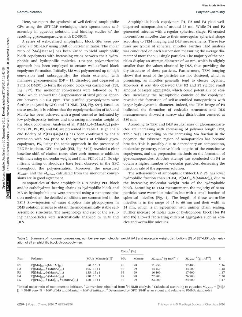
Table 1 Summary of monomer conversions, number average molecular weight (Mn) and molecular weight distributions (Đ) of SET-LRP polymeriz-ation of all amphiphilic block glycocopolymers
Run Polymer [MA] : [ManAc] : [I]a
Conv.b (%)
Mn,NMRc (g mol−1) Mn,GPC
d (g mol−1) ĐMA ManAc
P1 P((MA)76-b-(ManAc)15) 80 : 15 : 1 96 98 11 850 12 400 1.16P2 P((MA)103-b-(ManAc)15) 105 : 15 : 1 97 99 14 150 14 800 1.18P3 P((MA)130-b-(ManAc)15) 135 : 15 : 1 96 99 16 400 17 600 1.17P4 P((MA)206-b-(ManAc)15) 210 : 15 : 1 97 98 22 800 26 900 1.20P5 P((PEG)45-b-(MA)172-b-(ManAc)15) 180 : 15 : 1 96 99 23 800 24 600 1.17
a Initial molar ratio of monomers to initiator. b Conversions obtained from 1H NMR analysis. c Calculated according to equation Mn,NMR = ([M]0/[I] × NMR conv.% × MW of MA and ManAc) + MW of initiator. dDetermined by GPC (DMF as an eluent and relative to PMMA standards).
Communication Polymer Chemistry
6294 | Polym. Chem., 2016, 7, 6293–6296 This journal is © The Royal Society of Chemistry 2016
Ope
n A
cces
s A
rtic
le. P
ublis
hed
on 3
0 Se
ptem
ber
2016
. Dow
nloa
ded
on 2
7/06
/201
7 13
:42:
08.
Thi
s ar
ticle
is li
cens
ed u
nder
a C
reat
ive
Com
mon
s A
ttrib
utio
n-N
onC
omm
erci
al 3
.0 U
npor
ted
Lic
ence
.View Article Online
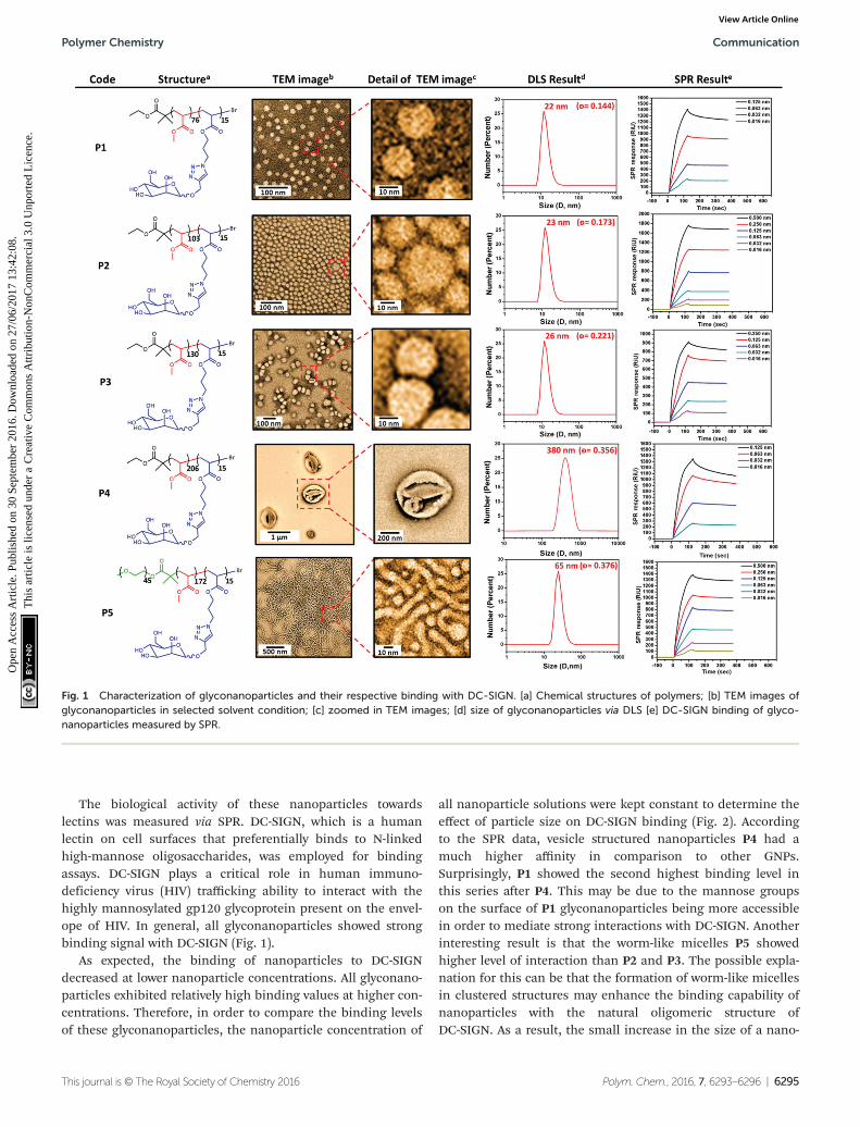
The biological activity of these nanoparticles towardslectins was measured via SPR. DC-SIGN, which is a humanlectin on cell surfaces that preferentially binds to N-linkedhigh-mannose oligosaccharides, was employed for bindingassays. DC-SIGN plays a critical role in human immuno-deficiency virus (HIV) trafficking ability to interact with thehighly mannosylated gp120 glycoprotein present on the envel-ope of HIV. In general, all glyconanoparticles showed strongbinding signal with DC-SIGN (Fig. 1).
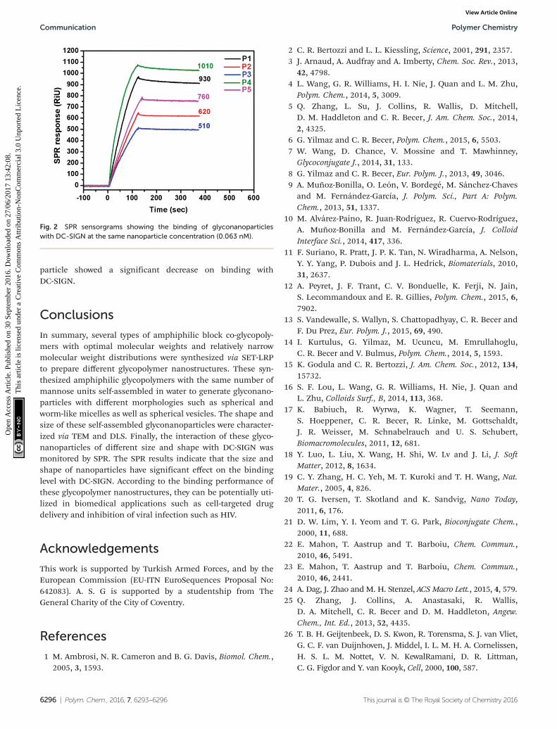
As expected, the binding of nanoparticles to DC-SIGNdecreased at lower nanoparticle concentrations. All glyconano-particles exhibited relatively high binding values at higher con-centrations. Therefore, in order to compare the binding levelsof these glyconanoparticles, the nanoparticle concentration of
all nanoparticle solutions were kept constant to determine theeffect of particle size on DC-SIGN binding (Fig. 2). Accordingto the SPR data, vesicle structured nanoparticles P4 had amuch higher affinity in comparison to other GNPs.Surprisingly, P1 showed the second highest binding level inthis series after P4. This may be due to the mannose groupson the surface of P1 glyconanoparticles being more accessiblein order to mediate strong interactions with DC-SIGN. Anotherinteresting result is that the worm-like micelles P5 showedhigher level of interaction than P2 and P3. The possible expla-nation for this can be that the formation of worm-like micellesin clustered structures may enhance the binding capability ofnanoparticles with the natural oligomeric structure ofDC-SIGN. As a result, the small increase in the size of a nano-
Fig. 1 Characterization of glyconanoparticles and their respective binding with DC-SIGN. [a] Chemical structures of polymers; [b] TEM images ofglyconanoparticles in selected solvent condition; [c] zoomed in TEM images; [d] size of glyconanoparticles via DLS [e] DC-SIGN binding of glyco-nanoparticles measured by SPR.
Polymer Chemistry Communication
This journal is © The Royal Society of Chemistry 2016 Polym. Chem., 2016, 7, 6293–6296 | 6295
Ope
n A
cces
s A
rtic
le. P
ublis
hed
on 3
0 Se
ptem
ber
2016
. Dow
nloa
ded
on 2
7/06
/201
7 13
:42:
08.
Thi
s ar
ticle
is li
cens
ed u
nder
a C
reat
ive
Com
mon
s A
ttrib
utio
n-N
onC
omm
erci
al 3
.0 U
npor
ted
Lic
ence
.View Article Online
particle showed a significant decrease on binding withDC-SIGN.
Conclusions
In summary, several types of amphiphilic block co-glycopoly-mers with optimal molecular weights and relatively narrowmolecular weight distributions were synthesized via SET-LRPto prepare different glycopolymer nanostructures. These syn-thesized amphiphilic glycopolymers with the same number ofmannose units self-assembled in water to generate glyconano-particles with different morphologies such as spherical andworm-like micelles as well as spherical vesicles. The shape andsize of these self-assembled glyconanoparticles were character-ized via TEM and DLS. Finally, the interaction of these glyco-nanoparticles of different size and shape with DC-SIGN wasmonitored by SPR. The SPR results indicate that the size andshape of nanoparticles have significant effect on the bindinglevel with DC-SIGN. According to the binding performance ofthese glycopolymer nanostructures, they can be potentially uti-lized in biomedical applications such as cell-targeted drugdelivery and inhibition of viral infection such as HIV.
Acknowledgements
This work is supported by Turkish Armed Forces, and by theEuropean Commission (EU-ITN EuroSequences Proposal No:642083). A. S. G is supported by a studentship from TheGeneral Charity of the City of Coventry.
References
1 M. Ambrosi, N. R. Cameron and B. G. Davis, Biomol. Chem.,2005, 3, 1593.
2 C. R. Bertozzi and L. L. Kiessling, Science, 2001, 291, 2357.3 J. Arnaud, A. Audfray and A. Imberty, Chem. Soc. Rev., 2013,
42, 4798.4 L. Wang, G. R. Williams, H. I. Nie, J. Quan and L. M. Zhu,
Polym. Chem., 2014, 5, 3009.5 Q. Zhang, L. Su, J. Collins, R. Wallis, D. Mitchell,
D. M. Haddleton and C. R. Becer, J. Am. Chem. Soc., 2014,2, 4325.
6 G. Yilmaz and C. R. Becer, Polym. Chem., 2015, 6, 5503.7 W. Wang, D. Chance, V. Mossine and T. Mawhinney,
Glycoconjugate J., 2014, 31, 133.8 G. Yilmaz and C. R. Becer, Eur. Polym. J., 2013, 49, 3046.9 A. Muñoz-Bonilla, O. León, V. Bordegé, M. Sánchez-Chaves
and M. Fernández-García, J. Polym. Sci., Part A: Polym.Chem., 2013, 51, 1337.
10 M. Alvárez-Paino, R. Juan-Rodríguez, R. Cuervo-Rodríguez,A. Muñoz-Bonilla and M. Fernández-García, J. ColloidInterface Sci., 2014, 417, 336.
11 F. Suriano, R. Pratt, J. P. K. Tan, N. Wiradharma, A. Nelson,Y. Y. Yang, P. Dubois and J. L. Hedrick, Biomaterials, 2010,31, 2637.
12 A. Peyret, J. F. Trant, C. V. Bonduelle, K. Ferji, N. Jain,S. Lecommandoux and E. R. Gillies, Polym. Chem., 2015, 6,7902.
13 S. Vandewalle, S. Wallyn, S. Chattopadhyay, C. R. Becer andF. Du Prez, Eur. Polym. J., 2015, 69, 490.
14 I. Kurtulus, G. Yilmaz, M. Ucuncu, M. Emrullahoglu,C. R. Becer and V. Bulmus, Polym. Chem., 2014, 5, 1593.
15 K. Godula and C. R. Bertozzi, J. Am. Chem. Soc., 2012, 134,15732.
16 S. F. Lou, L. Wang, G. R. Williams, H. Nie, J. Quan andL. Zhu, Colloids Surf., B, 2014, 113, 368.
17 K. Babiuch, R. Wyrwa, K. Wagner, T. Seemann,S. Hoeppener, C. R. Becer, R. Linke, M. Gottschaldt,J. R. Weisser, M. Schnabelrauch and U. S. Schubert,Biomacromolecules, 2011, 12, 681.
18 Y. Luo, L. Liu, X. Wang, H. Shi, W. Lv and J. Li, J. SoftMatter, 2012, 8, 1634.
19 C. Y. Zhang, H. C. Yeh, M. T. Kuroki and T. H. Wang, Nat.Mater., 2005, 4, 826.
20 T. G. Iversen, T. Skotland and K. Sandvig, Nano Today,2011, 6, 176.
21 D. W. Lim, Y. I. Yeom and T. G. Park, Bioconjugate Chem.,2000, 11, 688.
22 E. Mahon, T. Aastrup and T. Barboiu, Chem. Commun.,2010, 46, 5491.
23 E. Mahon, T. Aastrup and T. Barboiu, Chem. Commun.,2010, 46, 2441.
24 A. Dag, J. Zhao and M. H. Stenzel, ACS Macro Lett., 2015, 4, 579.25 Q. Zhang, J. Collins, A. Anastasaki, R. Wallis,
D. A. Mitchell, C. R. Becer and D. M. Haddleton, Angew.Chem., Int. Ed., 2013, 52, 4435.
26 T. B. H. Geijtenbeek, D. S. Kwon, R. Torensma, S. J. van Vliet,G. C. F. van Duijnhoven, J. Middel, I. L. M. H. A. Cornelissen,H. S. L. M. Nottet, V. N. KewalRamani, D. R. Littman,C. G. Figdor and Y. van Kooyk, Cell, 2000, 100, 587.
Fig. 2 SPR sensorgrams showing the binding of glyconanoparticleswith DC-SIGN at the same nanoparticle concentration (0.063 nM).
Communication Polymer Chemistry
6296 | Polym. Chem., 2016, 7, 6293–6296 This journal is © The Royal Society of Chemistry 2016
Ope
n A
cces
s A
rtic
le. P
ublis
hed
on 3
0 Se
ptem
ber
2016
. Dow
nloa
ded
on 2
7/06
/201
7 13
:42:
08.
Thi
s ar
ticle
is li
cens
ed u
nder
a C
reat
ive
Com
mon
s A
ttrib
utio
n-N
onC
omm
erci
al 3
.0 U
npor
ted
Lic
ence
.View Article Online