polymerase chain reaction-based detection of moniezia in sheep
TRANSCRIPT

Republic of Iraq
Ministry of Higher Education
& Scientific Research
University of Al-Qadisiyah
College of Veterinary Medicine
Polymerase chain reaction-based detection of
Moniezia in sheep
A Graduation Project Submitted to the Department Council of the
Internal and Preventive Medicine-College of Veterinary Medicine/
University of Al-Qadisiyah in a partial fulfillment of the requirements for
the Degree of Bachelor of Science in Veterinary Medicine and Surgery.
By
Rabab Hussain Hatem
Supervised by
Dr. Al-fatlawi M. A.
2018 A.D. 1439 A.H.

سورة طهمن

Certify of supervisor
I certify that the project entitled (Polymerase chain reaction-based
detection of Moniezia in sheep) was prepared by Rabab Hussain Hatem
under my supervision at the College of Veterinary Medicine / University
of Al-Qadisiyah.
Supervisor
Al-fatlawi Monyer Abd
Dept. of Veterinary microbiology
Coll.Of Vet .Med. / Univ. of Al-Qadisiyah.
01 / 03/ 2018

Certificate of Department
We certify that Rabab Hussain Hatem
Has finished her Graduation Project entitled (Polymerase chain reaction-
based detection of Moniezia in sheep) and candidate it for debating.
Instructor
Dr. Muthanna H. Hussain
01 /03/ 2018
Head of Dept of Int. and Prev. Med.
Dr. Muthanna H. Hussain
01 /03 / 2018

Dedication
To who I waited a long time to dedicate to
him my graduation project, Al-Imam Al-
mahdi….
To the memory of my great father….
To the source of kindness, the greater my
mother….
To the great supporter in my life, my
brothers and my sisters ….
To my best friends , Israa Ayad and Israa
Jawad ….
To my classmates , Nooraldeen Hakim and
Mortada Mohammed….
Rabab Al-zameli

Acknowledgement
I would like to thank merciful God for enabling
me to perform this work.
I would like to express my deeply thanks to my
supervisor Dr. Monyer Abd Al-Fatlawi for his
supervisor, constant help and endless
encouragement….
Rabab Al-zameli

Abstract
The sheep infestation by Moniezia spp, cestodes that
normally affect intestine of ruminants especially sheep, are
wide-spread-parasitic infestation. This study was initiated to
investigate the cestodes that infest sheep intestine. From Al-
Najaf City-Iraq, 50 sheep intestines were examined seeking for
tapeworms. After that searching, the results indicated that 13
sheep were infested with Moniezia spp. For studying the
morphological features of the tapeworms, 5 cestodes were
stained with modified Carmen stain, and the mature segment of
these worms showed the presence of genital pore, cirrus sac,
vitelline gland, testes, and inter-proglottid gland. For better
detection of the tapeworm, 4 samples out of 13 cestodes were
subjected to polymerase chain reaction (PCR) test.Using 18S
rRNA gene, the results of the PCR have confirmed the identity
of these tapeworms as Moniezia spp. This study indicates the
presence of Moniezia spp in the intestine of sheep that belong to

the city of Al-Najaf, Iraq. This issue should alarm the veterinary
officials to control and prevent such parasitic infestation.
List of contents
No. Subject Page
1 Introduction
2 Literature review
2-1 Morphological features
2-2 Digestive system
2-3 Natural environment
2-4 Life cycle
2-5 Reproduction
2-6 Pathogenesis
2-7 Diagnosis
2-8 Treatment
2-9 Moniezia expansa impct on the community
3 Materials and methods
3-1 Internal exploring and sample collection
3-2 Morphological examination
3-3 Polymerase chain reaction
3-4 The equipment, Instrument and kits
3-5 Primers of PCR
3-6 Chemicals used for PCR
3-7 Worm DNA extraction
3-8 Nanodrop for extracted DNA
3-9 PCR master mix preparation
3-10 PCR Thermocycler conditions
3-11 PCR product analysis
4 Results
5 Discussion
6 References

List of figures
1 The life cycle of Moniezia spp.
2 Moniezia spp. mature segment showed 1- Genital
pore 2- cirrus sac 3- Vitelline gland 4- testes 5-
Inter-proglottid gland.
3 Image of agarose gel electrophoresis that
demonstrates the amplification of the 18S rRNA
gene for Moniezia sp. M is the Ladder (2000-
100bp). Lanes 1 to 4are the amplification of this
gene the Moniezia spp. of sheep. Lanes 5 to 8 are
the amplification of this gene for Moniezia sp. of
cattle. The product size is at 743bp.
List of tables
1 mentions the equipment and instrument that were
used in this study.
2 refers to the kits that were used in the present study
3 shows the primers used for the PCR of the Moniezia spp.
4 reveals the chemical substances that were used in
the current study.

1. Introduction
The cestode namely Moniezia spp. are wide-spread tapeworms that
affect intestines of ruminants especially sheep. The worm infestation
allows huge economic losses in different countries of the world (Soulsby,
1982; Mazyad and el-Nemr, 2002). These worms characterized by the
presence of the featured scolex, neck and strobila. Moreover,these
tapeworms have unique scolex and neck that are small in size with long
chain of strobili. Anoplocephalidae is the family of these tapeworms and
descent from Cyclophyllidea order. In order to differentiate this genus
from other genera, the tapeworms have anterior, posterior, mature and
gravid segments. The sexual organs of Moniezia spp. are repeated for
each proglottid. As this worm needs an intermediate host, mites play as
an important source of infestation via grassing and then digesting of these
mites to release the active larvae which grow later to the adult stage of
these worms, Figure 1,(Denegri, Bernadina, Perez-Serrano, & Rodriguez-
Caabeiro, 1998). The disease caused by this worm is called monieziasis.
There are few species of Moniezia spp. with limited genetic studies
regarding 3 out of 7 species. These species are Moniezia expansa,

Moniezia benedeni and Moniezia monardi ((Ohtori, Aoki, & Itagaki,
2015). The current study was intended to detect and genetically identify
Moniezia spp. that affect sheep in Al-Najaf City, Iraq.
Figure 1: The life cycle of Moniezia spp.
(http://www.danekeclublambs.com/Tapeworms.html)

2. Review of literatures
2-1. Morphological Features
Moniezia expansa is a tapeworm that belongs to the Moniezia genus one
of the widely distributed double-pored ruminant (Gómez-Puerta et al.,
2008). In addition to its high incidence in sheep, Moniezia expansa was
also reported in swine and once unusual case in human (Gómez-Puerta et
al., 2008; el-Shazly et al., 2004). It is well known that Moniezia expansa
mainly infects young animals (Wymann, 2008). Moniezia expansa body
is segmented into several proglottids and characterized by the presence of
an anterior scolex that contains sensory organs, neck which connects the
premature segments and the strobilus which represents the longest part of
the body. The sensory organs in the scolex are responsible for tactile
stimulation, and they are close to the nerves that expand along the
parasite body (Brusca and Brusca, 1990). This Cestode size changes
through the life of the parasite to be about 8-10 meters in length and 1.5
centimeter in width, and the scolex is about 0.8 millimeter (Chilton, et al.,
2007). It does not contain rostellum and rostellar hooks, instead the
scolex carrys four suckers that are responsible for penetrating or holding
on to the host tissues (Mehlhorn, 2008). This parasite is monecious with
both genders sexual organs on each subject. The reproductive organs

locate laterally, and each proglottid is characterized by the presence of
cirrus pouches as well as genital pores and abundant testes. Despite the
fact that there are no studies about the behavior of the Moniezia expansa,
it is known that these worms are not able to travel due to the lack of cilia
in their bodies (Brusca and Brusca, 1990).
2-2. Digestive System
The body does not involve digestive organs, and it resembles a whole
inverted intestine since it is covered with microvilli that increase surface
through which absorbing takes place. They get their nutrition such as
body fluids by absorption through their tegument, or the external cover-
ing (Brusca and Brusca, 1990).
2-3. Natural Environment
There are three natural habitats for Moniezia expansa that can be
summarized as follw:
1. External habitat: represented by the ground.
2. The gastrointestinal organs of the intermediate host: represented by
the oribatid mite.
3. The definitive host: represented by the intestine of sheep, cattle and
goat.

2-4. Life Cycle
Life cycle of Moniezia expansa needs two hosts; final host that is sheep,
cattle or goat and intermediate host that is the oribatid mites (Elliott,
1986). In details, eggs go out of angulates intestine with the feces to be
eaten within one day by mites that are highly available in soil.Then, the
eggs reach the intestine of the mites where they develop and hatch into
oncospheres (larvae with 6 hooks). As their intermediate host, the eggs
stay in the mites’intestines for 1-3 months. After that, the oncospheres
pass through the haemocoel to grow into the cysticercoid stage. Finally,
ruminants graze on the mites infected grass and the cysticercoids get
released into the digestive system to be developed to full grown up
parasite within 5-6 weeks in the large intestine (Barriga, 1994). For the
life cycle to go on and on, the proglottids that involve the Moniezia ex-
pansa mature eggs are discarded with the animals’ feces to be consumed
and broken down by the mouth of the intermediate host (Barriga, 1994).
Interestingly, there are no studies about the life span of this tapeworm yet.
2-5. Reproduction
Moniezia expansa adult worm has both male and female reproductive
system as mentioned above. The mature proglottids can mate internally
since they have both sexes. They also can couple with other tapeworm to

reproduce. Reproduction produces gravid proglottids that pass out with
angulates feces (Melhorn, 2001). Dry conditions destroy the eggs unless
otherwise they were eaten by the soil mites. These tapeworms can
communicate with each other chemically in addition to the tactile and
chemical ways for perception communication.
2-6. Pathogenesis
No recorded signs of the Moniezia expansa infection in sheep and
ruminants generally. It is worth to notice that in intensive infection;the
disease might cause intestinal blockage, diarrhea and weight loss (Bauer,
1990).
2-7. Diagnosis
Since Moniezia expansa is asymptomatic, it is accidently detected on
parasitological postmortem fecal examination. Importantly, examiners
sometimes mix between Moniezia benedeni and Moniezia expansa due to
their similar interproglottidal glands. Accordingly and in order to have
very accurate results, examiners must be able to differentiate those glands
in the Moniezia expansa and Moniezia benedeni. Those glands have
rosette shape in the posterior part of the Moniezia expansa, but they make
striate line in the other species (Taylor, 1928).
Moniezia expansa infection can be diagnosed by either of the next ways:

1. Macroscopic examining of the anal area and fecal samples to
grossly detect the gravid proglottids since they are big enough to be
seen easily by naked eye.
2. Microscopic examining of fecal samples in laboratory to detect the
eggs of Moniezia expansa.
3. Enzyme Linked Immunosorbent Assay (ELIZA) (Jiménez et al.,
2007).
4. Polymerase Chain Reaction(PCR) (Khan et al., 2010).
2-8. Treatment
The following antiparasites are often prescribed to treat the Moniezia
expansa infection:
1. Niclosamide
2. Praziquantel (Bauer et al., 2007)
3. Albendazole (Bauer et al., 2007)
4. The compination of Praziquantel + Levamisole(Southworth et al.,
1996).
2-9. Moniezia expansa Impact on the Community
Moniezia expansa can impact the community by affecting the definitive
hosts such as sheep, cattle and goats. As mentioned above, those parasites
can cause blockage to the intestine under severe conditions. Moreover,

the infection can also cause diarrhea and weight loss especially in young
ruminants which ends up with economical stock breeding loss (Gomez-
Puerta and Denegre, 2008).

3. Materials and methods
3-1. Intestinal exploring and sample collection
This study was initiated to investigate the cestodes that infest sheep
intestine. From Al-Najaf City-Iraq, 50 sheep intestines were examined
seeking for tapeworms. After that searching, the results indicated that 13
sheep were infested with Moniezia spp.
3-2. Morphological examination
For studying the morphological features of the tapeworms, 5 cestodes
were stained with modified Carmen stain.
3-3. Polymerase chain reaction
To confirm the tapeworm identity, 4 samples out of 13 cestodes were
subjected to polymerase chain reaction (PCR) test using 18S rRNA gene.
3-4. The Equipment, instruments, and kits
Table 1 mentions the equipment and instrument that were used in this
study. Table 2 refers to the kits that were used in the present study.
Table 1: mentions the equipment and instrument that were used in this
study.

No. Equipment & instrument Company
1 High Speed Cold centrifuge Eppendorf /Germany
2 Incubator Mammert/Germany
3 Sensitive Balance Sartorius/Germany
4 Water Bath Mammert/Germany
5 Vortex CYAN/ Belgium
6 Micropipettes 5-50, 0.5-10,
100-1000µl CYAN/ Belgium
7 Refrigerator Concord /Lebanon
8 Thermocycler PCR MJ-Mini BioRad/ USA
9 Exispin centrifuge Bioneer/ Korea
10 Eppendorf tubes Bioneer/ Korea
11 Disposable syringe 10 ml,
5ml and 3ml Sterile EO. / China
12 Sterile test tube Superestar/ India
13 UV Transilluminator ATTA/ Korea
14 Gel electrophoresis Shandod Scientific/ UK
15 Digital camera Samsung/ china
Table 2: refers to the kits that were used in the present study.

No. Kit Company Country
1 gSYAN DNA Extraction Kit Geneaid USA
GST buffer
GSB buffer
W1 buffer
Wash buffer
Elution buffer
GD column
Collection tube 2ml
2 AccuPowerTM
PCR PreMix Bioneer Korea
Taq DNA polymerase
dNTPs (dATP, dCTP, dGTP,
dTTP)
Tris-HCl pH 9.0
KCl
MgCl2
Stabilizer and Tracking dye
3-5. Primers of the PCR

The primers were designed in the current study and were purchased from
(Bioneer Company, South Korea). The primers were deposited in the
ribosomal database under the number AY752651.1. Table 3 shows these
primers.
Table 3: shows the primers used for the PCR of the Moniezia spp.
Amplicon Sequence Primer
18S ribosomal
RNA gene
F TGCTACCCGCATGATGTTGT 1124bp
R ACACAGTTGGCTGCACTCTT
3-6. Chemicals used for the PCR
Table 4 reveals the chemical substances that were used in the current
study.
Table 4: reveals the chemical substances that were used in the current
study.
No. Chemical Company and Origin
1 Absolute Ethanol BDH (England)

2 Agarose BioBasic (Canada)
3 TBE buffer BioBasic (Canada)
5 Ehidium Bromide BioBasic (Canada)
6 Proteinase k BioBasic (Canada)
9 Free nuclease water Biolab/ USA
10 PCR water Bioneer (Korea)
3-7. Worm DNA Extraction
Using gSYAN DNA mini kit-based extraction kit (Geneaid, USA), the
genomic DNA was extracted from the tapeworm tissues and following
the manufacturer’s protocol as shown in the following steps:
1. From the tapeworm tissues, 200mg was placed in a 1.5ml tube, and
200ul of the GST buffer was added. The mix was homogenized
using micropestle.
2. Next, 30μl proteinase K was added and mixed by vortexing. Then,
the mix was incubated at 60℃ for 1 hour.
3. Later, 200μl of GSB was placed and vortexed vigorously, which
then were incubated at 70℃ for 15 minutes and inverted every 3
minutes until done.

4. After that, 200μl ethanol was added and immediately mixed by
shaking vigorously.
5. Then, DNA filter column was inserted into a 2 ml collection tube.
Next, the mixture containing the precipitant was transferred to the
column. Then, it was centrifuged at 10000 rpm for 5 minutes. The
2ml-collection tube was discarded and a new tube was used with
the column.
6. Following that, the filter column received 400μl of W1 buffer and
then was centrifuged at 10000 rpm for 30 seconds. The 2ml-
collection tube was disposed and a new one was used.
7. Later, the column received 600μl of Wash Buffer (ethanol). Then
was centrifuged at 10000rpm for 30 seconds. The flow-through
solution was disposed and the tube was used again.
8. Then, the tubes were centrifuged at 10000rpm for 3 minutes for
drying out purposes.
9. The filter column containing the DNA was transferred to a clean
1.5ml-centrifuge tube. These tubes received 50μl-pre-heated
elution buffer.
10. Finally, 5-minute standing for the tubes was given to increase the
absorption of the elution buffer by the matrix. Then, these tubes
were centrifuged at 10000 rpm for 30 seconds for the DNA to be
eluted.
3-8. Nanodrop for extracted DNA
The resulted DNA was checked for quality and quantity using
NanoDrop spectrophotometer (Thermo, USA), and the measured

quantity was resulted using the unit;ng/µL. The measured quality was
counted using the unit as purity by absorbance measurement at (260
/280 nm) as following steps:
1. When run the NanoDrop software, the suitable process was
selected, Nucleic acid, DNA process.
2. The measurement pedestals were cleaned using dry wipes several
times. Then,2μl of free-nuclease water was placed on the lower
measurement pedestal for blanking the reading.
3. After that, the moving arm was lowered,and the “Ok” button was
hit to run the process of the blank reading.Finally and to read the DNA
quality and quantity, the pedestals was cleaned using wipes, and 1μl
DNA was placed to run the process of measurement.
3-9. PCR master mix preparation
AccuPower PCR PreMix Kit was used to generate the PCR mastermix
following the manufacturer’s protocol, table 5.
Table 5: the PCR mastermix components
PCR Master mix Volume
DNA template 5µl
Forward primer
(10pmol) 1.5µl
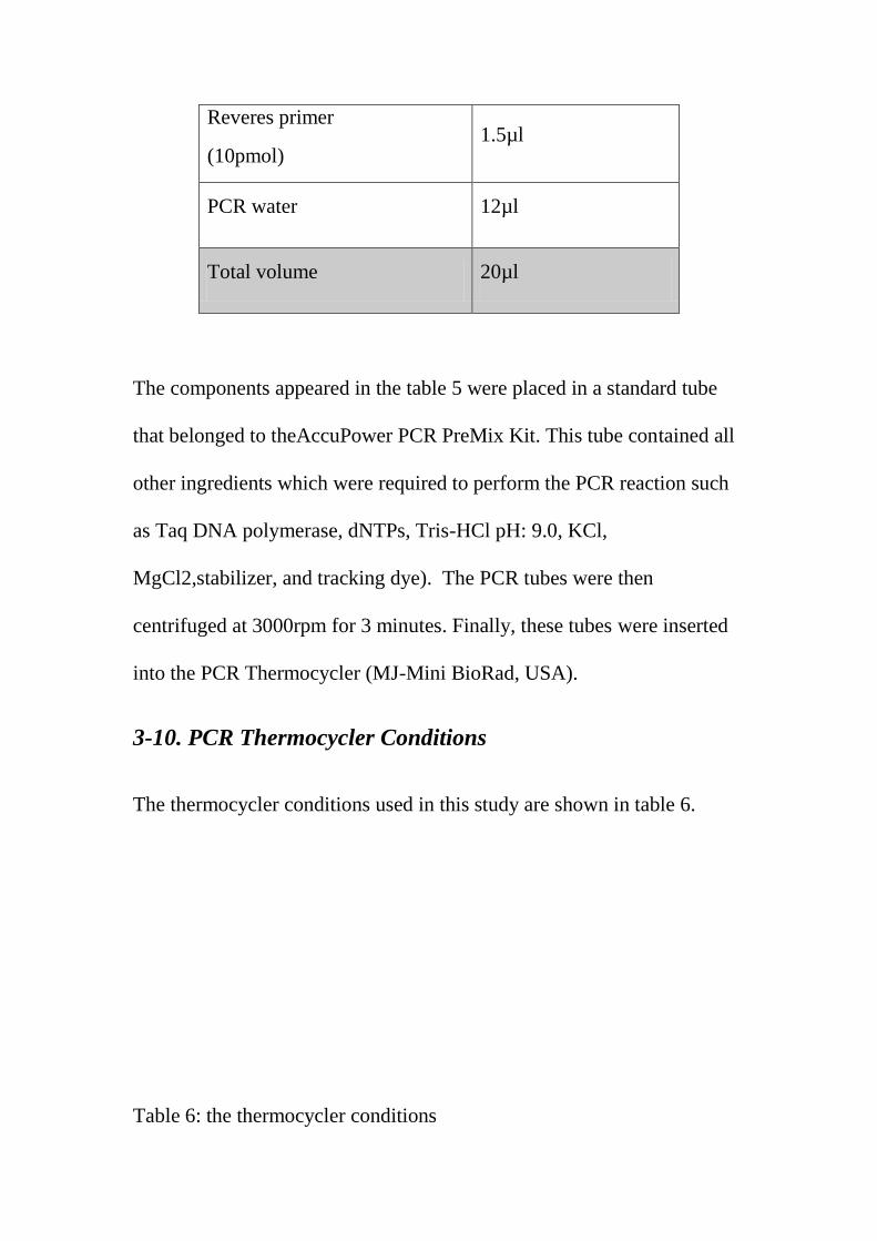
Reveres primer
(10pmol) 1.5µl
PCR water 12µl
Total volume 20µl
The components appeared in the table 5 were placed in a standard tube
that belonged to theAccuPower PCR PreMix Kit. This tube contained all
other ingredients which were required to perform the PCR reaction such
as Taq DNA polymerase, dNTPs, Tris-HCl pH: 9.0, KCl,
MgCl2,stabilizer, and tracking dye). The PCR tubes were then
centrifuged at 3000rpm for 3 minutes. Finally, these tubes were inserted
into the PCR Thermocycler (MJ-Mini BioRad, USA).
3-10. PCR Thermocycler Conditions
The thermocycler conditions used in this study are shown in table 6.
Table 6: the thermocycler conditions

PCR step Temp. Time repeat
Initial Denaturation 95C 5min 1
Denaturation 95C 30sec.
30cycle Annealing 58C 30sec
Extension 72C 1 min
Final extension 72C 5min 1
Hold 4C Forever -
3-11. PCR product analysis
Electrophoresis of agarose gel (1.5%) was used to analyze the PCR
products as appeared in the following steps:
1- Agarose(1.5%) powder was dissolved in 1X TBE using water bath at
100 °C for 15 minutes and later was left to cool down until 50°C.
2- After that, 3µl of ethidium bromide was added into the solution of the
agarose solution.
3- After fixing the comb in the tray, the solution of the agarose was
poured and was allowed to solidify for 15 minutes at room temperature.

After solidification of the solution, the comb was removed gently, and
10µl of the PCR products were pipetted into the pre-selected
wells.Ladder of 100bp size was pipetted at 5µl.
4- The gel was then covered by 1X TBE buffer. Electric current was
setup at 100 volt and 80A for 1hour.
5- Finally, the gel was visualized under UV Transilluminator.
4. Results
From Al-Najaf City-Iraq, 50 sheep intestines were examined
searching for tapeworms. After that searching, the results indicated that
13 sheep were infested with Moniezia spp. For studying the
morphological features of the tapeworms, 5 cestodes were stained with

modified Carmen stain, and the mature segment of these worms showed
the presence of genital pore, cirrus sac, vitelline gland, testes, and inter-
proglottid gland, figure 2. For better detection of the tapeworm, 4 samples
out of 13 cestodes were subjected to polymerase chain reaction (PCR)
test. Using 18S rRNA gene, the results of the PCR have confirmed the
identity of these tapeworms as Moniezia spp., table 3.
3
4
5
1


5. Discussion
The result of the current study has detected the presence of
Moniezia spp. in the intestine of sheep, and this is considered to the wide-
Figure 3: Image of agarose gel electrophoresis that
demonstrates the amplification of the 18S rRNA gene
for Moniezia sp. M is the Ladder (2000-100bp). Lanes
1 to 4are the amplification of this gene the Moniezia
spp. of sheep. Lanes 5 to 8 are the amplification of
this gene for Moniezia sp. of cattle. The product size
is at 743bp.

spread of this tapeworm in small ruminants. The finding that 13 sheep
were infested with Moniezia spp. indicates a major problem of how big is
the economic loss that might this worm introduce to the livestock
industry in south of Iraq(Diop et al., 2015). This worm has previously
been identified in camels in Al-Najaf and Al-Diwaniyah cities
(Anisimova, 2012), who found that the morbidity rates were 32.35% and
15.38% respectively. The current study result agrees with (Fadl et al.,
2011) that found that the prevalence of Moniezia spp. in sheep in
Baghdad regions was 0.9%. Increasing the occurrence rate of the
infestation is happened during summers; however; this increase could
start in spring when there are much numbers of mites that carry the
intermediate stage of this worm on grass (Fadl et al., 2011).
For studying the morphological features of the tapeworms, 5
cestodes were stained with modified Carmen stain, and the mature
segment of these worms showed the presence of genital pore, cirrus sac,
vitelline gland, testes, and inter-proglottid gland, figure 2.The
morphological feature results agree with the following literatures.
Moniezia expansa adult worm has both male and female reproductive
system as mentioned above. The mature proglottids can mate internally
since they have both sexes. They also can couple with other tapeworm to
reproduce. Reproduction produces gravid proglottids that pass out with

angulates feces (Melhorn, 2001).Moniezia expansa is a tapeworm that
belongs to the Moniezia genus one of the widely distributed double-pored
ruminant tapeworm that infects sheep, cattle, and goats (Denegre, 2008,
Gómez-Puerta et al., 2008). In addition to its high incidence in sheep,
Moniezia expansa was also reported in swine and once unusual case in
human (Gómez-Puerta et al., 2008; el-Shazly et al., 2004). It is well
known that Moniezia expansa mainly infects young animals (Wymann,
2008). Moniezia expansa body is segmented into several proglottids and
characterized by the presence of an anterior scolex that contains sensory
organs, neck which connects the premature segments, and the strobilus
which represents the longest part of the body. The sensory organs in the
scolex are responsible for tactile stimulation, and they are close to the
nerves that expand along the parasite body (Brusca and Brusca, 1990).
This Cestode size changes through the life of the parasite to be about 8-10
meters in length and 1.5 centimeter in width, and the scolex is about 0.8
millimeter (Chilton, et al., 2007). It does not contain rostellum and
rostellar hooks, instead the scolex carrys four suckers that are responsible
for penetrating or holding on to the host tissues (Mehlhorn, 2008). This
parasite is monecious with both genders sexual organs on each subject.
The reproductive organs locate laterally, and each proglottid is
characterized by the presence of cirrus pouches as well as genital pores
and abundant testes. Despite the fact that there are no studies about the

behavior of the Moniezia expansa, it is known that these worms are not
able to travel due to the lack of cilia in their bodies (Brusca and Brusca,
1990).
For better detection of the tapeworm, 4 samples out of 13
cestodes were subjected to polymerase chain reaction (PCR) test. Using
18S rRNA gene, the results of the PCR have confirmed the identity of
these tapeworms as Moniezia spp., table 3. It has been shown that PCR
could be utilized as a suitable molecular tool to identify the presence of
Moniezia spp. in sheep and goat (Nguyen et al., 2012). These current
study results allow researchers to use PCR as a trusted method to detect
this tapeworm in sheep. The results also alarm the veterinary officials to
place control and prevention procedures against the infestation by this
parasite.
6. References

1- Anisimova, E. I. (2012). Moniezia expansa ( Moniex , 1879 ) in
camels ( Camelus dromedarius ) in central Iraq. AlKufa Vet. J.,
14(3):111-116.
2- Denegri, G., Bernadina, W., Perez-Serrano, J., & Rodriguez-Caabeiro,
F. (1998). Anoplocephalid cestodes of veterinary and medical
significance: a review. Folia Parasitologica, 45(1), 1–8. Retrieved
from http://www.ncbi.nlm.nih.gov/pubmed/9516990
3- Diop, G., Yanagida, T., Hailemariam, Z., Menkir, S., Nakao, M., Sako,
Y., … Ito, A. (2015). Genetic characterization of Moniezia species in
Senegal and Ethiopia. Parasitology International, 64(5), 256–60.
https://doi.org/10.1016/j.parint.2015.02.008
4- Nguyen, T. D., Le, Q. D., Huynh, V. V., Nguyen, S. T., Nguyen, T. V.,
& Vu-Khac, H. (2012). The development of PCR methodology for
the identification of species of the tapeworm Moniezia from cattle,
goats and sheep in central Vietnam. Journal of Helminthology, 86(4),
426–429. https://doi.org/10.1017/S0022149X11000629
5- Ohtori, M., Aoki, M., & Itagaki, T. (2015). Sequence differences in the
internal transcribed spacer 1 and 5.8S ribosomal RNA among three
Moniezia species isolated from ruminants in Japan. The Journal of
Veterinary Medical Science, 77(1), 105–7.

https://doi.org/10.1292/jvms.14-0309
6- Soulsby EJL. Helminths, arthropods & protozoa of domesticated
animals. 7th ed. London: BailliereTindall; 1982.
7- Mazyad SAM, el-Nemr HI. The endoparasites of sheep and goats, and
shepherd in North Sinai Governorate, Egypt. J Egypt SocParasitol
2002;32:119–26.
8- Barriga, O. 1994. Veterinary Parasitology. Columbus: Greyden Press.
Bauer, C (1990). "Comparative efficacy of praziquantel, albendazole,
febantel and oxfendazole against Moniezia expansa".The Veterinary
Record. 127 (14): 353–4. PMID 2260242.
9- Brusca, R., G. Brusca. 1990. Invertebrates. Sunderland, MA: Sinauer
Associates.
10- Chilton, N., M. O'Callaghan, I. Beveridge, R. Andrews. 2007. Ge-
netic markers to distanguish Moniezia expansa from M. benedeni. Para-
sitology Research, 100: 1187.
11- Denegri, G; Bernadina, W; Perez-Serrano, J; Rodriguez-Caabeiro, F
(1998). "Anoplocephalid cestodes of veterinary and medical significance:
a review". Folia Parasitologica. 45 (1): 1–8. PMID 9516990.

12- Elliott, D.C. (1986). "Tapeworm (Moniezia expansa) and its effect on
sheep production: the evidence reviewed". New Zealand Veterinary
Journal. 34 (5): 61–5. doi:10.1080/00480169.1986.35289. PMID
16031272.
13- El-Shazly AM, Morsy TA, Dawoud HA (2004). "Human Monieziasis
expansa: the first Egyptian parastic zoonosis". J Egypt SocParasitol. 34
(2): 380–381. PMID 15287174.
14- Gomez-Puerta, Denegre. 2008. Occurrence of Moniezia expansa in
dometic pig. Veterinary Parasitology, 33: 191-194.
15- Gómez-Puerta, Luis Antonio; Lopez-Urbina, Maria Teresa; González,
Armando E. (2008). "Occurrence of Moniezia expansa (Rud, 1810)
Blanchard, 1891 (Cestoda: Anoplocephalidae) in domestic pig (Susscrofa
domestica Linnaeus, 1758) in Perú". Veterinary Parasitology. 158 (4):
380–1. doi:10.1016/j.vetpar.2008.08.019. PMID 19028016.
16- Jiménez AE et al (2007) Dynamics of infections with gastrointestinal
parasites and Dictyocaulusz viviparus in dairy and beef cattle from Costa
Rica. Vet Parasitol 148(3-4):262-271
17- Khan MN et al (2010) Gastrointestinal helminthiasis: prevalence and
associated determinants in domestic ruminants of district Toba Tek
Singh, Punjab, Pakistan. Parasitol Res 107(4):787-794

18- Mehlhorn H (2008). Encyclopedia of parasitology, Volume 1 (3rd
edn).Springer. ISBN 978-3-540-48994-8
19- Melhorn, H. 2001. Encyclopedic Reference of Parasitology. Berlin:
Springer.
20- Southworth, J.; Harvey, C.; Larson, S. (1996)."Use of praziquantel
for the control of Moniezia expansa in lambs".New Zealand Veterinary
Journal. 44 (3): 112–5. doi:10.1080/00480169.1996.35947. PMID
16031907.
21- Stancampiano L et al (2007) Parasites of the digestive tract in beef
cattle imported from France to Italy. Parassitologia 49(1-2):101-106
22- Taylor, E. 1928. Moniezia, a genus of cestode worms, and the pro-
posed reduction of its species to three. Proceedings of the US Natational
Museum, 74: 1-9.
23- Wymann MN et al (2008) Gastrointestinal parasite egg excretion in
young calves in periurban livestock production in Mali. Res Vet Sci
84(2):225-231
24- Fadl, S. R. Kalef, D.A., Abbas, S.M. 2011. Prevalence of Parasitic
Infection in Sheep From different Regionsin Baghdad.The Iraqi Journal
of Veterinary Medicine. 1:204-209