polymorphic exact tandem repeat a (petra): a newly
TRANSCRIPT
JOURNAL OF CLINICAL MICROBIOLOGY, Dec. 2009, p. 4006–4020 Vol. 47, No. 120095-1137/09/$12.00 doi:10.1128/JCM.01270-09Copyright © 2009, American Society for Microbiology. All Rights Reserved.
Polymorphic Exact Tandem Repeat A (PETRA): a Newly DefinedLineage of Mycobacterium tuberculosis in Israel Originating
Predominantly in Sub-Saharan Africa�
Paul J. Freidlin,1* Drora Goldblatt,1 Hasia Kaidar-Shwartz,1 and Efrat Rorman2
National Mycobacterium Reference Laboratory,1 National Public Health Laboratory,2 Ministry of Health, Tel-Aviv, Israel
Received 29 June 2009/Returned for modification 7 September 2009/Accepted 11 October 2009
As part of the Israel National Program for Prevention and Control of Tuberculosis, the molecular epide-miology of new tuberculosis cases is monitored. Prospective screening showed that about 20% of all new casesof culture-positive tuberculosis (43 of 222) in Israel in the year 2008 were caused by certain Mycobacteriumtuberculosis strains of the central Asian (CAS) spoligotype lineage. The identity and similarity of these strainsby mycobacterial interspersed repetitive-unit–variable-number tandem-repeat (MIRU-VNTR) typing form alineage we call PETRA for polymorphic at locus ETR A. The name PETRA was given to 79 strains we havefound since the year 2000, because the largest number of strains with MIRU-VNTR profiles identical otherthan at locus A formed three groups, including 5 of 10 strains that had deleted the ETR A region from theirgenomes. No PETRA strain was found to be multiple drug resistant (resistant to both isoniazid and rifampin[rifampicin]). Most patients (75% [58 of 77 patients of known origin]) infected with PETRA were of sub-Saharan African origins. The genotypes associated with the 79 PETRA lineage strains presented in this papersuggest that the PETRA lineage is a large, major contributor to new tuberculosis cases in Israel.
Mycobacterium tuberculosis is a major cause of world deathand morbidity due to bacterial infection (15). It is estimatedthat almost a third of the world’s population, 2 billion persons,has been exposed to M. tuberculosis and that M. tuberculosiscauses about 9 million persons to become ill each year; ofthese, nearly 2 million die from tuberculosis (15). Israel is acountry with a low incidence of tuberculosis, but the recentabsorption of almost a fifth of the population from countrieswith a high incidence of tuberculosis has presented a challengefor the control, prevention, and treatment of tuberculosis. Inparticular, the introduction of multiple-drug-resistant (MDR)M. tuberculosis primarily from former Soviet Union countries(unpublished data) and of the non-MDR but highly infectiouscentral Asian (CAS) spoligotype lineage MT, primarily fromsub-Saharan African (SSA) countries (this study), continues tochallenge the public health system in Israel.
CAS spoligotype sublineages (5, 6, 61) have been reportedto be prevalent in patients from SSA countries (3, 29, 58).Extrapulmonary tuberculosis has been reported from CASspoligotype M. tuberculosis (41). It was recently reported thatthe CAS spoligotype lineage contributes to the tuberculosisburden in the Middle East (3).
Genotyping on culture-positive Mycobacterium tuberculo-sis complex (MTC) has been performed at our NationalMycobacterium Reference Laboratory since 1997 using re-striction fragment length polymorphism (RFLP) typing (75,76) as the gold standard. Later, 43-spacer spoligotyping (19)was introduced, followed by typing of 16 mycobacterial in-terspersed repetitive-unit–variable-number tandem-repeat
(MIRU-VNTR) loci (12 MIRU-VNTR [47, 66] loci, 3 exacttandem-repeat [ETR] loci [25], and the QUB11b [68] locus).For the year 2000, a retrospective analysis of all new culture-positive pulmonary MTC cases in Israel was performed us-ing RFLP typing. From 2001 to 2005 and in 2007, MTCstrains were genotyped according to clinical and epidemio-logical needs. Most new MTC strains of 2006 were MIRU-VNTR genotyped. Since the beginning of 2008, all newculture-positive MTC cases are being MIRU-VNTR geno-typed in a prospective screen employing high-throughputcapillary electrophoresis with 16 capillaries (4, 65, 67, 68).
This paper presents the results obtained from the prospec-tive MIRU-VNTR molecular epidemiological screening of allpatients with new cases of culture-positive tuberculosis in Is-rael in the year 2008 and in addition some results obtainedfrom the retrospective screening of previous years in Israel.These results lead to the definition of a new M. tuberculosisMIRU-VNTR lineage (with CAS spoligotypes) which isnamed PETRA (for polymorphic ETR A) here. Evidence ispresented that the M. tuberculosis PETRA lineage is a majorcontributor of tuberculosis in Israel and is of predominantlySSA origins. Some PETRA lineage strains have deleted thechromosomal region containing locus ETR A.
MATERIALS AND METHODS
Growth of MTC and drug sensitivity tests. For the prospective screening of2008, analysis was conducted on all new culture-positive strains isolated frommaterial collected in 2008 and processed from 1 January 2008 through 28 Feb-ruary 2009. Retrospective screening of previous years, in whole or in part, wasconducted on the basis of clinical and epidemiological needs. M. tuberculosiscomplex (MTC) strains were grown on solid Lowenstein-Jensen medium usingstandard methods (37). Strains with MTC biochemical and physiological char-acteristics were confirmed as MTC by the AccuProbe (GenProbe) test. Since2007, a commercial strip test (Hain, Germany) (52) has been used by ourlaboratory for initial identification of MTC strains. We also use the commercialstrip test to screen for multiple-drug-resistant status (resistance to both isoniazid
* Corresponding author. Mailing address: National Public HealthLaboratory, Ministry of Health, 69 Ben-Tzvi, Tel-Aviv, Israel. Phone:972 3 5158687. Fax: 972 3 5185537. E-mail: [email protected].
� Published ahead of print on 21 October 2009.
4006
Dow
nloa
ded
from
http
s://j
ourn
als.
asm
.org
/jour
nal/j
cm o
n 03
Feb
ruar
y 20
22 b
y 12
1.16
9.11
9.18
.

and rifampin [rifampicin]). Commercial drug susceptibility test results were con-firmed and expanded by the resistance ratio method (37) for isoniazid, rifampin,streptomycin, ethambutol, and pyrazinamide. Mycobacterium bovis BCG P2 wasa kind gift from Hillel Bercovier, Hebrew University of Jerusalem. Mycobacte-rium tuberculosis strain MT14323 was a kind gift from the National Institute ofPublic Health and the Environment (RIVM), Bilthoven, The Netherlands. EveryMIRU-VNTR typing plate analyzed by capillary electrophoresis included strainMT14323 for typing at all loci as a control.
Extraction of Mycobacterium tuberculosis DNA. RFLP-grade DNA was ex-tracted by the international consensus standard method (75). Initial screening forMTC strains in our lab involves commercial strip testing (Hain, Germany), whichcan also detect simultaneous infection with about 30 species of nontuberculousmycobacteria. It was convenient and efficient to use a fast DNA extractionmethod for automated high-throughput prospective MIRU-VNTR screening ofall the MTC isolates from new patients. The fast DNA extraction method (usedfor strip testing; Hain, Germany) consisted of preparing a “crude” extract asfollows. Bacteria grown on solid medium were collected with an inoculation loopand suspended in approximately 300 �l of water (molecular biology grade). Thesuspension was incubated for 45 min at 95°C in a water bath, transferred to anultrasonic bath for 15 min, and then centrifuged in a microcentrifuge at full speedfor 5 min. The supernatant was transferred to a new tube and stored at �20°C.This paper deals only with new MTC isolates. The few cases of mixed infection(two MIRU-VNTR alleles at one or more loci) were dropped from this study.The fast DNA extraction method (amount used, 2 �l) was also successfullyemployed for spoligotyping (Ocimum Biosolution Company, India).
PCR and agarose gel electrophoresis. Quantitative agarose gel electrophoresiswas performed using simplex PCR catalyzed by Qiagen HotStarTaq polymeraseand accompanying reagents (catalog no. 203203; Qiagen) with the primers de-veloped by Supply et al. (66) and Frothingham and Meeker-O’Connell (25). Forexample, enough mix for 2 reaction mixture volumes for 2 mM MgCl2 contained5 �l of 10� HotStarTaq buffer (Qiagen), 4.8 �l of autoclaved H2O (catalog no.W4502; Sigma), 10 �l of 5� Q solution (Qiagen), 1 �l of 25 mM MgCl2(Qiagen), 4 �l of 2.5 mM deoxynucleoside triphosphates, and 0.2 �l of HotStar-Taq polymerase (5 U/�l) (Qiagen). To 12.5 �l of this reaction solution was added8.5 �l of DNA containing 0.5 �l of Hain extract or 10 ng of RFLP-grade DNA.Then 2 �l each of the forward and reverse primers was added for a final volumeof 25 �l. PCR was performed as follows. The lid was 99°C. PCR temperaturecycling was performed as follows: (i)15 min at 95°C; (ii) 40 cycles, with 1 cycleconsisting of 60 s at 94°C, 60 s at 59°C, and 90 s at 72°C; (iii) 10 min at 72°C; (iv)pause at 10°C (67). PCR product was stored at 2 to 8°C until use. Agarose gelelectrophoresis was set up and performed as follows. One hundred milliliters of2% LE agarose (SeaKem) in 1� Tris-borate-EDTA (Promega) was prepared.After the agarose was cooled to 65°C, 1 drop of ethidium bromide (0.25 �gAmresco) was added. This was poured into a 10- by 10-cm gel apparatus andallowed to solidify, and then the apparatus was loaded with samples which wereelectrophoresed (Bio-Rad apparatus) at 100 V for 90 min, which was immedi-ately followed by electrophoresis at 50 V for 45 min. The PCR products from 15different loci (or other arrangements of loci as needed) were electrophoresedalong with (i) a positive control (locus MIRU-VNTR 23 with MT14323 DNA),(ii) a negative control (locus MIRU-VNTR 23 with autoclaved water [catalog no.W4502; Sigma]), and (iii) three wells (the two outer wells and middle wellnumber 11) of base pair standards (Roche, 100-bp ladder, 100 to 1,500 bp). Inour hands, only the Roche bp ladder allowed quantitative results; a bp ladderfrom another company was not suitable. Results were analyzed by recording thedata with a Fuji/Pharmacia digital camera UV gel visualization instrument andcalculating (using Total Lab v2 image analysis software) the base pairs corre-sponding to the migration of fluorescent ethidium bromide-labeled ampliconDNA bands relative to the migration of the standards. Translation of base pairsinto copy numbers was done according to the values provided by Supply et al.(65, 66, 68). The meaning of “copy number” has been discussed by Skuce et al.(60). Another set of primers was made flanking the region encompassed by thestandard primers for locus ETR A, using Primer3 software (53), that were namedmla-1: forward mla-1 primer, ATA TCG ATA CAG CTA GGC ACT CCT;reverse mla-1 primer, GAA GTT CAA GAT TCC CGA TGT C. The mla-1forward and reverse primers flanked the standard primers for amplification ofthe ETR A locus in order to confirm that the inability to obtain ETR A ampli-cons from certain strains involved chromosomal deletions.
Bioinformatics. Tools provided by the National Center for BiotechnologyInformation (NCBI) were used to obtain and examine the sequences of strainsH37Rv Rv1917c and RD Rv1917c with chromosomal deletions and insertion ofIS6110. These tools were BLAST2 sequences, nucleotide, and entrez gene. Inaddition, the PubMed literature search tool was employed. All NCBI tools wereutilized at the central website, http://www.ncbi.nlm.nih.gov. Primers flanking the
standard ETR A locus region were constructed with the software programPrimer3 (53) found at the website http://frodo.wi.mit.edu. Note that the addressdoes not include www. The MIRU-VNTR and spoligotype profiles of thePETRA lineage were identified as being a sublineage of the CAS spoligotypelineage by comparison with data in the MIRU-VNTRplus database (5): http://www.miru-vntrplus.org/MIRU/index.faces. In addition, further CAS MIRU-VNTR types and associated spoligotypes were identified in the literature (3, 6, 7,58, 61, 62).
High-throughput capillary electrophoresis using the AB3100 Prism with 16capillaries. Capillary electrophoresis with 16 capillaries allowed the constructionof a high-throughput system based on multiplex PCR (4, 65, 67, 68) and auto-matic electrophoresis and recording of results. Analysis of results was facilitatedby GeneMapper software version 4 (Applied Biosystems), which after initialdetermination of offsets by comparison with agarose gel results and resultspublished in the literature (4, 65) and subsequent assignation of correspondingbins allowed speedy assignation of alleles (copy numbers) to peaks. The primerswere constructed and labeled by the method of Supply et al. (68) with thefollowing exceptions. MIRU 4 was prepared by the method of Supply et al. (67).ETR B was prepared by the method of Frothingham and Meeker-O’Connell (25)with the forward primer labeled with VIC. ETR C was prepared by the methodof Frothingham and Meeker-O’Connell (25) with the forward primer labeledwith 6-carboxyfluorescein (FAM). QUB11b was prepared by the method ofSupply et al. (68) but with the reverse primer labeled with PET (red color;Applied Biosystems). In addition, the MIRU 23 primers (68) were also madewith forward primer labeled with PET. Five colors were used: 6-FAM(blue) (represented by f in primer designations), VIC (green; Applied Biosys-tems) (represented by v in primer designations), NED (yellow; Applied Biosys-tems) (represented by n in primer designations), PET (red) (represented by p inprimer designations) and LIZ (orange, for the standard bp ladder LIZ1200purchased from Applied Biosystems). An identical MgCl2 concentration for allprimer sets was sought for multiplex PCR in order to maximize the high-through-put aspects of MIRU-VNTR screening. For our primer sets, the optimal con-centration was 3 mM MgCl2 using Qiagen HotStarTaq polymerase and kitcomponents of polymerase buffer, MgCl2 and Q buffer. These primer sets (pan-els) were as follows: (i) 4f-26v-40n (68), (ii) Cf-16v-31n, (iii) 2f-23v-39n (68), (iv)20f-24v-27n (68), (v) 10f-An, (vi) Bv-QUB11bp, and (vii) 23p. Primer set 7 wasof course for a simplex PCR, but 3 mM MgCl2 was also used for this PCR. Thewater used was autoclaved water (catalog no. W4502; Sigma). The deoxynucleo-side triphosphates were used at a final concentration of 0.2 mM each. The finalreaction mixture volume was 20 �l. PCR was performed as previously described(67). DNA for PCR was usually extracted by the fast DNA extraction method,although RFLP-grade DNA (2 ng in 2 �l) (75) and DNA extracted by heat (45min at 95°C) (47) could also be employed. The advantage of using the fast-method DNA extracts was to connect directly with the routine throughput of thediagnostic laboratory, which had switched to Hain diagnostic tests for identifi-cation of Mycobacterium species. Undiluted crude fast DNA extract (2 �l) wasused for primer sets 2, 4, and 5. For the remaining primer sets, crude fast DNAextract diluted 1:50 in 1� Tris-EDTA (Sigma) was employed. Mixes, reactionmixture volumes, and electrophoresis conditions were adapted from those usedby Supply et al. (68) and from the Institute Pasteur Manual kindly provided byPhilip Supply (65). Capillary electrophoresis runs were for 2,100 s using POP4(Applied Biosystems) and 36-cm-long capillaries. Currently, for capillary elec-trophoresis, we use an upgraded machine, Applied Biosystems 3130xl. The 96-well microplate setup for our routine consists of 14 strains analyzed in wells A1to A6, wells B1 to B6, to wells F7 to F12. Wells 1 to 6 and wells 7 to 12 containthe DNA for a given strain and multiplex panels 1 to 6, respectively. Wells G7 toG12 contain DNA from fast DNA extracted from strain MT14323. Wells H7 toH9 contain negative-control water instead of DNA, with multiplex panel 3. WellsH10 to H12 contain positive-control fast DNA extracted from strain MT14323with simplex panel 7. After PCR (PCR product could be stored at 2 to 8°C forat least 2 weeks), 1 �l of PCR product was added to 10 �l of mix for a totalvolume of 11 �l. The mix was composed of 8.5 volumes of formamide (HI-DIApplied Biosystems), 0.5 volume of LIZ1200 size standard (Applied Biosystems),and 0.5 volume of MIRU 23p MT14323 (1 part of MIRU 23p MT14323 dilutedwith 3 parts of autoclaved water [catalog no. W4502; Sigma]) (MIRU 23pMT14323 from one well of wells H10 to H12) as an internal standard. Thus, eachplate for electrophoresis has an MT14323 control for all loci (wells G7 to G12),and an internal MIRU 23p MT14323 control (red peak) in each well, in additionto the LIZ1200 size standard in each well. This is a high-throughput operation,using multidispenser pipettes (for example, to dispense the 10 �l of mix into eachone of the 96 wells of the electrophoresis plate) or multichannel pipettes when-ever possible (for example, to transfer 1 �l of PCR product from each well of thePCR plate to the corresponding well of the electrophoresis plate).
VOL. 47, 2009 PETRA 4007
Dow
nloa
ded
from
http
s://j
ourn
als.
asm
.org
/jour
nal/j
cm o
n 03
Feb
ruar
y 20
22 b
y 12
1.16
9.11
9.18
.
MIRU-VNTR results were stored in an Excel database (Microsoft) and in theBionumerics version 3.5 database (Applied Maths BVBA, Belgium) by associ-ating the database information with the genotypic profiles stored using thecharacters module. Clustering MIRU-VNTR results was performed by using thecategorical coefficient and the unweighted-pair group method using averagelinkages (UPGMA) algorithm to generate a dendrogram, or analysis was done bygenerating a minimum spanning tree (MST) using the following default options:coefficient, categorical; priority, highest number of single-locus variants (SLVs);creation of complexes, maximum neighbor distance two changes and minimumsize 2 types.
IS6110 RFLP typing. Restriction fragment length polymorphism typing wasperformed using standard international conventions (75) as detailed in theRIVM manual (76) kindly provided by Dick van Soolingen and Kristin Kremerof the RIVM, Bilthoven, The Netherlands. It is important to note that most ofthe electrophoresis is performed overnight at 20 V. Electrophoresis of PvuII-digested M. tuberculosis DNA is performed until a standard running marker bandhas migrated to 7.0 cm (�0.4 cm). The DNA fragments in the 0.8% agarose(SeaKem LE) gel are then vacuum blotted onto a nitrocellulose membrane(Hybond N�) and fixed to the membrane using a UV cross-linker instrument.The membrane is hybridized first with probe for insertion sequence IS6110 andthen, according to the protocol, hybridized with probe for the internal standardsthat were placed in every well. In addition to the internal standards, we run threeMT14323 M. tuberculosis standards (two outside lanes and the middle lane) onevery gel. The IS6110-containing band and internal standard bands were visual-ized using the GE Healthcare kit for enhanced chemiluminescence (ECL kit).The autoradiograms were scanned using international conventions (75, 76), andthe bands were stored in the Bionumerics database, version 3.5. RFLP profileswere stored in the Bionumerics database, version 3.5, associated with the corre-sponding strain information. Dendrograms were produced using the Dice coef-ficient with UPGMA clustering (results not shown).
Spoligotyping. Spacer oligonucleotide typing (spoligotyping) for 43 spacers ofthe direct repeat region (9, 56, 61) was performed using a kit according to themanufacturer’s instructions (Isogen/Omicium). The Taq polymerase (catalog no.M1861; Promega) was kept in storage buffer A. Results were visualized usingoctal or character representation of the 43 spacers (19). Spoligotyping resultswere stored in the Bionumerics database, version 3.5, using the characters mod-ule, and clustered using the categorical coefficient and the UPGMA algorithm, oranalyzed with the MST option using the default options described above.
RESULTS
PETRA lineage consensus MIRU-VNTR typing profile. Atotal of 79 cases were found to have the M. tuberculosisPETRA lineage profile described below (Table 1). The con-
sensus MIRU-VNTR profiles of 16 loci were as follows (shownin the form locus:copy number): 02:2; 04:2; 10:variable; 16:variable; 20:2; 23:5; 24:1; 26:variable; 27:3 (rarely 1 or 2);31:variable; 39:3; 40:variable; A:4, 6, or no amplicon; B:2(rarely 1); C:2; and QUB11b:variable. Most profiles come fromthe years 2000, 2006, and 2008 because those were the yearsthat systematic complete screening (retrospective for 2000[RFLP], prospective for year 2008 [MIRU-VNTR]) or partialscreening (retrospective for year 2006 [MIRU-VNTR]) wasundertaken. The MIRU-VNTR profiles are strikingly similarto the profiles exhibited by the 10 PETRA strains that lack theETR A locus (Table 2 and Fig. 1). The invariant allele for locus24, copy number 1, is characteristic of “modern” tuberculosis,and is reportedly typical for the CAS strains (64).
Drug sensitivity test results. No PETRA lineage memberwas multiple drug resistant (resistant to at least both isoni-azid and rifampin), nor were any PETRA lineage membersresistant to rifampin or ethambutol (Fig. 1). One strain wasresistant to isoniazid alone, 14 strains were resistant tostreptomycin alone, and 4 strains were resistant to bothisoniazid and streptomycin (Fig. 1). In summary, 19 of the78 strains (24%) for which information was available, wereresistant to one or more of the first-line antituberculosisdrugs isoniazid and/or streptomycin.
ETR A deletion in the PETRA lineage. The first 10 PETRAstrains listed in Table 2, missing locus A (mla) PETRA, failedto amplify an amplicon (primers mla-1) for a larger flankingETR A-containing region (larger by 258 bp). This highly sug-gests that there is a chromosomal deletion of the sequencecontaining the ETR A locus and of nearby flanking regions. Incontrast, the control strains all amplified the ETR A regionwith standard primers, as well as the larger ETR A-containingregion flanked by the mla-1 primers. Table 2 lists these controlstrains in the following order: (i) four PETRA family membersof ETR A copy number 4 or 6, (ii) two Beijing family memberswith PETRA-like MIRU-VNTR profiles, (iii) two Beijing fam-ily members with classic Beijing MIRU-VNTR profiles, (iv)
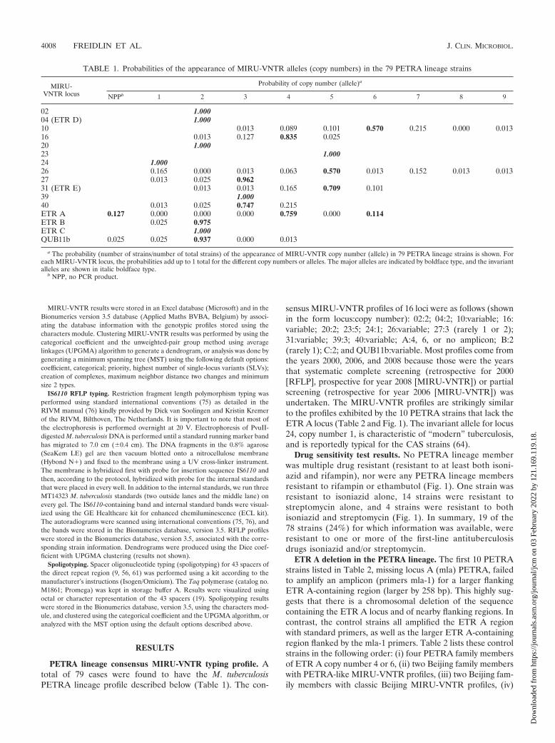
TABLE 1. Probabilities of the appearance of MIRU-VNTR alleles (copy numbers) in the 79 PETRA lineage strains
MIRU-VNTR locus
Probability of copy number (allele)a
NPPb 1 2 3 4 5 6 7 8 9
02 1.00004 (ETR D) 1.00010 0.013 0.089 0.101 0.570 0.215 0.000 0.01316 0.013 0.127 0.835 0.02520 1.00023 1.00024 1.00026 0.165 0.000 0.013 0.063 0.570 0.013 0.152 0.013 0.01327 0.013 0.025 0.96231 (ETR E) 0.013 0.013 0.165 0.709 0.10139 1.00040 0.013 0.025 0.747 0.215ETR A 0.127 0.000 0.000 0.000 0.759 0.000 0.114ETR B 0.025 0.975ETR C 1.000QUB11b 0.025 0.025 0.937 0.000 0.013
a The probability (number of strains/number of total strains) of the appearance of MIRU-VNTR copy number (allele) in 79 PETRA lineage strains is shown. Foreach MIRU-VNTR locus, the probabilities add up to 1 total for the different copy numbers or alleles. The major alleles are indicated by boldface type, and the invariantalleles are shown in italic boldface type.
b NPP, no PCR product.
4008 FREIDLIN ET AL. J. CLIN. MICROBIOL.
Dow
nloa
ded
from
http
s://j
ourn
als.
asm
.org
/jour
nal/j
cm o
n 03
Feb
ruar
y 20
22 b
y 12
1.16
9.11
9.18
.

BCG P2, and (v) MT14323. The QUB11b region, which islocated in gene Rv1917c upstream of the ETR A-containingregion, was with one exception (strain 0469-08) amplified bythe mla PETRA strains (Table 2). This suggests that deletionof the ETR A-containing region involved only partial deletionof the M. tuberculosis RD Rv1917c gene for at least 9 of the 10mla PETRA strains. Amplification of the QUB11b region forstrain 0469-08 failed in all independent experiments, and thereason for the possible QUB11b deletion needs to be deter-mined. Indeed, only sequencing of the regions spanning theETR A deletions will confirm and explain these deletions (forexample, see references 27 and 43). No data were gatheredconcerning the region farther downstream of the ETR A locus-containing region. It can be concluded that there is probably achromosomal deletion of the ETR A region in the Rv1917cgene of the 10 PETRA strains listed first in Table 2 and Fig. 1.Further investigation, precise determination of the regionsflanking the deletions, is needed to determine whether or notother genes are codeleted with the ETR A deletions. LocusETR B was amplified for all strains as a positive control, andthe expected ETR B PCR product was produced by all strains.
Bioinformatics search for examples of deleted ETR A re-gions. Two examples (unpublished) relevant to our work weresubmitted by other investigators to GenBank; these were ex-amples of the Rv1917c gene disrupted by insertion and dele-tion of the mobile element IS6110 (submitted 20 January 2005
to the National Institute for Medical Research, United King-dom, by C. Menendez). Isolate 9375 can be accessed with theidentifier GI:71056981, and isolate 8088 can be accessed withthe identifier GI:71056983. Both isolates were part of a clinicalstudy of M. tuberculosis strains in the South Asian communityof the United Kingdom. Using “BLAST2 sequences” to probethe Menendez et al. GenBank information for the IS6110 indelRv1917c sequences, it can be observed that isolate 9375 haddeleted the QUB11b and ETR A-containing regions from thegene. However, it retained at least some of the region aboveQUB11b, below ETR A, and between QUB11b and ETR A.Isolate 8088 had deleted the QUB11b and ETR A-containingregions from the gene, and it also retained at least some of theregions between QUB11b and ETR A and below ETR A.However, in contrast to isolate 9375, isolate 8088 had deletedall of the Rv1917c sequences found above the QUB11b region.This information combined with our results, although lackingdetailed sequences of the RD Rv1917c regions of mla PETRAisolates, suggests that Rv1917c is a target for IS6110 insertion/deletion, and that IS6110 insertion/deletion can cause the elim-ination of the ETR A locus (including the flanking standardprimers). The IS6110 mediated ETR-A deletions reported inGenBank appear to be different from the deletions observed inour PETRA ETR A-deleted isolates, since our isolates (withone exception) retained the QUB11b region. It is also inter-esting to note that the two isolates were obtained from the
TABLE 2. Evidence for deletion of ETR A in certain PETRA lineage strainsa
Status Strain
PCR product of ETR Aby AGEb
ETR A copyno. by CEc
PCR product ofmla-1 by AGE
PCR product of ETR Bby AGE Copy no. by CE
bp Copyno. Proto-bp bp Proto-bp bp Copy
no. Proto-bp ETR B QUB11b
mla PETRA 0433-00 NB NB NB 234 2 235 2 21178-00 NB NB NB 231 2 235 2 22136-00 NB NB NB 232 2 235 2 20545-06 NB NB NB 233 2 235 2 20958-06 NB NB NB 232 2 235 2 20170-08 NB NB NB 227 2 235 2 20469-08 NB NB NB 176 1 178 1 00804-08 NB NB NB 232 2 235 2 20937-08 NB NB NB 227 2 235 2 21158-08 NB NB NB 233 2 235 2 2
PETRA 1468-00 497 4 495 4 748 753 230 2 235 2 20428-05 637 6 645 6 887 903 230 2 235 2 20339-06 500 4 495 4 755 753 232 2 235 2 20382-08 491 4 495 4 748 753 229 2 235 2 2
Beijing (PETRA-like) 0674-06 494 4 495 4 753 753 231 2 235 2 20653-08 488 4 495 4 750 753 229 2 235 2 6
Beijing (classic) 0419-04 494 4 495 4 746 753 228 2 235 2 60944-06 497 4 495 4 746 753 236 2 235 2 6
Control BCG P2 573 5 570 5 840 828 410 5 406 5 3MT14323 421 3 420 3 672 678 232 2 235 2 2
a The chromosomal region containing ETR A in the putative PPE protein-coding RD Rv1917c gene is deleted in some PETRA lineage strains. PETRA lineagestrains lacking locus ETR A (missing locus A �mla� �no amplicons made on PCR with the standard primer pair�) also lack an enlarged region amplified with primers(mla-1) flanking the standard ETR A region primers. Note that the ETR A repeat size for a copy is 75 bp. Locus QUB11b is also found in M. tuberculosis RD Rv1917c,upstream of the ETR A locus. The flanking mla-1 primers enlarge the ETR A region by 258 bp (170 bp upstream and 88 bp downstream according to H37Rv sequencedata).
b The PCR product of ETR A by agarose gel electrophoresis (AGE) is shown. Abbreviations: bp, base pairs; proto-bp, prototype (expected) base pairs; copy no.,MIRU-VNTR copy number; NB, no band.
c The PCR product of ETR A by capillary electrophoresis (CE) is shown. NB, no band.
VOL. 47, 2009 PETRA 4009
Dow
nloa
ded
from
http
s://j
ourn
als.
asm
.org
/jour
nal/j
cm o
n 03
Feb
ruar
y 20
22 b
y 12
1.16
9.11
9.18
.

4010 FREIDLIN ET AL. J. CLIN. MICROBIOL.
Dow
nloa
ded
from
http
s://j
ourn
als.
asm
.org
/jour
nal/j
cm o
n 03
Feb
ruar
y 20
22 b
y 12
1.16
9.11
9.18
.

South Asian community and therefore may have been of CASspoligotype lineage. Neither transmissibility nor pathogenicitywas abrogated in M. tuberculosis PETRA lineage strains withETR A-deleted regions or in the two M. tuberculosis strainswith reported ETR A deletions in GenBank. It can be inferredfrom these results that IS6110 insertion/deletion is a possible,and even probable, cause of the PETRA lineage chromosomaldeletions of the RD Rv1917c ETR A-containing region. How-ever, definitive characterization will require sequencing of thepostdeletion chromosomal region.
Genotypic and demographic analyses of M. tuberculosisPETRA lineage strains. Figure 1 summarizes data for all 79PETRA strains including strain identification (in the formstrain number-year of isolation), country of origin of thepatient, type of tuberculosis specimen, resistance to first-lineantituberculosis drugs, MIRU-VNTR profiles for 16 loci (alsoshown in Fig. 2 and 3), MIRU-VNTR minimum spanning treegroup (MST shown in Fig. 4), 43-spacer octal spoligotypes(also shown in Fig. 2 and 3), 43-spacer character spoligotypes(0 for no hybridization, 1 for hybridization), and spoligotypeminimum spanning tree group (MST shown in Fig. 5). Figure1 is arranged in sections for convenient reference and can besummarized as a whole or by section. The top four sections arecomprised of four of the largest PETRA MIRU-VNTR iden-tity clusters. Subclusters that also share spoligotype identity areindicated to the right of the figure. The countries of origin of75% (58 of 77 for whom information was available) of thepatients infected with PETRA lineage strains were in sub-Saharan Africa. The patients were from the following countriesin SSA: Ethiopia (39 patients [51%]), Sudan (12 patients[16%]), and Eritrea (7 patients [9%]). The remaining patientswere from India (4 patients [5%]), Israel (6 patients [7.7%]),Nepal (3 patients [4%]) (total of 13 patients [16.7%] fromIndia, Israel, and Nepal) and then from Egypt, Georgia, Iran,Iraq, Jordan, and Yemen, each with 1 case (1.3% each) for atotal of 6 patients (7.8%) from the last six countries.
Of the 79 PETRA lineage strains shown in Figure 1, 23(29%) were isolated from various extrapulmonary specimens.Nine of the 10 strains lacking ETR A (90%) were isolated fromspecimens from patients with pulmonary tuberculosis, and 1(10%) was from an extrapulmonary specimen (Fig. 1, top sec-tion); therefore, the chromosomal deletions of ETR A did not
attenuate the affected strains. Possible confounding factorsthat prevent clear interpretation of these data would be thepatient’s genetic and health backgrounds.
The countries of origin of the patients infected with PETRAlineage strains from the year 2008 (Fig. 1) were predominantlySSA, accounting for 33 of the 41 patients (80%) of knownorigins. The patients were from the following countries in SSA:Ethiopia (14 patients [34.1%]), Sudan (12 patients [29.3%]),and Eritrea (7 patients [17.1%]). The remaining countries wereIndia (2 patients [4.9%]), Israel (3 patients [7.3%]), Nepal (2patients [4.9%]), and Jordan (1 patient [2.4%]). The tubercu-losis cases from patients from Sudan are recent cases, appear-ing only in the year 2008.
The top section of Fig. 1 shows the 10 PETRA lineagestrains lacking locus ETR A (missing locus a [mla]). Nine ofthese strains (the exception being strain 0958-06, from apatient originating from Iran) have the same spoligotypeand are of SSA origin. Five of these strains (strains 0433-00,0937-08, 1158-08, 1178-00, and 2136-00 [indicated by grayshaded background]) have identical spoligotypes andMIRU-VNTR profiles, and all were isolated from patientsoriginating from Ethiopia.
Continuing down the list of strains in Fig. 1, the next sectionshows 14 PETRA strains with MIRU-VNTR profiles identicalto the profiles of the 5 identical mla PETRA strains, exceptthat locus ETR A yields amplicons of copy number 4. Nine ofthese strains with amplicons of copy number 4 (A � 4) (strains0735-08, 0882-08, 1180-06, 1407-00, 1468-00, 2095-00, 0787-08,0382-08, and 0936-08) also have spoligotypes identical to thoseof the five identical mla PETRA strains and are indicated bygray shaded background. Seven of these nine strains are frompatients of SSA origins (five from Ethiopia, one from Eritrea,and one from Sudan), and two have non-SSA origins, one fromIndia and one from Jordan.
The next lower section of the Fig. 1 shows PETRA lineagestrains with MIRU-VNTR profiles identical to the profiles ofthe five identical ETR A-deleted PETRA strains, except thatlocus ETR A yields amplicons of copy number 6. Four of theseA � 6 strains (strains 0132-08, 0543-07, 0875-06, and 0448-08[indicated by gray shaded background]) also have spoligotypesidentical to those of the five identical ETR A-deleted PETRAstrains and nine identical A � 4 PETRA strains. These four
FIG. 1. Data for the 79 PETRA lineage strains of this study. The leftmost column (ID) contains the strain identification codes (a numberfollowed by the year of isolation). The next column shows the country of origin of the patient with tuberculosis as follows: IRN, Iran; ERI, Eritrea;ETH, Ethiopia; IND, India; JOR, Jordan; SUD, Sudan; NEP, Nepal; ISR, Israel; EGY, Egypt; YEM, Yemen; GEO, Georgia; IRQ, Iraq. The typeof tuberculosis specimen (TB type) is shown as follows: P, pulmonary; EP, extrapulmonary. In the RES column, resistance to first lineanti-tuberculosis drugs (RES) is shown as follows: I, isoniazid; S, streptomycin; n a, information not available. The MIRU-VNTR loci columnshows the MIRU-VNTR copy number profile for each strain, by locus in the following order: 2, 4, 10, 16, 20, 23, 24, 26, 27, 31, 39, 40, A, B, C,and QUB11b, which are clustered in Fig. 2. Note that 0 indicates an absence of the amplicon (terminology which facilitates data manipulation byExcel and Bionumerics). The column labeled MST MIRU Group shows the MIRU-VNTR minimum spanning tree (MST) group as visualized inFig. 4. The SPOLIGO Octal column shows the octal equivalent of the 43-spacer spoligotype. The column labeled SPOLIGO Spacers shows thecharacter representation of the 43-spacer spoligotype profiles for each strain, which are clustered in Fig. 3, as follows: 1 for hybridization and 0for no hybridization. The MST Spoligo Group column shows the spoligotype MST group as visualized in Fig. 5. The strains with gray shadedbackgrounds have identical spoligotypes and MIRU-VNTR types that vary only at locus ETR A. To the right of the figure, the MIRU-VNTRsubclusters that also share spoligotype identity (MIRU-spo-identity) are shown. ETR A amplicons of various copy numbers are shown (forexample, amplicons of copy number 4 are represented by A � 4). Since it is difficult in places to distinguish among zero, Q, and capital O, we notefor the sake of clarity that the country of origin for strain 0753-06 is Iraq (IRQ), the MST MIRU group for strain 2039-00 is A0, the MST MIRUgroup for strain 0346-08 is AO, the MST MIRU group for strain 0493-06 is AQ, the MST spoligo group for strain 0854-08 is Q, and the MSTspoligo group for strain 0346-08 is O.
VOL. 47, 2009 PETRA 4011
Dow
nloa
ded
from
http
s://j
ourn
als.
asm
.org
/jour
nal/j
cm o
n 03
Feb
ruar
y 20
22 b
y 12
1.16
9.11
9.18
.
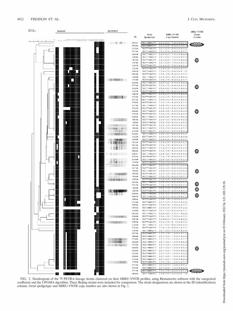
FIG. 2. Dendrogram of the 79 PETRA lineage strains clustered on their MIRU-VNTR profiles, using Bionumerics software with the categoricalcoefficient and the UPGMA algorithm. Three Beijing strains were included for comparison. The strain designations are shown in the ID (identification)column. Octal spoligotype and MIRU-VNTR copy number are also shown in Fig. 1.
4012 FREIDLIN ET AL. J. CLIN. MICROBIOL.
Dow
nloa
ded
from
http
s://j
ourn
als.
asm
.org
/jour
nal/j
cm o
n 03
Feb
ruar
y 20
22 b
y 12
1.16
9.11
9.18
.

FIG. 3. Dendrogram of the 79 PETRA lineage strains clustered on their 43 spacer spoligotype profiles, using Bionumerics software with thecategorical coefficient and the UPGMA algorithm. Three Beijing strains were included for comparison. Strain designations are shown in the ID(identification) column. Octal spoligotype and MIRU-VNTR copy number are also shown in Fig. 1.
VOL. 47, 2009 PETRA 4013
Dow
nloa
ded
from
http
s://j
ourn
als.
asm
.org
/jour
nal/j
cm o
n 03
Feb
ruar
y 20
22 b
y 12
1.16
9.11
9.18
.
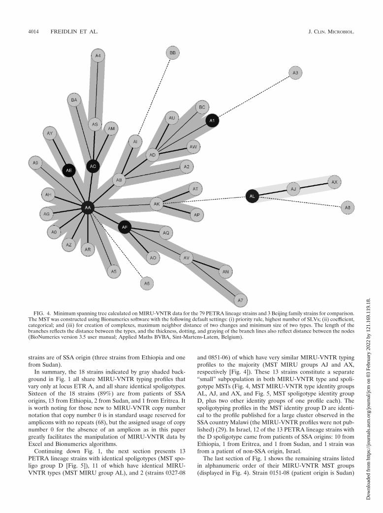
strains are of SSA origin (three strains from Ethiopia and onefrom Sudan).
In summary, the 18 strains indicated by gray shaded back-ground in Fig. 1 all share MIRU-VNTR typing profiles thatvary only at locus ETR A, and all share identical spoligotypes.Sixteen of the 18 strains (89%) are from patients of SSAorigins, 13 from Ethiopia, 2 from Sudan, and 1 from Eritrea. Itis worth noting for those new to MIRU-VNTR copy numbernotation that copy number 0 is in standard usage reserved foramplicons with no repeats (68), but the assigned usage of copynumber 0 for the absence of an amplicon as in this papergreatly facilitates the manipulation of MIRU-VNTR data byExcel and Bionumerics algorithms.
Continuing down Fig. 1, the next section presents 13PETRA lineage strains with identical spoligotypes (MST spo-ligo group D [Fig. 5]), 11 of which have identical MIRU-VNTR types (MST MIRU group AL), and 2 (strains 0327-08
and 0851-06) of which have very similar MIRU-VNTR typingprofiles to the majority (MST MIRU groups AJ and AX,respectively [Fig. 4]). These 13 strains constitute a separate“small” subpopulation in both MIRU-VNTR type and spoli-gotype MSTs (Fig. 4, MST MIRU-VNTR type identity groupsAL, AJ, and AX, and Fig. 5, MST spoligotype identity groupD, plus two other identity groups of one profile each). Thespoligotyping profiles in the MST identity group D are identi-cal to the profile published for a large cluster observed in theSSA country Malawi (the MIRU-VNTR profiles were not pub-lished) (29). In Israel, 12 of the 13 PETRA lineage strains withthe D spoligotype came from patients of SSA origins: 10 fromEthiopia, 1 from Eritrea, and 1 from Sudan, and 1 strain wasfrom a patient of non-SSA origin, Israel.
The last section of Fig. 1 shows the remaining strains listedin alphanumeric order of their MIRU-VNTR MST groups(displayed in Fig. 4). Strain 0151-08 (patient origin is Sudan)
FIG. 4. Minimum spanning tree calculated on MIRU-VNTR data for the 79 PETRA lineage strains and 3 Beijing family strains for comparison.The MST was constructed using Bionumerics software with the following default settings: (i) priority rule, highest number of SLVs; (ii) coefficient,categorical; and (iii) for creation of complexes, maximum neighbor distance of two changes and minimum size of two types. The length of thebranches reflects the distance between the types, and the thickness, dotting, and graying of the branch lines also reflect distance between the nodes(BioNumerics version 3.5 user manual; Applied Maths BVBA, Sint-Martens-Latem, Belgium).
4014 FREIDLIN ET AL. J. CLIN. MICROBIOL.
Dow
nloa
ded
from
http
s://j
ourn
als.
asm
.org
/jour
nal/j
cm o
n 03
Feb
ruar
y 20
22 b
y 12
1.16
9.11
9.18
.
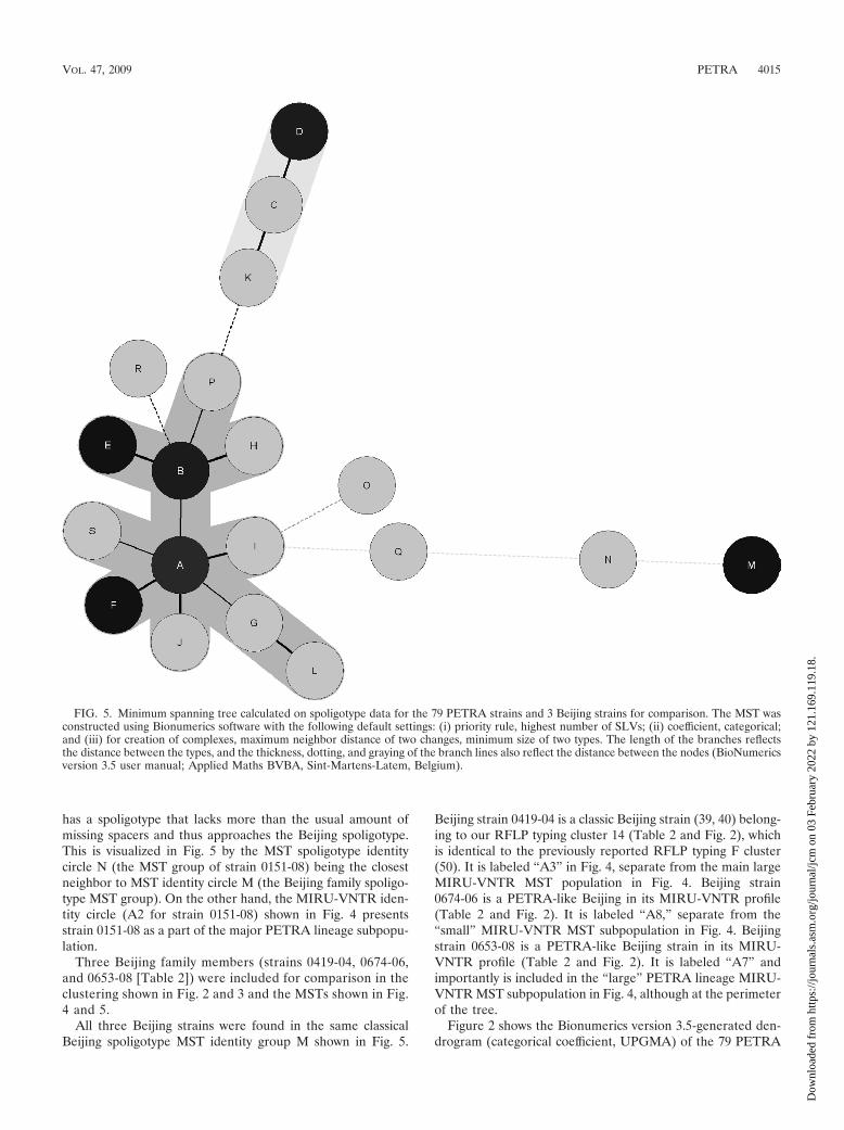
has a spoligotype that lacks more than the usual amount ofmissing spacers and thus approaches the Beijing spoligotype.This is visualized in Fig. 5 by the MST spoligotype identitycircle N (the MST group of strain 0151-08) being the closestneighbor to MST identity circle M (the Beijing family spoligo-type MST group). On the other hand, the MIRU-VNTR iden-tity circle (A2 for strain 0151-08) shown in Fig. 4 presentsstrain 0151-08 as a part of the major PETRA lineage subpopu-lation.
Three Beijing family members (strains 0419-04, 0674-06,and 0653-08 [Table 2]) were included for comparison in theclustering shown in Fig. 2 and 3 and the MSTs shown in Fig.4 and 5.
All three Beijing strains were found in the same classicalBeijing spoligotype MST identity group M shown in Fig. 5.
Beijing strain 0419-04 is a classic Beijing strain (39, 40) belong-ing to our RFLP typing cluster 14 (Table 2 and Fig. 2), whichis identical to the previously reported RFLP typing F cluster(50). It is labeled “A3” in Fig. 4, separate from the main largeMIRU-VNTR MST population in Fig. 4. Beijing strain0674-06 is a PETRA-like Beijing in its MIRU-VNTR profile(Table 2 and Fig. 2). It is labeled “A8,” separate from the“small” MIRU-VNTR MST subpopulation in Fig. 4. Beijingstrain 0653-08 is a PETRA-like Beijing strain in its MIRU-VNTR profile (Table 2 and Fig. 2). It is labeled “A7” andimportantly is included in the “large” PETRA lineage MIRU-VNTR MST subpopulation in Fig. 4, although at the perimeterof the tree.
Figure 2 shows the Bionumerics version 3.5-generated den-drogram (categorical coefficient, UPGMA) of the 79 PETRA
FIG. 5. Minimum spanning tree calculated on spoligotype data for the 79 PETRA strains and 3 Beijing strains for comparison. The MST wasconstructed using Bionumerics software with the following default settings: (i) priority rule, highest number of SLVs; (ii) coefficient, categorical;and (iii) for creation of complexes, maximum neighbor distance of two changes, minimum size of two types. The length of the branches reflectsthe distance between the types, and the thickness, dotting, and graying of the branch lines also reflect the distance between the nodes (BioNumericsversion 3.5 user manual; Applied Maths BVBA, Sint-Martens-Latem, Belgium).
VOL. 47, 2009 PETRA 4015
Dow
nloa
ded
from
http
s://j
ourn
als.
asm
.org
/jour
nal/j
cm o
n 03
Feb
ruar
y 20
22 b
y 12
1.16
9.11
9.18
.
family MIRU-VNTR profiles, along with their associated spo-ligotypes and partial RFLP typing profiles. Profiles from threeBeijing family strains (strains 0419-04, 0653-08, and 0674-06)were included for comparison. By comparison with the MIRU-VNTRplus database (5) and published MIRU-VNTR profilesand spoligotyping profiles (3, 6, 7, 61, 62), we determined thatour PETRA spoligotypes are identical to those found in theCAS spoligotype lineage and that our PETRA consensusMIRU-VNTR types (Table 1 and Fig. 1) therefore constitutea lineage of CAS MIRU-VNTR types. Our 16-locus MIRU-VNTR panel yielded nine MIRU-VNTR clusters (identicalMIRU-VNTR profiles), three of which clustered together dif-ferent spoligotypes (clusters 2, 3, and 4 of Fig. 2). Therefore,the 16-locus panel is less discriminatory than RFLP for molec-ular typing of PETRA lineage strains, although it still may havevalue for identifying epidemiological links (8, 74). A 24-locusMIRU-VNTR panel has recently been reported to separateeach of many different CAS spoligotypes (6). However, thefact that the 16-locus MIRU-VNTR panel gave nine clusterscontaining 52 strains, while clustering on spoligotypes (cat-egorical coefficient, UPGMA, Fig. 3) yielded six clusterscontaining 67 strains shows that the 16-locus MIRU-VNTRtyping is more discriminatory than the 43-spacer spoligotyp-ing for clustering PETRA lineage strains. Thirty-two of the43 PETRA lineage strains from 2008 (74%) were found ineight of the nine MIRU-VNTR clusters (Fig. 2). Each of the32 clustered PETRA 2008 strains was found in cluster withat least one other PETRA strain from 2008 (Fig. 2). This ishighly suggestive of recent transmission, but not necessarilytransmission in Israel. Twenty-six of the 32 clusteredPETRA 2008 strains (81%) were from patients of SSA or-igins (Fig. 1 and Fig. 2). As already noted, in 2008, therewere 33 SSA origin PETRA strains; therefore, 79% (26 of33) of these SSA origin PETRA strains were clustered byMIRU-VNTR typing (Fig. 1 and 2).
The MST calculated on 16-locus MIRU-VNTR typing re-sults, using Bionumerics version 3.5 default parameters, isshown in Fig. 4. The PETRA lineage strains were found in onelarge subpopulation and one small subpopulation as alreadydescribed concerning the results shown in Fig. 1. The 10PETRA strains with deleted ETR A were not separated into asubpopulation different from the “wild-type” PETRA strains(capable of producing ETR A amplicons), but they did formtheir own “branch” of the larger subpopulation (Fig. 1 and 4,MST identity circles AC, AM, AS, A4, and BA). These resultssupport the suitability of the 16-locus MIRU-VNTR panel forscreening for PETRA lineage membership. One Beijing familymember with a PETRA-like MIRU-VNTR profile (strain0653-08, identity circle A7) was also included in the PETRApopulation. This is not necessarily an artifact, and its implica-tions are considered below in the Discussion. On the otherhand, this indicates that the 16-locus MIRU-VNTR panel willin rare cases give unclear results or even false-positive resultswith respect to PETRA versus Beijing assignation. It is notcertain that even a 24-locus MIRU-VNTR screen (68, 80)could avoid this difficulty (31). Therefore, use of the 16-locusMIRU-VNTR panel (the original 12 standard loci, ETR lociA, B, and C, and QUB11b) necessitates complementaryscreening by 43-spacer spoligotyping for definitive identifica-
tion as a PETRA lineage (this paper) or Beijing family (10, 39,40, 77) strain.
The MST calculated on 43-spacer spoligotyping resultsusing Bionumerics version 3.5 default parameters, is shownin Fig. 5. The spoligotype MST divided the PETRA strainsinto one large subpopulation and one small subpopulationas already described concerning the results shown in Fig. 1.This division can also be visualized in the Fig. 3 dendrogramof spoligotype clusters. These two populations were essen-tially equivalent to the two major subpopulations of theMST based on MIRU-VNTR profiles. As expected, the Bei-jing strains formed another separate subpopulation (Fig. 5,MST spoligotype identity circle M; also see Fig. 3). PETRA43-spacer spoligotypes were clearly shown to be of CASlineage (3, 6, 7, 58, 61, 62), as visualized in Fig. 3 and Fig. 5.Since MIRU-VNTR resolves MST subpopulations similar tothe spoligotype-resolved MST subpopulations, it can be animportant aid in determining a spoligotype lineage, such asthe CAS lineage.
DISCUSSION
Discovery of PETRA. Prospective screening of every newculture-positive Mycobacterium tuberculosis complex case inIsrael in the year 2008 was a direct benefit of automatic, high-throughput, 16-capillary electrophoresis of multiplex panels of16 MIRU-VNTR loci (4, 47, 65, 67, 68). In addition, retro-spective screening had been undertaken (partially by agarosegel electrophoresis) of isolates submitted in previous years,primarily from the years 2000 (in which a complete retrospec-tive RFLP typing screen of new pulmonary Mycobacteriumtuberculosis was performed) and 2006 (a year of partial system-atic retrospective MIRU-VNTR screening). This screeningand especially the high-throughput MIRU-VNTR screeningenabled discovery and characterization of a major contributorto new tuberculosis cases in Israel, responsible for about 20%(43 of 222) of new culture-positive cases in 2008. The contrib-utor was found to be a new consensus MIRU-VNTR lineage(Table 1 and Fig. 1) of the central Asian (CAS) spoligotypelineage (5, 6, 61, 62) which was termed “PETRA” for “poly-morphic ETR A.”
CAS spoligotypes: considerations and origins with a focuson the PETRA lineage. Recently, other groups of investigatorshave reported that the CAS spoligotype lineage contributes tothe tuberculosis burdens of various nations in the Middle East(attributed to population influx from Africa [3]), Africa (29,58), and Asia (7, 33, 63). In Israel as reported in this paper, 77(out of a total of 79) of the PETRA lineage strains wereisolated from patients for whom information regarding countryorigin was available. Out of the 77 patients, 58 (75%) origi-nated from SSA. Interestingly, other investigators reporting onpossible venues for CAS genotypes did not observe significantnumbers of CAS genotypes in their sample populations fromTurkey (2, 21), Ethiopia (14), and East Asia (RD Rv1519deletion confers immune subverting phenotype [49]) (77).
PETRA strains were isolated from both pulmonary and ex-trapulmonary samples. Extrapulmonary tuberculosis has beenreported from CAS spoligotype MT (41). The role, if any, forPETRA lineage ETR A in extrapulmonary tuberculosis re-quires further work for elucidation.
4016 FREIDLIN ET AL. J. CLIN. MICROBIOL.
Dow
nloa
ded
from
http
s://j
ourn
als.
asm
.org
/jour
nal/j
cm o
n 03
Feb
ruar
y 20
22 b
y 12
1.16
9.11
9.18
.
Since the majority of our PETRA strains, 75%, originatefrom SSA countries, we anticipate that the distribution ofPETRA is global, although we are the first to describe thislineage. Indeed, 25% of the PETRA strains have non-SSAorigins (Fig. 1). A similar situation was recently described inwhich a Mycobacterium tuberculosis RD lineage was a majorcause of tuberculosis in Rio de Janeiro, Brazil (43), and wassubsequently found to be globally disseminated (27) as a sub-lineage of the Latin American-Mediterranean (LAM) Myco-bacterium tuberculosis spoligotype lineage. MIRU-VNTR com-bined with spoligotyping was recently used to define a newMycobacterium tuberculosis lineage responsible for over 40% ofsmear-positive pulmonary tuberculosis cases in Cameroon dur-ing the time period of the study (51).
Usually, screening for Beijing family members on the rec-ommended loci (40) allows differentiation of Beijing andPETRA lineage members. However, certain strains give incon-clusive results and require spoligotyping to resolve the issue.This is reasonable, as clustering on MIRU-VNTR loci (24 loci)has shown that Beijing and CAS lineages are closely related (4,68). It has been suggested that the CAS lineage is the ancestorto the Beijing lineage (61). One of our PETRA strains (strain0151-08) has a spoligotype that closely resembles that of Bei-jing members (Fig. 1, 2, 3, and 5); this spoligotype has beenassigned to the CAS lineage (5).
Putative mechanism for deletion of ETR A in PETRAstrains. IS6110 has been inferred to insert into preferentialnonrandom sites (23) and to mediate certain deletion polymor-phisms in Mycobacterium tuberculosis (22, 24, 57). IS6110-me-diated deletion provides a likely mechanism for the deletionsof ETR A in M. tuberculosis RD Rv1917c in the IS6110 indelsequences submitted by Menendez et al. to GenBank, GI:71056981 and GI:71056983. The Menendez et al. submissionswere sequences recovered from virulent clinical isolates andthus complement our findings that PETRA isolates with de-leted ETR A remain virulent. However, the deletions of ETRA in PETRA lineage members must be characterized in se-quence-specific detail before any firm conclusions can bereached concerning the mechanisms responsible for the dele-tions. IS6110-mediated deletion of the RvD2 region in clinicalisolates did not attenuate virulence, and by inference, deletionof RvD2 was not responsible for attenuation of H37Ra versusthe virulent H37Rv (42, 81). Therefore, attenuation of viru-lence very much depends on which sequences are deleted. Forexample, deletion (not necessarily involving IS6110) of RD1from M. bovis BCG (13) is the primary cause of attenuation ofBCG (34). The effect of IS6110 insertion is further complicatedby the ability of IS6110 to provide a promoter sequence for thetranscription of genes (9). Small differences in genotypes fromstrains isolated from the same patient (46, 57) may haveIS6110-mediated associations (57).
From our limited RFLP typing data, it can be seen thatPETRA lineage strains have many copies of IS6110 (profileswith many bands [Fig. 2 and 3]). However, it is interesting thatdespite the many different RFLP typing profiles with largenumbers of bands (from strains with large numbers of chro-mosomal copies of IS6110), the PETRA MIRU-VNTR lineageis the first lineage and so far the only lineage to be shown toinclude a significant number of strains that lack ETR A.
Effect of the ETR A locus on the putative PPE34 protein.The ETR A region is found in the gene Rv1917c which is acoding sequence for the putative transmembrane PPE34 pro-tein (PPE stands for Pro-Pro-Glu) (17, 55, 79). Thus, ourdiscovery of PETRA strains that delete the ETR A regionsuggests the intriguing, but currently speculative, possibilitythat the PETRA lineage is subject to evolutionary pressure onits putative PPE34 protein.
As reported in this work, PETRA is the first MIRU-VNTRlineage, a sublineage of the CAS spoligotype lineage, shown tobe clearly predisposed toward chromosomal deletions of theRv1917c ETR A locus (Table 2 and Fig. 1, 2, and 3). Unlike thePPE gene-associated deletion events that resulted in attenu-ated nonpathogenic M. bovis BCG (12, 13, 32), the deletion ofputative PPE34-coding, Rv1917c ETR A-containing chromo-somal regions reported herein left the virulence of thesePETRA lineage members intact, although possibly modified. Atotal of 10 ETR A-deleted PETRA strains were observed, 5during the prospective screen of 2008, 2 from 2006, and 3 fromthe year 2000. About 12% of the PETRA lineage members in2008 (5 of 43) were missing locus ETR A (mla in Table 2 andFig. 1). Loss of ETR A has immediate implications for stan-dardized MIRU-VNTR epidemiological screening, for whichETR A is one of the recommended screening loci (68).
Previous works reported that the Rv1917c gene is tran-scribed (mRNA was detected) in M. tuberculosis (55, 79). How-ever, to date, there is no evidence that the putative M. tuber-culosis PPE34 protein is transmembrane and surface exposed,although indirect evidence with a recombinant PPE34 in M.smegmatis suggests this behavior (55). Bioinformatics predic-tion of PPE protein structure also suggests that the putativePPE34 when found in M. tuberculosis will be transmembraneand surface bound. Many of the MPTR PPE proteins have apredicted beta-barrel outer membrane protein structure (re-viewed in reference 26). An obvious but important question iswhether there is a difference in PPE34 expression, location,antigenic potential, or function between PETRA strains withETR A deletions compared to the PETRA strains that possessETR A. Zvi et al. (82) have reviewed antigenic and functionalM. tuberculosis PPE proteins that could serve as vaccine can-didates. It would be desirable to know whether putative PPE34peptides are suitable candidates for vaccines.
The fact that in some PETRA lineage strains, the ETR Alocus and flanking primers are deleted with the PETRA-asso-ciated deletion of part of the chromosomal region encom-passed by M. tuberculosis RD Rv1917c results in the failure todetect the ETR A locus in epidemiologic MIRU-VNTR PCRscreening. Deletion of the target region has been reported tocause the failure of a commercial assay to detect Mycobacte-rium tuberculosis DNA in sputum samples (28) and to causenegative results when attempting to detect the mtp40 sequence(plcA) by PCR (78). As a positive contribution, deletion pro-filing has been used to type Mycobacterium tuberculosis com-plex species (16, 35, 38) and also has shown the close relation-ship between the Beijing and CAS lineages (7).
The role of PPE proteins in antigenic variation of Mycobac-terium tuberculosis has been strongly suspected at least sincethe publication of the whole-genome sequence of M. tubercu-losis H37Rv (17). Whole-genome comparisons of fully se-quenced strain H37Rv with fully sequenced strain H37Ra (81)
VOL. 47, 2009 PETRA 4017
Dow
nloa
ded
from
http
s://j
ourn
als.
asm
.org
/jour
nal/j
cm o
n 03
Feb
ruar
y 20
22 b
y 12
1.16
9.11
9.18
.
and fully sequenced strain CDC 1551 (24) have shown that thePPE gene family significantly contributes to the observed poly-morphism. In three other whole-genome comparison studies,technical issues related to methodology prevented informativeresults on PPE gene family members (36, 54, 71). The 68-member glycine-rich PPE (Pro-Pro-Glu motif) family was oneof the new families revealed in the first published analysis ofthe complete genome of Mycobacterium tuberculosis H37Rv(17). The ETR A locus (25) is found in the translated openreading frame of the H37Rv Rv1917c gene coding for M.tuberculosis Rv1917c mRNA (55, 79) and for the putativePPE34 protein (17, 26, 55). Rv1917c has been assigned to themajor polymorphic tandem repeat (MPTR) subfamily (17, 26).It has been suggested (26) that the PPE MPTR subfamilyoriginated from a duplication from ESAT-6 (esx) gene clusterregion 5. The ESAT-6 system is well-known for being deletedfrom M. bovis BCG in the RD1 deletion, which is largelyresponsible for the attenuation of BCG (12, 32). In addition,this provides the basis for a commercial test employing se-lected RD1 peptides for diagnosis of active tuberculosis (30).Mycobacterial “core” genes (45) and the Rv1917c gene (26)have been searched for in nontuberculous mycobacteria. Someapparent homology to Rv1917c was found in Mycobacteriumasiaticum, Mycobacterium gordonae, Mycobacterium marinum,and Mycobacterium ulcerans, but no detailed comparisons weremade to the M. tuberculosis Rv1917c gene (26).
Various PPE proteins have been shown to be immunogenic(for example, PPE41 [reviewed in reference 1], PPE44 [11],PPE55 [59], and PPE68 [20]), cell surface associated (for ex-ample, recombinant PPE34 in Mycobacterium smegmatis [55]),and secreted (for example, PPE41 [1]). Clusters of PE and PPEgenes of Mycobacterium tuberculosis have been reported to beorganized in operons (73). As already mentioned, PE (forexample, PE-PGRS33 [70]) and PPE (for example, putativePPE34 [55]) proteins may be surface expressed and potentiallycontribute a large amount of the antigenic variation that affectsimmunity (17, 82). A recent paper reporting genome-widetranscriptional profiling of chronic tuberculosis (69) did notfocus on the PE/PPE protein families. However, an earlierpaper summarizing whole-genome Mycobacterium tuberculosisPE/PPE expression patterns under various environmental con-ditions (79), reported that expression of the gene for PPE34was repressed on day 14 of stationary phase.
Previous reports on the structure, location, antigenic poten-tial, and function of PPE proteins and the proclivity of theirDNA coding regions to be associated with chromosomal dele-tions suggest that it is likely that the Rv1917c putative PPE34protein will be transmembrane, surface exposed, antigenic, andfunctional. If further investigation shows this to be the case,then the chromosomal deletions of ETR A in RD Rv1917ccould indicate that M. tuberculosis PETRA is under evolution-ary pressure at PPE34. Such pressure could occur throughnormal immune surveillance or host genetic variability in in-nate immunity such as that recently documented for Toll-likereceptors (48) or perhaps variability in other receptors, such asdectin-1 (72), or even other types of genetic variability (18).The possible interaction between host genotype and M. tuber-culosis genotype has been noted by the authors of a recentarticle (44).
Conclusions. Culture-positive tuberculosis in Israel includestwo challenging groups of strains. These are (i) the multipledrug resistance-associated Beijing family strains (unpublisheddata), primarily from former Soviet Union countries, and (ii)the PETRA MIRU-VNTR lineage (this paper): non-MDRcentral Asian (CAS) spoligotype lineage M. tuberculosis, pri-marily from sub-Saharan African countries, causing about 20%of new cases of tuberculosis in Israel in 2008.
The genotypes associated with the 79 PETRA lineage strainspresented in this paper strengthen the conviction that there isa large, ongoing spread (but not necessarily transmitted, inwhole or in part, in Israel) of tuberculosis caused by thePETRA lineage.
ACKNOWLEDGMENTS
This work was performed under the auspices of the Israel NationalProgram for the Prevention and Control of Tuberculosis run by theMinistry of Health. We thank Daniel Chemtob, Head of the Depart-ment of Tuberculosis and AIDS, for his support.
REFERENCES
1. Abdallah, A. M., T. Verboom, F. Hannes, M. Safi, M. Strong, D. Eisenberg,R. J. Musters, C. M. Vandenbroucke-Grauls, B. J. Appelmelk, J. Luirink,and W. Bitter. 2006. A specific secretion system mediates PPE41 transport inpathogenic mycobacteria. Mol. Microbiol. 62:667–679.
2. Aktas, E., T. Zozio, F. B. Comert, C. Kulah, O. Aydin, N. Rastogi, and C.Sola. 2007. A first insight into the genetic diversity and population structureof Mycobacterium tuberculosis in Zonguldak, Turkey. Clin. Microbiol. Infect.14(1):55–59.
3. Al-Hajoj, S. A. M., T. Zozio, F. Al-Rabiah, V. Mohammad, M. Al-Nasser, C.Sola, and N. Rastogi. 2007. First insight into the population structure ofMycobacterium tuberculosis in Saudi Arabia. J. Clin. Microbiol. 45:2467–2473.
4. Allix, C., P. Supply, and M. Fauville-Dufaux. 2004. Utility of fast mycobac-terial interspersed repetitive unit-variable number tandem repeat genotypingin clinical mycobacteriological analysis. Clin. Infect. Dis. 39:783–789.
5. Allix-Beguec, C., D. Harmesen, T. Weniger, P. Supply, and S. Niemann.2008. Evaluation and strategy for use of MIRU-VNTRplus, a multifunctionaldatabase for online analysis of genotyping data and phylogenetic identifica-tion of Mycobacterium tuberculosis complex isolates. J. Clin. Microbiol. 46:2692–2699.
6. Allix-Beguec, C., M. Fauville-Dufaux, and P. Supply. 2008. Three-year pop-ulation-based evaluation of standardized mycobacterial interspersed repeti-tive-unit–variable-number tandem-repeat typing of Mycobacterium tubercu-losis. J. Clin. Microbiol. 46:1398–1406.
7. Banu, S., S. V. Gordon, S. Palmer, R. Islam, S. Ahmed, K. M. Alam, S. T.Cole, and R. Brosch. 2004. Genotypic analysis of Mycobacterium tuberculosisin Bangladesh and prevalence of the Beijing strain. J. Clin. Microbiol. 42:674–682.
8. Barlow, E. L., D. M. Gascoyne-Binzi, S. H. Gillespie, A. Dickens, S. Qamer,and P. M. Hawkey. 2001. Comparison of variable number tandem repeat andIS6110-restriction fragment length polymorphism analyses for discriminationof high- and low-copy-number IS6110 Mycobacterium tuberculosis isolates.J. Clin. Microbiol. 39:2453–2457.
9. Beggs, M. L., K. D. Eisenach, and M. D. Cave. 2000. Mapping of IS6110insertion sites in two epidemic strains of Mycobacterium tuberculosis. J. Clin.Microbiol. 38:2923–2928.
10. Bifani, P. J., B. Mathema, N. E. Kurepina, and B. N. Kreiswirth. 2002.Global dissemination of the Mycobacterium tuberculosis W-Beijing familystrains. Trends Microbiol. 10:45–51.
11. Bonanni, D., L. Rindi, N. Lari, and C. Garzelli. 2005. Immunogenicity ofmycobacterial PPE44 (Rv2770c) in Mycobacterium bovis BCG-infected mice.J. Med. Microbiol. 54:443–448.
12. Brodin, P., L. Majlessi, L. Marsollier, M. I. De Jonge, D. Bottai, C. Deman-gel, J. Hinds, O. Neyrolles, P. D. Butcher, C. Leclerc, S. T. Cole, and R.Brosch. 2006. Dissection of ESAT-6 system 1 of Mycobacterium tuberculosisand impact on immunogenicity and virulence. Infect. Immun. 74:88–98.
13. Brosch, R., S. V. Gordon, T. Garnier, K. Eiglmeier, W. Frigui, P. Valenti,S. D. Santos, S. Duthoy, C. Lacroix, C. Garcia-Pelayo, J. K. Inwald, P. Golby,J. N. Garcia, R. G. Hewinson, M. A. Behr, M. A. Quail, C. Churcher, B. G.Barrell, J. Parkhill, and S. T. Cole. 2007. Genome plasticity of BCG andimpact on vaccine efficacy. Proc. Natl. Acad. Sci. USA 104:5596–5601.
14. Bruchfeld, J., G. Aderaye, I. B. Palme, B. Bjorvatn, S. Ghebremichael, S.Hoffner, and L. Lindquist. 2002. Molecular epidemiology and drug resis-tance of Mycobacterium tuberculosis isolates from Ethiopian pulmonary tu-
4018 FREIDLIN ET AL. J. CLIN. MICROBIOL.
Dow
nloa
ded
from
http
s://j
ourn
als.
asm
.org
/jour
nal/j
cm o
n 03
Feb
ruar
y 20
22 b
y 12
1.16
9.11
9.18
.
berculosis patients with and without human immunodeficiency virus infec-tion. J. Clin. Microbiol. 40:1636–1643.
15. Centers for Disease Control and Prevention. 2007. World TB Day—March24, 2007. MMWR Morb. Mortal. Wkly. Rep. 56:245.
16. Cole, S. T. 2002. Comparative and functional genomics of the Mycobacteriumtuberculosis complex. Microbiology 148:2919–2928.
17. Cole, S. T., R. Brosch, J. Parkhill, T. Garnier, C. Churcher, D. Harris, S. V.Gordon, K. Eiglmeier, S. Gas, C. E. Barry III, F. Tekaia, K. Badcock, D.Basham, D. Brown, T. Chillingworth, R. Connor, R. Davies, K. Devlin, T.Feltwell, S. Gentles, N. Hamlin, S. Holroyd, T. Hornsby, K. Jagels, A. Krogh,J. McLean, S. Moule, L. Murphy, K. Oliver, J. Osborne, M. A. Quail, M.-A.Rajandream, J. Rogers, S. Rutter, K. Seeger, J. Skelton, R. Squares, S.Squares, J. E. Sulston, K. Taylor, S. Whitehead, and B. G. Barrell. 1998.Deciphering the biology of Mycobacterium tuberculosis from the completegenome sequence. Nature 393:537–544.
18. Cooke, G. S., S. J. Campbell, S. Bennett, C. Lienhardt, K. P. W. J. McAdam,G. Sirugo, O. Sow, P. Gustafson, F. Mwangulu, P. van Helden, P. Fine, E. G.Hoal, and A. V. S. Hill. 2008. Mapping of a novel susceptibility locus suggestsa role for MC3R and CTSZ in human tuberculosis. Am. J. Respir. Crit. CareMed. 178:203–207.
19. Dale, J. W., D. Brittain, A. A. Cataldi, D. Cousins, J. T. Crawford, J. Driscoll,H. Heersma, T. Lillbaek, T. Quitugua, N. Rastogi, R. A. Skuce, C. Sola, D.van Soolingen, and V. Vincent. 2001. Spacer oligonucleotide typing of bac-teria of the Mycobacterium tuberculosis complex: recommendations for stan-dardised nomenclature. Int. J. Tuberc. Lung Dis. 5:216–219.
20. Demangel, C., P. Brodin, P. J. Cockle, R. Brosch, L. Majlessi, C. Leclerc, andS. T. Cole. 2004. Cell envelope protein PPE68 contributes to Mycobacteriumtuberculosis RD1 immunogenicity independently of a 10-kilodalton culturefiltrate protein and ESAT-6. Infect. Immun. 72:2170–2176.
21. Durmaz, R., T. Zozio, S. Gunal, C. Allix, M. Fauville-Dufaux, and N. Ras-togi. 2007. Population-based molecular epidemiological study of tuberculosisin Malatya, Turkey. J. Clin. Microbiol. 45:4027–4035.
22. Fang, Z., C. Doig, D. T. Kenna, N. Smittipat, P. Palittapongarnpim, B. Watt,and K. J. Forbes. 1999. IS6110-mediated deletions of wild-type chromo-somes of Mycobacterium tuberculosis. J. Bacteriol. 181:1014–1020.
23. Fang, Z., D. T. Kenna, C. Doig, D. N. Smittipat, P. Palittapongarnpim, B.Watt, and K. J. Forbes. 2001. Molecular evidence for independent occur-rence of IS6110 insertions at the same sites of the genome of Mycobacteriumtuberculosis in different clinical isolates. J. Bacteriol. 183:5279–5284.
24. Fleischmann, R. D., D. Alland, J. A. Eisen, L. Carpenter, O. White, J.Peterson, R. DeBoy, R. Dodson, M. Gwinn, D. Haft, E. Hickey, J. F. Kolonay,W. C. Nelson, L. A. Umayam, M. Ermolaeva, S. L. Salzberg, A. Delcher, T.Utterback, J. Weidman, H. Khouri, J. Gill, A. Mikula, W. Bishai, W. R.Jacobs, Jr., J. C. Bernter, and C. M. Fraser. 2002. Whole-genome compar-ison of Mycobacterium tuberculosis clinical and laboratory strains. J. Bacte-riol. 184:5479–5490.
25. Frothingham, R., and W. A. Meeker-O’Connell. 1998. Genetic diversity inthe Mycobacterium tuberculosis complex based on variable numbers of tan-dem DNA repeats. Microbiology 144:1189–1196.
26. Gey van Pittius, N. C., S. L. Sampson, H. Lee, Y. Kim, D. van Helden, andR. M. Warren. 2006. Evolution and expansion of the Mycobacterium tuber-culosis PE and PPE multigene families and their association with the dupli-cation of the ESAT-6 (esx) gene cluster regions. BMC Evol. Biol. 6:95.
27. Gibson, A. L., R. C. Huard, N. C. Gey van Pittius, L. C. O. Lazzarini, F.Driscoll, N. Kurepina, T. Zozio, C. Sola, S. M. Spindola, A. L. Kritski, D.Fitzgerald, K. Kremer, H. Mardassi, P. Chitale, J. Brinkworth, D. G. deViedma, B. Gicuel, J. W. Pape, D. van Soolingen, B. N. Kreiswirth, R. M.Warren, P. D. van Helden, N. Rastogi, P. N. Suffys, J. Lapa e Silva, and J. L.Ho. 2008. Application of sensitive and specific molecular methods to uncoverglobal dissemination of the major RDRio sublineage of the Latin American-Mediterranean Mycobacterium tuberculosis spoligotype family. J. Clin. Mi-crobiol. 46:1259–1267.
28. Gilpin, C. M., D. J. Dawson, G. O’Kane, J. G. Armstrong, and C. Coulter.2002. Failure of commercial ligase chain reaction to detect Mycobacteriumtuberculosis DNA in sputum samples from a patient with smear-positivepulmonary tuberculosis due to a deletion of the target region. J. Clin. Mi-crobiol. 40:2305–2307.
29. Glynn, J. R., A. C. Crampin, H. Traore, S. Chaguluka, D. T. Mwafulirwa, S.Alghamdi, B. M. M. Ngwira, M. D. Yates, F. D. Drobniewski, and P. E. M.Fine. 2008. Determinants of cluster size in large, population-based molecularepidemiology study of tuberculosis, northern Malawi. Emerg. Infect. Dis.14:1060–1066.
30. Goletti, D., D. Vincenti, S. Carrara, O. Butera, F. Bizzoni, G. Bernardini, M.Amicosante, and E. Girardi. 2005. Selected RD1 peptides for active tuber-culosis diagnosis: comparison of a gamma interferon whole-blood enzyme-linked immunosorbent assay and an enzyme-linked immunospot assay. Clin.Diagn. Lab. Immunol. 12:1311–1316.
31. Hanekom, M., G. D. van der Spuy, N. C. Gey van Pittius, C. R. E. McEvoy,K. G. P. Hoek, S. L. Ndabambi, A. M. Jordaan, T. C. Victor, P. D. vanHelden, and R. M. Warren. 2008. Discordance between mycobacterialinterspersed repetitive-unit–variable-number tandem-repeat typing andIS6110 restriction fragment length polymorphism genotyping for analysis of
Mycobacterium tuberculosis Beijing strains in a setting of high incidence oftuberculosis. J. Clin. Microbiol. 46:3338–3345.
32. Harboe, M., T. Oettinger, H. G. Wiker, I. Rosenkrands, and P. Andersen.1996. Evidence for occurrence of the ESAT-6 protein in Mycobacteriumtuberculosis and virulent Mycobacterium bovis and for its absence in Myco-bacterium bovis BCG. Infect. Immun. 64:16–22.
33. Hasan, T., M. Tanveer, A. Kanji, Q. Hasan, S. Ghebremichael, and R.Hasan. 2006. Spoligotyping of Mycobacterium tuberculosis isolates from Pa-kistan reveals predominance of Central Asian strain 1 and Beijing isolates.J. Clin. Microbiol. 44:1763–1768.
34. Hsu, T., S. M. Hingley-Wilson, B. Chen, M. Chen, A. Z. Dai, P. M. Morin,C. B. Marks, J. Padiyar, C. Goulding, M. Gingery, D. Eisenberg, R. G.Russell, S. C. Derrick, F. M. Collins, S. L. Morris, C. H. King, and W. R.Jacobs, Jr. 2003. The primary mechanism of attenuation of bacillusCalmette-Guerin is a loss of secreted lytic function required for invasion oflung interstitial tissue. Proc. Natl. Acad. Sci. USA 100:12420–12425.
35. Huard, R. C., L. C. de Oliveira Lazzarini, W. R. Butler, D. van Soolingen,and J. L. Ho. 2003. PCR-based method to differentiate the subspecies of theMycobacterium tuberculosis complex on the basis of genomic deletions.J. Clin. Microbiol. 41:1637–1650.
36. Kato-Maeda, M., J. T. Rhee, T. R. Gingeras, H. Salamon, J. Drenkow, N.Smittipat, and P. M. Small. 2001. Comparing genomes within the speciesMycobacterium tuberculosis. Genome Res. 11:547–554.
37. Kent, P. T., and G. P. Kubica. 1985. Public health mycobacteriology—aguide for the level III laboratory. Centers for Disease Control, Public HealthService, U.S. Department of Health and Human Services, Atlanta, GA.
38. Kong, Y., M. D. Cave, L. Zhang, B. Foxman, C. F. Marrs, J. H. Bates, andZ. H. Yang. 2006. Population-based study of deletions in five differentgenomic regions of Mycobacterium tuberculosis and possible clinical rele-vance of the deletions. J. Clin. Microbiol. 44:3940–3946.
39. Kremer, K., J. R. Glynn, T. Lillebaek, S. Niemann, N. E. Kurepina, B. N.Kreiswirth, P. J. Bifani, and D. van Soolingen. 2004. Definition of theBeijing/W lineage of Mycobacterium tuberculosis on the basis of geneticmarkers. J. Clin. Microbiol. 42:4040–4049.
40. Kremer, K., B. K. Y. Au, P. C. W. Yip, R. Skuce, P. Supply, K. M. Kam, andD. van Soolingen. 2005. Use of variable-number tandem-repeat typing todifferentiate Mycobacterium tuberculosis Beijing family isolates from HongKong and comparison with IS6110 restriction fragment length polymorphismtyping and spoligotyping. J. Clin. Microbiol. 43:314–320.
41. Lari, N., L. Rindi, R. Cristofani, N. Rastogi, E. Tortoli, and C. Garzelli. 2009.Association of Mycobacterium tuberculosis complex isolates of BOVIS andCentral Asian (CAS) genotypic lineages with extrapulmonary disease. Clin.Microbiol. Infect. 15:538–543.
42. Lari, N., L. Rindi, and C. Garzelli. 2001. Identification of one insertion siteof IS6110 in Mycobacterium tuberculosis H37Ra and analysis of the RvD2deletion in M. tuberculosis clinical isolates. J. Med. Microbiol. 50:805–811.
43. Lazzarini, L. C. O., R. C. Huard, N. L. Boechat, H. M. Gomes, M. C.Oelemann, N. Kurepina, E. Shashkina, F. C. Q. Mello, A. L. Gibson, M. J.Virginio, A. G. Marsico, W. R. Butler, B. N. Kreiswirth, P. N. Suffys, J. R.Lapa e Silva, and J. L. Ho. 2007. Discovery of a novel Mycobacteriumtuberculosis lineage that is a major cause of tuberculosis in Rio de Janeiro,Brazil. J. Clin. Microbiol. 45:3891–3902.
44. Ma, X., Y. Liu, B. B. Gowen, E. A. Graviss, A. G. Clark, and J. M. Musser.2007. Full-exon resequencing reveals Toll-like receptor variants contributeto human susceptibility to tuberculosis disease. PLoS One 2:e1318.
45. Marmiesse, M., P. Brodin, C. Buchrieser, C. Gutierrez, N. Simoes, V. Vin-cent, P. Glaser, S. T. Cole, and R. Brosch. 2004. Macro-array and bioinfor-matic analyses reveal mycobacterial ‘core’ genes, variation in the ESAT-6gene family and new phylogenetic markers for the Mycobacterium tubercu-losis complex. Microbiology 150:483–496.
46. Martin, A., M. Herranz, M. J. R. Serrano, E. Bouza, and D. G. de Viedma.2007. Rapid clonal analysis of recurrent tuberculosis by direct MIRU-VNTRtyping on stored isolates. BMC Microbiol. 7:73–79.
47. Mazars, E., S. Lesjean, A.-L. Banuls, M. Gilbert, V. Vincent, B. Gicquel, M.Tibayrenc, C. Locht, and P. Supply. 2001. High-resolution minisatellite-based typing as a portable approach to global analysis of Mycobacteriumtuberculosis molecular epidemiology. Proc. Natl. Acad. Sci. USA 98:1901–1906.
48. Misch, E. A., and T. R. Hawn. 2008. Toll-like receptor polymorphisms andsusceptibility to human disease. Clin. Sci. 114:347–360.
49. Newton, S. M., R. J. Smith, K. A. Wilkinson, M. P. Nicol, N. J. Garton, K. J.Staples, G. R. Stewart, J. R. Wain, A. R. Martineau, S. Fandrich, T. Smallie,B. Foxwell, A. Al-Obaidi, J. Shafi, K. Rajakumar, B. Kampmann, P. W.Andrew, L. Ziegler-Heitbrock, M. R. Barer, and R. J. Wilkinson. 2006. Adeletion defining a common Asian lineage of Mycobacterium tuberculosisassociates with immune subversion. Proc. Natl. Acad. Sci. USA 103:15594–15598.
50. Niemann, S., S. Rusch-Gerdes, and E. Richter. 1997. IS6110 fingerprintingof drug-resistant Mycobacterium tuberculosis strains isolated in Germanyduring 1995. J. Clin. Microbiol. 35:3015–3020.
51. Niobe-Eyangoh, S. N., C. Kuaban, P. Sorlin, J. Thonnon, V. Vincent, andM. C. Gutierrez. 2004. Molecular characteristics of strains of the Cameroon
VOL. 47, 2009 PETRA 4019
Dow
nloa
ded
from
http
s://j
ourn
als.
asm
.org
/jour
nal/j
cm o
n 03
Feb
ruar
y 20
22 b
y 12
1.16
9.11
9.18
.
family, the major group of Mycobacterium tuberculosis in a country with ahigh prevalence of tuberculosis. J. Clin. Microbiol. 42:5029–5035.
52. Richter, E., M. Weizenegger, S. Rusch-Gerdes, and S. Niemann. 2003. Eval-uation of genotype MTBC assay for differentiation of clinical Mycobacteriumtuberculosis complex isolates. J. Clin. Microbiol. 41:2672–2675.
53. Rozen, S., and H. J. Skaletsky. 2000. Primer3 on the WWW for general usersand for biologist programmers, p. 365–386. In S. Krawetz and S. Misener(ed.) Bioinformatics methods and protocols: methods in molecular biology.Humana Press, Totowa, NJ.
54. Salamon, H., M. Kato-Maeda, P. M. Small, J. Drenkow, and T. R. Gingeras.2000. Detection of deleted genomic DNA using a semiautomated computa-tional analysis of GeneChip data. Genome Res. 10:2044–2054.
55. Sampson, S. L., P. Lukey, R. M. Warren, P. D. van Helden, M. Richardson,and M. J. Everett. 2001. Expression, characterization and subcellular local-ization of the Mycobacterium tuberculosis PPE gene Rv1917c. Tuberculosis81:305–317.
56. Sampson, S. L., R. M. Warren, M. Richardoson, T. C. Victor, A. M. Jordaan,G. D. van der Spuy, and P. D. Helden. 2003. IS6110-mediated deletionpolymorphism in the direct repeat region of clinical isolates of Mycobacte-rium tuberculosis. J. Bacteriol. 185:2856–2866.
57. Sampson, S. L., M. Richardson, P. D. van Helden, and R. M. Warren. 2004.IS6110-mediated deletion polymorphism in isogenic strains of Mycobacte-rium tuberculosis. J. Clin. Microbiol. 42:895–898.
58. Sharaf-Eldin, G. S., N. S. Saeed, M. E. Hamid, A. M. Jordaan, G. D. Van derSpuy, R. M. Warren, P. D. Van Helden, and T. C. Victor. 2002. Molecularanalysis of clinical isolates of Mycobacterium tuberculosis collected frompatients with persistent disease in the Khartoum region of Sudan. J. Infect.44:244–251.
59. Singh, K. K., Y. Dong, S. A. Patibandla, D. N. McMurray, V. K. Arora, andS. Laal. 2005. Immunogenicity of the Mycobacterium tuberculosis PPE55(Rv3347c) protein during incipient and clinical tuberculosis. Infect. Immun.73:5004–5014.
60. Skuce, R. A., T. P. McCorry, J. F. McCarroll, S. M. M. Roring, A. N. Scott,D. Brittain, S. L. Hughes, R. G. Hewinson, and S. D. Neill. 2002. Discrim-ination of Mycobacterium tuberculosis complex bacteria using novel VNTR-PCR targets. Microbiology 148:519–528.
61. Sola, C., I. Filliol, E. Legrand, S. Lesjean, C. Locht, P. Supply, and N.Rastogi. 2003. Genotyping of the Mycobacterium tuberculosis complex usingMIRUs: association with VNTR and spoligotyping for molecular epidemi-ology and evolutionary genetics. Infect. Genet. Evol. 3:125–133.
62. Streicher, E. M., T. C. Victor, G. van der Spuy, C. Sola, N. Rastogi, P. D. vanHelden, and R. M. Warren. 2007. Spoligotype signatures in the Mycobacte-rium tuberculosis complex. J. Clin. Microbiol. 45:237–240.
63. Sun, Y. J., R. Bellamy, A. S. G. Lee, S. T. Ng, S. Ravindran, S. Y. Wong, C.Locht, P. Supply, and N. I. Paton. 2004. Use of mycobacterial interspersedrepetitive unit–variable-number tandem repeat typing to examine geneticdiversity of Mycobacterium tuberculosis in Singapore. J. Clin. Microbiol. 42:1986–1993.
64. Sun, Y. J., A. S. Lee, S. T. Ng, S. Ravindran, K. Kremer, R. Bellamy, S. Y.Wong, D. van Soolingen, P. Supply, and N. I. Paton. 2004. Characterizationof ancestral Mycobacterium tuberculosis by multiple genetic markers andproposal of genotyping strategy. J. Clin. Microbiol. 42:5058–5064.
65. Supply, P. 2005. Multilocus variable number tandem repeat genotyping ofMycobacterium tuberculosis. Technical guide. Institut de Biologie/InstitutPasteur de Lille, Lille, France.
66. Supply, P., E. Mazars, S. Lesjean, V. Vincent, B. Gicquel, and C. Locht. 2000.Variable human minisatellite-like regions in the Mycobacterium tuberculosisgenome. Mol. Microbiol. 36:762–771.
67. Supply, P., S. Lesjean, E. Savine, K. Kremer, D. van Soolingen, and C.Locht. 2001. Automated high-throughput genotyping for study of global
epidemiology of Mycobacterium tuberculosis based on mycobacterial inter-spersed repetitive units. J. Clin. Microbiol. 39:3563–3571.
68. Supply, P., C. Allix, S. Lesjean, M. Cardoso-Oelemann, S. Rusch-Gerdes, E.Willery, E. Savine, P. de Haas, H. van Deutekom, S. Roring, P. Bifani, N.Kurepina, B. Kreiswirth, C. Sola, N. Rastogi, V. Vatin, M. C. Gutierrez, M.Fauville, S. Niemann, R. Skuce, K. Kremer, C. Locht, and D. van Soolingen.2006. Proposal for standardization of optimized mycobacterial interspersedrepetitive unit–variable-number tandem repeat typing of Mycobacterium tu-berculosis. J. Clin. Microbiol. 44:4498–4510.
69. Talaat, A. M., S. K. Ward, C.-W. Wu, E. Rondon, C. Tavano, J. P. Bannan-tine, R. Lyons, and S. A. Johnston. 2007. Mycobacterial bacilli are metabol-ically active during chronic tuberculosis in murine lungs: insights from ge-nome-wide transcriptional profiling. J. Bacteriol. 189:4265–4274.
70. Talarico, S., M. D. Cave, C. F. Marrs, B. Foxman, L. Zhang, and Z. Yang.2005. Variation of the Mycobacterium tuberculosis PE_PGRS33 gene amongclinical isolates. J. Clin. Microbiol. 43:4954–4960.
71. Tsolaki, A. G., A. E. Hirsh, K. DeRiemer, J. A. Enciso, M. Z. Wong, M.Hannan, Y. L. G. de la Salmoniere, K. Aman, M. Kato-Maeda, and P. M.Small. 2004. Functional and evolutionary genomics of Mycobacterium tuber-culosis: insights from genomic deletions in 100 strains. Proc. Natl. Acad. Sci.USA 101:4865–4870.
72. Tsoni, S. V., and G. D. Brown. 2008. Beta-glucose and dectin-1. Ann. N. Y.Acad. Sci. 1143:45–60.
73. Tundup, S., Y. Akhter, D. Thiagarajan, and S. E. Hasnain. 2006. Clusters ofPE and PPE genes of Mycobacterium tuberculosis are organized I operons:evidence that PE Rv2431c is co-transcribed with PPE Rv2430c and theirgene products interact with each other. FEBS Lett. 20:1285–1293.
74. van Deutekom, H., P. Supply, P. E. de Haas, E. Willery, S. P. Hoijng, C.Locht, R. A. Coutinho, and D. van Soolingen. 2005. Molecular typing ofMycobacterium tuberculosis by mycobacterial interspersed repetitive unit–variable-number tandem repeat analysis, a more accurate method for iden-tifying epidemiological links between patients with tuberculosis. J. Clin.Microbiol. 43:4473–4479.
75. van Embden, J. D. A., M. D. Cave, J. T. Crawford, J. W. Dale, K. D.Eisenach, B. Gicquel, P. Hermans, C. Martin, R. McAdam, T. M. Shinnick,and P. M. Small. 1993. Strain identification of Mycobacterium tuberculosis byDNA fingerprinting: recommendations for a standardized methodology.J. Clin. Microbiol. 31:406–409.
76. van Soolingen, D., and K. Kremer. 1999. Manual for restriction fragmentlength polymorphism (RFLP) typing. RIVM, Bilthoven, The Netherlands.
77. van Soolingen, D., L. Qian, P. E. W. de Haas, J. T. Douglas, H. Traore, F.Portaels, H. Z. Qing, D. Enkhsaikan, P. Nymadawa, and J. D. A. vanEmbden. 1995. Predominance of a single genotype of Mycobacterium tuber-culosis in countries of East Asia. J. Clin. Microbiol. 33:3234–3238.
78. Vera-Cabrera, L., M. A. Hernandez-Vera, O. Welsh, W. M. Johnson, and J.Castro-Garza. 2001. Phospholipase region of Mycobacterium tuberculosis is apreferential locus for IS6110 transposition. J. Clin. Microbiol. 39:3499–3504.
79. Voskuil, M. I., D. Schnappinger, R. Rutherford, Y. Liu, and G. K. Schoolnik.2004. Regulation of the Mycobacterium tuberculosis PE/PPE genes. Tuber-culosis 84:256–262.
80. Wada, T., S. Maeda, A. Hase, and K. Kobayashi. 2007. Evaluation of variablenumbers of tandem repeat as molecular epidemiological markers of Myco-bacterium tuberculosis in Japan. J. Med. Microbiol. 56:1052–1057.
81. Zheng, H., L. Lu, B. Wang, S. Pu, X. Zhang, G. Zhu, W. Shi, L. Zhang, H.Wang, S. Wang, G. Zhao, and Y. Zhang. 2008. Genetic basis of virulenceattenuation revealed by comparative genomic analysis of Mycobacteriumtuberculosis strain H37Ra versus H37Rv. PloS One 3:e2375.
82. Zvi, A., N. Ariel, J. Fulkerson, J. C. Sadoff, and A. Shafferman. 2008. Wholegenome identification of Mycobacterium tuberculosis vaccine candidates bycomprehensive data mining and bioinformatic analyses. BMC Med. Genom-ics 1:18.
4020 FREIDLIN ET AL. J. CLIN. MICROBIOL.
Dow
nloa
ded
from
http
s://j
ourn
als.
asm
.org
/jour
nal/j
cm o
n 03
Feb
ruar
y 20
22 b
y 12
1.16
9.11
9.18
.