polypeptides, systems, and methods useful for detecting
TRANSCRIPT

University of KentuckyUKnowledge
Chemistry Faculty Patents Chemistry
6-18-2013
Polypeptides, Systems, and Methods Useful forDetecting GlucoseSylvia DaunertUniversity of Kentucky, [email protected]
Kendrick TurnerUniversity of Kentucky
Smita JoelUniversity of Kentucky
Laura Rowe
Right click to open a feedback form in a new tab to let us know how this document benefits you.
Follow this and additional works at: https://uknowledge.uky.edu/chemistry_patents
Part of the Chemistry Commons
This Patent is brought to you for free and open access by the Chemistry at UKnowledge. It has been accepted for inclusion in Chemistry Faculty Patentsby an authorized administrator of UKnowledge. For more information, please contact [email protected].
Recommended CitationDaunert, Sylvia; Turner, Kendrick; Joel, Smita; and Rowe, Laura, "Polypeptides, Systems, and Methods Useful for Detecting Glucose"(2013). Chemistry Faculty Patents. 5.https://uknowledge.uky.edu/chemistry_patents/5

(12) United States Patent Daunert et a].
US008465981B2
US 8,465,981 B2 Jun. 18, 2013
(10) Patent N0.: (45) Date of Patent:
(54)
(75)
(73)
(21)
(22)
(86)
(87)
(65)
(60)
(51)
(52)
(58)
(56)
4,703,756 A 5,001,054 A
POLYPEPTIDES, SYSTEMS, AND METHODS USEFUL FOR DETECTING GLUCOSE
Inventors: Sylvia Daunert, Coral Gables, FL (US); Kendrick Turner, Washington, DC (US); Smita Joel, Miami, FL (US); Laura Rowe, Harrodsburg, KY (US)
Assignee: University of Kentucky Research Foundation, Lexington, KY (U S)
Notice: Subject to any disclaimer, the term of this patent is extended or adjusted under 35 U.S.C. 154(b) by 268 days.
Appl. No.: 12/672,403
PCT Filed: Aug. 6, 2008
PCT No.:
§ 371 (00)’ (2), (4) Date:
PCT/US2008/072354
Feb. 1, 2011
PCT Pub. No.: WO2009/021052
PCT Pub. Date: Feb. 12, 2009
Prior Publication Data
US 2011/0117661A1 May 19,2011
Related U.S. Application Data
Provisional application No. 60/954,269, ?led on Aug. 6, 2007, provisional application No. 60/954,348, ?led on Aug. 7, 2007.
Int. Cl. G01N 33/66 (2006.01) G01N 33/68 (2006.01) G01N 33/50 (2006.01) G01N 21/76 (2006.01) C07K 14/00 (2006.01) U.S. c1. USPC ................. .. 436/95; 436/56; 436/63; 436/86;
436/89; 436/164; 436/172; 422/8205; 422/8208; 435/14; 530/350
Field of Classi?cation Search USPC ................. .. 436/56, 63, 86, 89, 95, 164, 166,
436/172; 422/82.05, 82.08; 435/6.1, 14; 530/350; 600/365
See application ?le for complete search history.
References Cited
U.S. PATENT DOCUMENTS
11/1987 Gough et al. 3/1991 Wagner
5,165,407 A 11/1992 Wilson et al. 5,342,789 A 8/1994 Chick et al. 6,197,534 B1 3/2001 LakoWicZ et al. 6,231,733 B1 5/2001 Nilsson et al. 6,239,255 B1 5/2001 Furlong et al. 6,277,627 B1 8/2001 Hellinga 6,432,723 B1 8/2002 Plaxco et al. 6,521,446 B2 2/2003 Hellinga 6,855,556 B2 2/2005 Amiss et al. 6,977,180 B2 12/2005 Hellinga et al. 7,064,103 B2 6/2006 Pitner et al. 7,146,203 B2 12/2006 Botvinick et al. 7,163,511 B2 1/2007 Conn et al. 7,169,600 B2 1/2007 Hoss et al. 7,183,068 B2 2/2007 Burson et a1. 7,256,038 B2 8/2007 Daugherty et al. 7,629,172 B2 * 12/2009 Alarcon et al. ............... .. 436/95 7,851,593 B2 * 12/2010 Hsieh et al. ...... .. 530/350
2001/0039350 A1* 11/2001 Thorwart et al. ........... .. 548/128 2002/0004217 A1 1/2002 Hellinga 2003/0018438 A1* 1/2003 Nestor et al. .................. .. 702/27
2003/0130167 A1 7/2003 Pitner et al. 2003/0134346 A1 7/2003 Amiss et al. 2003/0153026 A1 8/2003 Alarcon et al. 2003/0232383 A1 12/2003 Daunert et al. 2004/0118681 A1 6/2004 Hellinga et al. 2005/0014290 A1 1/2005 Hsieh et al. 2005/0042704 A1 2/2005 Alarcon et al. 2005/0148003 A1* 7/2005 Keith et al. ..................... .. 435/6 2006/0216752 A1 9/2006 Pitner et al. 2006/0216753 A1 9/2006 Pitner et al. 2006/0280652 A1 12/2006 Pitner et al. 2007/0136825 A1 6/2007 Frommer et al. 2007/0161047 A1 7/2007 Zhong et al. 2008/0044856 A1* 2/2008 Amiss et al. ............... .. 435/69.1
FOREIGN PATENT DOCUMENTS
WO 2004101769 11/2004
OTHER PUBLICATIONS
Salins et al. In Chemical and Biological Sensors for Environmental Monitoring, ACS Symposium Series, American Chemical Society: Washington, DC, Chapter 6, pp. 87-101, Aug. 15, 2000* Siegrist, et al.; Continuous glucose sensor using novel genetically engineered binding polypeptides towards in vivo applications; Sen sors and Actuators; B 149; 2010; pp. 51-58.
* cited by examiner
Primary Examiner * Maureen Wallenhorst
(74) Attorney, Agent, or Firm * Stites & Harbison PLLC; Mandy Wilson Decker
(57) ABSTRACT
The presently-disclosed subject matter provides biosensors for detecting molecules of interest. The biosensors include a polypeptide capable of selectively-binding glucose, Wherein the polypeptide molecule is selected from: an unnatural ana logue of Wildtype glucose binding protein; a fragment of Wild type glucose binding protein; and an unnatural analogue frag ment of Wild type glucose binding protein.
22 Claims, 6 Drawing Sheets

US. Patent Jun. 18, 2013 Sheet 1 of6 US 8,465,981 B2
FIG. 2C FIG.2B

US. Patent Jun. 18, 2013 Sheet 2 of6 US 8,465,981 B2
170 160 £ 150 r
140 -
13o - o
120 - ,
110 - ‘
100 ~ 0
90 ~ . '
so . . . 1
Fluorescence Intensity (counts)
|og(Glucose mM)
FIG. 3
410
350
320
290 O
250 .
230
200 . . . .
0 -2 -4 -6 -8
log (Glucose, M)
FIG. 4

US. Patent Jun. 18, 2013 Sheet 3 of6 US 8,465,981 B2
180 110
160 f 150 i 140
130 § 5 120 110 i 100 i
“ITCO ‘M10300 1mm "UOCZ'IF' log (Glucose, M)
FIG. 5
200
J-b-l NQQ OOO i-O-I ll v-O-4 Fluorescence intensity (counts) -\ d A h 0| 0': O O O
0
PM
130 § 120 I . I I .
log (Glucose, M)
FIG. 6
US. Patent Jun. 18, 2013 Sheet 4 of6 US 8,465,981 B2
120 E 0 ° 5 no . 3 :100 .
90 -
2 so -
8 5 70 -
§ 60 - O
E I I I
0 4 -6 -a
log (Glucose, M)
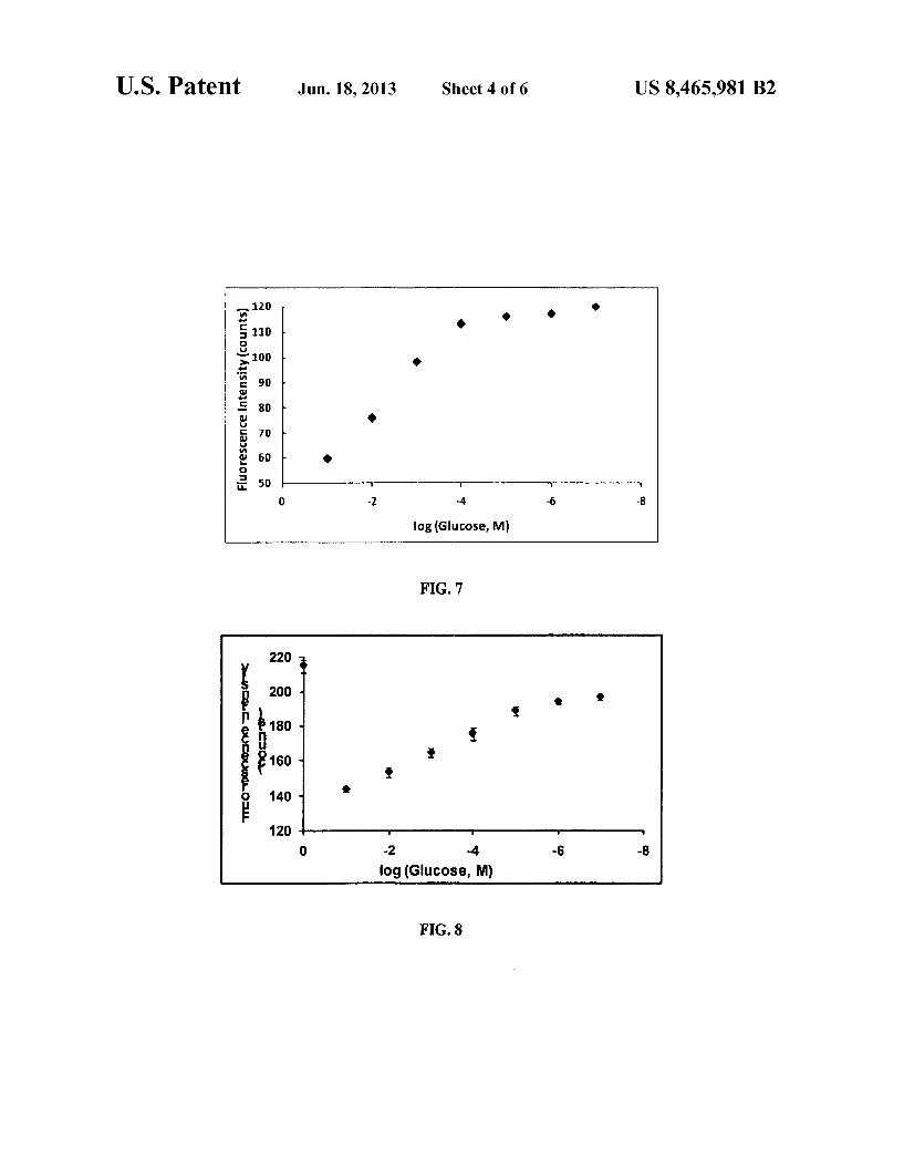
FIG. 7
g 220 i‘, B 200 - .
Q g £180 — g
B Q g (9160 < 9
E 140 ' I’ I I
o -2 -4 -s -8
log (Glucose, M)
FIG. 8

US. Patent Jun. 18, 2013 Sheet 5 of6 US 8,465,981 B2
-1.75
-2
-2.25
_25 —GRP M 1 with 5,5,5 trifluoroleucine
-2.75 i 1 . . .
O 20 40 60 80 Temperature (' C)
FIG. 10
-8
-10 m
-12
—GBP -14
-16 t -——r I r v
0 20 40 6O 80
Temperature (' C)
FIG. 11


US 8,465,981 B2 1
POLYPEPTIDES, SYSTEMS, AND METHODS USEFUL FOR DETECTING GLUCOSE
RELATED APPLICATIONS
This application claims priority from US. Provisional Application Ser. Nos. 60/954,269 ?led on Aug. 6, 2007, and 60/954,348 ?led on Aug. 7, 2007, the entire disclosures of Which are incorporated herein by this reference.
TECHNICAL FIELD
The presently-disclosed subject matter relates to biosen sors, and systems and methods useful for detecting glucose.
BACKGROUND
Monitoring blood glucose levels in a diabetic individual is important for maintaining metabolic control in the individual. Currently-available systems and methods of monitoring glu cose levels require extraction of blood from the diabetic indi vidual at multiple times during each day, e.g., ?nger prick. As such, only information about blood glucose levels at discrete time points is made available. Additionally, extracting blood for use in testing can be painful, and as a result, can lead to loW compliance or non-compliance by diabetic individuals.
Efforts have therefore been made to develop an effective system and method for monitoring blood glucose levels in a continuous and less invasive manner. Biosensors have been proposed for use in the in vivo, continuous detection of blood analyte levels. A biosensor includes an element capable of speci?cally detecting an analyte of interest, alloWing a mea surable signal to be produced, Which can be correlated to analyte concentration.
Biosensors for detecting glucose can include an element that selectively binds glucose. For example, Wild type glucose binding protein (WtGBP) is capable of binding glucose. GBP of Escherichia coli is a 33 kDa periplasmic binding protein. GBP consists of tWo distinctly similarly folded globular domains that are connected to each other by three peptide segments. The sugar binding site is located in the cleft of the protein formed betWeen the tWo domains. The binding of glucose is accompanied by a conformational change of the protein at the hinge region. In the open form of GBP (in the absence of glucose) the tWo domains are far apart and the cleft is exposed to the solvent, While in the closed form the glucose is engulfed in the cleft.
Although biosensors have made use of certain WtGBPs, such biosensors have drawbacks. For example, GBPs are not very stable at room temperature. As such, there are apparent problems associated biosensors that make use of WtGBP for monitoring blood glucose levels in diabetic patients. Further more, currently-available systems and methods for continu ously-detecting glucose are associated With a signi?cant inability to detect glucose levels in the hypoglycemic ranges in a reliable manner. Further, these available systems also suffer from short lifespans and are not easily used for routine clinical use. In addition, the use of current WtGBPs for con tinuous blood glucose monitoring for implantable or catheter based devices is complicated by the need for sterilization. The development of novel proteins With improved thermal and chemical stability Will lead to easier and more cost-effective means of sterilization.
Accordingly, there is a need in the art for systems and methods for detecting glucose levels in a subject in a continu
20
25
30
35
45
50
55
60
65
2 ous manner, at ambient temperatures, With operability for clinical use, reliability, speci?city, and sensitivity.
SUMMARY
The presently-disclosed subject matter meets some or all of the above-identi?ed needs, as Will become evident to those of ordinary skill in the art after a study of information provided in this document.
This Summary describes several embodiments of the pres ently-disclosed subject matter, and in many cases lists varia tions and permutations of these embodiments. This Summary is merely exemplary of the numerous and varied embodi ments. Mention of one or more representative features of a
given embodiment is likeWise exemplary. Such an embodi ment can typically exist With or Without the feature(s) men tioned; likeWise, those features can be applied to other embodiments of the presently-disclosed subject matter, Whether listed in this Summary or not. To avoid excessive repetition, this Summary does not list or suggest all possible combinations of such features. The presently-disclosed subject matter provides, in some
embodiments, a biosensor for detecting a molecule of inter est. In some embodiments, the biosensor comprises an iso lated polypeptide molecule capable of selectively-binding glucose. In some embodiments, the polypeptide molecule is selected from: an unnatural analogue of Wild type glucose binding protein; a fragment of Wild type glucose binding protein; and an unnatural analogue fragment of Wild type glucose binding protein. The biosensor further comprises a label associated With the polypeptide molecule, Wherein binding of glucose to the polypeptide molecule causes the label to generate a signal, such that the glucose can be detected.
In some embodiments, the polypeptide molecule is a frag ment of Wild type glucose binding protein. In some embodi ments, the polypeptide molecule is a fragment of the amino acid sequence of SEQ ID NO: 1, Wherein up to about 13 amino acids are truncated from the N-terminus and/or Wherein up to about 83 amino acids are truncated from the C-terminus of SEQ ID NO: 1. In some embodiments, the polypeptide molecule is a fragment of the amino acid sequence of SEQ ID NO: 5, Wherein up to about 12 amino acids are truncated from the N-terminus and/ or Wherein about 99 amino acids are truncated from the C-terminus of SEQ ID NO: 5. In some embodiments, the polypeptide molecule, comprises the amino acid sequence of SEQ ID NOs: 1-7.
In some embodiments, the polypeptide molecule is an unnatural analogue of Wild type glucose binding protein, Wherein one or more natural amino acids of Wild type glucose binding protein are replaced With one or more unnatural amino acids. In some embodiments, the one or more natural amino acids of Wild type glucose binding protein are replaced With one or more unnatural amino acids as set forth in Table
A. In some embodiments, the polypeptide molecule is an unnatural analogue or an unnatural analogue fragment, Wherein one or more natural leucines are replaced With unnatural leucines. In some embodiments, one or more natu
ral leucines are replaced With 5,5,5-tri?uoroleucine. In some embodiments, one or more natural leucines are replaced With
5,5,5-tri?uoro-DL-leucine. In some embodiments, one or more natural tryptophans are replaced With 5-?uorotryp tophan. In some embodiments, one or more natural tryp tophans are replaced With 5-?uoro-L-tryptophan.
In some embodiments, the polypeptide molecule is stable over a Wider range of temperatures relative to WtGBP. In some

US 8,465,981 B2 3
embodiments, the polypeptide molecule is stable at tempera tures betWeen about 55° C. and about 75° C.
In some embodiments, the label is a ?uorophore. In some embodiments, label is the ?uorophore MDCC. In some embodiments, binding of glucose to the polypeptide mol ecule causes the label to generate a detectably altered ?uo rescence intensity of the ?uorophore such that the glucose can be detected. In some embodiments, the ?uorescence intensity can be correlated to the concentration of the glucose in a sample.
In some embodiments, the label is attached to an amino acid site of the polypeptide molecule selected from Asp 13, Asp 44, Val 68, Ala 71, Phe 90, Lys 92, Glu 93, Pro 94, Lys 97,Gly 109, Thr 110,Asp 111,Gly 151, Pro 153,Ala 155,Asp 184, Asp 212, Leu 255, Ala 258,Asn 260, Ala 262, Arg 292, Val 293, and Pro 294.
In some embodiments of the presently-disclosed subject matter, a method for detecting a glucose molecule is pro vided. In some embodiments, the method comprises contact ing a biosensor disclosed herein With a sample of interest; detecting the signal; and collecting and displaying the signal With a detection and data collection device, to thereby detect the glucose. In some embodiments, the glucose is continu ously detected.
BRIEF DESCRIPTION OF THE FIGURES
FIG. 1A is a rendering of the three-dimensional structure of the polypeptide of SEQ ID NO: 1. The 3D structures Were created using a protein structure modeling program called DS VieWer Pro. Structural information for proteins is available online through the RCSB Protein Data Bank. This structural information is available in the form of a ?le that contains the coordinates in three dimensions for proteins such as they have been published in current literature sources. Protein structure ?les are available by PDB#. In this document, the PDB ?les used are as folloWs: #1GLG(WtGBP fromE. cali) and #2H3H (WtGBP from T marilima), FIG. 1A and FIG. 2A, respec tively.
FIG. 1B is a rendering of the three-dimensional structure of the polypeptide of SEQ ID NO: 2. The structure shoWn is a truncated form of the complete WtGBP from E. coli shoWn in FIG. 1A, in Which the dark gray portion represents the struc ture of the truncated form speci?ed by the ?gure and the light gray portion represents the portion of the WtGBP that is being omitted.
FIG. 1C is a rendering of the three-dimensional structure of the polypeptide of SEQ ID NO: 3. The structure shoWn is a truncated form of the complete WtGBP from E. coli shoWn in FIG. 1A, in Which the dark gray portion represents the struc ture of the truncated form speci?ed by the ?gure and the light gray portion represents the portion of the WtGBP that is being omitted.
FIG. 1D is a rendering of the three-dimensional structure of the polypeptide of SEQ ID NO: 4. The structure shoWn is a truncated form of the complete WtGBP from E. coli shoWn in FIG. 1A, in Which the dark gray portion represents the struc ture of the truncated form speci?ed by the ?gure and the light gray portion represents the portion of the WtGBP that is being omitted.
FIG. 2A is a rendering of the three-dimensional structure of the polypeptide of SEQ ID NO: 5. The 3D structures Were created using DS VieWer Pro.
FIG. 2B is a rendering of the three-dimensional structure of the polypeptide of SEQ ID NO: 6. The structure shoWn is a truncated form of the complete WtGBP from T maritime shoWn in FIG. 2A, in Which the dark gray portion represents
20
25
30
35
40
45
50
55
60
65
4 the structure of the truncated form speci?ed by the ?gure and the light gray portion represents the portion of the WtGBP that is being omitted.
FIG. 2C is a rendering of the three-dimensional structure of the polypeptide of SEQ ID NO: 7. The structure shoWn is a truncated form of the complete WtGBP from T maritime shoWn in FIG. 2A, in Which the dark gray portion represents the structure of the truncated form speci?ed by the ?gure and the light gray portion represents the portion of the WtGBP that is being omitted.
FIG. 3 is a dose response curve for glucose With an unnatu ral analogue of a WtGBP, including the isolated polypeptide of SEQ ID NO: 1, Where natural tryptophans are replaced With 5 -?uorotryptophans;
FIG. 4 is a dose response curve for glucose With an unnatu ral analogue fragment of a WtGBP, including the isolated polypeptide of SEQ ID NO: 2, Where natural tryptophans are replaced With 5-?uorotryptophans;
FIG. 5 is a dose response curve for glucose With an unnatu ral analogue fragment of a WtGBP, including the isolated polypeptide of SEQ ID NO: 3, Where natural tryptophans are replaced With 5-?uorotryptophans;
FIG. 6 is a dose response curve for glucose With an unnatu ral analogue of a WtGBP, including the isolated polypeptide of SEQ ID NO: 1, Where natural leucines are replaced With 5,5,5-tri?uoroleucines;
FIG. 7 is a dose response curve for glucose With an unnatu ral analogue fragment of a WtGBP, including the isolated polypeptide of SEQ ID NO: 2, Where natural leucines are replaced With 5,5,5-tri?uoroleucines;
FIG. 8 is a dose response curve for glucose With an unnatu ral analogue fragment of a WtGBP, including the isolated polypeptide of SEQ ID NO: 3, Where natural leucines are replaced With 5,5,5-tri?uoroleucines;
FIG. 9 is a dose response curve for glucose With an unnatu ral analogue of WtGBP including unnatural tryptophan, immobiliZed on a ?ber optic tip;
FIG. 10 is a circular dichroism (CD) spectrum of an unnatural analogue of a WtGBP, including the isolated polypeptide of SEQ ID NO: 2, Where natural leucines are replaced With 5,5,5-tri?uoroleucines; and
FIG. 11 is a CD spectrum of a WtGBP, including the iso lated polypeptide of SEQ ID NO: 1.
FIG. 12 are tWo graphs shoWing a comparison of the ?uo rescence emission intensity of GBP and GRP-7-AZatryp tophan.
FIG. 13 is a CD spectrum shoWing a comparison of the ?uorescence emission intensity over a range of temperatures of GRP-7-AZatryptophan Without (light grey line) or With (dark grey line, shifted right toWard higher temperature) bound glucose.
BRIEF DESCRIPTION OF THE SEQUENCE LISTING
SEQ ID NO: 1 includes an amino acid sequence for Wild type glucose binding protein (WtGBP) from Escherichia coli (GenBank Accession number X05646). The disclosed sequence provided is Without the N-terminal signaling pep tide. SEQ ID NO: 2 includes an amino acid sequence for tGBPl,
including amino acids 14-296 of SEQ ID NO: 1. SEQ ID NO: 3 includes an amino acid sequence for tGBP2,
including amino acids 14-256 of SEQ ID NO: 1. SEQ ID NO: 4 includes an amino acid sequence for tGBP3,
including amino acids 14-226 of SEQ ID NO: 1.

US 8,465,981 B2 5
SEQ ID NO: 5 includes an amino acid sequence for Wild type glucose binding protein (WtGBP) from Thermologa marilima (GenBank® Accession No. NPi227930), Where the ?rst 31 amino acids making up the N-terminal signaling peptide have been removed. SEQ ID NO: 6 includes an amino acid sequence for tGRP4,
including amino acids 13-253 of SEQ ID NO: 5. SEQ ID NO: 7 includes an amino acid sequence for tGRP5,
including amino acids 13-205 of SEQ ID NO: 5.
DESCRIPTION OF EXEMPLARY EMBODIMENTS
The details of one or more embodiments of the presently disclosed subject matter are set forth in this document. Modi ?cations to embodiments described in this document, and other embodiments, Will be evident to those of ordinary skill in the art after a study of the information provided in this document. The information provided in this document, and particularly the speci?c details of the described exemplary embodiments, is provided primarily for cleamess of under standing and no unnecessary limitations are to be understood therefrom. Some of the polynucleotide and polypeptide sequences
disclosed herein are cross-referenced to GENBANK® acces sion numbers. The sequences cross-referenced in the GEN BANK® database are expressly incorporated by reference as are equivalent and related sequences present in GENBANK® or other public databases. Also expressly incorporated herein by reference are all annotations present in the GENBANK® database associated With the sequences disclosed herein.
While the folloWing terms are believed to be Well under stood by one of ordinary skill in the art, the folloWing de?ni tions are set forth to facilitate explanation of the presently disclosed subject matter.
Unless de?ned otherWise, all technical and scienti?c terms used herein have the same meaning as commonly understood by one of ordinary skill in the art to Which the presently disclosed subject matter belongs. Although any methods, devices, and materials similar or equivalent to those described herein can be used in the practice or testing of the presently disclosed subject matter, representative methods, devices, and materials are noW described.
Following long-standing patent laW convention, the terms “a”, “an”, and “the” refer to “one or more” When used in this application, including the claims. Thus, for example, refer ence to “a cell” includes a plurality of such cells, and so forth.
Unless otherWise indicated, all numbers expressing quan tities of ingredients, properties such as reaction conditions, and so forth used in the speci?cation and claims are to be understood as being modi?ed in all instances by the term “about”. Accordingly, unless indicated to the contrary, the numerical parameters set forth in this speci?cation and claims are approximations that can vary depending upon the desired properties sought to be obtained by the presently-disclosed subject matter. As used herein, the term “about,” When referring to a value
or to an amount of mass, Weight, time, volume, concentration or percentage is meant to encompass variations of in some embodiments 120%, in some embodiments 110%, in some embodiments 15%, in some embodiments 11%, in some embodiments 10.5%, and in some embodiments 10.1% from the speci?ed amount, as such variations are appropriate to perform the disclosed method.
The presently-disclosed subject matter includes isolated polypeptides useful for detecting glucose. The presently-dis
20
25
30
35
40
45
50
55
60
65
6 closed subject matter further includes biosensors, systems, and methods for detecting glucose. The presently-disclosed subject matter includes isolated
polypeptide molecules that are capable of selectively-binding glucose, and biosensors that include isolated polypeptide molecules that are capable of selectively-binding glucose. In some embodiments, the isolated polypeptide molecule is a fragment of a Wild type glucose binding protein (WtGBP). In some embodiments, the isolated polypeptide molecule is an unnatural analogue of WtGBP. In some embodiments, the isolated polypeptide molecule is an unnatural analogue frag ment of WtGBP.
The terms “polypeptide, protein,” and “peptide,” Which are used interchangeably herein, refer to a polymer of the 20 protein amino acids, including modi?ed amino acids (e.g., phosphorylated, glycated, etc.) and amino acid analogs, regardless of siZe or function. Although “protein” is often used in reference to relatively large polypeptides, and “pep tide” is oftenused in reference to small polypeptides, usage of these terms in the art overlaps and varies. The term “peptide” as used herein refers to peptides, polypeptides, proteins, and fragments of proteins, unless otherWise noted. The terms “protein”, “polypeptide”, and “peptide” are used inter changeably herein When referring to a gene product and frag ments thereof. Thus, exemplary polypeptides include gene products, naturally occurring proteins, homologs, orthologs, paralogs, fragments, and other equivalents, variants, frag ments, and analogs of the foregoing. In some embodiments, the term polypeptide includes a conservatively substituted variant. The term “conservatively substituted variant” refers to a
peptide comprising an amino acid residue sequence that dif fers from a reference peptide by one or more conservative amino acid substitution, and maintains some or all of the activity of the reference peptide as described herein. A “con servative amino acid substitution” is a substitution of an amino acid residue With a functionally similar residue. Examples of conservative substitutions include the substitu tion of one non-polar (hydrophobic) residue such as isoleu cine, valine, leucine or methionine for another; the substitu tion of one polar (hydrophilic) residue for another such as betWeen arginine and lysine, betWeen glutamine and aspar agine, betWeen glycine and serine; the substitution of one basic residue such as lysine, arginine or histidine for another; or the substitution of one acidic residue, such as aspartic acid or glutamic acid for another. The term “isolated,” When used in the context of an isolated
polypeptide, is a polypeptide that, by the hand of man, exists apart from its native environment and is therefore not a prod uct of nature. As used herein, the term “selectively-bind” refers to an
interaction betWeen glucose and a binding site of a polypep tide molecule. In some embodiments, the interaction betWeen glucose and the binding site can be identi?ed as “selective” if: the equilibrium dissociation constant (K) is about the same or less than the Kd of glucose and a reference polypeptide binding site; the equilibrium inhibitor dissociation constant (K1) is about the same or less than the K- of glucose and a reference polypeptide binding site; or the effective concen tration at Whichbinding of glucose is inhibited by 50% (ECSO) is about the same or less than the EC5O of glucose and a reference polypeptide binding site. For example, glucose selectively binds an isolated polypeptide molecule that is a fragment, unnatural analogue, or unnatural analogue frag ment of Wild type Glucose Binding Protein (WtGBP) if the Kd, Ki, and/or EC5O is about the same or less than the Kd of glucose and WtGBP.

US 8,465,981 B2 7
In some embodiments, the interaction between glucose and the binding site can be identi?ed as “selective” When the equilibrium dissociation constant (Kd) is less than about 100 nM, 75 nM, 50 nM, 25 nM, 20 nM, 10 nM, 5 nM, or 2 nM. In some embodiments, the interaction betWeen glucose and the binding site can be identi?ed as “selective” When the equilib rium inhibitor dissociation constant (K) is less than about is less than about 100 11M, 75 11M, 50 11M, 25 11M, 20 11M, 10 11M, 5 11M, or 2 11M, When competing With glucose. In some embodiments, the interaction betWeen glucose and the bind ing site can be identi?ed as “selective” When the effective concentration at Which glucose binding is inhibited by 50% (ECSO) is less than about 500 11M, 400 11M, 300 11M, 100 11M, 50 11M, 25 11M, or 10 11M. The terms “polypeptide fragment” or “fragment”, When
used in reference to a polypeptide, refers to a polypeptide in Which amino acid residues are absent as compared to the full-length reference polypeptide itself, but Where the remain ing amino acid sequence is usually identical to the corre sponding positions in the reference polypeptide. Such dele tions can occur at the amino-terminus or carboxy-terminus of the reference polypeptide, or alternatively both. A fragment can retain one or more of the biological activities of the reference polypeptide. In some embodiments, a fragment can comprise a domain or feature, and optionally additional amino acids on one or both sides of the domain or feature, Which additional amino acids can number from 5, 10, 15, 20, or more residues.
As used herein, the term “unnatural analogue” refers to a polypeptide Wherein one or more natural amino acids are replaced With unnatural amino acids, relative to a reference polypeptide. Examples of unnatural amino acids are set forth in Table A, provided herein beloW. As used herein, the term “unnatural analogue fragment”
refers to a polypeptide fragment Wherein one or more natural amino acids are replaced With unnatural amino acids, relative to a reference polypeptide. As used herein, WtGBP refers to a reference protein includ
ing the amino acid residues of a full-length Wild type glucose binding protein. A full-length Wild type glucose binding pro tein Will be knoWn to those of ordinary skill in the art. Some embodiments of the polypeptide molecule of the presently disclosed subject matter can be described With reference to the amino acid residues of a WtGBP. In some embodiments, the WtGBP can be a full-length WtGBP from E. coli (Gen Bank® Accession No. X05646). In some embodiments, the WtGBP can be the polypeptide of SEQ ID NO: 1. In some embodiments, the WtGBP can be a full-length WtGBP from T marilima (GenBank® Accession No. NPi227930). In some embodiments, the WtGBP can be the polypeptide of SEQ ID NO: 5. In some embodiments, the WtGBP can be a full-length WtGBP from T hermus lhermophilus (GenBank® Accession No. YPi004303).
In some embodiments of the presently-disclosed subject matter, the isolated polypeptide molecule is a fragment of WtGBP that selectively binds glucose. For example, in some embodiments, the polypeptide molecule comprises a frag ment of WtGBP from E. coli including amino acids 14-296, set forth in SEQ ID NO: 2 (referred to herein as tGBPl). For another example, in some embodiments, the polypeptide molecule comprises a fragment of WtGBP from E. coli including amino acids 14-256, set forth in SEQ ID NO: 3 (referred to herein as tGBP2). For yet another example, in some embodiments, the polypeptide molecule comprises a fragment of WtGBP from E. coli including amino acids 14-226, set forth in SEQ ID NO: 4 (referred to herein as tGBP3). For another example, in some embodiments, the
20
25
30
35
40
45
50
55
60
65
8 polypeptide molecule comprises a fragment of WtGBP from T marilima including amino acids 13-253, set forth in SEQ ID NO: 6 (referred to herein as tGBP4). For another example, in some embodiments, the polypeptide molecule comprises a fragment of WtGBP from T marilima including amino acids 13-205, set forth in SEQ ID NO: 7 (referred to herein as tGBP5).
It is contemplated that isolated polypeptide molecules of the presently-disclosed subject matter can be fragments and/ or unnatural analogues of WtGBP for various species, or an isolated polypeptide molecule having a three-dimensional structure that is suf?ciently similar to such a WtGBP or func tional fragment thereof, such that it is capable of speci?cally binding glucose. Three-dimensional structures of the isolated polypeptide molecules of SEQ ID NOS: 1-7 are provided in FIGS. 1A-2C. These 3D structures Were created using a pro tein structure modeling program called DS Viewer Pro. Struc tural information for proteins is available online through the RCSB Protein Data Bank. This structural information is available in the form of a ?le that contains the coordinates in three dimensions for proteins such as they have been pub lished in current literature sources. Protein structure ?les are available by PDB#. In the present context, the PDB ?les used are as folloWs: #IGLG (WtGBP from E. coli) and #2H3H (WtGBP from T marilima). These structures are shoWn in FIG. 1A and FIG. 2A respectively. Structures shoWn in FIG. 1B, FIG. 1C, and FIG. ID are truncated forms of the complete WtGBP from E. coli shoWn in FIG. 1A in Which the dark gray portion represents the structure of the truncated form speci ?ed by that ?gure and the light gray portion represents the portion of the WtGBP that is being omitted. Likewise, FIG. 2B and FIG. 2C represent truncated forms of the complete WtGBP from T maritime shoWn in FIG. 2A in Which the dark gray portion represents the structure of the truncated form speci?ed by that ?gure and the light gray portion represents the portion of the WtGBP that is being omitted.
In some embodiments, the polypeptide molecule of the presently-disclosed subject matter comprises a fragment of WtGBP, Whereinup to 1, 2, 3, 4, 5, 6, 7, 8, 9, 10, 11, 12, 13, 14, 15, 16, 17, 18, 19, 20, 21, 22, 23, 24, or 25 amino acids are truncated from the N-terminus of the WtGBP. In some embodiments, the polypeptide molecule comprises a frag ment ofWtGBP, Wherein up to about 1, 2, 3, 4, 5, 6, 7, 8, 9, 10, 11, 12, 13, 14, 15, 16, 17, 18, 19, 20, 21, 22,23, 24, 25, 26, 27, 28, 29, 30, 31, 32, 33, 34, 35, 36, 37, 38, 39, 40, 41, 42, 43, 44, 45, 46, 47, 48, 49, 50, 51, 52, 53, 54, 55, 56, 57, 58, 59, 60, 61, 62, 63, 64, 65, 66, 67, 68, 69, 70, 71, 72, 73, 74, 75, 76, 77, 78, 79, 80, 81, 82, 83, 84, 85, 86, 87, 88, 89, 90, 91, 92, 93, 94, 95, 96, 97, 98, 99, 100, 101, 102, 103, 104, 105, 106, 107, 108, 109, or 1 10 amino acids are truncated from the C-terminus of WtGBP.
Without Wishing to be bound by theory, it is believed that certain amino acids are useful for enhancing functionality of fragments of WtBGP provided from particular species. For example, amino acids 14,91, 152, 158, 211, 236, and 256 of WtGBP from E. coli (SEQ ID NO: 1) are believed to be involved in the binding of glucose. As such, in some embodi ments, the isolated polypeptide molecule is a fragment of WtGBP from E. coli (SEQ ID NO: 1) that includes amino acids 14, 91, 152, 158, 211, 236, and 256. In some embodi ments, the isolated polypeptide molecule is a fragment of WtGBP from E. coli (SEQ ID NO: 1) that includes at least 1, 2, 3, 4, 5, or 6 of the amino acids selected from amino acids 14, 91, 152, 158, 211, 236, and 256. Similarly, amino acid 13 ofWtGBP from T marilima (SEQ ID NO: 5) is believed to be involved in the binding of glucose. As such, in some embodi

US 8,465,981 B2
ments, the isolated polypeptide molecule is a fragment of WtGBP from T marilima (SEQ ID NO: 5) that includes amino acid 13.
In some embodiments of the presently-disclosed subject
10 polypeptide molecule comprises the sequence of WtGBP, Wherein one or more natural amino acids are replaced With one or more unnatural amino acids set forth in Table A. In some embodiments, the WtGBP can be from E. coli. In some
matter, the isolated polypeptide molecule is an unnatural 5 embodiments, the WtGBP can be from T marilima. In some analogue of WtGBP. For example, in some embodiments, the embodiments, the WtGBP can be from T lhermophilus.
TABLE A
Structure of
Natural Amino Acid Unnatural Amino Acid Unnatural Amino Acid
Alanine 2-Pyridylalanine
Alanine 3-Pyridylalanine —
N\ /
HZN
OH
O
Alanine 4-Pyridylalanine
Alanine p-Iodophenylalanine I
HZN
OH
O
Alanine p-Brornophenylalanine Br
HZN
OH

US 8,465 ,981 B2 1 1 12
TABLE A-continued
Sttucture of Natural Arnino Acid Unnatural Arnino Acid Unnatural AIHlHO Acid
Alanine L-2-Arninobutyric acid H NH;
H3C on “I”
O
Glycine L-Allylglycine /
O
HZN
OH
Glycine L-Propargylglycine %
O
HZN
Glycine 2-Methoxy-phenylglycine
0/ OH
HZN
O
Glycine 3-Thienylglycine / S
OH
HZN
O
Leucine 5,5,5-trifluoro-DL-leucine C133
0%} O I Lysine D-Lysine HZN H O
H2NCH2(CH2)2CH2C:— C — OH
Phenylalanine Alpha-methyl-L phenylalanine
H N 2 0
HO
Phenylalanine p-Arnino-DL-phenylalanine NH; O


US 8,465,981 B2 15 16
TABLE A-continued
Structure of Natural Amino Acid Unnatural Amino Acid Unnatural AIHIHO Acid
Tyrosine 3-Fluoro-L-tyrosine O
OH
HZNhI“. H
HO F
Valine D-Norvaline
In some embodiments, the polypeptide molecule com prises the sequence of a fragment of WtGBP, wherein one or more natural amino acids are replaced With one or more
unnatural amino acids set forth in Table A. In some embodi ments, the WtGBP can be from E. coli. In some embodiments, the WtGBP can be from T marilima. In some embodiments, the WtGBP can be from T lhermophilus. In some embodi ments, the polypeptide molecule comprises the sequence of SEQ ID NO: 1, Wherein one or more natural amino acids are replaced With one or more unnatural amino acids set forth in
Table A. In some embodiments, the polypeptide molecule comprises the sequence of SEQ ID NO: 2, Wherein one or more natural amino acids are replaced With one or more
unnatural amino acids set forth in Table A. In some embodi ments, the polypeptide molecule comprises the sequence of SEQ ID NO: 3, Wherein one or more natural amino acids are replaced With one or more unnatural amino acids set forth in Table A. In some embodiments, the polypeptide molecule comprises the sequence of SEQ ID NO: 4, Wherein one or more natural amino acids are replaced With one or more
unnatural amino acids set forth in Table A. In some embodi ments, the polypeptide molecule comprises the sequence of SEQ ID NO: 5, Wherein one or more natural amino acids are replaced With one or more unnatural amino acids set forth in
Table A. In some embodiments, the polypeptide molecule comprises the sequence of SEQ ID NO: 6, Wherein one or more natural amino acids are replaced With one or more
unnatural amino acids set forth in Table A. In some embodi ments, the polypeptide molecule comprises the sequence of SEQ ID NO: 7, Wherein one or more natural amino acids are replaced With one or more unnatural amino acids set forth in Table A.
In some embodiments of the presently-disclosed subject matter, the isolated polypeptide molecule is an unnatural analogue of WtGBP, including one or more of the folloWing substitutions:
(i) at least one alanine is replaced With an unnatural ala nine;
(ii) at least one glycine is replaced With an unnatural gly cine;
(iii) at least one lysine is replaced With an unnatural lysine; (iv) at least one phenylalanine is replaced With an unnatural
phenylalanine;
25
30
35
40
45
50
55
60
65
(V) at least one proline is replaced With an unnatural pro line;
(vi) at least one tyrosine is replaced With an unnatural tyrosine;
(vii) at lest one valine is replaced With an unnatural valine; (viii) at least one leucine is replaced With an unnatural
leucine; and (ix) at least one tryptophan is replaced With an unnatural
tryptophan. In some embodiment, at least one alanine is replaced With
an unnatural alanine selected from: 2-Pyridylalanine, 3-Py ridylalanine, 4-Pyridylalanine, p-Iodophenylalanine, p-Bro mophenylalanine, and L-2-Aminobutyric acid.
In some embodiment, at least one glycine is replaced With an unnatural glycine selected from: L-Allylglycine, L-Prop argylglycine, 2-MethoXy-phenylglycine, and 3-Thienylgly cine.
In some embodiments, at least one lysine is replaced With D-lysine.
In some embodiments, at least one phenylalanine is replaced With an unnatural phenylalanine selected from: Alpha-methyl-L-phenylalanine, and p-Amino-DL-phenyla lanine
In some embodiments, at least one proline is replaced With an unnatural proline selected from: alpha-BenZyl-proline HCl, and gamma-(3-?uoro-benZyl)-L-proline-HCl.
In some embodiments, at least one tyrosine is replaced With 3-Fluoro-L-tyrosine.
In some embodiments, at least one valine is replaced With D-norvaline.
In some embodiments, at least one leucine is replaced With 5,5,5-tri?uoro-DL-leucine.
In some embodiments, at least one tryptophan is replaced With an unnatural tryptophan selected from: 5-HydroXy-L tryptophan, 5-?uoro-L-tryptophan, 7-aZatryptophan, 6-aZa tryptophan, and amino tryptophans.
In some embodiments, the polypeptide molecule is a WtGBP or fragment thereof, Wherein one or more natural leucines are replaced With unnatural leucines. Without Wish ing to be bound by theory, it is believed that replacing one or more natural leucines With unnatural leucines confers enhanced thermal and chemical stability to the resulting polypeptide analogue. In some embodiments, all natural leu

US 8,465,981 B2 17
cines are replaced With unnatural leucines. In some embodi ments, the unnatural leucines are 5,5,5-tri?uoro-DL-leucine.
In some embodiments, the polypeptide molecule com prises the sequence of SEQ ID NO: 1, Wherein one or more natural leucines are replaced With unnatural leucines. In some embodiments, the polypeptide molecule comprises the sequence of SEQ ID NO: 1, Wherein one or more natural leucines are replaced With 5,5,5-tri?uoro-DL-leucine. In some embodiments, the polypeptide molecule comprises the sequence of SEQ ID NO: 1, Wherein all of the natural leucines are replaced With 5,5,5-tri?uoro-DL-leucine.
In some embodiments, the polypeptide molecule com prises the sequence of SEQ ID NO: 2, Wherein one or more natural leucines are replaced With unnatural leucines. In some embodiments, the polypeptide molecule comprises the sequence of SEQ ID NO: 2, Wherein one or more natural leucines are replaced With 5,5,5-tri?uoro-DL-leucine. In some embodiments, the polypeptide molecule comprises the sequence of SEQ ID NO: 2, Wherein all of the natural leucines are replaced With 5,5,5-tri?uoro-DL-leucine.
In some embodiments, the polypeptide molecule com prises the sequence of SEQ ID NO: 3, Wherein one or more natural leucines are replaced With an unnatural leucines. In some embodiments, the polypeptide molecule comprises the sequence of SEQ ID NO: 3, Wherein one or more natural leucines are replaced With 5,5,5-tri?uoro-DL-leucine. In some embodiments, the polypeptide molecule comprises the sequence of SEQ ID NO: 3, Wherein all of the natural leucines are replaced With 5,5,5-tri?uoro-DL-leucine.
In some embodiments, the polypeptide molecule com prises the sequence of SEQ ID NO: 4, Wherein one or more natural leucines are replaced With an unnatural leucines. In some embodiments, the polypeptide molecule comprises the sequence of SEQ ID NO: 4, Wherein one or more natural leucines are replaced With 5,5,5-tri?uoro-DL-leucine. In some embodiments, the polypeptide molecule comprises the sequence of SEQ ID NO: 4, Wherein all of the natural leucines are replaced With 5,5,5-tri?uoro-DL-leucine.
In some embodiments, the polypeptide molecule com prises the sequence of SEQ ID NO: 5, Wherein one or more natural leucines are replaced With an unnatural leucines. In some embodiments, the polypeptide molecule comprises the sequence of SEQ ID NO: 5, Wherein one or more natural leucines are replaced With 5,5,5-tri?uoro-DL-leucine. In some embodiments, the polypeptide molecule comprises the sequence of SEQ ID NO: 5, Wherein all of the natural leucines are replaced With 5,5,5-tri?uoro-DL-leucine.
In some embodiments, the polypeptide molecule com prises the sequence of SEQ ID NO: 6, Wherein one or more natural leucines are replaced With unnatural leucines. In some embodiments, the polypeptide molecule comprises the sequence of SEQ ID NO: 6, Wherein one or more natural leucines are replaced With 5,5,5-tri?uoro-DL-leucine. In some embodiments, the polypeptide molecule comprises the sequence of SEQ ID NO: 6, Wherein all of the natural leucines are replaced With 5,5,5-tri?uoro-DL-leucine.
In some embodiments, the polypeptide molecule com prises the sequence of SEQ ID NO: 7, Wherein one or more natural leucines are replaced With unnatural leucines. In some embodiments, the polypeptide molecule comprises the sequence of SEQ ID NO: 7, Wherein one or more natural leucines are replaced With 5,5,5-tri?uoro-DL-leucine. In some embodiments, the polypeptide molecule comprises the sequence of SEQ ID NO: 7, Wherein all of the natural leucines are replaced With 5,5,5-tri?uoro-DL-leucine.
In some embodiments, the polypeptide molecule is a WtGBP or fragment thereof, Wherein one or more natural
20
25
30
35
40
45
50
55
60
65
18 tryptophans are replaced With unnatural tryptophans. Without Wishing to be bound by theory, and With reference to the description herein of biomarkers including an isolated polypeptide molecule that is capable of emitting a signal When binding glucose, it is believed that replacing one or more natural tryptophans With unnatural tryptophans confers an enhanced ability of the resulting biomarker to emit a sig nal, e.g., ?uoresce. For example, as shoWn in FIG. 12, the incorporation of 7-AZatryptophan in GRP enhances the inten sity of ?uorescence emission. Further, as shoWn in FIG. 13 the incorporation of 7-AZatryptophan in GRP results in a clearly distinguishable ?uorescence signature betWeen 7-AZatryptophan GRP With and Without bound glucose across different temperatures. In some embodiments all natu ral tryptophans are replaced With unnatural tryptophans. In some embodiments, unnatural tryptophans are selected from: 5-hydroxy-L-tryptophan, and 5-?uoro-L-tryptophan. In some embodiments, the unnatural tryptophans are 5-?uorot ryptophan.
In some embodiments, the polypeptide molecule com prises the sequence of SEQ ID NO: 1, Wherein one or more natural tryptophans are replaced With unnatural tryptophans. In some embodiments, the polypeptide molecule comprises the sequence of SEQ ID NO: 1, Wherein one or more natural tryptophans are replaced With unnatural tryptophan selected from: S-hydroXy-L-tryptophan, and 5-?uoro-L-tryptophan. In some embodiments, the polypeptide molecule comprises the sequence of SEQ ID NO: 1, Wherein one or more natural tryptophans are replaced With 5-?uorotryptophan. In some embodiments, the polypeptide molecule comprises the sequence of SEQ ID NO: 1, Wherein all of the natural tryp tophans are replaced With S-?uorotryptophan.
In some embodiments, the polypeptide molecule com prises the sequence of SEQ ID NO: 2, Wherein one or more natural tryptophans are replaced With unnatural tryptophans. In some embodiments, the polypeptide molecule comprises the sequence of SEQ ID NO: 2, Wherein one or more natural tryptophans are replaced With unnatural tryptophan selected from: S-hydroXy-L-tryptophan, and 5-?uoro-L-tryptophan. In some embodiments, the polypeptide molecule comprises the sequence of SEQ ID NO: 2, Wherein one or more natural tryptophans are replaced With 5-?uorotryptophan. In some embodiments, the polypeptide molecule comprises the sequence of SEQ ID NO: 2, Wherein all of the natural tryp tophans are replaced With 5-?uorotryptophan.
In some embodiments, the polypeptide molecule com prises the sequence of SEQ ID NO: 3, Wherein one or more natural tryptophans are replaced With unnatural tryptophans. In some embodiments, the polypeptide molecule comprises the sequence of SEQ ID NO: 3, Wherein one or more natural tryptophans are replaced With unnatural tryptophan selected from: S-hydroXy-L-tryptophan, and 5-?uoro-L-tryptophan. In some embodiments, the polypeptide molecule comprises the sequence of SEQ ID NO: 3, Wherein one or more natural tryptophans are replaced With 5-?uorotryptophan. In some embodiments, the polypeptide molecule comprises the sequence of SEQ ID NO: 3, Wherein all of the natural tryp tophans are replaced With 5-?uorotryptophan.
In some embodiments, the polypeptide molecule com prises the sequence of SEQ ID NO: 4, Wherein one or more natural tryptophans are replaced With unnatural tryptophans. In some embodiments, the polypeptide molecule comprises the sequence of SEQ ID NO: 4, Wherein one or more natural tryptophans are replaced With unnatural tryptophan selected from: S-hydroXy-L-tryptophan, and 5-?uoro-L-tryptophan. In some embodiments, the polypeptide molecule comprises the sequence of SEQ ID NO: 4, Wherein one or more natural

US 8,465,981 B2 19
tryptophans are replaced With 5-?uorotryptophan. In some embodiments, the polypeptide molecule comprises the sequence of SEQ ID NO: 4, Wherein all of the natural tryp tophans are replaced With 5-?uorotryptophan.
In some embodiments, the polypeptide molecule com prises the sequence of SEQ ID NO: 5, Wherein one or more natural tryptophans are replaced With unnatural tryptophans. In some embodiments, the polypeptide molecule comprises the sequence of SEQ ID NO: 5, Wherein one or more natural tryptophans are replaced With unnatural tryptophan selected from: 5-hydroxy-L-tryptophan, and 5-?uoro-L-tryptophan. In some embodiments, the polypeptide molecule comprises the sequence of SEQ ID NO: 5, Wherein one or more natural tryptophans are replaced With 5-?uorotryptophan. In some embodiments, the polypeptide molecule comprises the sequence of SEQ ID NO: 5, Wherein all of the natural tryp tophans are replaced With 5-?uorotryptophan.
In some embodiments, the polypeptide molecule com prises the sequence of SEQ ID NO: 6, Wherein one or more natural tryptophans are replaced With unnatural tryptophans. In some embodiments, the polypeptide molecule comprises the sequence of SEQ ID NO: 6, Wherein one or more natural tryptophans are replaced With unnatural tryptophan selected from: 5-hydroxy-L-tryptophan, and 5-?uoro-L-tryptophan. In some embodiments, the polypeptide molecule comprises the sequence of SEQ ID NO: 6, Wherein one or more natural tryptophans are replaced With 5-?uorotryptophan. In some embodiments, the polypeptide molecule comprises the sequence of SEQ ID NO: 6, Wherein all of the natural tryp tophans are replaced With 5-?uorotryptophan.
In some embodiments, the polypeptide molecule com prises the sequence of SEQ ID NO: 7, Wherein one or more natural tryptophans are replaced With unnatural tryptophans. In some embodiments, the polypeptide molecule comprises the sequence of SEQ ID NO: 7, Wherein one or more natural tryptophans are replaced With unnatural tryptophan selected from: 5-hydroxy-L-tryptophan, and 5-?uoro-L-tryptophan. In some embodiments, the polypeptide molecule comprises the sequence of SEQ ID NO: 7, Wherein one or more natural tryptophans are replaced With 5-?uorotryptophan. In some embodiments, the polypeptide molecule comprises the sequence of SEQ ID NO: 7, Wherein all of the natural tryp tophans are replaced With 5-?uorotryptophan.
In some embodiments, the polypeptide molecule com prises a WtGBP or fragment thereof, Wherein at least one natural leucine is replaced With 5,5,5-tri?uoro-DL-leucine, and at least one natural tryptophan is replaced With an unnatu ral tryptophan selected from 5-hydroxy-L-tryptophan, and 5-?uoro-L-tryptophan. In some embodiments, at least one natural leucine is replaced With 5,5,5-tri?uoro-DL-leucine, and at least one natural tryptophan is replaced With 5-?uoro L-tryptophan. In some embodiments, all natural leucines are replaced With 5 ,5 ,5 -tri?uoro -DL-leucine, and all natural tryp tophans are replaced With 5-?uoro-L-tryptophan.
In some embodiments, When more than one of a particular natural amino acid is replaced With an unnatural amino acid, different unnatural amino acids can be used. For one example, in some embodiments, an unnatural analogue can include a ?rst natural tryptophan replaced With 5-hydroxy-L-tryp tophan, and a second natural tryptophan replaced With 5-?uoro-L-tryptophan.
In some embodiments of the presently-disclosed subject matter, the isolated polypeptide molecule is an unnatural analogue fragment of WtGBP that selectively binds glucose. In embodiments Where the polypeptide molecule comprises an unnatural analogue fragment of WtGBP, natural amino acids can be replaced With unnatural amino acids, as
20
25
30
35
40
45
50
55
60
65
20 described above With reference to unnatural analogues of WtGBP. In some embodiments, the polypeptide molecule comprises a fragment of WtGBP, Wherein at least one natural amino acid is replaced With an unnatural amino acid, such as an unnatural amino acid set forth in Table A. For another example, in some embodiments, the polypeptide molecule comprises a fragment of SEQ ID NOs: 2, 3, 4, 5, 6, or 7, Wherein at least one natural amino acid is replaced With an unnatural amino acid, such as an unnatural amino acid set forth in TableA. For another example, in some embodiments, the polypeptide molecule comprises a fragment of WtGBP Wherein at least one leucine is replaced With 5,5,5-tri?uoro DL-leucine, and/or at least one tryptophan is replaced With 5-?uoro-L-tryptophan. In some embodiments, the polypep tide molecule comprises a fragment of WtGBP Wherein all natural leucines are replaced With 5,5,5-tri?uoro-DL-leucine, and/or all natural tryptophans are replaced With 5-?uoro-L tryptophan.
In some embodiments, the isolated polypeptide molecule is a fragment, unnatural analogue of WtGBP, or an unnatural analogue fragment of WtGBP, Wherein one or more amino acids have been added to the N-terminus and/or the C-termi nus of the isolated polypeptide molecule. For example, in some embodiments, Met-Arg can be added to the N-terminus of the isolated polypeptide molecule. For another example, in some embodiments, Arg-Ser-His-His-His-His-His-His can be added to the C-terminus of the isolated polypeptide mol ecule. The presently-disclosed subject matter includes a biosen
sor capable of detecting glucose. As used herein, the term “detect” means to determine quantitatively and/or qualita tively. The biosensor includes an isolated polypeptide mol ecule that is capable of selectively binding glucose, and a label capable of generating a signal When the isolated polypeptide molecule binds glucose. The isolated polypep tide molecule of the biosensor can be an isolated polypeptide molecule as described above. The label canbe associated With a binding site of the isolated polypeptide molecule, Which label is capable of generating a signal. For example, the label can be a ?uorescent label, e.g., ?uorophore, or an electro chemical label. As used herein, the terms “label” and “labeled” refer to the
attachment of a moiety, capable of detection by spectro scopic, radiologic, or other methods, to a probe molecule. Thus, the terms “label” or “labeled” refer to incorporation or attachment, optionally covalently or non-covalently, of a detectable marker into a molecule, such as a polypeptide. Various methods of labeling polypeptides are knoWn in the art and can be used. Speci?c examples are described herein. Fluorescent probes that can be utiliZed include, but are not limited to ?uorescein isothiocyanate; ?uorescein dichlorotri aZine and ?uorinated analogs of ?uorescein; naphtho?uores cein carboxylic acid and its succinimidyl ester; carbox yrhodamine 6G; pyridyloxaZole derivatives; Cy2, 3, 3.5, 5, 5.5, and 7; phycoerythrin; phycoerythrin-Cy conjugates; ?uorescent species of succinimidyl esters, carboxylic acids, isothiocyanates, sulfonyl chlorides, and dansyl chlorides, including propionic acid succinimidyl esters, and pentanoic acid succinimidyl esters; succinimidyl esters of carboxytet ramethylrhodamine; rhodamine Red-X succinimidyl ester; Texas Red sulfonyl chloride; Texas Red-X succinimidyl ester; Texas Red-X sodium tetra?uorophenol ester; Red-X; Texas Red dyes; tetramethylrhodamine; lissamine rhodamine B; tetramethylrhodamine; tetramethylrhodamine isothiocy anate; naphtho?uoresceins; coumarin derivatives (e. g.,

US 8,465,981 B2 21
hydroxycoumarin, aminocoumarin, and methoxycoumarin); pyrenes; pyridyloxazole derivatives; dapoxyl dyes; Cascade Blue and Yellow dyes; benzofuran isothiocyanates; sodium tetra?uorophenols; 4,4-di?uoro-4-bora-3a,4a-diaza-s-in dacene; ALEXA FLUORS® (e.g., 350, 430,488, 532, 546, 555, 568, 594, 633, 647, 660, 680, 700, and 750); green ?uorescent protein; yelloW ?uorescent protein; and fruit ?uo rescent proteins. The peak excitation and emission Wave lengths Will vary for these compounds and selection of a particular ?uorescent probe for a particular application can be made in part based on excitation and/or emission Wave
lengths. In some embodiments, the isolated polypeptide molecule
is a fragment, an unnatural analogue, or an unnatural ana
logue fragment of a WtGBP; and the label is attached to an amino acid residue of the isolated polypeptide molecule. The label can be attached to one of the folloWing amino acid sites close to the binding site of the isolated polypeptide molecule, and identi?ed With reference to WtGBP of SEQ ID NO: 1 : Asp 13,Asp 44, Val 68, Ala 71, Phe 90, Lys 92, Glu 93, Pro 94, Lys 97, Gly 109, Thr 110, Asp 111, Gly 151, Pro 153, Ala 155, Asp 184, Asp 212, Leu 255, Ala 258, Asn 260, Ala 262, Arg 292, Val 293, and Pro 294. The label can be a ?uorophore, such as a ?uorophore selected from: BODIPY 630/650 (Bro momethyl); ALEXA FLUORS® 680 679/702 (Maleimide); NBD 478/541 (Haloacetamide); PyMPO 415/570 (Haloac etamide); Texas Red 595/615 (Maleimide); and MDCC (N [2-(1 -maleimidyl(ethyl] -7-(diethylamino)coumarin-3 -car boxamide).
The biosensors of the presently-disclosed subject matter can be utilized in a number of different capacities in order to detect glucose, both in vitro and in vivo. As one non-limiting example, the biosensor can be coupled With a catheter for continuous in vivo detection of glucose in a body of a subject. Any knoWn catheter suitable for implantation in a body canbe utilized With the biosensors disclosed herein, including but not limited to catheter systems disclosed in International Patent Application No. PCT/US08/732338 to Daunert et al. entitled “DEVICE FOR DETECTION OF MOLECULES OF INTEREST,” claiming priority from US. Provisional Application Ser. Nos. 60/954,269 and 60/954,348, and ?led on Aug. 6, 2008 (hereinafter referred to as the “Daunert et al. Application), Which is incorporated herein by reference in its entirety.
In addition to coupling With catheter systems, the biosen sors disclosed herein can also be utilized in several other systems for glucose detection. The incorporation of non natural amino acids into single locations Within glucose rec ognition peptides can alloW for site-speci?c labeling With ?uorophores, electrochemical tags, etc., due to the enhanced functional diversity that non-natural amino acids have in comparison to their canonical counterparts. This site speci?c potential provides for greater control and analytical reproduc ibility When developing glucose biosensors With non-natural glucose recognition peptides. The incorporation of non-natu ral amino acids further permits for the site-speci?c immobi lization of these glucose recognition peptides, Which can decrease the activity loss that is commonly encountered upon peptide immobilization. The retention of this glucose respon sive activity can be useful in optimizing both hydrogel-cath eter based glucose sensors disclosed in the Daunert et al. Application and in developing alternative sensor designs.
20
25
30
35
40
45
50
55
60
65
22 For example, the biosensors disclosed herein comprising
novel glucose recognition peptides can be employed for the development of sensing systems on non-catheter platform systems. For example, the biosensors disclosed herein can be used to quantitate glucose levels on both microtiter plate and miniaturized micro?uidics platforms, Which are popular in high-throughput screening, clinical laboratory practice, and in the development of point-of-care diagnostic equipment. Additionally, presently-disclosed biosensors can be immobi lized on affordable and robust paper strips, Whose visible color change Would correlate to glucose levels. These paper strips Would be a practical and competitive option for patient self-monitoring of glucose levels. Moreover, the dynamic range of the glucose recognition peptides of the presently disclosed subject matter can permit salivary analysis of glu cose levels, instead of the painful and invasive ?nger prick method commonly used today.
In addition, the non-natural glucose recognition peptides disclosed herein and contained Within hydrogel, as disclosed for example in the Daunert et al. Application can also be used for the development and improvement implantable drug delivery devices and contact lens glucose sensors. As an example of a potential use in implantable drug delivery devices, the change in ?uorescent or electrical signal caused by glucose binding to the appropriately labeled glucose rec ognition peptides can be translated to the opening and closing of a reversible insulin-containing drug reservoir. In this man ner glucose levels can be both monitored and corrected in diabetic patients With a glucose responsive glucose recogni tion peptide-hydrogel derived device. Additionally, current contact lens glucose sensors have a sensing plastic chip incor porated into the regular corrective lens. This plastic chip changes colors via holographic sensing methods and boron containing ?uorophores. These color changes are visible to the Wearer, With different colors corresponding to different glucose levels in tears, thus alerting the patient if insulin is needed. One advantage the biosensors described herein have over this current contact lens scheme is that the glucose sensor disclosed herein can be hydrogel based. Hydrogels are more Water and oxygen permeable than the plastic chips that are currently used, and this permeability is quite important for both the comfort and long term optical health of the contact lens Wearer.
The presently-disclosed subj ect matter is further illustrated by the folloWing speci?c but non-limiting examples. The folloWing examples may include compilations of data that are representative of data gathered at various times during the course of development and experimentation related to the presently-disclosed subject matter.
EXAMPLES
Expression and Labeling of Isolated Polypeptide Mol ecules. In order to obtain the gene for insertion into an expres sion vector, PCR ampli?cation Was employed. Primers Were designed to amplify isolated polypeptide molecules of inter est, e.g., polypeptide molecules of SEQ ID NOS: 1-7, and a fragment including amino acids 87-271 of SEQ ID NO: 1. Table B shoWs data from the fragment containing amino acids 87-271 from E. coli. This fragment shoWed large error and no clear trend in response to glucose, Which demonstrates that rational design of truncated forms of WtGBPs is necessary to render a responsive glucose recognition peptide.

US 8,465,981 B2 23
TABLE B
Glucose Fluorescence
Concentration Intensity Standard
(M) (AVG) Deviation
0 485.282 17.004
1.00E-08 442.682 32.888
1.00E-07 463.041 20.018
1.00E-06 497.146 22.400
1.00E-05 478.877 36.678
1.00E-04 467.383 41.020
1.00E-03 468.792 23.450
Once the PCR ampli?ed gene fragments Were obtained, they Were inserted into expression vector pQE70. The con structed expression plasmids Were then transformed into the auxotrophic strain of E. coli. Protein expression Was per formed using medium shift method. A single freshly trans formed colony Was used to inoculate 5 mL of M9 media supplemented With 0.4% glucose; 1 mM MgSO4; 0.1 mM CaCl2; 1 mM Thiamine; 0.1 volume of a solution containing 0.01% (W/v) each of 20 amino acids, less any amino acid that is to be replaced by unnatural amino acids (e.g., each of 19 amino acids Where one is to be replaced); 40 ug/mL of the remaining amino acid(s) (e.g., 20”’ amino acid), and appro priate amount of antibiotic. T his culture Was alloWed to groW overnight at 37° C., 250 rpm. 500 mL culture containing the same ingredients Was inoculated With the overnight groWn 5 mL culture. This culture Was then groWn until the optical density (OD6OO) Was about 0.5. The cells Were then centri fuged at 10000 rpm for 10 min, 250 C., the supernatant Was discarded and the cells Were resuspended in a 0.9% NaCl solution for Washing, and this Was repeated three times. The cells Were then resuspended in 500 mL M9 minimal media Which Was supplemented With 0.4% glucose, 1 mM MgSO4, 0.1 mM CaCl2, 1 mM Thiamine, 0.1 volume of a solution containing 0.01% (W/v) each of 20 amino acids less any amino acid(s) to be replaced by unnatural amino acids (e.g., 19 amino acids), appropriate amount of antibiotic, and groWn at 37° C. for 30 min to further deplete natural amino acid that has to be replaced by the unnatural amino acid. 1 mM IPTG Was then added With 0.1 mM of unnatural amino acid to the culture and Was groWn overnight at 37° C. The cells Were harvested by centrifugation and the protein Was puri?edusing Ni-NTA resin since GBP mutant has His-tag. The puri?ed protein Was then labeled With ?uorophore MDCC (N-[2-(1 maleimidyl(ethyl]—7-(diethylamino)coumarin-3-carboxam ide), e.g., labeled at cysteine-152 of SEQ ID NO: 1.
Fragments of WtGBP in Response to Glucose. In order to check for activity of the labeled tGBPs, an assay Was per formed. Upon glucose binding, the proteins should undergo a conformational change, resulting in a change in the local environment of the ?uorophore molecule, Which leads to a change in the ?uorescence signal observed. The assay Was carried out by preparing glucose solutions of concentrations ranging from 1><10_lM to 1><10_6M. These glucose standards Were then added to 180 pL samples of the labeled tGBPs at a concentration of 1><10_7M. The resulting ?uorescence inten sity Was measured. The results obtained from one study including the isolated polypeptide molecules of SEQ ID NO: 2 (tGBPl) and SEQ ID NO: 3 (tGBP2) are presented in Table C.
20
25
30
35
40
45
50
55
60
65
24 TABLE c
Fluorescence signal quenching in response to varying concentration of glucose obtained from tGBPs.
Percent Fluorescence Quenching
Glucose, M tGBPl tGBP2
1x 10’6 —0.09539 1.748188 1 x 10’5 2.907571 2.794934 1 x 1041 2.432862 5.87441 1 x 10’3 7.5538 8.966095 1 x 10’2 14.60875 7.50211 1 x 10’1 20.36978 6.265703
Unnatural Analogues of WtGBP With Unnatural Tryp tophans. Unnatural analogues of WtGBP from E. coli Were produced, Where natural tryptophans Were replaced With unnatural tryptophans. Tryptophan auxotrophic strain of E. coli (#27873) Was obtained. Plasmid placI and plasmid pQE70 containing the genetic sequence of the WtGBP mutated at 152 (containing a cysteine in this position for ?uorophore attachment) Were transformed into the tryp tophan auxotrophic strain of E. coli. Protein expression Was performed using medium shift method. A single freshly trans formed colony Was used to inoculate 5 mL of M9 media supplemented With 0.4% glucose, 1 mM MgSO4, 0.1 mM CaCl2, 1 mM Thiamine, 0.1 volume of a solution containing 0.01% (W/v) each of 18 amino acids (less Trp and Leu), 40 ug/mL Leu, 40 ug/mL Trp, 100 ug/mL Ampicillin, and 35 ug/mL Chloramphenicol.
This culture Was alloWed to groW overnight at 37° C., 250 rpm. 500 mL culture containing the same ingredients Was inoculated With the overnight groWn 5 mL culture. This cul ture Was then groWn till the optical density (OD6OO) Was about 0.5. The cells Were then centrifuged at 10000 rpm for 10 min, 250 C., the supernatant Was discarded and the cells Were resuspended in a 0.9% NaCl solution for Washing, and this Was repeated three times. The cells Were then resuspended in 500 mL M9 minimal media Which Was supplemented With 0.4% glucose, 1 mM MgSO4, 0.1 mM CaCl2, 1 mM Thia mine, 0.1 volume of a solution containing 0.01% (W/v) each of 18 amino acids (less Trp and Leu), 40 ug/mL Leu, 100 ug/mL Ampicillin and 35 ug/mL Chloramphenicol, and groWn at 37° C. for 30 min to further deplete natural tryp tophan. 1 mM IPTG Was then added With 0.1 mM 5-Fluorot ryptophan to the culture and Was groWn overnight at 37° C. The cells Were harvested by centrifugation and the protein Was puri?ed using Ni-NTA resin since the produced mutant has a His-tag. The puri?ed protein Was then labeled With ?uorophore MDCC (N-[2-(1-maleimidyl)ethyl]—7-(diethy lamino)coumarin-3-carboxamide).
Unnatural Analogues of WtGBP With Unnatural Leucines. Unnatural analogues of WtGBP from E. coli Were produced, Where natural leucines Were replaced With unnatural leucines. Leucine auxotrophic strain of E. coli HB101F Was used for groWing the glucose recognition protein With unnatural leu cines. The procedure for groWing and harvesting the cells Was similar to the procedure described above With reference to unnatural analogues With unnatural tryptophans, except that no Chloramphenicol Was added to the cultures.
Unnatural Analogues of WtGBP in Response to Glucose. Labeled protein Was diluted to 5x10‘7 M and 180 pL of this diluted labeled protein Was incubated With 20 pL of different concentrations of glucose (10'2 M-10'8 M) for 15 min at room temperature. Fluorescence of the samples Was then measured using a Carry Eclipse Fluorimeter.

US 8,465,981 B2 25
FIG. 3 is a dose response curve for glucose With an unnatu ral analogue of a WtGBP including unnatural tryptophan. The unnatural analogue of WtGBP is the isolated polypeptide of SEQ ID NO: 1, Where all natural tryptophans are replaced With 5-?uorotryptophans. A dose-dependent change in ?uo rescence intensities observed When the unnatural analogue is contacted With increasing concentrations of glucose.
FIG. 4 is a dose response curve for glucose With an unnatu ral analogue fragment of a WtGBP including unnatural tryp tophan. The unnatural analogue fragment of WtGBP is the isolated polypeptide of SEQ ID NO: 2, Where all natural tryptophans are replaced With 5-?uorotryptophans. A dose dependent change in ?uorescence intensities observed When the unnatural analogue fragment is contacted With increasing concentrations of glucose.
FIG. 5 is a dose response curve for glucose With an unnatu ral analogue fragment of a WtGBP including unnatural tryp tophan. The unnatural analogue fragment of WtGBP is the isolated polypeptide of SEQ ID NO: 3, Where all natural tryptophans are replaced With 5-?uorotryptophans. A dose dependent change in ?uorescence intensities observed When the unnatural analogue fragment is contacted With increasing concentrations of glucose.
FIG. 6 is a dose response curve for glucose With an unnatu ral analogue of a WtGBP including unnatural leucine. The unnatural analogue of WtGBP is the isolated polypeptide of SEQ ID NO: 1, Where all natural leucines are replaced With 5,5,5-tri?uoroleucines. A dose-dependent change in ?uores cence intensities observed When the unnatural analogue is contacted With increasing concentrations of glucose.
FIG. 7 is a dose response curve for glucose With an unnatu ral analogue fragment of a WtGBP including unnatural leu cine. The unnatural analogue fragment of WtGBP is the iso lated polypeptide of SEQ ID NO: 2, Where all natural leucines are replaced With 5,5,5-tri?uoroleucines. A dose-dependent change in ?uorescence intensities observed When the unnatu ral analogue fragment is contacted With increasing concen trations of glucose.
FIG. 8 is a dose response curve for glucose With an unnatu ral analogue fragment of a WtGBP including unnatural leu cine. The unnatural analogue fragment of a WtGBP is the isolated polypeptide of SEQ ID NO: 3, Where all natural leucines are replaced With 5,5,5-tri?uoroleucines. A dose dependent change in ?uorescence intensities observed When the unnatural analogue fragment is contacted With increasing concentrations of glucose.
Labeled protein Was immobilized in an acrylamide based hydrogel and the hydrogel Was then immobilized on the tip of an optical ?ber. The tip of the optical ?ber With immobilized hydrogel Was then dipped in glucose solutions of different concentration made in 10 mM HEPES, 0.2 mM CaCl2, pH 8.0 buffer and the change in ?uorescence intensity Was measured using an Ocean Optics ?ber optic instrument. FIG. 9 is a dose response curve for glucose in buffer With an unnatural ana
20
25
30
40
45
26 logue of WtGBP including unnatural tryptophan, immobi lized on a ?ber optic tip. A dose-dependent change in ?uo rescence intensities observed When the unnatural analogue fragment is contacted With increasing concentrations of glu cose.
Circular dichroism (CD) spectra Were obtained to deter mine the structural integrity at different temperatures, i.e., thermal stability, of an unnatural analogue fragment of a WtGBP including unnatural leucines, as compared to a WtGBP. FIG. 10 is a circular dichroism spectrum of an unnatural analogue fragment of a WtGBP. The unnatural ana logue of WtGBP is the isolated polypeptide of SEQ ID NO: 2, Where all natural leucines are replaced With 5,5,5-tri?uoro leucines. FIG. 11 is a CD spectrum of a WtGBP, including the isolated polypeptide of SEQ ID NO: 1. The CD spectrum of FIG. 11 shoWs that the WtGBP is stable Within the tempera ture range of about 350 C. to about 55° C. The CD spectrum of FIG. 10 shoWs that the unnatural analogue including unnatural leucines has better thermal stability, shoWing sta bility over a Wider range of about 350 C. to about 75° C. Method of Detecting Molecules of Interest An exemplary method for continuous detection of a mol
ecule of interest includes initially immersing a tip of an opti cal ?ber having a biosensor positioned on said tip into a sample. The biosensor includes an isolated peptide having a binding site that selectively binds said molecule of interest. Upon binding the molecule of interest, the peptide emits a signal that is transmitted by said optical ?ber, to thereby detect said molecule of interest. The method further includes collecting the signal With a detection and data collection device operably connected With the optical ?ber and then correlating the signal to an amount of the molecule of interest. The amount of the molecule of interest can then be displayed. In some embodiments, the molecule of interest can be glucose or IL-6, and the biosensor peptide can be a glucose binding protein or an IL-6-speci?c antibody, respectively.
In this exemplary embodiment, the peptide has high stabil ity at physiological conditions and is incorporated Within an optically transparent hydrogel positioned on said distal tip. The biosensor includes a label associated With the binding site. The label generates the signal and is selected from a ?uorescent label and an electrochemical label. When the molecule of interest is glucose, the hydrogel can
include a precursor solution of bisacrylamide, acrylamide, buffer solution, glycerol, glucose binding protein, acrylic acid, and 2,2-Diethoxyacetaphenone. The precursor solution can be UV polymerized for approximately 30 minutes to form the hydrogel incorporated With the peptide.
Finally, the sample can be a blood stream or body ?uid in a body of a subject, in vivo, or can be an in vitro sample.
It Will be understood that various details of the presently disclosed subject matter can be changed Without departing from the scope of the subject matter disclosed herein. Fur thermore, the foregoing description is for the purpose of illustration only, and not for the purpose of limitation.
SEQUENCE LISTING
SEQ ID NO 1
LENGTH: 309
TYPE: PRT
ORGANISM: E. Coli
<400> SEQUENCE: l


