polyphosphate storage during sporulation in the gram ... · purified bacillus cereus, bacillus...
TRANSCRIPT
Polyphosphate Storage during Sporulation in the Gram-NegativeBacterium Acetonema longum
Elitza I. Tocheva,a Anne E. Dekas,c* Shawn E. McGlynn,c Dylan Morris,a Victoria J. Orphan,c Grant J. Jensena,b
Division of Biology,a Howard Hughes Medical Institute,b and Geological and Planetary Sciences,c California Institute of Technology, Pasadena, California, USA
Using electron cryotomography, we show that the Gram-negative sporulating bacterium Acetonema longum synthesizes high-density storage granules at the leading edges of engulfing membranes. The granules appear in the prespore and increase in sizeand number as engulfment proceeds. Typically, a cluster of 8 to 12 storage granules closely associates with the inner spore mem-brane and ultimately accounts for �7% of the total volume in mature spores. Energy-dispersive X-ray spectroscopy (EDX)analyses show that the granules contain high levels of phosphorus, oxygen, and magnesium and therefore are likely composed ofpolyphosphate (poly-P). Unlike the Gram-positive Bacilli and Clostridia, A. longum spores retain their outer spore membraneupon germination. To explore the possibility that the granules in A. longum may be involved in this unique process, we imagedpurified Bacillus cereus, Bacillus thuringiensis, Bacillus subtilis, and Clostridium sporogenes spores. Even though B. cereus andB. thuringiensis contain the ppk and ppx genes, none of the spores from Gram-positive bacteria had granules. We speculate thatpoly-P in A. longum may provide either the energy or phosphate metabolites needed for outgrowth while retaining an outermembrane.
Bacteria have the ability to store energy and nutrients such ascarbon, phosphate, and nitrogen in the form of granules
(1). Inorganic phosphorus (Pi) is stored in the form ofpolyphosphate (poly-P), chains of tens to hundreds of Pi resi-dues, linked by high-energy phosphoanhydride bonds (2). Avariety of roles for poly-P granules have been suggested in cellmembrane formation, transcriptional and enzymatic regula-tion, stress and stationary-phase responses, and cation seques-tration (3). Even though the mechanism underlying poly-Paccumulation is not clearly understood, the principal enzymesinvolved in the metabolism of poly-P in bacteria have beenidentified: two classes of poly-P kinases (PPK1 and PPK2) po-lymerize the terminal phosphate of ATP onto a poly-P chainand can also work in reverse to generate ATP from poly-P, andexopolyphosphatase (PPX) hydrolyzes the terminal phosphatefrom linear poly-P (4). Genes encoding PPK are present inmany bacteria, including various human pathogens (5). Dele-tion of ppk affects growth, motility, quorum sensing, biofilmformation, and virulence (4, 6, 7). In the opportunistic patho-gen Bacillus cereus, the �ppx mutant was also impaired in sporu-lation (8).
Sporulation is a complex morphological process performed bysome members of the phylum Firmicutes when nutrients are lim-ited (9). The process begins with an asymmetric cell division, fol-lowed by the engulfment of the smaller compartment by the big-ger, mother cell (10). At the end of sporulation, two membranesand numerous protective layers surround the mature spore.When the conditions are favorable again, the spore germinatesand a new cell is released via outgrowth (10). Our previous studieson sporulation revealed that, unlike Bacilli and Clostridia, thenoncanonical Gram-negative organism A. longum retains bothspore membranes during outgrowth (11). Here, we describe howduring sporulation, Acetonema longum also forms small densebodies at the leading edges of engulfing membranes. The numberand size of these bodies increase as engulfment proceeds, reachinga final number of 8 to 12 per mature spore. Using bioinformatics,nanoscale secondary ion mass spectrometry (NanoSIMS), elec-
tron cryotomography (ECT), and energy-dispersive X-ray spec-troscopy (EDX), we identify these bodies as poly-P storage gran-ules (SGs) and discuss their possible roles in sporulation.
MATERIALS AND METHODSSample preparation. Acetonema longum strain APO-1 cells were grown asdescribed previously (12). Sporulating cells were harvested from culturesentering stationary phase. Bacillus subtilis, B. cereus, and Bacillus thurin-giensis cells were grown in one-fourth Luria-Bertani medium (Life Tech-nologies), and sporulation was induced by suspending exponential-phasecells in sporulation medium (13). One liter of sporulation medium con-tains 3 �M FeCl3 · 6H2O, 40 mM MgCl2 · 6H2O, 38 mM MnCl2 · 4H2O,0.01 M NH4Cl, 75 mM Na2SO4, 0.12 M NH4NO3, 0.05 M KH2PO4, 0.25mM morpholinepropanesulfonic acid (MOPS), pH 7.5, 0.02% glutamicacid, 0.1 mM CaCl2, 4 mM MgSO4. Pure spores from A. longum, B. sub-tilis, B. cereus, and B. thuringiensis were harvested by centrifugation andpurified from mother cells as described previously (14). Clostridiumsporogenes spores were a kind gift from Adrian Ponce. Cells and sporeswere grown and purified as described previously (15).
Electron cryotomography. Pure spores and cells were prepared forECT by plunge freezing in nitrogen-cooled liquid ethane. Images andtilt-series were collected on an FEI Polara (FEI Company, Hillsboro, OR)300-kV field emission gun (FEG) transmission electron microscopeequipped with a Gatan energy filter and a lens-coupled 4k-by-4k Ultra-Cam camera (Gatan, Pleasanton, CA) or a Titan Krios (FEI Company,Hillsboro, OR) microscope equipped with a Gatan energy filter and a K2Summit direct detector (Gatan, Pleasanton, CA). Samples were imaged
Received 16 June 2013 Accepted 20 June 2013
Published ahead of print 28 June 2013
Address correspondence to Grant J. Jensen, [email protected].
* Present address: Anne E. Dekas, Chemical Sciences Division, Lawrence LivermoreNational Laboratory, Livermore, California, USA.
Supplemental material for this article may be found at http://dx.doi.org/10.1128/JB.00712-13.
Copyright © 2013, American Society for Microbiology. All Rights Reserved.
doi:10.1128/JB.00712-13
3940 jb.asm.org Journal of Bacteriology p. 3940–3946 September 2013 Volume 195 Number 17
on February 1, 2019 by guest
http://jb.asm.org/
Dow
nloaded from
with 200 e�/Å2, a defocus of �10 �m, and a tilt range from �60 to �60°.Three-dimensional reconstructions and segmentations were producedwith IMOD (16).
Traditional electron microscopy (EM) of pure A. longum spores.Pure spores were chemically fixed based on protocols developed by Saba-tini et al. (17). Briefly, primary fixation was done in 2.5% glutaraldehydein buffer A (0.1 M sodium cacodylate buffer, pH 7.2). After three consec-utive rinses in buffer A, the spores were fixed with 1% osmium tetroxide inbuffer A. Epon epoxy resin was sequentially dissolved in 50%, 70%, and100% and infiltrated into the spores. Once fully infiltrated, the resin wascured at 60°C for 2 days and then sectioned with an ultramicrotome. Thethickness of the sections was 300 nm. Projection images were collected ona Tecnai T12 electron microscope.
NanoSIMS of A. longum spores. Sections were prepared as describedabove for traditional EM analysis. NanoSIMS analyses were performedusing a Cameca nanoscale secondary ion mass spectrometry (NanoSIMS)50L instrument (Gennevilliers, France). A primary Cs� ion beam wasfocused to an �100-nm spot size and scanned over the sample in 256 by256 pixel rasters to generate secondary ions. Dwell time was 1 to 5 ms perpixel, and raster size was 3 by 3 �m. Five secondary ions (12C�, 16O2
�,31P�, 32S�, and 14N12C�) were collected simultaneously using electronmultipliers.
EDX analysis of whole A. longum spores. EDX analysis was per-formed on an FEI Titan 80- to 300-kV scanning transmission electronmicroscope equipped with an Oxford Instruments System Detector 7773(FEI Company, Hillsboro, OR). Mature hydrated spores from A. longum
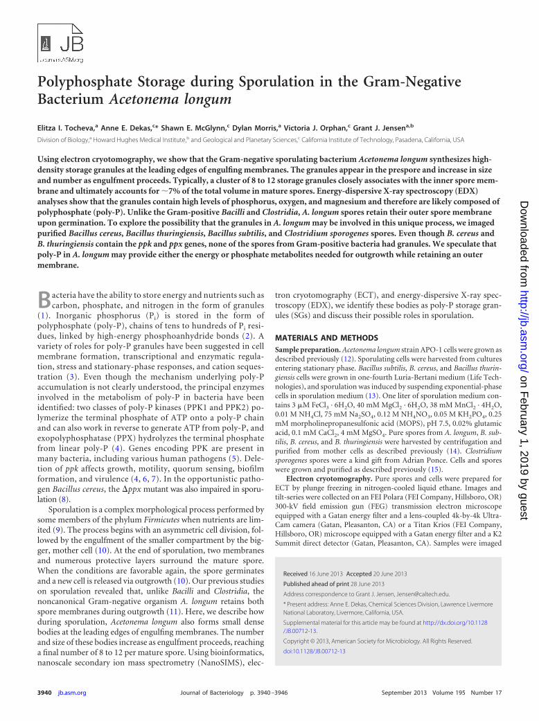
FIG 1 Storage granule formation during sporulation in A. longum. (A) Tomographic slices through vegetative cell; (B) sporulating cell during early stages ofengulfment; (C and D) sporulating cell during later stages of engulfment; (E) mature spore; (F) germinating cell. Abbreviations: S, spore; M, mother cell; SG,storage granule; IM, inner membrane, OM, outer membrane. Bar, 200 nm.
Poly-P Storage during Sporulation in A. longum
September 2013 Volume 195 Number 17 jb.asm.org 3941
on February 1, 2019 by guest
http://jb.asm.org/
Dow
nloaded from
were placed on carbon-coated copper grids and air dried. The FEI trans-mission EM (TEM) imaging and analysis (TIA) software package was usedto acquire data from a point measurement over a storage granule andfrom an area over a whole spore. EDX spectra of different areas werecollected at 80 kV and a dosage of �50 e�/Å2.
Homology searches. Several kinds of storage granules (SGs) have beendescribed in bacteria, including glycogen, poly-�-hydroxybutyrate (PHB),polyphosphate, “sulfur-rich” granules, and “nitrogen-rich” granules (18–22).We searched for genes encoding enzymes associated with the production ofstorage granules in the genomes of A. longum and a number of “control”species, including Ralstonia eutropha (known to form PHB granules) (23),Caulobacter crescentus (known to form polyphosphate granules) (24), Esche-richia coli (known to store glycogen) (25), and Allochromatium vinosum(known to form sulfur globules). The well-known endospore-forming spe-cies B. subtilis and C. sporogenes were also included as controls. To analyze thedistribution of enzymes responsible for storage granule formation, we con-ducted BLAST searches of the several genomes using the blastp program withlow-complexity filtering disabled and a strict E value threshold of 1e�10 (26).The query proteins used for these searches for glycogen storage were GlgBI(NP_629578.1) and GlgBII (NP_631386.1); those for PHB granules werePhaC (YP_726471.1), PhaP (YP_001171240.1), PhaZ1 (YP_725659.1), andPhaZ2 (YP_727307.1); those for polyphosphate storage were PPK1(NP_416996.1) and PPX (NP_416997.1); and those for sulfur globules wereSgpA (YP_003443861.1) and SgpB (YP_003442351.1).
Homology searches for just the poly-P enzymes in all sequenced spo-rulating bacteria were performed using the Pfam domains PF02503,PF03976, and PF02541 for PPK1, PPK2, and PPX, respectively.
RESULTSECT of sporulating A. longum cells reveals storage granules. Asdescribed in the work of Tocheva et al., cryotomograms of �250
A. longum cells were recorded at different stages of sporulation(11). Dense storage granules (SGs) were rarely observed (�1%) invegetative cells of A. longum (Fig. 1A) but were consistently foundin all prespores at the leading edges of engulfing membranes dur-ing early stages of engulfment (Fig. 1B). The number and size ofthe SGs increased as sporulation proceeded (Fig. 1C and D). Mea-surements of the distance of the SGs to the closest leading edge ofengulfing membranes show a range of distances (from 67 nm to272 nm), with the closest SG located 71 � 7 nm from a leadingedge. At the end of engulfment, all mature A. longum spores typ-ically contained 8 to 12 storage granules with diameters of 40 to120 nm, accounting for �7% of the spore volume (Fig. 1E). Inmature spores, the SGs remained clustered but were no longerproximal to the inner spore membrane. The SGs persistedthroughout germination and outgrowth (Fig. 1F), though no spe-cific localization with respect to the newly emerging cell was ap-parent.
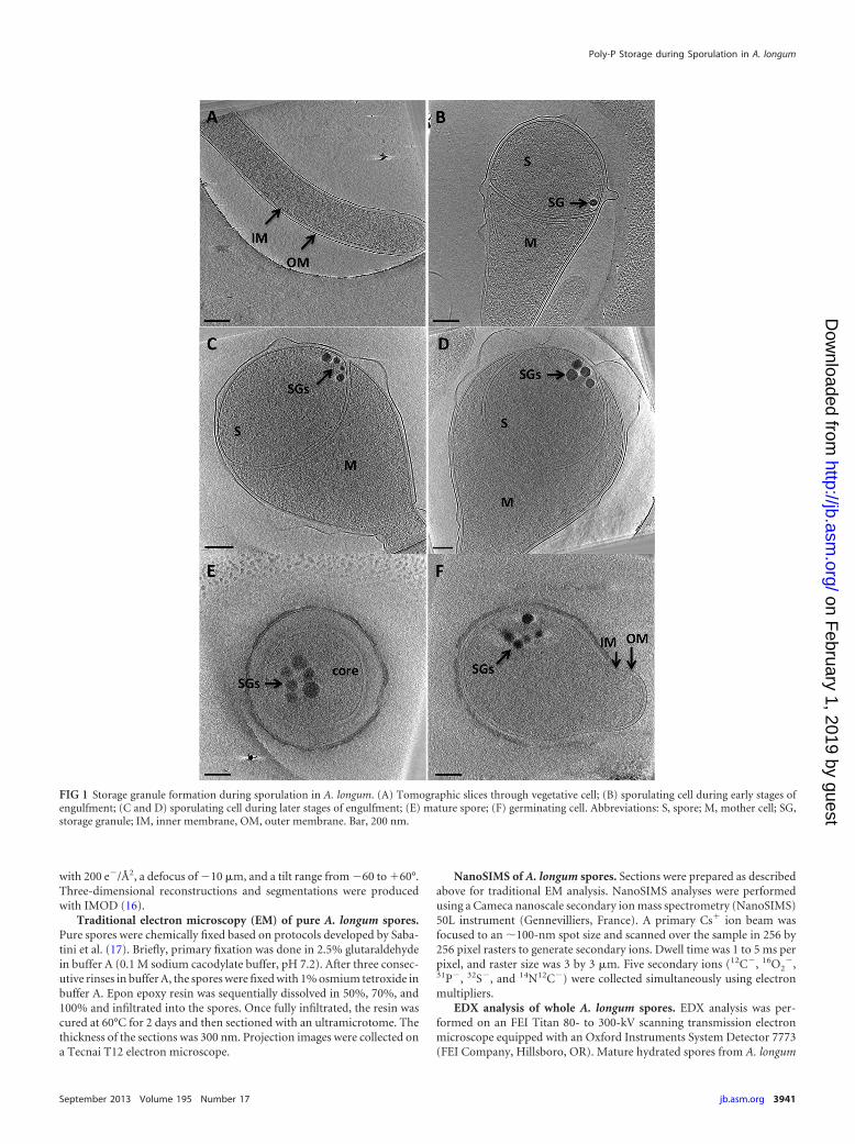
Appearance of SGs. The SGs in A. longum appeared dense andgrossly spherical, surrounded by an even denser shell (Fig. 2).Compared to other organisms, they closely resembled the size,density, and shape of the poly-P storage granules observed in Cau-lobacter crescentus and other organisms (see Fig. S4B in the sup-plemental material) (24). The shell around the SGs was discontin-uous, only partially covering the granule (Fig. 2C; white arrowsindicate the presence and black arrows indicate the absence of theprotein layer). No patterns in the positions of the shell patcheswere recognized. In contrast to reports of an apparent membra-nous shell surrounding poly-P storage granules in Agrobacterium
FIG 2 Structural features of the storage granules in A. longum. (A) Segmentation of the sporulating A. longum cell from Fig. 1D shows that 9 storage granules(represented as colored spheres) are clustered together and pressed against the inner spore membrane (green). Bar, 200 nm. (B) The storage granules are locatedclose to the leading edge of the engulfing membrane. Colored stars correspond to the colors of the storage granules in panel A. (C) The granules exhibit a varietyof roughly spherical shapes and are surrounded by a patchy surface layer. White arrows indicate areas of the presence of a proteinaceous layer; black arrowsindicate the absence of a layer. Bar, 50 nm.
Tocheva et al.
3942 jb.asm.org Journal of Bacteriology
on February 1, 2019 by guest
http://jb.asm.org/
Dow
nloaded from
tumefaciens and Rhodospirillum rubrum (27), the surroundingshell in A. longum was both discontinuous and variable in thick-ness and was therefore likely proteinaceous. The cores of the SGsappeared granular and void of internal organization. Fouriertransforms of the images also failed to reveal internal order (datanot shown).
Traditional EM of mature A. longum spores. Traditional EMmethods failed to preserve the SG consistently (see Fig. S1 in thesupplemental material). Using the same preparation method andsections, sometimes the SGs were retained, and other times theywere lost, leaving “holes” in the section that are a well-knownartifact of chemical fixation and alcohol dehydration (28). Theinconsistency of SG preservation with traditional EM methodscomplicated elemental data acquisition, and precautions weretherefore taken to analyze only dense (preserved) granules.
Mature spores from Bacilli and Clostridia lack SGs. In orderto explore the role of poly-P SGs in sporulation in general, maturespores of other sporulating bacteria were also imaged with ECT. Incontrast to A. longum, mature B. subtilis, B. cereus, B. thuringiensis,and C. sporogenes lacked dense SGs (see Fig. S2 in the supplemen-tal material).
Elemental mapping of A. longum spores using NanoSIMS.To investigate the intracellular elemental distribution in a spore,thin sections of three A. longum spores were analyzed with nano-scale secondary ion mass spectrometry (NanoSIMS). Areas of in-creased phosphorus concentration were observed within thespores (see Fig. S3 in the supplemental material). Peaks were alsovisible in the 31P�/14N12C� ratio image, in patterns different fromthose seen for the other elements (data not shown), demonstrat-ing that they were not an artifact of sample topology or unevengeneration of secondary ions (see Fig. S3C). Due to the lowersensitivity of NanoSIMS for phosphorus, the 31P� signal for DNAand RNA from the core of mature spores was not detected.
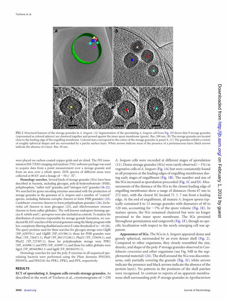
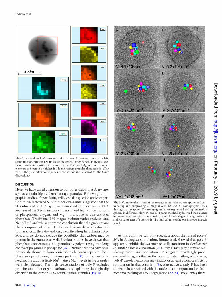
EDX. To further explore the elemental composition of the SGs,EDX was employed. A. longum spores and SGs were identifiedusing scanning-transmission electron microscopy (Fig. 3). EDXspectra were then collected and showed elevated counts for O, P,and Mg but not Na, S, Cl, Ca, Mn, and Cu within SGs. The countsfor phosphorus in the granules were �3-fold greater than thosefor magnesium but half those for oxygen (Fig. 3). The copper andsome of the carbon detected likely came from the EM grid. Thedistribution and overall shape of the storage granule cluster cor-related well with elevated signals for P, O, and Mg ions in arealanalyses (Fig. 4).
Bioinformatics. A. longum possesses the genes known to me-diate storage of polyphosphate and glycogen but not sulfur orPHB (see Fig. S4A in the supplemental material). While PPKand PPX are present in some Bacilli (B. cereus and B. thurin-giensis, also imaged with tomography), most Clostridia (forexample, C. sporogenes) and some Bacilli (for example, B. sub-tilis) lacked the genes associated with poly-P formation. A.longum and Pelosinus fermentans were the only Gram-negativeendospore-forming Firmicutes that had been sequenced, andboth had ppk and ppx genes.
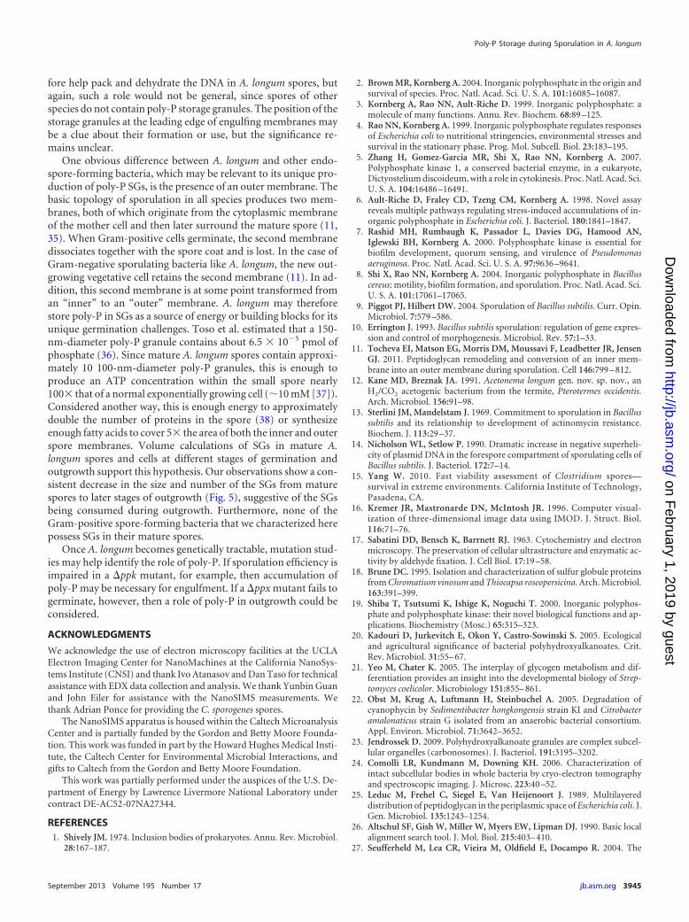
Volume calculations of SGs. To examine the fate of the SGsduring germination and outgrowth, we performed volume calcu-lations of the SGs in mature spores and germinating and outgrow-ing A. longum cells (n 25) (Fig. 5). Our results show that whilemature spores had clusters of SGs occupying �5 106 nm3, thevolume and number of the SGs gradually decreased in germinat-ing cells (cells with hydrolyzed cortex, Fig. 5C and D) to �3.5 106 nm3. The volume and number of SGs continued to decreaseduring initial stages of outgrowth (Fig. 5E and F) and were ulti-mately the lowest in cells at later stages of outgrowth (total volumeof �1.2 106 nm3).
FIG 3 High-dose EDX point analysis of a storage granule. The elemental compositions within a storage granule and a random location outside the granulebut within the spore core are shown in red and blue, respectively. Major peaks are assigned. Data show elevated levels of P, O, and Mg in the storage granulecompared to the spore core. The inset shows a scanning-transmission EM image of the air-dried spore used for imaging, with crosses marking thepositions analyzed.
Poly-P Storage during Sporulation in A. longum
September 2013 Volume 195 Number 17 jb.asm.org 3943
on February 1, 2019 by guest
http://jb.asm.org/
Dow
nloaded from
DISCUSSION
Here, we have called attention to our observation that A. longumspores contain highly dense storage granules. Following tomo-graphic studies of sporulating cells, visual inspection and compar-ison to characterized SGs in other organisms suggested that theSGs observed in A. longum were enriched in phosphorus. EDXanalyses of the SGs in mature spores showed high concentrationsof phosphorus, oxygen, and Mg2� indicative of concentratedphosphate. Traditional EM images, bioinformatics analyses, andNanoSIMS analysis support the conclusion that the granules arelikely composed of poly-P. Further analysis needs to be performedto characterize the ratio and lengths of the phosphate chains in theSGs, and we do not exclude the possibility that carbon may bepresent in the granules as well. Previous studies have shown thatphosphate concentrates into granules by polymerizing into longchains of polyanionic phosphate (29). Divalent cations have beenpreviously shown to form ionic bonds between separate phos-phate groups, allowing for denser packing (30). In the case of A.longum, the cation is likely Mg2�, since Mg2� levels in the granuleswere also elevated. The high concentration of poly-P excludesproteins and other organic carbon, thus explaining the slight dipobserved in the carbon EDX counts within granules (Fig. 4).
At this point, we can only speculate about the role of poly-PSGs in A. longum sporulation. Boutte et al. showed that poly-Pappears to inhibit the swarmer-to-stalk transition in Caulobactersp. under glucose exhaustion (31). Poly-P may play a similar reg-ulatory role during sporulation in A. longum. Interestingly, previ-ous work suggests that in the opportunistic pathogen B. cereus,poly-P depolymerization may induce or at least promote efficientsporulation in that organism (8). Alternatively, poly-P has beenshown to be associated with the nucleoid and important for chro-mosomal packing or DNA segregation (32–34). Poly-P may there-
FIG 4 Lower-dose EDX area scan of a mature A. longum spore. Top left,scanning-transmission EM image of the spore. Other panels, individual ele-ment distributions within the scanned area. P, O, and Mg but not the otherelements are seen to be higher inside the storage granules than outside. (The“K” in the panel titles corresponds to the atomic shell assessed for the X-raydispersion.)
FIG 5 Volume calculations of the storage granules in mature spores and ger-minating and outgrowing A. longum cells. (A and B) Tomographic slicesthrough mature spores. The storage granules are segmented and represented asspheres in different colors. (C and D) Spores that had hydrolyzed their cortexbut maintained an intact spore coat. (E and F) Early stages of outgrowth. (Gand H) Late stages of outgrowth. The total volume of the SGs is shown in eachpanel.
Tocheva et al.
3944 jb.asm.org Journal of Bacteriology
on February 1, 2019 by guest
http://jb.asm.org/
Dow
nloaded from
fore help pack and dehydrate the DNA in A. longum spores, butagain, such a role would not be general, since spores of otherspecies do not contain poly-P storage granules. The position of thestorage granules at the leading edge of engulfing membranes maybe a clue about their formation or use, but the significance re-mains unclear.
One obvious difference between A. longum and other endo-spore-forming bacteria, which may be relevant to its unique pro-duction of poly-P SGs, is the presence of an outer membrane. Thebasic topology of sporulation in all species produces two mem-branes, both of which originate from the cytoplasmic membraneof the mother cell and then later surround the mature spore (11,35). When Gram-positive cells germinate, the second membranedissociates together with the spore coat and is lost. In the case ofGram-negative sporulating bacteria like A. longum, the new out-growing vegetative cell retains the second membrane (11). In ad-dition, this second membrane is at some point transformed froman “inner” to an “outer” membrane. A. longum may thereforestore poly-P in SGs as a source of energy or building blocks for itsunique germination challenges. Toso et al. estimated that a 150-nm-diameter poly-P granule contains about 6.5 10�5 pmol ofphosphate (36). Since mature A. longum spores contain approxi-mately 10 100-nm-diameter poly-P granules, this is enough toproduce an ATP concentration within the small spore nearly100 that of a normal exponentially growing cell (�10 mM [37]).Considered another way, this is enough energy to approximatelydouble the number of proteins in the spore (38) or synthesizeenough fatty acids to cover 5 the area of both the inner and outerspore membranes. Volume calculations of SGs in mature A.longum spores and cells at different stages of germination andoutgrowth support this hypothesis. Our observations show a con-sistent decrease in the size and number of the SGs from maturespores to later stages of outgrowth (Fig. 5), suggestive of the SGsbeing consumed during outgrowth. Furthermore, none of theGram-positive spore-forming bacteria that we characterized herepossess SGs in their mature spores.
Once A. longum becomes genetically tractable, mutation stud-ies may help identify the role of poly-P. If sporulation efficiency isimpaired in a �ppk mutant, for example, then accumulation ofpoly-P may be necessary for engulfment. If a �ppx mutant fails togerminate, however, then a role of poly-P in outgrowth could beconsidered.
ACKNOWLEDGMENTS
We acknowledge the use of electron microscopy facilities at the UCLAElectron Imaging Center for NanoMachines at the California NanoSys-tems Institute (CNSI) and thank Ivo Atanasov and Dan Taso for technicalassistance with EDX data collection and analysis. We thank Yunbin Guanand John Eiler for assistance with the NanoSIMS measurements. Wethank Adrian Ponce for providing the C. sporogenes spores.
The NanoSIMS apparatus is housed within the Caltech MicroanalysisCenter and is partially funded by the Gordon and Betty Moore Founda-tion. This work was funded in part by the Howard Hughes Medical Insti-tute, the Caltech Center for Environmental Microbial Interactions, andgifts to Caltech from the Gordon and Betty Moore Foundation.
This work was partially performed under the auspices of the U.S. De-partment of Energy by Lawrence Livermore National Laboratory undercontract DE-AC52-07NA27344.
REFERENCES1. Shively JM. 1974. Inclusion bodies of prokaryotes. Annu. Rev. Microbiol.
28:167–187.
2. Brown MR, Kornberg A. 2004. Inorganic polyphosphate in the origin andsurvival of species. Proc. Natl. Acad. Sci. U. S. A. 101:16085–16087.
3. Kornberg A, Rao NN, Ault-Riche D. 1999. Inorganic polyphosphate: amolecule of many functions. Annu. Rev. Biochem. 68:89 –125.
4. Rao NN, Kornberg A. 1999. Inorganic polyphosphate regulates responsesof Escherichia coli to nutritional stringencies, environmental stresses andsurvival in the stationary phase. Prog. Mol. Subcell. Biol. 23:183–195.
5. Zhang H, Gomez-Garcia MR, Shi X, Rao NN, Kornberg A. 2007.Polyphosphate kinase 1, a conserved bacterial enzyme, in a eukaryote,Dictyostelium discoideum, with a role in cytokinesis. Proc. Natl. Acad. Sci.U. S. A. 104:16486 –16491.
6. Ault-Riche D, Fraley CD, Tzeng CM, Kornberg A. 1998. Novel assayreveals multiple pathways regulating stress-induced accumulations of in-organic polyphosphate in Escherichia coli. J. Bacteriol. 180:1841–1847.
7. Rashid MH, Rumbaugh K, Passador L, Davies DG, Hamood AN,Iglewski BH, Kornberg A. 2000. Polyphosphate kinase is essential forbiofilm development, quorum sensing, and virulence of Pseudomonasaeruginosa. Proc. Natl. Acad. Sci. U. S. A. 97:9636 –9641.
8. Shi X, Rao NN, Kornberg A. 2004. Inorganic polyphosphate in Bacilluscereus: motility, biofilm formation, and sporulation. Proc. Natl. Acad. Sci.U. S. A. 101:17061–17065.
9. Piggot PJ, Hilbert DW. 2004. Sporulation of Bacillus subtilis. Curr. Opin.Microbiol. 7:579 –586.
10. Errington J. 1993. Bacillus subtilis sporulation: regulation of gene expres-sion and control of morphogenesis. Microbiol. Rev. 57:1–33.
11. Tocheva EI, Matson EG, Morris DM, Moussavi F, Leadbetter JR, JensenGJ. 2011. Peptidoglycan remodeling and conversion of an inner mem-brane into an outer membrane during sporulation. Cell 146:799 – 812.
12. Kane MD, Breznak JA. 1991. Acetonema longum gen. nov. sp. nov., anH2/CO2 acetogenic bacterium from the termite, Pterotermes occidentis.Arch. Microbiol. 156:91–98.
13. Sterlini JM, Mandelstam J. 1969. Commitment to sporulation in Bacillussubtilis and its relationship to development of actinomycin resistance.Biochem. J. 113:29 –37.
14. Nicholson WL, Setlow P. 1990. Dramatic increase in negative superheli-city of plasmid DNA in the forespore compartment of sporulating cells ofBacillus subtilis. J. Bacteriol. 172:7–14.
15. Yang W. 2010. Fast viability assessment of Clostridium spores—survival in extreme environments. California Institute of Technology,Pasadena, CA.
16. Kremer JR, Mastronarde DN, McIntosh JR. 1996. Computer visual-ization of three-dimensional image data using IMOD. J. Struct. Biol.116:71–76.
17. Sabatini DD, Bensch K, Barrnett RJ. 1963. Cytochemistry and electronmicroscopy. The preservation of cellular ultrastructure and enzymatic ac-tivity by aldehyde fixation. J. Cell Biol. 17:19 –58.
18. Brune DC. 1995. Isolation and characterization of sulfur globule proteinsfrom Chromatium vinosum and Thiocapsa roseopersicina. Arch. Microbiol.163:391–399.
19. Shiba T, Tsutsumi K, Ishige K, Noguchi T. 2000. Inorganic polyphos-phate and polyphosphate kinase: their novel biological functions and ap-plications. Biochemistry (Mosc.) 65:315–323.
20. Kadouri D, Jurkevitch E, Okon Y, Castro-Sowinski S. 2005. Ecologicaland agricultural significance of bacterial polyhydroxyalkanoates. Crit.Rev. Microbiol. 31:55– 67.
21. Yeo M, Chater K. 2005. The interplay of glycogen metabolism and dif-ferentiation provides an insight into the developmental biology of Strep-tomyces coelicolor. Microbiology 151:855– 861.
22. Obst M, Krug A, Luftmann H, Steinbuchel A. 2005. Degradation ofcyanophycin by Sedimentibacter hongkongensis strain KI and Citrobacteramalonaticus strain G isolated from an anaerobic bacterial consortium.Appl. Environ. Microbiol. 71:3642–3652.
23. Jendrossek D. 2009. Polyhydroxyalkanoate granules are complex subcel-lular organelles (carbonosomes). J. Bacteriol. 191:3195–3202.
24. Comolli LR, Kundmann M, Downing KH. 2006. Characterization ofintact subcellular bodies in whole bacteria by cryo-electron tomographyand spectroscopic imaging. J. Microsc. 223:40 –52.
25. Leduc M, Frehel C, Siegel E, Van Heijenoort J. 1989. Multilayereddistribution of peptidoglycan in the periplasmic space of Escherichia coli. J.Gen. Microbiol. 135:1243–1254.
26. Altschul SF, Gish W, Miller W, Myers EW, Lipman DJ. 1990. Basic localalignment search tool. J. Mol. Biol. 215:403– 410.
27. Seufferheld M, Lea CR, Vieira M, Oldfield E, Docampo R. 2004. The
Poly-P Storage during Sporulation in A. longum
September 2013 Volume 195 Number 17 jb.asm.org 3945
on February 1, 2019 by guest
http://jb.asm.org/
Dow
nloaded from
H(�)-pyrophosphatase of Rhodospirillum rubrum is predominantly lo-cated in polyphosphate-rich acidocalcisomes. J. Biol. Chem. 279:51193–51202.
28. Dubochet J, Sartori Blanc N. 2001. The cell in absence of aggregationartifacts. Micron 32:91–99.
29. Kulaev I, Vagabov V, Kulakovskaya T. 1999. New aspects of inorganicpolyphosphate metabolism and function. J. Biosci. Bioeng. 88:111–129.
30. Parsons AJ, Ahmed I, Rudd CD, Cuello GJ, Pellegrini E, Richard D,Johnson MR. 2010. Neutron scattering and ab initio molecular dynamicsstudy of cross-linking in biomedical phosphate glasses. J. Phys. Condens.Matter 22:485403. doi:10.1088/0953-8984/22/48/485403.
31. Boutte CC, Henry JT, Crosson S. 2012. ppGpp and polyphosphatemodulate cell cycle progression in Caulobacter crescentus. J. Bacteriol. 194:28 –35.
32. Kahng LS, Shapiro L. 2003. Polar localization of replicon origins in themultipartite genomes of Agrobacterium tumefaciens and Sinorhizobiummeliloti. J. Bacteriol. 185:3384 –3391.
33. Fraley CD, Rashid MH, Lee SS, Gottschalk R, Harrison J, Wood PJ,Brown MR, Kornberg A. 2007. A polyphosphate kinase 1 (ppk1) mutant
of Pseudomonas aeruginosa exhibits multiple ultrastructural and func-tional defects. Proc. Natl. Acad. Sci. U. S. A. 104:3526 –3531.
34. Butan C, Hartnell LM, Fenton AK, Bliss D, Sockett RE, SubramaniamS, Milne JL. 2011. Spiral architecture of the nucleoid in Bdellovibrio bac-teriovorus. J. Bacteriol. 193:1341–1350.
35. Tocheva EI, Lopez-Garrido J, Hughes HV, Fredlund J, Kuru E, Van-nieuwenhze MS, Brun YV, Pogliano K, Jensen GJ. 2013. Peptidoglycantransformations during Bacillus subtilis sporulation. Mol. Microbiol. 88:673– 686.
36. Toso DB, Henstra AM, Gunsalus RP, Zhou ZH. 2011. Structural, massand elemental analyses of storage granules in methanogenic archaeal cells.Environ. Microbiol. 13:2587–2599.
37. Bennett BD, Kimball EH, Gao M, Osterhout R, Van Dien SJ, Rabinow-itz JD. 2009. Absolute metabolite concentrations and implied enzymeactive site occupancy in Escherichia coli. Nat. Chem. Biol. 5:593–599.
38. Piques M, Schulze WX, Hohne M, Usadel B, Gibon Y, Rohwer J, StittM. 2009. Ribosome and transcript copy numbers, polysome occupancyand enzyme dynamics in Arabidopsis. Mol. Syst. Biol. 5:314. doi:10.1038/msb.2009.68.
Tocheva et al.
3946 jb.asm.org Journal of Bacteriology
on February 1, 2019 by guest
http://jb.asm.org/
Dow
nloaded from