pomegranate seed oil nanoemulsions for the prevention and...
TRANSCRIPT

Nanomedicine: Nanotechnology, Biology, and Medicine10 (2014) 1353–1363
Original Article
Pomegranate seed oil nanoemulsions for the prevention and treatment ofneurodegenerative diseases: the case of genetic CJD
Michal Mizrahi, Bsca,1, Yael Friedman-Levi, PhDa,1, Liraz Larush, PhDb, Kati Frid, Bsca,Orli Binyamin, MSca, Dvir Dori, MDa, Nina Fainstein, PhDa, Haim Ovadia, PhDa,
Tamir Ben-Hur, Md, PhDa, Shlomo Magdassi, PhDb, Ruth Gabizon, PhDa,⁎aDepartment of Neurology, The Agnes Ginges Center for Human Neurogenetics, Hadassah University Hospital, Jerusalem, Israel
bCasali Institute of Chemistry, The Hebrew University of Jerusalem, Jerusalem, Israel
Received 8 November 2013; accepted 24 March 2014
nanomedjournal.com
Abstract
Neurodegenerative diseases generate the accumulation of specific misfolded proteins, such as PrPSc prions or A-beta in Alzheimer'sdiseases, and share common pathological features, like neuronal death and oxidative damage. To test whether reduced oxidation altersdisease manifestation, we treated TgMHu2ME199K mice, modeling for genetic prion disease, with Nano-PSO, a nanodroplet formulation ofpomegranate seed oil (PSO). PSO comprises large concentrations of a unique polyunsaturated fatty acid, Punicic acid, among the strongestnatural antioxidants. Nano-PSO significantly delayed disease presentation when administered to asymptomatic TgMHu2ME199K mice andpostponed disease aggravation in already sick mice. Analysis of brain samples revealed that Nano-PSO treatment did not decrease PrPSc
accumulation, but rather reduced lipid oxidation and neuronal loss, indicating a strong neuroprotective effect. We propose that Nano-PSOand alike formulations may be both beneficial and safe enough to be administered for long years to subjects at risk or to those alreadyaffected by neurodegenerative conditions.
From the Clinical Editor: This team of authors report that a nanoformulation of pomegranade seed oil, containing high levels of a strongantioxidant, can delay disease onset in a mouse model of genetic prion diseases, and the formulation also indicates a direct neuroprotective effect.© 2014 Elsevier Inc. All rights reserved.
Key words: Neurodegeneration; Oxidation; Prion; PSO; Nanoparticles
Neurodegenerative diseases are late onset fatal disorders thataffect large numbers of individuals in our society.1 Since clinicalsigns typically present after considerable irreversible loss ofbrain cells have occurred, therefore there is a clear unmet neednot only for delaying disease progression in diagnosed patientsbut also for preventing disease manifestation in subjects at risk.Ongoing efforts in the search of disease modifying agents aremainly focused on screening for molecules that can dismantle orinhibit the formation of misfolded “key” disease proteinsaggregates,2,3 which individually characterize each of theseconditions, such as Amyloid β in Alzheimer's disease (AD), αsynuclein in Parkinson's disease (PD) and PrPSc in prion diseases
Funding: This project was funded by a grant from the Israel ScienceFoundation (ISF) and by the Agnes Ginges Center. At present time, acommercial entity (Granalix) is being formed based on the presented results
⁎Corresponding author. Department of Neurology, Hadassah UniversityHospital, Jerusalem, Israel.
E-mail address: [email protected] (R. Gabizon).1 Equal contribution.
Please cite this article as: Mizrahi M, et al, Pomegranate seed oil nanoemulsionsgenetic CJD. Nanomedicine: NBM 2014;10:1353-1363, http://dx.doi.org/10.10
http://dx.doi.org/10.1016/j.nano.2014.03.0151549-9634/© 2014 Elsevier Inc. All rights reserved.
such as Creutzfeldt Jacob disease (CJD),4,5 considered the hallmarkof neurodegeneration.6,7
A complementary concept would be to look for therapeutictargets common to all neurodegenerative diseases, as is the casefor sensitivity to oxidative stress.8 In fact, most of the aggregatedkey disease proteins mentioned above are oxidized,9,10 and inprion diseases oxidation of Met residues in PrP helix 3 precedesthe acquisition of protease resistance by this protein.11 Also brainlipids are oxidized in these and other brain diseases,12,13
suggesting lipid oxidation may play an important role in thepathogenesis of neurodegenerative diseases.14-16 Indeed, oxidizedphospholipids generate compounds such as 4-oxo-2-nonenal andacrolein, which are predominantly toxic to brain cells.17 Consistentwith this, we propose to investigate whether safe α-oxidantreagents could be beneficial for individuals at risk of developingneurodegenerative conditions, constituting today a large fraction ofthe world population.1,18-22
To this effect, we tested whether administration of pome-granate seed oil (PSO), either in its natural form added to food oras a water soluble Nano-emulsion, can delay the clinical advance
for the prevention and treatment of neurodegenerative diseases: the case of16/j.nano.2014.03.015

Table 1Score of disease signs.
Score Disease signs
1 Initial hind limb weakness1.5 Partial hind limb weakness2 Significant hind limb weakness/paralysis2.5 +Legs clasping3 +Full paralysis in one limb3.5 +Significant weakness at the other hind foot4 Full paralysis in both limbs5 Death
1354 M. Mizrahi et al / Nanomedicine: Nanotechnology, Biology, and Medicine 10 (2014) 1353–1363
and ameliorate prion and neurodegeneration pathologicalfeatures in TgMHu2ME199K mice,23 which model for geneticCJD (gCJD) linked to the E200K PrP mutation.24 As of today,therapeutic intervention in all forms of human prion diseases hadfailed.25-28 PSO comprises a unique component, punicic acid(PA), a conjugated polyunsaturated fatty acid considered asone of the strongest natural antioxidants.29 Unsaturated fattyacids such as linoleic acid, similar to PA, were shown to readilycross the blood brain barrier (BBB).30-32 PA is present only inPSO (60-80%) and in Trichosanthes kirilowii (40%),33 andwas effective in protecting tissue lipid profiles in inflammatorydisease models.34 PSO lack of toxicity and partial bioavailabilitywas already established in humans.33 An additional antioxidant,β-sitosterol, which was demonstrated to accumulate in the plasmamembrane of brain cells,35 is present in PSO at significantlyhigher concentrations as compared to oils from other plants,36
indicating PSO may constitute a natural compound with strongerantioxidant activities than its individual components.
To increase the bioavailability and activity of PSO, wegenerated water soluble nanoemulsions hereby denominatedNano-PSO.37 This approach, as is the case for delivery systemssuch as phospholipid micelles or nanoparticles,38-40 may changethe target and distribution of the oil components betweendifferent organs, thereby enabling a longer circulation whichmay increase the levels of PA available to pass the BBB.
TgMHu2ME199K mice23 express human-mouse chimericE199K PrP on a null (for homozygous) or a wt PrP (forheterozygous) background. Mice from both lines suffer fromprogressive neurological symptoms as early as 5-6 months ofage and deteriorate to a terminal condition several monthsthereafter, concomitant with the accumulation of a truncatedform of PK resistant.23,41 TgMHu2ME199K mice exhibittypical pathological features of human CJD and of generalneurodegeneration.21 This model therefore represents themost stringent challenge for candidate therapies in neurode-generative diseases.
We show here that administration of PSO significantlydelayed disease onset in TgMHu2ME199K mice, constituting aproof of concept that a natural antioxidant may fight neurode-generation. Nano-PSO delayed disease onset and progression ina considerably faster mode and lower dose than natural PSO.Most important, it was only Nano-PSO and not PSO that couldprevent further advance of disease when administrated to alreadysick TgMHu2ME199K mice. No aberrant side effect wasobserved in the time frame (months) and PSO doses used inthese experiments. Analysis of brain samples revealed that whileaccumulation of PK resistant PrP was not affected by PSOformulations, brains from treated mice exhibited a strongneuroprotection effect, as seen by decreased lipid oxidation andneuronal loss, as well as increased synapthophysin expressionand neurogenesis.
Methods
Animal experiments
All animal experiments were conducted under the guidelinesand supervision of the Hebrew University Ethical Committee,
which approved of the methods employed in this project(Permit Number: MD-11746-5).
Treatment of TgMHu2ME199K mice
PSO and Nano-PSO were administrated to TgMHu2-ME199K mice modeling for E200K CJD expressing human-mouse chimeric E199K PrP on a null (for homozygous) or a wtPrP (for heterozygous) background,23 as described in Table 2.In the PSO experiments,1-3 mouse pelleted feed to which PSOwas added as described below was unlimitedly administered toyoung and older mice as applicable for as long as described inthe table. Nano-PSO was administered either by gavage 5 timesa week as in experiment 4 (150 μl/day), or by adding Nano-PSOto the mice drinking water (experiment 5). At the end of theexperiments, mice were sacrificed and their brains processedfor pathological and biochemical experiments.
Mice scoring for disease signs
TgMHu2ME199K mice were followed twice a week for theappearance of spontaneous neurological disease. Mice werescored for disease severity and progression according to the scaledescribed here and in Table 1. No clinical signs: 0; Initial hind limbsweakness = 1; Partial hind limbs weakness = 1.5; Significant hindlimb/s weakness or paralysis = 2; Significant hind limb/s weaknessor paralysiswith significant legs clasping = 2.5; Full paralysis in onelimb = 3; Full paralysis in one limb, and significant weakness at theother hind foot = 3.5; Full paralysis in both limbs = 4; Death = 5.
Mice were sacrificed at designated time points when required,according to the ethical requirements of the Hebrew UniversityAnimal Authorities when they were too sick or paralyzed to reachfood and water, or after losing 20% body weight.
Preparation of PSO-enriched food
1 kg of mouse pelleted food (Harlan, Taklad) was dissolved inwater and subsequently supplemented with 25 ml of PSO (Flavex,Germany). Mixture was next reassembled and dehydrated as pellets.
Preparation of O/W pomegranate oil nanoemulsion by sonication
A nanoemulsion with 10.8% oil fraction was prepared asfollows: 1.56 g of Pomegranate oil, 0.65 g of Tween 80, and0.39 g of glyceryl monooleate were mixed by a magnetic stirrerfor 20 min. 2.5 g from the above mixture was mixed by amagnetic stirrer with 0.277 g of glycerol, for 15 min. 2 g of theglycerol mixture was then added drop wise to 8 g of deionized

Table 2Groups in in vivo experiments.
Treatment Exp # Geneticbackground
Gender Number of miceexperiment/control
Days oftreatment
PSO 1 Tg/wt Male 5/4 86-270PSO 2 Tg/ko Male 8/7 76-270PSO 3 Tg/ko Female 6/7 190-270Nano-PSO 4 Tg/ko Female 6/6 70-270Nano-PSO 5 Tg/ko Male 11/10 236-300
1355M. Mizrahi et al / Nanomedicine: Nanotechnology, Biology, and Medicine 10 (2014) 1353–1363
water. A crude white emulsion was obtained. At the second stagethis crude emulsion was sonicated using a horn sonicator(model Vibra-Cell, Sonics & Materials Inc., USA) for 10 min at750 W. The samples were cooled in an ice water bath during thesonication process. A bluish nano emulsion was obtained.
Dynamic light scattering
Droplets size measurements were performed with a ZetasizerNano-S (Malvern Instruments Ltd., Worcestershire, UK). Sizemeasurements were performed in triplicates after dilution of theemulsion in water. The coarse white emulsion droplets size was inthe range of few microns. The average droplets size of the O/Wnano emulsion used in these experiments was 135 ± 12 nm.
Statistical analysis
The survival curves were compared using the Kaplan–Meieranalysis with log rank test calculatingΧ squares on one degree offreedom, P values and medians were calculated and are statedthrough the manuscript.
The clinical score severity curves in mice were comparedbetween control and experimental groups using Mann–Whitneytest (two tailed) and performed on the means + SEM (number ofmice in each experimental group are detailed in Table 2).
Western blot analysis
Brains from TgMHu2ME199Kmice at the designated end pointof the experiments were homogenized at 10% (W/V) in 10 mMTris–HCl, pH 7.4 and 0.3 M sucrose. For Proteinase K digestions,30 μl of 10% brain homogenates extracted with 2% sarkosyl on icewas incubated with 40 mg/ml Proteinase K for 30 min at 37 °C.Samples were boiled in the presence of SDS, subjected to SDSPAGE and immunoblotted with α-PrP pAb RTC.11
Immunocytochemistry
Four μm thick sections of formalin fixed, paraffinembedded brains of treated and untreated TgMHu2ME199Kmice were evaluated for the levels of oxidized phospholipidswith EO6 mAb (Avanti) and for the levels of neuronal synapseswith an α-Synaptophysin pAb (Novus).
Neurogenesis
For identifying proliferating brain cells (neurogenesis),10 months old wt, as well as Nano-PSO treated and untreatedTgMHu2ME199K mice were injected intraperitoneally withBromodeoxyuridine (BrdU, Sigma-Aldrich, 50 μg/1 g bodyweight) for 7 consecutive days. Subsequently, mice were
anesthetized with a lethal dose of pentobarbital and brains wereperfused via the ascending aorta with ice-cold PBS followedby cold 4% paraformaldehyde. Tissues were deep frozen in liquidnitrogen, and next serial 10 μM coronal sections were immuno-stained for BrdU (rat α-BrdU, Serotec) as previously described.42
Results
Delay of disease onset following administration of PSO in foodto young and asymptomatic TgMHu2ME199K mice
Groups of asymptomatic TgMHu2ME199K mice(TgMHu2ME199K/wt and TgMHu2ME199K/KO),23 as wellas 7 months old TgMHu2ME199K/KO mice already presentingsignificant neurological signs (see Table 1 for description ofdisease scores, and Table 2 for details about the experimentalgroups) were fed either with regular rodent food or with foodenriched with pomegranate seed oil (PSO) at a concentrationof 25 ml oil/kg. Since mice consume about 3-4 g of food/day,we may assume treated mice received about 100 μl of PSO perday or 700 μl of PSO/week. Disease progression in eachTgMHu2ME199K mouse was evaluated by frequent scoring ofclinical signs, and by calculating twice a week the average ofgroup scores in the treated animals as compared to untreatedlittermate groups generated from the same male and several ofits sibling females. Administration of PSO in food to youngTgMHu2ME199K mice was initiated just before they reached3 months of age, the first time point in which PK resistant PrP canbe easily detected in the still asymptomatic TgMHu2ME199Kmice.23,41 Controls and treated mice from the same experimentwere sacrificed simultaneously (at 270 days of age) and thereaftertheir brains processed for biochemical and pathological analysis. Inthe older mice, treatment commenced at 200 days (average scorein the group = 2) and continued for 9 weeks before termination ofthe experiment. No adverse effects were observed in any ofthe mice following the long term administration with the oilenriched food.
Figure 1 shows the effect of PSO administration on the clinicalpresentation and advance of the disease in TgMHu2ME199Kmice. Results were similar for treatment of heterozygous andhomozygous TgMHu2ME199K mice (panels AI and BI),consistent with our recent results indicating that wt PrP does notparticipate in disease presentation in the genetic mice,41 and showa significant delay in disease presentation in the treated mice(P b 0.02 for both panels A and B, see methods for description ofstatistical analysis). To better understand the meaning of this resultfor medical practice, we evaluated the progression of disease forindividual mice at two clinical time points, as represented by thepercentage of mice under score 2 (A and B panels II), or underscore 2.5 (A and B panels III). We found that PSO administrationin food could confer a beneficial effect to most treated mice whendata was estimated for the more advance stage, as depicted by the2.5 score (difference in medians between treated and untreatedgroups were 60 and 80 days (panelsA andB) (P = 0.04,χ2 = 3.9;P = 0.02,χ2 = 4.1)). At score 2, less than 50%of themice reactedto the treatment, as indicated by a median difference of only 10or 20 days respectably (P N 0.05). These results indicate that thebeneficial effect of PSO on disease presentation (for most mice)

Figure 1. Natural PSO delays disease onset in TgMHu2ME199K mice. Young TgMHu2ME199K/wt and TgMHu2ME199K/ko mice or 190 days oldTgMHu2ME199K/KOmice were treated with regular or with PSO enriched food for the designated time course (Table 2). Mice were scored for disease signs asdescribed in Table 1. Figures (I) in panels (A), (B) and (C): Average group score as related to age of mice. Figures (II) in panels (A) and (B): % of mice underscore 2 as related to age of mice. Figures (III) in panels (A) and (B): Percentage of mice under score 2.5 as related to age of mice. Figure (II) in panel (C): % ofmice aggravated by 0.5 score.
1356 M. Mizrahi et al / Nanomedicine: Nanotechnology, Biology, and Medicine 10 (2014) 1353–1363
can be observed only after long term consumption of the oil, andin many cases manifest when disease is already well advanced.As for the treatment of older and sick mice, Figure 1, C shows nodifference in the average group scores or on the progression ofdisease for individual mice between PSO treated and untreatedmice 200 days old sick mice (panels I and II).
Overall, these findings constitute the proof of conceptthat PSO may serve as an anti-prion/neuroprotective compound,but also present the limitations of such treatment; it requireslong term administration of the anti-oxidant from the subclinicalstage, while results may be apparent only when subjects arealready affected.
Increased delay of disease onset and of disease progressionfollowing the administration of Nano-PSO to young and oldTgMHu2ME199K mice
To generate a more effective and bioavailable formulation,we emulsified PSO to form oil nanodroplets with an averagediameter of approximately 150 nm (see methods), herebydenominated Nano-PSO. As stated in the introduction, suchformulations may allow for a longer circulation time of the nowdispersed drug and thereby larger activity.43 Figure 2, A presents
the results of an experiment comparable to those depicted inFigure 1, A and B for PSO enriched food. In this case, Nano-PSO was administrated to TgMHu2ME199K/KO mice from70 to 270 days by gavage (15 μl PSO/day; 5 days a week or75 μl/week). As in the previous experiment, treated and untreatedmice were sacrificed at the same time point, to comparepathological parameters. The dose of PSO in the Nano-PSOformulation used in this experiment constitutes approximately10% of the PSO administered as enriched food in the experimentsdescribed in Figure 1. Panel AI of Figure 2 shows a significantdifference between the average group score of disease as relatedto the time of Nano-PSO administration (P b 0.001). Whencomparing effect on individual mice, panel A (II and III) indicatethat administration of Nano-PSO resulted in a more rapid responseto treatment as compared to TgMHu2ME199K mice treated withPSO enriched food. Already at score 1 (panel II), which is a verymild and early diagnosed disease condition (see Table 1) there is a40 days difference between the median of treated and untreatedmice (P = 0.002, χ2 = 9.55). In addition, the graph in panel IIIshows that while the median difference between the treatedand untreated groups at score 2 was only 65 days (P = 0.0082,χ2 = 6.98), 50% of the treated mice never reached score 2 duringthe duration of the experiment. In fact, none of the mice treated
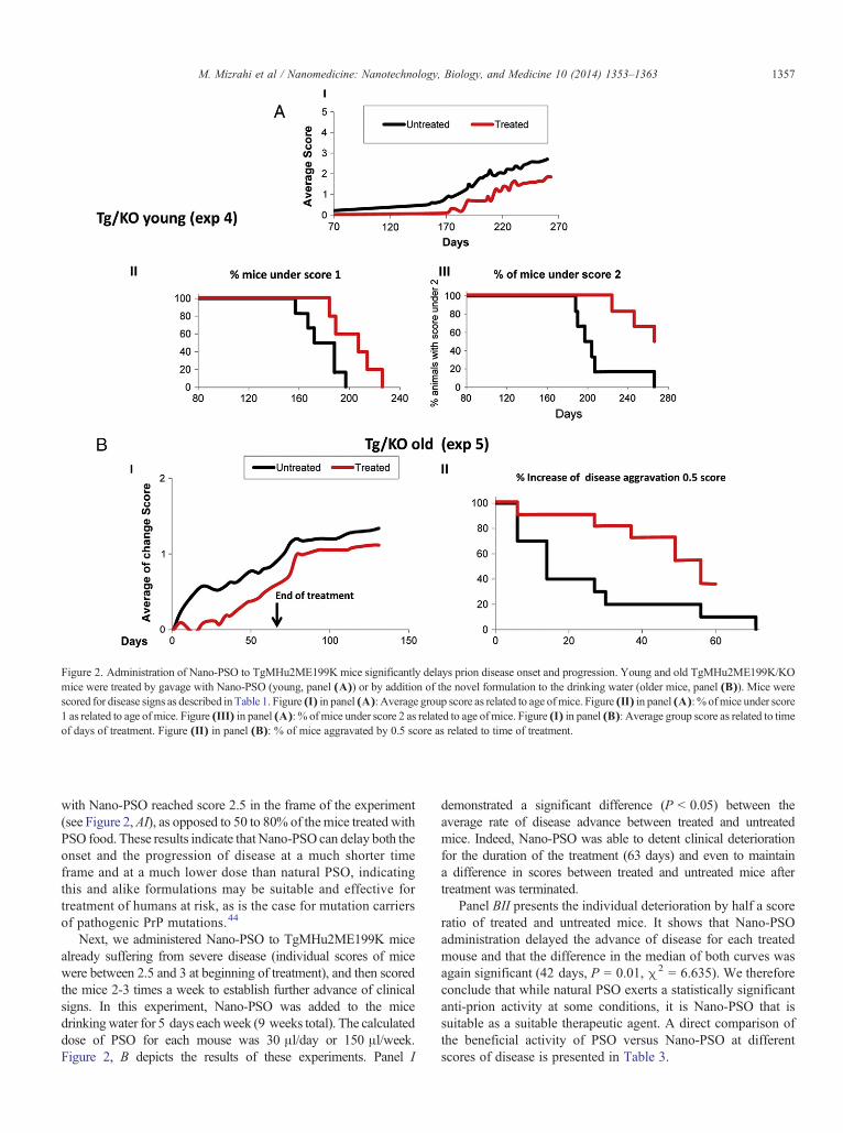
Figure 2. Administration of Nano-PSO to TgMHu2ME199K mice significantly delays prion disease onset and progression. Young and old TgMHu2ME199K/KOmice were treated by gavage with Nano-PSO (young, panel (A)) or by addition of the novel formulation to the drinking water (older mice, panel (B)). Mice werescored for disease signs as described in Table 1. Figure (I) in panel (A): Average group score as related to age ofmice. Figure (II) in panel (A): % ofmice under score1 as related to age ofmice. Figure (III) in panel (A): % of mice under score 2 as related to age ofmice. Figure (I) in panel (B): Average group score as related to timeof days of treatment. Figure (II) in panel (B): % of mice aggravated by 0.5 score as related to time of treatment.
1357M. Mizrahi et al / Nanomedicine: Nanotechnology, Biology, and Medicine 10 (2014) 1353–1363
with Nano-PSO reached score 2.5 in the frame of the experiment(see Figure 2,AI), as opposed to 50 to 80% of themice treated withPSO food. These results indicate that Nano-PSO can delay both theonset and the progression of disease at a much shorter timeframe and at a much lower dose than natural PSO, indicatingthis and alike formulations may be suitable and effective fortreatment of humans at risk, as is the case for mutation carriersof pathogenic PrP mutations.44
Next, we administered Nano-PSO to TgMHu2ME199K micealready suffering from severe disease (individual scores of micewere between 2.5 and 3 at beginning of treatment), and then scoredthe mice 2-3 times a week to establish further advance of clinicalsigns. In this experiment, Nano-PSO was added to the micedrinkingwater for 5 days eachweek (9 weeks total). The calculateddose of PSO for each mouse was 30 μl/day or 150 μl/week.Figure 2, B depicts the results of these experiments. Panel I
demonstrated a significant difference (P b 0.05) between theaverage rate of disease advance between treated and untreatedmice. Indeed, Nano-PSO was able to detent clinical deteriorationfor the duration of the treatment (63 days) and even to maintaina difference in scores between treated and untreated mice aftertreatment was terminated.
Panel BII presents the individual deterioration by half a scoreratio of treated and untreated mice. It shows that Nano-PSOadministration delayed the advance of disease for each treatedmouse and that the difference in the median of both curves wasagain significant (42 days, P = 0.01, χ2 = 6.635). We thereforeconclude that while natural PSO exerts a statistically significantanti-prion activity at some conditions, it is Nano-PSO that issuitable as a suitable therapeutic agent. A direct comparison ofthe beneficial activity of PSO versus Nano-PSO at differentscores of disease is presented in Table 3.

Table 3Comparing the clinical effect of the effect of PSO to Nano-PSO.
Experimental group Parameter measured PSO Nano-PSO
Young, asymptomatic Δ = The difference in medians betweentreated and untreated groups
Score 1 = no difference Score 1 = 40 daysP b 0.002
Score 2 = 10 days At score 2 ≫ 65⁎ daysP b 0.082
Score 2.5 = 80 days⁎P b 0.04
No score 2.5 in⁎ treated mice
Old, symptomatic Δ = The difference in deteriorationby half a score ratio
No difference 42 days ⁎P b 0.01
⁎ Statistically significant.
1358 M. Mizrahi et al / Nanomedicine: Nanotechnology, Biology, and Medicine 10 (2014) 1353–1363
No reduction in disease related PrP in PSO/Nano-PSO treatedTgMHu2ME199K mice
Disease related PrP forms, mostly resistant to digestion byproteinase K (PK), are the main markers of prion diseases, andare also considered the major/only components of the infectiousprion agents.4 In our TgMHu2ME199K model, mutant PrP canbe detected as a PK resistant and truncated form (PrPST) alreadyat 3 months of age, when mice are still asymptomatic for thedisease. PK resistant PrP levels increase with age, in parallel withdisease progression.41 Figure 3 shows individual brain extractsof TgMHu2ME199K mice treated with PSO or Nano-PSO(from experiments 2 and 4, 270 days at end point) whenimmunoblotted with α PrP pAb RTC.11 No difference in thetotal PrP levels was observed in the corresponding groups beforePK digestion, indicating that administration of PSO in both forms(panel A as PSO, panel B as Nano-PSO) had no effect on PrPexpression. Most important, no reduction in PK resistant PrP wasobserved in the treated brains in spite of the profound difference indisease score between treated and untreatedmice (Figures 1 and 2).Actually, the signal for PK resistant PrP even increased slightlyin some of the treated mice, as was the case for scrapie infectedmice treated with Simvastatin.45 This implies that PSO does notinterfere with the pathological process or aberrant metabolicpathway which leads to the accumulation of disease related PrP inTgMHu2ME199K mice, but may rather induce neuroprotectivepathways that manifest as reduced cell death. Such a mechanismmay even explain an apparent increase in prion protein accumulationin the treated brains.
PSO formulations protect neurons from death
Next, we immunostained paraffin embedded brain slices fromwt mice as well as from treated and untreated TgMHu2ME199Kmice with an α-synapthophysin antibody. The samples used werefrom experiments 2 (PSO) and 4 (Nano-PSO), as in theexperiments described in Figure 3. Synapthophysin is an integralmembrane protein located in presynaptic vesicles.46,47 Decreasedlevels of this and other synaptic proteins are considered a generalmarker of neurodegeneration48-50 and were observed in the brainsof CJD patients, bovine spongiform encephalopathy (BSE)infected cattle and scrapie infected mice.51-54
Figure 4, A shows that while synapthophysin levels werelargely reduced in the untreated TgMHu2ME199K mice, as seenby the reduce number of brown dots, as compared to age matched
wt mice, such immunostaining was significantly restored in thetreated mice from the parallel groups for both experiments. Thisindicates that early treatment with PSO formulations may preventnot only the advance of clinical signs but also the presentation ofgeneral neurodegenerative features of prion and other neurode-generative diseases, as is the case of synapthophysin expression.
Next, and to further establish whether PSO formulationscan inhibit neuronal death, we counted in age matched wt,as well as in treated and untreated TgMHu2ME199Kmice frozensections the number of cells in the CA1 and CA3 regions of thehippocampus, as manifested by DAPI staining. Figure 4, B showsthe results of such an experiments for sick TgMHu2ME199Kmice treated with and without Nano-PSO (experiment 5).Indeed, while the brains from the sick TgMHu2ME199K micepresented significant neuronal loss as compared to those ofwtmice(see thinner blue sections and numbers in the graph bellow), suchreduction was partially prevented by the Nano-PSO treatment.Indeed, differences between cell numbers in treated and untreatedsections, as well as between non-treated to wt mice were statisticallysignificant (P b 0.01 or P b 0007, respectively). Death of neuronsin the hippocampus was shown to correlate with dementia,55
suggesting both that our TgMHu2ME199K mice model is a goodmodel of neurodegeneration and that Nano-PSO exerts a strongneuroprotective affect in the most severe conditions, as representedby treatment of already sick TgMHu2ME199K mice.
Nano-PSO may restore hippocampal neurogenesis in sickTgMHu2ME199K mice
Numerous new studies demonstrate that during normal aging,certain areas of the brain retain pluripotent precursors with thecapacity of self-renewal.56,57 This feature, also known as adultneurogenesis, may be impaired in neurodegenerative diseasessuch as CJD and AD.58,59 To evaluate levels of neurogenesisin treated and untreated TgMHu2ME199K mice, we injectedBromodeoxyuridine (BrdU), a synthetic analog of thymidinecommonly used in the detection of proliferating cells in livingtissues60 to wt and TgMHu2ME199Kmice of several ages as wellas to Nano-PSO treated TgMHu2ME199K mice (experiment 5).Mice were sacrificed a week after the first injection and newneurons in the granular zone of the dentate gyrus were detected infrozen sectionswith anα-BrdU antibody. Figure 4,C shows that at4 months of age, when the TgMHu2ME199K mice were stillasymptomatic, there was no significant difference in the number ofnew cells between their brains and those of wt mice. Contrarily, in

Figure 3. No reduction in disease related PrP in PSO/Nano-PSO treated Tg mice: Brain extracts of mice sacrificed at the end point of experiments 2 and 4 weredigested in the presence and absence of PK, as described in the methods, and immunoblotted with α PrP pAb RTC.11 Panel (A): PSO treated mice, last 2 lanesrepresent scrapie infected brains (RML, and wt mice). Panel (B): Nano-PSO treated mice, last 2 lanes represent scrapie infected brains (RML, and wt mice).
1359M. Mizrahi et al / Nanomedicine: Nanotechnology, Biology, and Medicine 10 (2014) 1353–1363
older mice already presenting signs of disease (6 and 10 months ofage) the number of new cells in TgMHu2ME199K brains wassignificantly lower that the corresponding new cells in agematchedwt mice (P b 0.002), indicating reduced neurogenesis, at leastat this age, is a feature of neurodegeneration more than a propertyof aging.While only marginally significant (P b 0.12), the numberof new cells in Nano-PSO treated mice was higher than in the non-treated mice, indicating a possible additional neuroprotectiveproperty of Nano-PSO treatment.
PSO protects cell lipids from oxidative insults
As reviewed above, lipid and protein oxidation are importantfeatures of all neurodegenerative diseases.13,12,61 To establishwhether Nano-PSO can reduce lipid oxidation, we immuno-stained paraffin embedded brain slices brain from wt and formcontrol and treated TgMHu2ME199K mice with EO6, an anti-oxidized phospholipids mAb62 which was shown to detectoxidized lipids in MS plaques.13 In Figure 5, we show that whilewe could not detect any α-EO6 immunostaining in wt mice ofdifferent ages (no dispersed brown color or specific features),TgMHu2ME199K mice brain sections from experiment 4presented strong EO6 immunostaining in all cerebellar layers(dispersed brown in general and in cerebellum's Purkinje cells).These results are consistent with our previous findings, indicatinga large sensitivity of TgMHu2ME199K mice Purkinje cells todisease aggravation.63 Our results also show that Nano-PSOtreated sections present considerable lower levels of EO6 staining,
both general and cell specific, as compared to the parallel untreatedbrains. In samples from experiment 5, mice treated when alreadysick, we show that EO6 may also recognize star like plaques in thebrains of the untreated, but not in the treated TgMHu2ME199Kmice. These results demonstrate that lipid oxidation may bean important feature of prion diseases and that Nano-PSOformulations can reduce/prevent such oxidation, concomitantlywith inhibition of cell death and disease aggravation.
Discussion
A significant number of natural anti-oxidants are ubiquitouslypresent in a healthy human diet. Many of them, such asSulforaphane from Broccoli, Curcumin and EGCG from greentea were recognized for their neuroprotective properties in cellsand tested in appropriate animal models.64-66 However, their invivo activity was limited by the sub-pharmacological dosespresented in food, their poor bioavailability to humans, rapidchemical degradation and reduced distribution to differentorgans in the body, in particular the CNS. In this work, andafter PSO by itself was found to be clinically active in its naturaloil form, we made an effort to overcome such limitations bytailoring a more active formulation in the form of Nano-PSO.
We show here that administration of PSO in food may delay theonset of spontaneous genetic prion disease in the TgMHu2ME199Kmouse line, constituting a proof of concept that natural antioxidants

Figure 4. Neuroprotection of PSO formulations (A) PSO and Nano-PSO restore synapthophysin expression in sick mice: Paraffin embedded brain slices of wt,as well as PSO and Nano-PSO treated and untreated TgMHu2ME199K/KO mice (experiments 2 and 4), were immunostained with an α-synapthophysinantibody. Magnification in all pictures is ×20. (B) Nano-PSO protects hippocampal cells from death: Frozen sections the number of cells in the CA1 and CA3regions of the hippocampus, as manifested by DAPI staining. Statistical analysis of average from 4 mice in each group was performed by T-test.(C) Neurogenesis in treated and untreated TgMHu2ME199K mice: Slides of mice treated with BrdU and subsequently stained with an α-BrdU antibody werecounted for reacting cells. Average number of cells in slides of 4 mice was plotted in the graph. Statistical significance was evaluated by T-test.
1360 M. Mizrahi et al / Nanomedicine: Nanotechnology, Biology, and Medicine 10 (2014) 1353–1363
may exert a beneficial neuroprotective effect. However, it was onlyNano-PSO, a novel formulation inwhich natural PSOwas convertedinto soluble nanodroplets, which generated an impressive clinicaleffect at amuch lower PSOdose for both prevention and treatment ofprogressive prion disease in the TgMHu2ME199Kmice. Our resultsalso show that the mechanism of activity of PSO formulations wasnot prion specific, or at least not PrP specific, since the long termadministration of the oil did not affect the expression of mutantPrP or its accumulation as a PK resistant PrP form. Rather than aspecific anti-prion effect, these PSO formulations, concomitant withan impressive clinical outcome, presented a wide neuroprotectiveeffect, in the form of reduced neuronal death and lipid oxidationand increased neurogenesis, which may be valuable also for thetreatment of an array of neurodegenerative conditions. Indeed thecombination of an unsaturated fatty acid that can most probablycross the BBB,30 and the increased bioavailability conferred by thenanoparticle formulation,37 may explain the impressive neuropro-tective activity of Nano-PSO.
The search for disease modifiers for the treatment ofneurodegenerative diseases is focused mostly on screening for
reagents such as antibodies or small molecules that may dismantleor inhibit the aggregation of misfolded specific key proteins, as isthe case for PrPSc in prion diseases,67 A-beta in AD and synucleinin Parkinson's disease,68-71 or reduce the expression of the normalkey proteins, in the form of siRNAs72 or other molecules.73 Whilethese pathways may well lead to effective drugs, we propose analternative approach in the form of brain protection from commonneurodegeneration features, as is the case for neuroinflammationand oxidative stress. Indeed, Nano-PSO generated a strong clinicaleffect with an excellent safety profile which may allow itsprolonged administration to individuals at risk. The formulationdescribed here, oil nanodroplets of a specific size, may be furtherimproved by optimizing particle size, Nanoemulsion ingredients,stability features and physicochemical principles of preparation.Pharmacokinetics experiments in progress in our laboratory mayhelp to fine-tune the most active formulations, before we engage inclinical trials.
Indeed, since Nano-PSO can most probably be classified as a“safe” reagent, even in the levels of safety of a food supplement,such trials may come about soon enough. A comprehensive clinical

Figure 5. PSO protects brain lipids from oxidation: Paraffin embedded slides from Nano-PSO treated and untreated mice (experiments 4 and 5) wereimmunostained with the EO6 antibody. Initial magnification in all pictures was ×20.
1361M. Mizrahi et al / Nanomedicine: Nanotechnology, Biology, and Medicine 10 (2014) 1353–1363
trial for CJD patients was recently described.28 While the results fordoxycyclinewere negative, the experimental designmay be useful inour and other cases. In addition, we speculate that once sophisticateddisease specific reagents become available, they may well becombined with general neuroprotective agents such as Nano-PSOfor the best possible outcome in patients and at risk individuals.
References
1. Hamacher M, Meyer HE, Marcus K. New access to Alzheimer's andother neurodegenerative diseases. Expert Rev Proteomics 2007;4(5):591-4.
2. Aguzzi A, O'Connor T. Protein aggregation diseases: pathogenicity andtherapeutic perspectives. Nat Rev Drug Discov 2010 Mar;9(3):237-48.
3. Morales R, Green KM, Soto C. Cross currents in protein misfoldingdisorders: interactions and therapy. CNS Neurol Disord Drug Targets2009 Nov;8(5):363-71.
4. Prusiner SB. Novel proteinaceous infectious particles cause scrapie.Science 1982 Apr 9;216(4542):136-44.
5. Olanow CW, Prusiner SB. Is Parkinson's disease a prion disorder? ProcNatl Acad Sci U S A 2009 Aug 4;106(31):12571-2.
6. Prusiner SB. Biology and genetics of prions causing neurodegeneration.Annu Rev Genet 2013;47:601-23.
7. Ashe KH, Aguzzi A. Prions, prionoids and pathogenic proteins inAlzheimer disease. Prion 2013;7(1):55-9.
8. Butterfield DA, Kanski J. Brain protein oxidation in age-relatedneurodegenerative disorders that are associated with aggregated proteins.Mech Ageing Dev 2001;122(9):945-62.
9. Hajieva P, Behl C. Antioxidants as a potential therapy against age-relatedneurodegenerative diseases: amyloid beta toxicity and Alzheimer's disease.Curr Pharm Des 2006;12(6):699-704.
10. Grossmann ME, Mizuno NK, Schuster T, Cleary MP. Punicic acid is anomega-5 fatty acid capable of inhibiting breast cancer proliferation.Int J Oncol 2010;36(2):421-6.
11. Canello T, Frid K, Gabizon R, Lisa S, Friedler A, Moskovitz J, et al.Oxidation of helix-3 methionines precedes the formation of PK resistantPrP. PLoS Pathog 2010;6(7):e1000977.
12. Uttara B, Singh AV, Zamboni P, Mahajan RT. Oxidative stressand neurodegenerative diseases: a review of upstream anddownstream antioxidant therapeutic options. Curr Neuropharmacol2009;7(1):65-74.
13. Haider L, Fischer MT, Frischer JM, Bauer J, Hoftberger R, Botond G,et al. Oxidative damage in multiple sclerosis lesions. Brain 2011;134(Pt 7):1914-24.
14. Perluigi M, Coccia DA, Butterfield R. 4-Hydroxy-2-nonenal, a reactiveproduct of lipid peroxidation, and neurodegenerative diseases: a toxiccombination illuminated by redox proteomics studies. Antioxid RedoxSignal 2012;17(11):1590-609.
15. Adibhatla RM, Hatcher JF. Lipid oxidation and peroxidation in CNShealth and disease: from molecular mechanisms to therapeuticopportunities. Antioxid Redox Signal 2010;12(1):125-69.
16. Reed TT. Lipid peroxidation and neurodegenerative disease. Free RadicBiol Med 2011;51(7):1302-19.
17. Singh M, Dang TN, Arseneault M, Ramassamy C. Role of by-productsof lipid oxidation in Alzheimer's disease brain: a focus on acrolein.J Alzheimers Dis 2010;21(3):741-56.
18. Mizuno Y, Hattori N, Kubo S, Sato S, Nishioka K, Hatano T, et al.Progress in the pathogenesis and genetics of Parkinson's disease. PhilosTrans R Soc Lond B Biol Sci 2008;363(1500):2215-27.
19. Shoulson I, Young AB. Milestones in huntington disease. Mov Disord2011;26(6):1127-33.
20. Guo Q, Wang Z, Li H, Wiese M, Zheng H. APP physiological andpathophysiological functions: insights from animal models. Cell Res2012;22(1):78-89.
21. KovacsGG, Seguin J,Quadrio I,HoftbergerR,Kapas I, Streichenberger N,et al. Genetic Creutzfeldt-Jakob disease associated with the E200Kmutation: characterization of a complex proteinopathy. Acta Neuropathol2010;121:39-57.
22. Hsiao K, Meiner Z, Kahana E, Cass C, Kahana I, Avrahami D, et al.Mutation of the prion protein in Libyan Jews with Creutzfeldt-Jakobdisease. N Engl J Med 1991;324(16):1091-7.

1362 M. Mizrahi et al / Nanomedicine: Nanotechnology, Biology, and Medicine 10 (2014) 1353–1363
23. Friedman-Levi Y, Meiner Z, Canello T, Frid K, Kovacs GG, Budka H,et al. Fatal prion disease in a mouse model of genetic E200KCreutzfeldt-Jakob disease. PLoS Pathog 2011;7(11):e1002350.
24. Meiner Z, Gabizon R, Prusiner SB. Familial Creutzfeldt-Jakob disease.Codon 200 prion disease in Libyan Jews. Medicine (Baltimore) 1997;76(4):227-37.
25. Stewart LA, Rydzewska LH, Keogh GF, Knight RS. Systematic review oftherapeutic interventions in human prion disease. Neurology 2008;70(15):1272-81.
26. Geschwind MD. Clinical trials for prion disease: difficult challenges, buthope for the future. Lancet Neurol 2009;8(4):304-6.
27. Otto M, Cepek L, Ratzka P, Doehlinger S, Boekhoff I, Wiltfang J, et al.Efficacy of flupirtine on cognitive function in patients with CJD: Adouble-blind study. Neurology 2004;62(5):714-8.
28. Haik S, Marcon G,Mallet A, Tettamanti M,Welaratne A, Giaccone G, et al.Doxycycline in Creutzfeldt-Jakob disease: a phase 2, randomised, double-blind, placebo-controlled trial. Lancet Neurol 2014;13(2):150-8.
29. Schubert SY, Lansky EP, Neeman I. Antioxidant and eicosanoid enzymeinhibition properties of pomegranate seed oil and fermented juiceflavonoids. J Ethnopharmacol 1999;66(1):11-7.
30. Dhopeshwarkar GA, Mead JF. Uptake and transport of fatty acids intothe brain and the role of the blood–brain barrier system. Adv Lipid Res1973;11:109-42.
31. Spector R. Fatty acid transport through the blood–brain barrier.J Neurochem 1988;50(2):639-43.
32. Avellini L, Terracina L, Gaiti A. Linoleic acid passage through theblood–brain barrier and a possible effect of age. Neurochem Res1994;19(2):129-33.
33. Yuan G, Sinclair AJ, Xu C, Li D. Incorporation and metabolism ofpunicic acid in healthy young humans.Mol Nutr Food Res 2009;53(10):1336-42.
34. Saha G, Ghosh M. Antioxidant effect of vegetable oils containingconjugated linolenic acid isomers against induced tissue lipidperoxidation and inflammation in rat model. Chem Biol Interact2011;190(2-3):109-20.
35. Shi C, Wu F, Zhu XC, Xu J. Incorporation of beta-sitosterol into themembrane increases resistance to oxidative stress and lipidperoxidation via estrogen receptor-mediated PI3K/GSK3beta signaling.Biochim Biophys Acta 2013;1830(3):2538-44.
36. Kaufman M, Wiesman Z. Pomegranate oil analysis with emphasis onMALDI-TOF/MS triacylglycerol fingerprinting. J Agric Food Chem2007;55(25):10405-13.
37. Sawant RR, Torchilin VP. Multifunctionality of lipid-core micellesfor drug delivery and tumour targeting. Mol Membr Biol 2010;27(7):232-46.
38. Merian J, Boisgard R, Decleves X, Theze B, Texier I, Tavitian B.Synthetic lipid nanoparticles targeting steroid organs. J Nucl Med2013;54(11):1996-2003.
39. Margulis-Goshen K, Magdassi S. Formation of simvastatin nanoparticlesfrom microemulsion. Nanomedicine 2009;5(3):274-81.
40. Kim D, Park JH, Kweon DJ, Han GD. Bioavailability of nanoemulsifiedconjugated linoleic acid for an antiobesity effect. Int J Nanomedicine2013;8:451-9.
41. Friedman-Levi L, Mizrahi M, Frid K, Binyamin O, Gabizon R. PrPST, asoluble, protease resistant and truncated PrP form features in thepathogenesis of a genetic prion disease. PLoS One 2013;8(7):e69583,http://dx.doi.org/10.1371/journal.pone.0069583.
42. Fainstein N, Cohen ME, Ben-Hur T. Time associated decline inneurotrophic properties of neural stem cell grafts render them dependent onbrain region-specific environmental support. Neurobiol Dis2012;49C:41-8.
43. Wang S, Su R, Nie S, Sun M, Zhang J, Wu D, et al. Applicationof nanotechnology in improving bioavailability and bioactivity of diet-derived phytochemicals. J Nutr Biochem 2013;25(4):363-76.
44. Hsiao K, Prusiner SB. Inherited human prion diseases. Neurology1990;40(12):1820-7.
45. Haviv Y, Avrahami D, Ovadia H, Ben-Hur T, Gabizon R, Sharon R.Induced neuroprotection independently from PrPSc accumulation in amouse model for prion disease treated with simvastatin. Arch Neurol2008;65(6):762-75.
46. Wiedenmann B, Franke WW. Identification and localization ofsynaptophysin, an integral membrane glycoprotein of Mr 38,000characteristic of presynaptic vesicles. Cell 1985;41(3):1017-28.
47. Wiedenmann B, Franke WW, Kuhn C, Moll R, Gould VE. Synapto-physin: a marker protein for neuroendocrine cells and neoplasms. ProcNatl Acad Sci U S A 1986;83(10):3500-4.
48. Zhan SS, Beyreuther K, Schmitt HP. Quantitative assessment ofthe synaptophysin immuno-reactivity of the cortical neuropilin various neurodegenerative disorders with dementia. Dementia1993;4(2):66-74.
49. Masliah E, Terry R. The role of synaptic proteins in the pathogenesis ofdisorders of the central nervous system. Brain Pathol 1993;3(1):77-85.
50. Lassmann H, Fischer P, Jellinger K. Synaptic pathology of Alzheimer'sdisease. Ann N Y Acad Sci 1993;695:59-64.
51. Clinton J, Forsyth C, Royston MC, Roberts GW. Synaptic degenerationis the primary neuropathological feature in prion disease: a preliminarystudy. Neuroreport 1993;4(1):65-8.
52. Ferrer I. Synaptic pathology and cell death in the cerebellum inCreutzfeldt-Jakob disease. Cerebellum 2002;1(3):213-22.
53. Miyashita M, Stierstorfer B, Schmahl W. Neuropathological findings inbrains of Bavarian cattle clinically suspected of bovine spongiformencephalopathy. J Vet Med B Infect Dis Vet Public Health 2004;51(5):209-15.
54. Cunningham C, Deacon R, Wells H, Boche D, Waters S, Diniz CP,et al. Synaptic changes characterize early behavioural signsin the ME7 model of murine prion disease. Eur J Neurosci 2003;17(10):2147-55.
55. Price JL, Ko AI, Wade MJ, Tsou SK, McKeel DW, Morris JC. Neuronnumber in the entorhinal cortex and CA1 in preclinical Alzheimerdisease. Arch Neurol 2001;58(9):1395-402.
56. Maslov AY, Barone TA, Plunkett RJ, Pruitt SC. Neural stem celldetection, characterization, and age-related changes in the subventricularzone of mice. J Neurosci 2004;24(7):1726-33.
57. Eriksson PS, Perfilieva E, Bjork-Eriksson T, Alborn AM, Nordborg C,Peterson DA, et al. Neurogenesis in the adult human hippocampus. NatMed 1998;4(11):1313-7.
58. Fuster-Matanzo A, Llorens-Martin M, Hernandez F, Avila J. Roleof neuroinflammation in adult neurogenesis and Alzheimer disease:therapeutic approaches. Mediators Inflamm 2013;2013:260925.
59. Rozemuller AJ, Jansen C, Carrano A, van Haastert ES, Hondius D, vander Vies SM, et al. Neuroinflammation and common mechanism inAlzheimer's disease and prion amyloidosis: amyloid-associated proteins,neuroinflammation and neurofibrillary degeneration. Neurodegener Dis2012;10(1-4):301-4.
60. Kuhn HG, Cooper-Kuhn CM. Bromodeoxyuridine and the detection ofneurogenesis. Curr Pharm Biotechnol 2007;8(3):127-31.
61. Freixes M, Rodriguez A, Dalfo E, Ferrer I. Oxidation, glycoxidation,lipoxidation, nitration, and responses to oxidative stress in the cerebralcortex in Creutzfeldt-Jakob disease. Neurobiol Aging 2006;27(12):1807-15.
62. Palinski W, Horkko S, Miller E, Steinbrecher UP, Powell HC, CurtissLK, et al. Cloning of monoclonal autoantibodies to epitopes of oxidizedlipoproteins from apolipoprotein E-deficient mice. Demonstration ofepitopes of oxidized low density lipoprotein in human plasma. J ClinInvest 1996;98(3):800-14.
63. Canello T, Friedman-Levi Y, Mizrahi M, Binyamin O, Cohen E,Frid K, et al. Copper is toxic to PrP-ablated mice and exacerbatesdisease in a mouse model of E200K genetic prion disease. Neurobiol Dis2012;45(3):1010-7.
64. Han JM, Lee YJ, Lee SY, Kim EM, Moon Y, Kim HW, et al. Protectiveeffect of sulforaphane against dopaminergic cell death. J Pharmacol ExpTher 2007;321(1):249-56.

1363M. Mizrahi et al / Nanomedicine: Nanotechnology, Biology, and Medicine 10 (2014) 1353–1363
65. Hatcher H, Planalp R, Cho J, Torti FM, Torti SV. Curcumin: fromancient medicine to current clinical trials. Cell Mol Life Sci 2008;65(11):1631-52.
66. Choi YT, Jung CH, Lee SR, Bae JH, Baek WK, Suh MH, et al.The green tea polyphenol (−)-epigallocatechin gallate attenuatesbeta-amyloid-induced neurotoxicity in cultured hippocampal neurons.Life Sci 2001;70(5):603-14.
67. Lu D, Giles K, Li Z, Rao S, Dolghih E, Gever JR, et al. Biaryl amidesand hydrazones as therapeutics for prion disease in transgenic mice.J Pharmacol Exp Ther 2012;347(2):325-38.
68. Schonberger O, Horonchik L, Gabizon R, Papy-Garcia D, Barritault D,Taraboulos A. Novel heparan mimetics potently inhibit the scrapie prionprotein and its endocytosis. Biochem Biophys Res Commun 2003;312(2):473-9.
69. Rinne JO, Brooks DJ, Rossor MN, Fox NC, Bullock R, Klunk WE, et al.11C-PiB PET assessment of change in fibrillar amyloid-beta load in
patients with Alzheimer's disease treated with bapineuzumab: a phase 2,double-blind, placebo-controlled, ascending-dose study. Lancet Neurol2010;9(4):363-72.
70. Brazier MW, Mot AI, White AR, Collins SJ. Immunotherapeuticapproaches in prion disease: progress, challenges and potential directions.Ther Deliv 2013;4(5):615-28.
71. PeretzD,WilliamsonRA,KanekoK,Vergara J, Leclerc E, Schmitt-UlmsG,et al. Antibodies inhibit prion propagation and clear cell cultures of prioninfectivity. Nature 2001;412(6848):739-43.
72. White MD, Mallucci GR. RNAi for the treatment of prion disease: awindow for intervention in neurodegeneration? CNS Neurol DisordDrug Targets 2009;8(5):342-52.
73. Karapetyan YE, Sferrazza GF, Zhou M, Ottenberg G, Spicer T,Chase P, et al. Unique drug screening approach for prion diseasesidentifies tacrolimus and astemizole as antiprion agents.Proc Natl Acad SciU S A 2013;110(17):7044-9.