possible protective effect of unani formulation …...1 possible protective effect of unani...
TRANSCRIPT

1
Possible Protective effect of Unani formulation against
Isoproterenol induced toxicity in SVEC cells
Study Background:
Main objective of the present study is to screen the protective efficiency of Khamira
Gaozaban Ambari Jadwar Ood Saleeb Wala (KGAJOSW) a Unani formulation by
using an experimentally induced toxicity model. Study mainly focused on the protective
efficacy of the Unani formulation “KGAJOSW” with special emphasis to Isoproterenol
(ISO) induced toxicity. This formulation has been referred as a “cardio tonic”, both in
ancient (Uddin Mk, 1967) and recent literatures (Ahmad et al, 2010) of Unanipathy. The
outcome of this study will deliver the protective and non-toxic nature of the KGAJOSW.
This will enhance the medicinal properties of other Unani formulation and making them
non-toxic.
Existing medicines and therapeutic procedures are target specific and have several post
therapeutic complications like hypertension, Obesity, etc. Recent studies demonstrate
that non-invasive treatment is the better way to treat and control development of chronic
diseases like atherosclerosis. Due to these undesirable effects of synthetic drugs, the
world population is turning towards medicines derived from locally available native
extracts of edible plant parts and plants products, like Unani and Ayurveda.
In this context it is proposed to investigate the following objectives, which emphasis the
lethality, ROS scavenging and expression of SOD-2 protein by KGAJOSW. The
outcome of this study will focus on the mode of action and brief mechanisms operative
at the “cardio tonic” level as affirmed for KGAJOSW in Unani pharmacopeia.
Objectives:
To identify the lethal dose of Isoproterenol and Unani formulation
To measure Reactive Oxygen Species (ROS) levels
To check the Superoxide dismutase2 (SOD2) protein and RNA expression
Wound healing assay to know the cell proliferation
Study design:
We come-up with a pilot study to screen the protective as well as non-toxic effects of
said Unani formulation. To achieve this we adopted a model of Isoproterenol (ISO)
induced toxicity which is widely used to study the myocardial infarction in cells and
animals. In this study we want to induce toxicity in simian virus derived murine
endothelial cell line (SVEC) cells by treating with ISO and we were assuming this
toxicity could be rescued by treating with Khamira (see the Schematic representation).
We designed four experimental conditions like Untreated- SVEC cells without any
treatment, ISO- SVEC cells receiving Isoproterenol, Khamira- SVEC cells receiving
Unani formulation, ISO+Khamira- SVEC cells receiving both Isoproterenol and
Khamira.
author/funder. All rights reserved. No reuse allowed without permission. The copyright holder for this preprint (which was not peer-reviewed) is the. https://doi.org/10.1101/062802doi: bioRxiv preprint

2
Materials and Methods
Materials: SVEC cells, Isoproterenol, KGAJOSW- Unani formulation (Hamdard
pharmaceuticals), DMEM, 10% FBS, Western Blotting apparatus, TRIzol reagent,
DCF-DA, MTT, Krebs-Ringer Solution [HEPES Buffered] and common chemicals and
reagents.
Cell culture:
Cell lines used were: SVEC (from Maneesha Inamdar, MBGU, JNCASR, Bangalore).
All cell lines were cultured in DMEM containing 10% FBS (cat. no. 10270, Gibco-BRL,
USA) and 2 mM Glutamax (cat. no. 35050, Invitrogen, Carlsbad, USA,).
author/funder. All rights reserved. No reuse allowed without permission. The copyright holder for this preprint (which was not peer-reviewed) is the. https://doi.org/10.1101/062802doi: bioRxiv preprint

3
Table 1. Composition of KGAJOSW
Ingredients Quantity
Gaozaban (Borago officinalis)
Gul-e-Gaozaban (Borago officinalis)
Kishneez khushik (Coriandrum sativum)
Aabresham (Bombbyx mori)
Behman surkh (Salvia haematodes)
Behman safaid (Centaurea behen)
Sandal safaid (Santalum album)
Badranjboya (Mellisa paraviflora)
Tukhm-e-Balango (Lallemantia royleana)
Tukm-e-Faranjmushk (Ocimum gratissimum)
Ustukhuddus (Lavendula stoechas)
Toodari Safaid (cheiranthus cheiri)
Toodari Surkh (Mathiola incana)
Amber (Ambra grasea)
Jadwar (Delphinium denudatum)
Ood Saleeb (Paeonia emodi)
Arq-e-Keora (Pandanus tectorius)
Warq-e-Nuqra (silver)
Warq-e-Tila (Gold)
Nabat Safaid
30 g
10 g
10 g
10g
10 g
10 g
10 g
10 g
10 g
10 g
10 g
10 g
10 g
2 g
10 g
10 g
10 ml
6 g
6 g
1 kg
Methods:
MTT assay:
MTT ( 3-(4,5-dimethylthiazol-2-yl)-2,5-diphenyltetrazolium bromide) assay, SVEC
cells were plated at a density of 500-10,000 cells in 96 well plate and incubated at 37ºC,
5% CO2 O/N to allow the cells to attach to wells. ISO, Khamira and ISO+Khamira were
added at different concentrations and incubated for 4 h and 24 h. MTT was added to a
final concentration of 0.5 mg/ml and incubated at 37ºC, 5% CO2 for 3 h to allow MTT
to be metabolized. Media was removed and 200 µl of DMSO was added to resuspend
formazan (MTT metabolic product) and kept on a shaking table for 5 min. Absorbance
was read at 560nm and background at 660 nm was substracted. Absorbance is related
with cell quantity. Assay was done in triplicates.
Measurement of intracellular ROS
DCF-DA is a non-specific probe for ROS to produce fluorescent 2'-7'-
dichlorofluorescin. The acetate groups of the DCF-DA are cleaved in vivo by indigenous
esterases, and subsequent oxidation by intracellular peroxides produces the fluorescent
DCF-DA (Thannickal and Fanburg, 2000), which can be detected by flow cytometry.
Human ESCs were mechanically detached from culture dishes. Colonies of hESCs were
treated with trypsin/EDTA to make single cells. Cells were washed with Krebs-Ringer-
HEPES (KRH) buffer containing 1 mM calcium, 0.1 mM glucose, and 0.2% BSA.
These cell suspensions were incubated with 10 µM DCF-DA in a 37℃ water bath for 30
min. To prevent degradation of DCF-DA from lights, we performed the reaction in the
darkness.
Krebs-Ringer Solution [HEPES Buffered]
120 mM NaCl, 5 mM KCl, 2 mM CaCl2, 1 mM MgCl2, 25 mM sodium bicarbonate,
5.5 mM HEPES, 1 mM D-glucose, pH 7.2 ± 0.15. +0.2%BSA
author/funder. All rights reserved. No reuse allowed without permission. The copyright holder for this preprint (which was not peer-reviewed) is the. https://doi.org/10.1101/062802doi: bioRxiv preprint

4
Table: 2 Krebs-Ringer Modified Buffer (KRB)
Reagent Final concentration
NaCl 135 mM
KCl 5 m M
MgSO4 1 m M
K2HPO4 0.4 mM
Glucose 5.5 m M
HEPES 20 m M
Adjust the pH to 7.4. Most experiments are performed in buffer supplemented with 1
mM CaCl2 and add 0.2% BSA
Wound healing Assay:
SVECs were grown to 100% confluence till the cell monolayer was wounded using a
200 μl pipette tip, gently washed with PBS and incubated with fresh media
supplemented with 5% FBS and 1,5 and 10 mg/mL Conc. of Khamira respectively.
Cells were monitored using IX70 inverted fluorescence microscope (Olympus, Tokyo,
Japan) and imaged at different time intervals using a CCD camera (CoolSNAP; Roper
Scientific, Inc, Trenton, NJ). The rate of wound closure was measured by calculating the
distance covered.
Western Blotting:
For protein lysates, cells were pelleted, washed with PBS and lysed in a buffer
containing 25mM HEPES, 150mMNaCl, 1%NP-40, 10%glycerol, 10mM MgCl2, 1mM
EDTA, 1mM Sodium ortho vanadate and10 μg/ml protease inhibitor cocktail (SIGMA
Chemical Co., USA)on ice and clarified by centrifugation. Protein was estimated using
Bradford reagent and 50 μg of lysate was loaded on 12% SDS-PAGE. Gel was
electroblotted on PVDF membrane, probed with specific antibody and developed using
ECL chemiluminescence (Biorad,US).
Total RNA isolation:
Total RNA was isolated from SVEC cell lines using TRI reagent following the
manufacturer’s protocol (Sigma). 1-2 x 10⁶ cells were dislodged from culture dish,
washed with PBS and lysed in 1 ml of TRI reagent and allowed to stand for 5 min at
room temperature (RT). 200 µl of distilled chloroform was added, mixed vigorously by
vortexing and kept at RT for 15 min. The resulting mixture was centrifuged at 12,000 x
g (Sigma 3K30) for 15 min at 4°C. The upper aqueous phase was carefully transferred
to another tube and RNA was precipitated by the addition of 500 µl isopropanol
followed by thorough mixing and incubation at RT for 10 min. RNA was pelleted by
centrifuging at 12,000 x g for 10min at 4°C and washed twice with 75% ethanol. The
RNA pellet obtained was allowed to air dry to remove traces of ethanol and resuspended
in 30 µl of DEPC treated water. The concentration of RNA was determined by using
Nanodrop by taking absorbance of 1:500 dilution of the RNA at 260 nm and calculated
author/funder. All rights reserved. No reuse allowed without permission. The copyright holder for this preprint (which was not peer-reviewed) is the. https://doi.org/10.1101/062802doi: bioRxiv preprint

5
by using the formula: Concentration of RNA in µg/ml = A260 x 40 x dilution factor.
RNA preparation was checked on 1.0% agarose gel.
First strand cDNA synthesis:
1 μg of RNA was used for cDNA preparation using First strand cDNA synthesis kit
according to manufacturer’s instructions (Fermentas). The reaction mix (Reaction
mix-1) containing 1 µg of RNA and oligo dT was made upto 11 μl with DEPC
water. Reaction was prepared on ice, mixed gently and incubated at 70°C for 5 min.
After incubation, other components (Reaction mix-2) i.e 5X reaction buffer,
RNAse out RNAse inhibitor and dNTP mix were added. The components were mixed
gently and incubated at 42°C for 2 min. 3 μl reverse transcriptase enzyme was added
to make up the reaction volume to 20 μl. The reaction was incubated at 42°C for 50
minutes, 70°C for 15 minutes followed quick chill and add 30 μl of nuclease free water.
Table 3: Reaction mix-2
Reaction mix Volume
Reaction mix 1 8.0 µl
5X reaction buffer 4.0 µl
RNAseout 0.5 µl
dNTP mix (2 mM) 5.0 µl
Total 17.0 μl
Reverse Transcriptase (Superscript) 3.0 μl
Total reaction mix 20.0 μl
*Reaction mix 1: 3 µg of RNA, Oligo dT 1 µl, Nuclease free water = total vol 8 µl
Quantitative Real-Time RT-PCR Assay:
Real-time RT-PCR reactions were performed using Real Time PCR Detection System
(Light Cycler System, Roche Diagnostics, Germany) with a mixture composed of SYBR
Green PCR Master Mix, primers and cDNA. The reactions were carried out for 40
cycles at 95°C denaturation for 10 sec, 55°C annealing for 30 sec and 72°C elongation
for 30 sec. The primer sequences were as follows: SOD-2, forward primer- 5’-
CAGACCTGCCTTACGACTATGG-3’ and reverse primer-5’-
CTCGGTGGCGTTGAGATTGTT-3’; GAPDH, forward primer -5’-
AGGTCGGTGTGAACGGATTTG-3’ and reverse primer- 5’-
TGTAGACCATGTAGTTGAGGTCA-3’. The copy numbers of SOD-2 mRNA were
quantified by determining the Cp values, followed by normalization to GAPDH.
Results and Discussion:
Cell viability Assay:
To check for cell viability, SVEC cells were treated with ISO and Khamira at different
concentrations individually. Cells were treated with different concentrations of ISO i.e.,
100 µM, 150 µM, 200 µM and 250 µM for 24 h. Similarly another set of cells were
treated with different concentration of Khamira i.e., 1, 10, 20 and 40 mg/mL for 24 h.
Later MTT assay was performed to observe the concentration at which ISO and
Khamira were toxic for the cells. Earlier studies have reported toxicity of ISO at 180
author/funder. All rights reserved. No reuse allowed without permission. The copyright holder for this preprint (which was not peer-reviewed) is the. https://doi.org/10.1101/062802doi: bioRxiv preprint

6
mg/Kg body weight in rat models. Till to date nobody reported any significant effect of
Khamira on Cell lines. Hence in this study we made an attempt to see the effect of
Khamira on SVEC cells.
Result shows that ISO at 200 µM and 250 µM shows severe mortality (Figure 1.A)
when compared to untreated cells. Khamira at 40 mg/mL shows high mortality (Figure
1.B) rate when compared to other concentrations. Based on these results we have chosen
to use 150 µM of ISO and 5 and 10 mg/mL of Khamira for further studies.
ROS assay:
ROS generated by mitochondrial respiration play an important role in maintaining
cellular functions. Increased levels of ROS develops an imbalance between oxidnat and
anitoxidnat status that furhter leads to xoidative stress. The production of ROS as
natural by products of metabolism is a consequence of using oxygen as an electron
acceptor, and when an imbalance in the redox homeostasis occurs, ROS are a
considerable cause of DNA damage. The major elements casuing oxidative stress
include ROS. We there fore characterized ROS production in the ISO and Khamira
treated cells. ISO treatment significantly stimulated ROS production, which was abated
by Khamira (Fig.2A - D), suggesting that Khamira played a protective role via the
inhibition of ROS production. The FITC-A vlaues for Untreated, ISO, Khamira and
ISO+Khamira are like 59,468; 67,754; 60,872; and 63,984 respectively. We assume that
the biochemical basis behind the decreased ROS levels in Khamira treatement could be
through increasing endogenous antioxidant levels.
The source of ROS is the key determinant of its subsequent effects. Collectively, the
present study showed that Khamira ameliorated pathological alternations via specific
inhibition of ROS production. However, the relationships between antiooxidant
enzymes and Khamira are still unknown. It has been speculated that Khamira may
author/funder. All rights reserved. No reuse allowed without permission. The copyright holder for this preprint (which was not peer-reviewed) is the. https://doi.org/10.1101/062802doi: bioRxiv preprint

7
reduce the lipid levels or cardiotonic specific to strenthening the heart muscles, to inhibit
cardiacpathology, which is supported by a our study. However, the mechanism remains
to be further elucidated.
author/funder. All rights reserved. No reuse allowed without permission. The copyright holder for this preprint (which was not peer-reviewed) is the. https://doi.org/10.1101/062802doi: bioRxiv preprint

8
author/funder. All rights reserved. No reuse allowed without permission. The copyright holder for this preprint (which was not peer-reviewed) is the. https://doi.org/10.1101/062802doi: bioRxiv preprint

9
Wound healing assay:
The wound-healing assay is simple, inexpensive, and one of the earliest developed
methods to study directional cell migration in vitro. This method mimics cell
migration during wound healing in vivo. The basic steps involve creating a "wound"
in a cell monolayer, capturing the images at the beginning and at regular intervals
during cell migration to close the wound, and comparing the images to quantify the
migration rate of the cells.
Wound healing assay was performed to check if Khamira induced cell proliferation.
SVEC cells treated with Khamira showed rapid increase in cell proliferation rates to
cover the wound when compared to control cells which migrated at a slower rate to
close the wound (Figure 3A &3B). Fig.3.C shows that Khamira with 5 mg/mL shows a
rapid wound healing ability than the other concentrations. MTT assay and Wound
healing assay showed increased cell proliferation and migration in Khamira treatment.
The biochemical signalling behind this phenomenon is yet to be explored. However, it
might be due to a yet to be known defence mechanism to overcome the toxicity or the
ability to heal the wound rapidly.
author/funder. All rights reserved. No reuse allowed without permission. The copyright holder for this preprint (which was not peer-reviewed) is the. https://doi.org/10.1101/062802doi: bioRxiv preprint

10
author/funder. All rights reserved. No reuse allowed without permission. The copyright holder for this preprint (which was not peer-reviewed) is the. https://doi.org/10.1101/062802doi: bioRxiv preprint
11
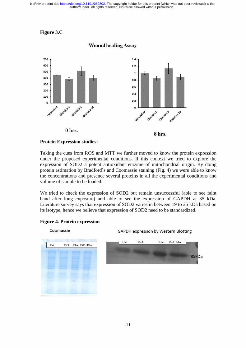
Figure 3.C
Protein Expression studies:
Taking the cues from ROS and MTT we further moved to know the protein expression
under the proposed experimental conditions. If this context we tried to explore the
expression of SOD2 a potent antioxidant enzyme of mitochondrial origin. By doing
protein estimation by Bradford’s and Coomassie staining (Fig. 4) we were able to know
the concentrations and presence several proteins in all the experimental conditions and
volume of sample to be loaded.
We tried to check the expression of SOD2 but remain unsuccessful (able to see faint
band after long exposure) and able to see the expression of GAPDH at 35 kDa.
Literature survey says that expression of SOD2 varies in between 19 to 25 kDa based on
its isotype, hence we believe that expression of SOD2 need to be standardized.
Figure 4. Protein expression
author/funder. All rights reserved. No reuse allowed without permission. The copyright holder for this preprint (which was not peer-reviewed) is the. https://doi.org/10.1101/062802doi: bioRxiv preprint
12
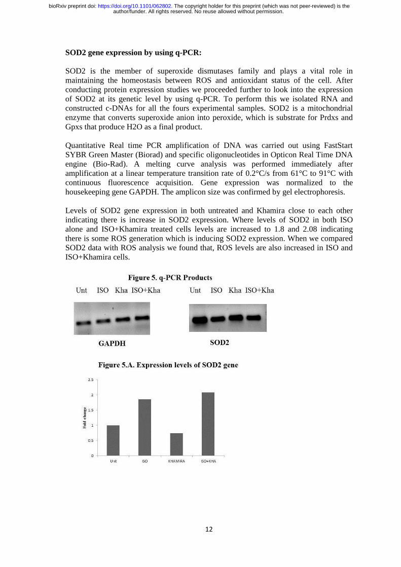
SOD2 gene expression by using q-PCR:
SOD2 is the member of superoxide dismutases family and plays a vital role in
maintaining the homeostasis between ROS and antioxidant status of the cell. After
conducting protein expression studies we proceeded further to look into the expression
of SOD2 at its genetic level by using q-PCR. To perform this we isolated RNA and
constructed c-DNAs for all the fours experimental samples. SOD2 is a mitochondrial
enzyme that converts superoxide anion into peroxide, which is substrate for Prdxs and
Gpxs that produce H2O as a final product.
Quantitative Real time PCR amplification of DNA was carried out using FastStart
SYBR Green Master (Biorad) and specific oligonucleotides in Opticon Real Time DNA
engine (Bio-Rad). A melting curve analysis was performed immediately after
amplification at a linear temperature transition rate of 0.2°C/s from 61°C to 91°C with
continuous fluorescence acquisition. Gene expression was normalized to the
housekeeping gene GAPDH. The amplicon size was confirmed by gel electrophoresis.
Levels of SOD2 gene expression in both untreated and Khamira close to each other
indicating there is increase in SOD2 expression. Where levels of SOD2 in both ISO
alone and ISO+Khamira treated cells levels are increased to 1.8 and 2.08 indicating
there is some ROS generation which is inducing SOD2 expression. When we compared
SOD2 data with ROS analysis we found that, ROS levels are also increased in ISO and
ISO+Khamira cells.
author/funder. All rights reserved. No reuse allowed without permission. The copyright holder for this preprint (which was not peer-reviewed) is the. https://doi.org/10.1101/062802doi: bioRxiv preprint

13
Conclusion:
This pilot study shows that Khamira is nontoxic to SVEC cells up to a concentration of
20 mg/mL after this concentration cells are unable to survive. We also observed that the
150 µM concentration of Isoproterenol induces mild lethality and anything above this
concentration leading to increased toxicity and induces death. During this study we
found rapid filling of cells in wounded region of Khamira with 5mg/mL concentration
than that of 10 mg/mL. In this entire study we adopted a method of Khamira co-
treatment but not pre-treatment. When we look into the ROS values we assume that
Khamira is able to maintain the sufficient endogenous antioxidant status that is useful to
rescue cells from ROS or free radicals. But when we observe the levels of SOD2 it is
evident that Khamira might be acting either on signalling events prior to ROS or after
ROS generation. The reason for this statement is SOD2 levels increase when there is
elevated ROS generation. However, this is a pilot study to look into the possible
protective role of Khamira in experimentally induced toxic conditions. More controlled
in vivo and in vitro studies are required to prove the exact mechanism lying behind
increased cell viability and wound healing assay.
References:
Uddin MK. Bayaz-e-kabir.vol.2 Delhi: Dafter-ul-Masehi; 1967. 56-67.
Sayeed Ahmed, Shabana rehman, Aftab Ahmed, Khalid M Siddiqui and Tamanna
Jahangir. Khamira’s a natural cardio tonic: an overview. J. Pharm. Bioall. 2010; 2:93-
99.
Thannickal and Fanburg. Reactive oxygen species in cell signaling. Am J Physiol Lung
Cell Mol Physiol. 2000 Dec; 279(6):L1005-28.
author/funder. All rights reserved. No reuse allowed without permission. The copyright holder for this preprint (which was not peer-reviewed) is the. https://doi.org/10.1101/062802doi: bioRxiv preprint