postpartum cervical repair in mice: a morphological
TRANSCRIPT

Archived version from NCDOCKS Institutional Repository http://libres.uncg.edu/ir/asu/
Postpartum Cervical Repair In Mice:A Morphological Characterization And Potential Role
For Angiogenic Factors
By: Chishimba Mowa, Takako Ohashi, & Robert Stanley
AbstractThe cervix undergoes marked mechanical trauma during delivery of the baby at birth. As such, a timely and complete tissue repair postpartum is necessary to prevent obstetrical complications, such as cervicitis, ectropion, hemorrhage, repeated miscarriages or abortions and possibly pre-
term labor and malignancies. However, our knowledge of normal cervical repair is currently incomplete and factors that influence repair are unclear. Here, we characterize the
morphological and angiogenic profile of postpartum repair in mice cervix during the first 48 h of postpartum. The key findings presented here are: (1) cervical epithelial folds and size are
diminished during the first 48 h of postpartum repair, (2) hypoxic inducible factor 1a, vascular endothelial growth factor (VEGF), and VEGF receptor 1 expression are pronounced early in
postpartum cervical repair, and (3) VEGF receptor 2 gene and protein expressions are variable. We conclude that postpartum cervical repair involves gross and microscopic changes and is linked to expression of angiogenic factors. Future studies will assess the suitability of these factors, identified in the present study, as potential markers for determining the phase of
postpartum cervical repair in obstetrical complications, such as cervical lacerations.
Chishimba, Mowa & Takako, Ohashi & Stanley, Robert (2015) "Postpartum Cervical Repair In Mice: A Morphological Characterization And Potential Role For Angiogenic Factors" Cell and Tissue Research vol. 362 issue. 1 pp. 253-263 [DOI: 10.1007/s00441-015-2184-x] Version of Record Available @ www.springer.com
Introduction
The cervix undergoes marked changes during pregnancy, aswell as significant trauma during normal vaginal delivery ofthe baby at birth (parturition) and immediately postpartum(after birth) (Timmons et al. 2010). As such, a timely andcomplete postpartum tissue repair is necessary to prevent ob-stetrical complications, such as cervicitis, ectropion, hemor-rhage, repeated miscarriages or abortions and, possibly, pre-term labor and malignancies (Fahmy et al. 1991). For in-stance, during pregnancy, the cervix has to contain and with-stand an ever increasing gravitational force exerted by therapidly growing fetus in order to ensure that it (the fetus) isheld in utero (Timmons et al. 2010), whereas at parturition,under the intense force of the contracting uterus, the ripenedcervical tissue is forced to expand and dilate to ultimatelyallow passage of the fetus through the birth canal(Mahendroo 2012). Immediately following these dramaticmechanical tissue Bassaults^, the cervix undergoes an exten-sive postpartum reconstruction and healing phase that restoresit (the cervix) to its non-pliable non-pregnant state (Timmonsand Mahendroo 2007). Collectively, these changes (gestation-al and postpartum) are termed cervical remodeling (CR),which is divided into four distinct yet overlapping phases,namely softening, ripening, dilation and postpartum repair(Read et al. 2007; Word et al. 2007). To date, most of thestudies have focused on the first three phases of CR, almostat the exclusion of postpartum repair.
Postpartum repair consists of a set of complex biologicalprocesses that ultimately restores the cervix to its original non-pregnant state, and thus ensures normal cervical function forsubsequent pregnancies (Timmons et al. 2010; Mahendroo2012). This final phase of CR is generally characterized asan inflammatory and wound-healing response (Timmons andMahendroo 2007; Bauer et al. 2009), as demonstrated by

studies that utilized gene microarrays. These studies haveshown that a variety of factors, including pro-inflammatoryfactors, metalloproteases, proteins involved in extracellularmatrix (ECM) synthesis, and genes governing epithelial dif-ferentiation pathways are all up-regulated postpartum(Timmons and Mahendroo 2007; Gonzalez et al. 2009).Immune cells, including neutrophils, eosinophils, and bothM1 and M2 macrophages, have all been shown to increasepostpartum compared to earlier phases of remodeling(Timmons et al. 2009, 2010; Mahendroo 2012). This inflam-matory response could serve to promote repair of the cervixafter parturition and/or serve a protective role of the birth canalagainst environmental hazards, such as infection (Gonzalezet al. 2009). Because inflammation and wound healing areintricately linked to vascular events, factors that regulate thisprocess, notably vascular endothelial growth factor (VEGF)(Nguyen et al. 2012), also likely influence postpartum cervicalrepair. Of note, VEGF is expressed in normal, pregnant andcancerous cervices in rodents and humans.
The importance of the role of VEGF in wound healing hasbeen well documented and characterized (Galiano et al. 2004;Mowa et al. 2008a; Alberts et al. 2008; Bao et al. 2009).VEGF controls wound healing by inducing various vascularprocesses, including angiogenesis (capillary growth), vaso-permeability, and recruitment of immune cells, as well as ep-ithelization and collagen deposition (Bao et al. 2009), the hall-mark of the healing response (Mahendroo 2012). For instance,induction of capillary growth during wound repair meets thegrowing demands for oxygen and nutrients, as well as remov-al of waste products (Alberts et al. 2008). Indeed, inhibition ofangiogenesis impairs wound healing, demonstrating the vitalrole of VEGF in this (healing) process (Galiano et al. 2004;Bao et al. 2009). Factors that induce VEGF expression duringwound healing include growth factors (transforming growthfactor, hepatocyte growth factor, keratinocyte growth factor,etc.), pro-inflammatory cytokines, such as interleukin-1 (IL-1)and tumor necrosis factor alpha (TNFα), and, notably, hypox-ia, i.e., low oxygen (Eming and Krieg 2006). Cells in a hyp-oxic environment will accumulate the intracellular transcrip-tion factor, hypoxia inducible factor-1 alpha (HIF-1α), whichis the most potent inducer of VEGF transcription (Majmundaret al. 2010). Following injury, VEGF is first secreted by hyp-oxic cells and will establish a gradient of VEGF, mirroring thegradient of hypoxia (Alberts et al. 2008). Activated platelets,and immune cells, i.e. macrophages and neutrophils, expressVEGF receptor 1 (VEGFR1) and respond chemotactically toVEGF, and will migrate towards the VEGF source at the siteof injury and/or hypoxia (Bao et al. 2009; Eming and Krieg2006). Macrophages and neutrophils will in turn stimulateangiogenesis by further secreting VEGF and TNFα that willalso stimulate VEGF production in keratinocytes and fibro-blasts (Bao et al. 2009). Although the role of VEGF in thecervical events during pregnancy has been studied (Nguyen
et al. 2012; Eming and Krieg 2006; Mowa et al. 2004, 2008b;Donnelly et al. 2013), to date, no study has examined its role,if any, in postpartum cervical repair.
The presence and profile of VEGF and its key tyrosinekinase receptors have been characterized in the uterine cervixof rodents (mice and rats), and human and VEGF-related genesin rats have been delineated using DNAmicroarray analysis inpregnant and non-pregnant conditions (Nguyen et al. 2012;Mowa et al. 2004, 2008a, b; Donnelly et al. 2013; and unpub-lished data). The effects of VEGF are largely mediated byVEGFR-1 (or fms-related tyrosine kinase-1, flt-1) and VEGFR-2, which have different signaling properties and are largelyexpressed by endothelial cells (Mowa et al. 2004).
Since postpartum repair is considered a pro-inflammatorywound healing response (Mahendroo 2012; Timmons andMahendroo 2007; Bao et al. 2009), and VEGF is known toinduce inflammation in the cervix (Nguyen et al. 2012) andplays roles in: (1) cervical remodeling (CR) during pregnancyin rodents (Mowa et al. 2008b; Donnelly et al. 2013), and (2)general wound healing (Bao et al. 2009; Eming and Krieg2006), here we speculate that VEGF plays a role in the post-partum repair phase of CR. In this study, we sought to deter-mine: (1) the gross morphological changes of the postpartumcervix, and (2) the expression patterns of HIF-1α, VEGF, andthe VEGF receptors (VEGFR-1 and VEGFR-2), during thefirst 2 days in mice.
Materials and methods
Animals and postpartum time intervals
Pregnant mice (C57BL6/129SvEv; Charles River, n=3~5 pertechnique of each time-point, except for H&E staining andconfocal immunofluorescence, where n=1) were used in thepresent study during the time points specified here: The timeof the first pup was noted as time 0 h and the mothers weresacrificed at an 8-h interval, after parturition, beginning with0 h, i.e., 0, 8, 16, 24, 32, 40 and 48 h. The average intervalbetween the birth of the first and last pup was within 1 h, withan average litter size between 8–12 pups. All animals wereeuthanized by a lethal injection of sodium pentobarbital(150 mg/kg bw, i.p.) and transcardially perfused with 0.9 %saline solution. Cervices were carefully harvested under a ste-reomicroscope, to avoid contamination with vaginal and uter-ine tissues, and the tissues were then processed and analyzedaccordingly, including using scanning electron and light mi-croscopy imaging, gene expression analysis using real-timePCR, and protein expression analysis using Western blot andconfocal immunofluorescence. All animals were housed un-der the following conditions: constant room temperature (RT);a 12:12-h light:dark cycle, and free access to water and food.All experiments were performed in accordance with the

Institutional Animal Care and Use Committee (IACUC) ofAppalachian State University and the NIH guidelines (NIHpublication number 86-23), with efforts made to minimizeboth number of animals used and animal suffering.
Scanning electron microscopy (SEM)
SEM was employed to observe the surface of cervical epithe-lia and any changes occurring during the postpartum period.
Harvested cervices were immediately immersed in a 2.5 %glutaraldehyde, 0.1 M phosphate buffer solution (PBS) andthe tissues were then dehydrated in a graded series of ethanoland dried using a critical point drying apparatus (PolaronInstruments, Laughton, East Sussex, UK). The dry samplesweremounted on aluminum stubs with carbon adhesive paper,sputter-coated in gold and viewed with the SEM (FEI,Hillsboro, OR, USA). The cervical epithelium was evaluatedfor various features, including, but not limited to, involutions,tissue size, overall appearance of cells, presence of macro-phages and paracellular spaces.
Confocal immunofluorescence
Confocal immunofluorescence was employed to complementWestern blot data of the proteins of interest (VEGF, VEGFreceptors, HIF-1α), as well as determine their cellular locali-zation in the cervix.
Harvested cervices were immersed in a 10 % formalin(pH 7.2) aqueous solution. The tissues were fixed in formalinat 4 °C for 3–4 days and then transferred into saturated sucrosedissolved in 0.1 M PBS, for at least 2 days prior to sectioning(12 μm) with the Cryostat (Leica, Buffalo Grove, IL, USA) at−30 °C. After sectioning, the slides were either stained imme-diately or stored at –20 °C until staining. For staining, thesections were initially incubated with 10% normal goat serumin 0.1 M PBS for 20 min at RT in order to block non-specificprotein binding. Next, the sections were washed 3 times in0.1 M PBS and then incubated overnight at 4 °C with dilutedprimary antibodies of interest at 0.5 μg/mL (Santa CruzBiotech, Santa Cruz, CA, USA). The next day, the sectionswere washed 3 times with 0.1 M PBS Buffer followed imme-diately by incubation in diluted fluoro-tagged secondary anti-body at 0.5 μg/mL (Santa Cruz Biotech) for 1 h at RT.Thereafter, the sections were washed 3 times with 0.1 MPBS, counterstained with 5 μMSytoxGreen™, mounted withaqueous Ultracruz Mounting Medium (Santa Cruz Biotech),and then examined with a confocal microscope (Carl Zeiss,Peabody, MA, USA).
Gene expression studies (real time PCR)
To perform gene expression studies, total RNA was first ex-tracted, reverse-transcribed into cDNA and then amplified.
Harvested cervices were snap-frozen and then either totalRNAwas extracted immediately or the tissues were stored at−80 °C until processing. Tissues were first homogenized inQiazol Lysis reagents (Qiagen, Valencia, CA, USA) and thentotal RNAwas extracted using RNeasyMini Kit (Qiagen) andits quality and quantity determined using the Nanodrop appa-ratus (Thermo Scientific, Waltham, MA, USA). If of goodquality and quantity, the total RNA (1000 ng) was then reversetranscribed using a cocktail containing the following: MgCl2,reverse transcriptase, RT buffer, dNTP, RNase inhibitor,RNase-free water and random hexamers, in order to generatean equivalent amount of total cDNA using a thermocycler(Eppendorf, Hamburg, Germany) at the following settings:25 °C for 10 min, 42 °C for 2 h, 95 °C for 5 min, and 4 °Cfor storage, according to protocol. The cDNAwas then used toamplify specific gene sequences using real-time PCR ampli-fication (e.g., VEGF, VEGFR1, VEGFR2, HIF-1α, andGus-β as a normalizer) in order to determine the relative ex-pression of these genes in all the time points during the 48 hafter the onset of parturition. We used commercially pre-designed and optimized real time PCR probes (AppliedBiosystems, Carlsbad, CA, USA). The PCR reactions wereset up in 96-well plate in a volume of 25 μl per well, withthe following components: 1 μl of cDNA, 12.5 μl of 2×Taqman Universal PCR Mastermix, 1.25 μl of 20× Assays-on-demand Gene Mix (e.g. VEGF), and 10.25 μl of RNase-free water. DNA amplification was performed using the ABI7300 HT Real-Time PCR machine (Applied Biosystems.Carlsbad, CA, USA) with the GeneAmp 7300 HT sequencedetection system software (Perkin-Elmer, Waltham, MA,USA), with the settings as follows: 95 °C for 10 min, and then40 cycles of 95 °C for 15 s, and 60 °C for 60 s.
Western blot analysis
Western blot analysis was used to confirm relative amounts ofthe proteins of interest, at the tissue level.
Harvested cervices were snap-frozen and stored at −80 °Cuntil processing. Total protein was extracted using the proteininhibitor cocktail and Cell Lysis buffer (Sigma Aldrich, St.Louis, MO, USA), and quantified with a bicinchoninic acid(BCA) assay (Thermo Scientific). Next, 10 μg of protein sam-ples, along with a set of standard protein ladders (Bio-Rad,Hercules, CA, USA), were loaded on a 4-12 % Bis-Tris Gel(Invitrogen, Waltham, MA, USA). The gel was electropho-resed at 75 V until the dye was close to the bottom of thegel. Thereafter, the protein was transferred to a PVDF mem-brane using the iBlot apparatus (Invitrogen, Waltham, MA,USA), and the membrane was incubated overnight withBlotto (5 % dehydrated milk in Tris Buffered Saline withTween) solution at 4 °C. The following day, the membranewas incubated with the diluted primary antibody dissolved inBlotto (Santa Cruz Biotech), and left to incubate overnight on

a shaker at RT. Thereafter, the membrane was washed threetimes in 1× TBST for 20 min, after which the membrane wasincubated with the diluted secondary antibody, i.e., donkeyanti-rabbit IgG HRP conjugated and StrepTactin HRP anti-bodies (Santa Cruz Biotech) for 1 h at RT on a shaker. Themembrane was then washed three times with 1× TBST andone wash with 1× TBS. Horseradish peroxidase solution (Bio-Rad) was added to the membrane for 5 min and the excesssolution was dripped off. The membranes were then exposedto x-ray film (GE, Fairfield, CT, USA) in a dark room for anoptimal period (~30 s–1 min), and the film was developed andanalyzed for presence of the target proteins (VEGF, VEGFR1,VEGFR-2, HIF-1α, and β actin as a normalizer). The densi-tometry was quantified using ImageJ software (NIH,Bethesda, MD, USA).
Statistical analysis
Data for western blot and real-time PCR analyses were ana-lyzed using Student's t test and ANOVA, followed byScheffe's F test for multiple comparisons. P values of<0.05were considered to be statistically significant.
Results
The murine cervix undergoes pronounced morphologicalchanges during the first 48 h postpartum
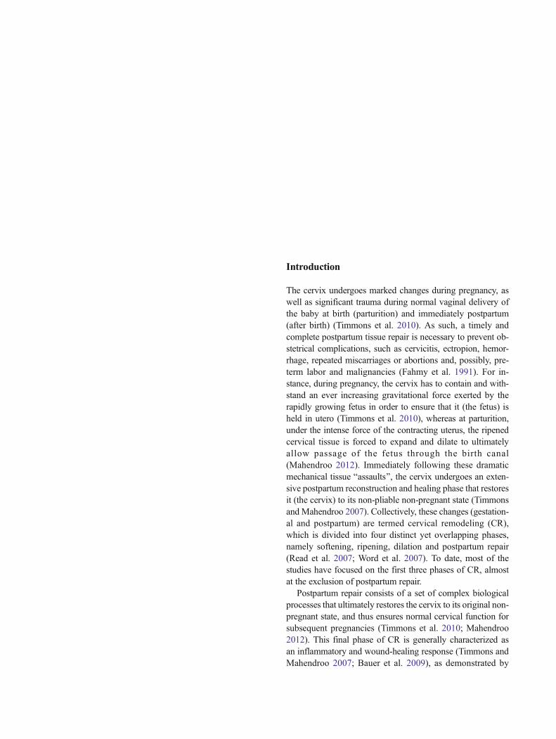
SEM analysis of cervices obtained from mice at 0, 24, and48 h postpartum displayed distinct morphological features atthe time points of interest (Fig. 1). At 0 h, the cervical epithe-lium displayed folds (Fig. 1a, b) and micro-folds (Fig. 1c, d),as well as less prominent epithelial cell borders, compared toboth 24- and 48-h postpartum. A layer of unidentified extra-cellular matrix or cell debris appeared at 24 h postpartum(Fig. 1g, h), but disappeared by 48 h postpartum. At both24- and 48-h postpartum, the presence of Bpits^ or gaps inthe cervical epithelium was observed and the cell–cell borderswere more pronounced than at 0 h (Fig. 1f, g, j, k). In order tofurther characterize the morphological changes of the postpar-tum cervix, we performed H&E staining, which revealedfolded cervical epithelium at 0 h PP (Fig. 2a). At 24 h PP,the layer of unidentified cellular debris reported earlier underSEM data was observed (Fig. 2d), but not at 48 h PP (Fig. 2g),consistent with the SEM findings.
Fig. 1 Scanning electron microscopy images of the chronological profileof postpartum mouse cervical tissue. Low magnification (×50) imagesreveal an obvious decrease in size from 0 to 48 h postpartum (PP) (a, e,i). Cervical epithelial folds, as well as diminished cell–cell borders, were
prominent at 0 h PP at higher magnifications (×500–×5,000). Anunidentified Bcovering^ was observed at 24 h PP, but was absent at 0 hand had disappeared by 48 h PP. n=3
Gene expression in the murine cervix postpartum
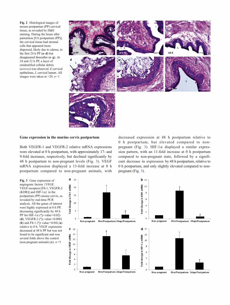
Both VEGFR-1 and VEGFR-2 relative mRNA expressionswere elevated at 0 h postpartum, with approximately 17- and9-fold increases, respectively, but declined significantly by48 h postpartum to non-pregnant levels (Fig. 3). VEGFmRNA expression displayed a 13-fold increase at 0 hpostpartum compared to non-pregnant animals, with
decreased expression at 48 h postpartum relative to0 h postpartum, but elevated compared to non-pregnant (Fig. 3). HIF-1α displayed a similar expres-sion pattern, with an 11-fold increase at 0 h postpartumcompared to non-pregnant state, followed by a signifi-cant decrease in expression by 48 h postpartum, relative to0 h postpartum, and only slightly elevated compared to non-pregnant (Fig. 3).
Fig. 2 Histological images ofmouse postpartum (PP) cervicaltissue, as revealed by H&Estaining. During the hours afterparturition [0 h postpartum (PP)],the cervical tissue had stromalcells that appeared moredispersed, likely due to edema, inthe first 24 h PP (a–d) butdisappeared thereafter (e–g). At24 and 32 h PP, a layer ofunidentified cellular debris(arrows) was observed. E cervicalepithelium, L cervical lumen. Allimages were taken at ×20. n=1
Fig. 3 Gene expression ofangiogenic factors {VEGF,VEGF receptors [Flt-1, VEGFR-2(KDR)] and HIF-1α} in thepostpartum (PP) mouse cervix, asrevealed by real-time PCRanalysis. All the genes of interestwere highly expressed at 0 h PP,decreasing significantly by 48 hPP for HIF-1α (*p value=0.02)(d), VEGFR-2 (*p value=0.008)(b) and Flt-1 (*p value=0.04) (a)relative to 0 h. VEGF expressiondecreased at 48 h PP but was notfound to be significant and wasseveral folds above the control(non-pregnant animals) (c). n=5

Protein expression in the mouse cervix postpartum
VEGF protein expression was found to be at the highest levelat 0 h postpartum, with levels decreasing approximately 2 foldby 16 h postpartum, followed by a slight decrease for theremaining time intervals (Fig. 4a). VEGFR-1 was found tobe at its highest expression at 8 h postpartum, with levelsdecreasing in the remainder of time intervals, save for a slightincrease at 24 h postpartum, relative to 16 h postpartum(Fig. 4d). It should be noted that the fold change from thehighest expression (8 h postpartum) to the lowest expression(48 h) was less than half. In contrast, the other receptor VEGFR-2, was found to increase steadily during the postpartumtime intervals; with the lowest levels at 0 h, steadily increasingthereafter and leveling off at 40-48 h postpartum (Fig. 4c).HIF-1α protein expression pattern was similar to VEGF, i.e.,it was elevated at 0 h postpartum, and was at its lowest at 48 hpostpartum (Fig. 4b). However, there was an increase in ex-pression from 16 to 24 h postpartum, remaining elevated at32 h, and falling off again by 40 h postpartum (Fig. 4b).
Protein expression in the mouse cervix postpartum,as revealed by confocal immunofluorescence
The highest levels of VEGF were observed at 0 h postpartum(Fig. 5a), with the lowest levels occurring at 48 h postpartum(Fig. 5g). The most pronounced expressions of VEGF werelocalized on the apical side of the epithelium, regardless of the
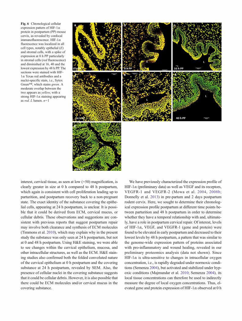
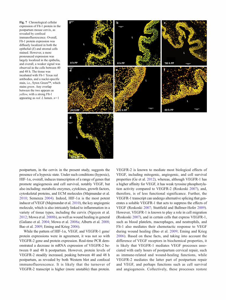
time interval or point. It is noteworthy that the expression ofVEGF was readily detected in the layer of cell debris observedat 24 h postpartum (Fig. 5d) and 32 h postpartum (Fig. 5e).HIF-1α expression mirrored that of VEGF, i.e., the highestlevels were at 0 h postpartum (Fig. 5a), lowest at 48 h(Fig. 5g), with expression preferentially localized at the apicalside of the cervical epithelium at each time interval. Also, HIF-1α expression was similar to VEGF in that it was also readilyexpressed in the layer of cell debris at 24 h postpartum(Fig. 5d) and 32 h postpartum (Fig. 5e). The VEGF receptorsdemonstrate different patterns of expression postpartum.VEGFR-1 expression mirrored that of VEGF and HIF-1α,with expression clearly at its highest at 0 h postpartum(Fig. 6a), lowest at 48 h postpartum (Fig. 6g), and localizedto the apical side of the epithelium and in the cell debris layer atboth 24 h postpartum (Fig. 6d) and 32 h postpartum (Fig. 6e).Overall, intensity of VEGFR-2 immunostaining increased overtime, particularly from 16 h postpartum in epithelia and stro-mal cells, with the weakest signal appearing between 0-8 h andslightly diminishing at 48 h (Fig. 7) (Fig. 8).
Discussion
Of the four phases of cervical remodeling, postpartum repair isthe least studied and, to date, there has been no comprehensivestudy that has focused exclusively on this fourth and finalphase of cervical remodeling. A comprehensive knowledge
Fig. 4 Chronological expressionpatterns of VEGF, HIF-1α, Flt-1,and VEGFR-2 (KDR) proteins inpostpartum mouse cervix, asdetermined by Western blotanalysis. a VEGF proteinexpression decreased over timebetween 0 and 48 h postpartum(PP) (*p value=4.56 × 10⁻⁸); bHIF-1α protein expressiondecreased between 0 and 16 h butsurged to 8 h levels between 24and 32 h, and decreased steadilythereafter, reaching its lowestlevels by 48 h PP (*p value=3.08 × 10⁻⁸); c VEGFR-2 proteinexpression pattern was opposed tothat of VEGF, i.e., it steadilyincreased over time, plateauing by40–48 h PP (*p value=0.0005);and d Flt-1 protein expressionpattern overall showed a similarpattern to VEGF, i.e., decreasedsteadily between 0 and 48 h PP(*p value=0.008). β actin wasused as the normalizer. n=5

of this phase of CR is potentially vital as it may improve ourunderstanding of obstetrical complications that may be asso-ciated with this period, such as cervicitis, ectropion, hemor-rhage, repeated miscarriages or abortions and, possibly, pre-term labor and malignancies (Fahmy et al. 1991). The presentstudy is the first comprehensive postpartum study and uses avariety of techniques to examine the morphological and mo-lecular changes in the mouse cervix during the first 48 h ofpostpartum. The key findings of the present data are: (1) epi-thelial folds and cervical size progressively diminish during
postpartum repair, (2) HIF-1α, VEGF, and VEGFR-1 expres-sion are pronounced early in postpartum cervical repair, and(3) VEGFR-2 gene and protein expressions are variable.
The current SEM data show the presence of cervical epi-thelial folds at 0 h postpartum. These findings are consistentwith our most recent work that utilized SEM andbromodeoxyuridine (BrdU) staining showing cervical epithe-lial proliferation leading up to parturition, as well as in non-pregnant ovariectomized mice that were treated with exoge-nous VEGF (Mowa et al. 2008b; Donnelly et al. 2013). Of
Fig. 5 Chronological cellularexpression pattern of VEGF inpostpartum mouse cervix, asrevealed by confocalimmunofluorescence. VEGFimmune-expression was initiallyconcentrated in cervical epitheliacells (b, b’, 0 h), spreading later tothe other cell types (stromal) (c,8 h) and diminishing in intensityby 24 h and thereafter (d–f). Thesections were stained with VEGFTexas red antibodies and a nuclei-specific stain, i.e., Sytox Green™,which stains green. The overlapbetween the two appears asyellow. n=1
interest, cervical tissue, as seen at low (×50) magnification, isclearly greater in size at 0 h compared to 48 h postpartum,which again is consistent with cell proliferation leading up toparturition, and postpartum recovery back to a non-pregnantstate. The exact identity of the substance covering the epithe-lial cells, appearing at 24 h postpartum, is unclear. It is possi-ble that it could be derived from ECM, cervical mucus, orcellular debris. These observations and suggestions are con-sistent with previous reports that suggest postpartum repairmay involve both clearance and synthesis of ECM molecules(Timmons et al. 2010), which may explain why in the presentstudy the substance was only seen at 24 h postpartum, but notat 0 and 48 h postpartum. Using H&E staining, we were ableto see changes within the cervical epithelium, mucosa, andother intracellular structures, as well as the ECM. H&E stain-ing studies also confirmed both the folded convoluted natureof the cervical epithelium at 0 h postpartum and the coveringsubstance at 24 h postpartum, revealed by SEM. Also, thepresence of cellular nuclei in the covering substance suggeststhat it could be cellular debris. However, it is also possible thatthere could be ECM molecules and/or cervical mucus in thecovering substance.
We have previously characterized the expression profile ofHIF-1α (preliminary data) as well as VEGF and its receptors,VEGFR-1 and VEGFR-2 (Mowa et al. 2004, 2008b;Donnelly et al. 2013) in pre-partum and 2 days postpartumrodent cervix. Here, we sought to determine their chronolog-ical expression profile postpartum at different time points be-tween parturition and 48 h postpartum in order to determinewhether they have a temporal relationship with and, ultimate-ly, have a role in postpartum cervical repair. Of interest, levelsof HIF-1α, VEGF, and VEGFR-1 (gene and protein) werefound to be elevated in early postpartum and decreased to theirlowest levels by 48 h postpartum, a pattern that was similar tothe genome-wide expression pattern of proteins associatedwith pro-inflammatory and wound healing, revealed in ourpreliminary proteomics analysis (data not shown). SinceHIF-1α is ultra-sensitive to changes in intracellular oxygenconcentration, i.e., is rapidly degraded under normoxic condi-tions (Semenza 2004), but activated and stabilized under hyp-oxic conditions (Majmundar et al. 2010; Semenza 2004), itslocal tissue concentrations can therefore be used to indirectlymeasure the degree of local oxygen concentrations. Thus, el-evated gene and protein expression of HIF-1α observed at 0 h
Fig. 6 Chronological cellularexpression pattern of HIF-1αprotein in postpartum (PP) mousecervix, as revealed by confocalimmunofluorescence. HIF-1αfluorescence was localized in allcell types, notably epithelial (E)and stromal cells, with a spike ofexpression at 8 h PP particularlyin stromal cells (red fluorescence)and diminished at 16, 40 and thelowest expression by 48 h PP. Thesections were stained with HIF-1α Texas red antibodies and anuclei-specific stain, i.e., SytoxGreen™, which stains green. Amoderate overlap between thetwo appears as yellow, with astrong HIF-1α staining appearingas red. L lumen. n=1
postpartum, in the cervix in the present study, suggests thepresence of a hypoxic state. Under such conditions (hypoxic),HIF-1α, overall, induces transcription of a range of genes thatpromote angiogenesis and cell survival, notably VEGF, butalso including: metabolic enzymes, cytokines, growth factors,cytoskeletal proteins, and ECM molecules (Majmundar et al.2010; Semenza 2004). Indeed, HIF-1α is the most potentinducer of VEGF (Majmundar et al. 2010), the key angiogenicmolecule, which is also intricately linked to inflammation in avariety of tissue types, including the cervix (Nguyen et al.2012;Mowa et al. 2008b), as well as wound healing in general(Galiano et al. 2004; Mowa et al. 2008a; Alberts et al. 2008;Bao et al. 2009; Eming and Krieg 2006).
While the pattern of HIF-1α, VEGF, and VEGFR-1 gene/protein expressions were in agreement, it was not so withVEGFR-2 gene and protein expression. Real-time PCR dem-onstrated a decrease in mRNA expression of VEGFR-2 be-tween 0 and 48 h postpartum. However, protein levels ofVEGFR-2 steadily increased, peaking between 40 and 48 hpostpartum, as revealed by both Western blot and confocalimmunofluorescence. It is likely that the turnover ofVEGFR-2 transcript is higher (more unstable) than protein.
VEGFR-2 is known to mediate most biological effects ofVEGF, including mitogenic, angiogenic, and cell survivalproperties (Ge et al. 2012), whereas, although VEGFR-1 hasa higher affinity for VEGF, it has weak tyrosine phosphoryla-tion activity compared to VEGFR-2 (Roskoski 2007), and,therefore, is of less functional significance. Further, theVEGFR-1 transcript can undergo alternative splicing that gen-erates a soluble VEGFR-1 that acts to suppress the effects ofVEGF (Roskoski 2007; Stuttfield and Ballmer-Hofer 2009).However, VEGFR-1 is known to play a role in cell migration(Roskoski 2007), and in certain cells that express VEGFR-1,such as blood platelets, macrophages, and neutrophils, andFlt-1 also mediates their chemotactic response to VEGFduring wound healing (Bao et al. 2009; Eming and Krieg2006). Based on these facts, and taking into account thedifference of VEGF receptors in biochemical properties, itis likely that VEGFR-1 mediates VEGF processes asso-ciated with early hours of postpartum cervical repair, suchas immune-related and wound-healing functions, whileVEGFR-2 mediates the latter part of postpartum repairand VEGF, and perhaps functions such as cell survivaland angiogenesis. Collectively, these processes restore
Fig. 7 Chronological cellularexpression of Flt-1 protein in thepostpartum mouse cervix, asrevealed by confocalimmunofluorescence. Overall,Flt-1 protein expression wasdiffusely localized in both theepithelial (E) and stromal cellsstained. However, a morepronounced expression waslargely localized in the epithelia,and overall, a weaker signal wasobserved in the cells between 40and 48 h. The tissue wasincubated with Flt-1 Texas redantibodies, and a nuclei-specificstain, i.e., Sytox Green™, whichstains green. Any overlapbetween the two appears asyellow, with a strong Flt-1appearing as red. L lumen. n=1

the cervix back to its non-pregnant state. Future functionalstudies are needed to test these speculations.
Hypoxia is known to be involved in various physiologicaland/or pathological responses, but may also play a role inparturition. For instance, in the high altitude mountain rangesof the Andes, expectant mothers are believed to induce child-birth at term by climbing to high elevations with a loweroxygen environment or hypoxia. Similarly, it is likely thathypoxia could account for the increased risk in preterm laborassociated with smoking. Interestingly, our recent preliminaryproteomics data revealed two hypoxia-induced proteins thatwere highly expressed at 0 h postpartum. Transgelin is anactin-crosslinking protein known to be expressed in fibro-blasts and smooth muscle (Kim et al. 2009, 2012), and isstrongly expressed in both the uterus and cervix. Recentstudies by Kim et al. (2012) demonstrated that transgelin isup-regulated by hypoxia, independent of HIF-1α, and alsoactivates the insulin-like growth factor receptor 1 (IGFR1)signaling pathway under hypoxic stress. The IGFR1 pathwayhas recently been shown to promote cell survival under hyp-oxic stress (Kim et al. 2012; Gariboldi et al. 2010) by up-regulating HIF-1α gene transcription (Gariboldi et al. 2010)
and stabilizing HIF-1α protein (Piecewicz et al. 2012). Thisup-regulation and stabilization of HIF-1α by the IGFR1 axishas been shown to induce VEGF expression during embryon-ic vasculogenesis (Piecewicz et al. 2012), and angiogenesis inmalignant tumor cells under hypoxic stress (Gariboldi et al.2010). Whether transgelin acts upstream of HIF-1α andVEGF in postpartum cervical repair is for now speculative.Like transgelin, vimentin has also been associated with hyp-oxia. For instance, it has been shown to redistribute among theendothelial cell into stable structures in response to hypoxia(Lui et al. 2010), is involved in general wound healing (Rogelet al. 2011), and is known to play a role in migrating cells,including fibroblasts, macrophages, endothelial cells, and in-vasive cancer cells (Lui et al. 2010; Rogel et al. 2011; Heflandet al. 2011; Glaser-Gabay et al. 2011). Further studies areneeded to examine the roles of these hypoxia-induced proteinsin the postpartum phase of cervical remodeling. Postpartumrepair has been previously characterized as a proinflammatoryand wound-healing process (Timmons and Mahendroo 2007;Bauer et al. 2009; Gonzalez et al. 2009). Our current data ofpostpartum cervical tissue are consistent with the earlierstudies.
Fig. 8 Chronological cellularexpression pattern of VEGFR-2protein in the postpartum mousecervix, as revealed by confocalimmunofluorescence. Overall, thesignal of VEGFR-2 stainingincreased over time, particularlyfrom 16 h postpartum in epithelia(E) and stromal cells, with theweakest signal appearing between0–8 h and slightly diminishing at48 h. The tissue was incubatedwith VEGFR-2 Texas redantibodies, and a nuclei-specificstain, i.e., Sytox Green™, whichstains green. Any overlapbetween the two appears asyellow, with a strong VEGFR-2signal appearing as red. L lumen.n=1

In summary, our data provide the first expression profile ofVEGF, its receptors (Flt-1 and VEGFR-2), and HIF-1α andlink their pattern of expression to hypoxia-related proteins andmorphological changes. The limitation of the current studyincludes small sample size and lack of a detailed immuno-chemical evaluation of angiogenic factors, particularlymarkers of angiogenesis, such as endothelial markers.
Acknowledgment Funding for the present study was provided by theCollege of Arts and Sciences, Appalachian State University, Boone,NC, USA.
References
Alberts B, Johnson A, Lewis J, Raff M, Roberts K, Walter P (2008)Molecular biology of the cell. Garland Science, New York, pp1417–1482
Bao P, Kodra A, Tomic-Canic M, Golinko MS, Ehrlich HP, Brem H(2009) The role of vascular endothelial growth factor in woundhealing. J Surg Res 15:347–358
Bauer M, Mazza E, Jabareen M, Sultan L, Bajka M, Lang U,Zimmermann R, Holzapfel GA (2009) Assessment of the in vivobiomechanical properties of the human uterine cervix in pregnancyusing the aspiration test a feasibility study. Eur J Obstet Gynecol144S:S77–S81
Donnelly S, Nguyen BT, Rhyne S, Estes J, Jesmin S, Mowa CN (2013)Vascular endothelial growth factor induces growth of the uterinecervix and immune cell recruitment in mice. J Endocrinol 217:83–94
Eming SA, Krieg T (2006) Molecular mechanisms of VEGF-A actionduring tissue repair. J Invest Dermatol 11:79–86
Fahmy K, el-Gazar A, Sammour M, Nosair M, Salem A (1991)Postpartum colposcopy of the cervix injury and healing. Int JGynaecol Obstet 34:133–137
Galiano RD, Tepper OM, Pelo CR, Bhatt KA, Callaghan M, Bastidas N,Bunting S, Steinmetz HG, Gurtner GC (2004) Topical vascular en-dothelial growth factor accelerates diabetic wound healing throughincreased angiogenesis and by mobilizing and recruiting bonemarrow-derived cells. Am J Pathol 164:1935–1947
Gariboldi MB, Ravizza R, Monti E (2010) The IGFR1 inhibitor NVP-AEW541 disrupts a pro-survival and pro-angiogenic IGF-STAT3-HIF1 pathway in human glioblastoma cells. Biochem Pharmacol 80:455–462
GeX, ZhaoL,HeL,ChenW,LiX (2012)Vascular endothelial growth factorreceptor 2 (VEGFR2, Flk-1/KDR) protects HEK293 cells againstCoCl2-induced hypoxic toxicity. Cell Biochem Funct 30:151–157
Glaser-Gabay L, Raiter A, Battler A, Hardy B (2011) Endothelial cellsurface vimentin binding peptide induces angiogenesis underhypoxic/ischemic conditions. Microvasc Res 82:221–226
Gonzalez JM, Xu H, Chai J, Ofori E, Elovitz MA (2009) Preterm andterm cervical ripening in CD1 mice (Mus musculus): similar or di-vergent molecular mechanisms? Biol Reprod 81:1226–1232
Hefland BT, Mendez MG, Murthy SN, Shumaker DK, Grin B,Mahammad S, Aebi U, Wedig T, Wu YI, Hahn KM, Inagaki M,Herrmann H,Goldman RD (2011) Vimentin organizationmodulatesthe formation of lamellipodia. Mol Biol Cell 22:1274–1289
Kim TR, Moon JH, Lee HM, Cho EW, Paik SG, Kim IG (2009)SM22alpha inhibits cell proliferation and protects against anticancerdrugs and gamma-radiation in HepG2 cells: involvement ofmetallothioneins. FEBS Lett 583:3356–3362
Kim TR, Cho EW, Paik SG, Kim IG (2012) Hypoxia-induced SM22α inA549 cells activates the IGF1R/PI3K/Akt pathway, conferring cel-lular resistance against chemo-and radiation therapy. FEBS Lett586:303–309
Lui T, Guevara OE, Warburton RR, Hill NS, Gaestel M, Kayyali US(2010) Regulation of vimentin intermediate filaments in endothelialcells by hypoxia. Am J Physiol Cell Physiol 299:C363–C373
Mahendroo M (2012) Cervical remodeling in term and preterm birth:insights from an animal model. Reproduction 143:429–438
Majmundar AJ, Wong WJ, Simon MC (2010) Hypoxia inducible factorsand the response to hypoxic stress. Mol Cell 40:294–309
Mowa CN, Jesmin S, Sakuma I, Usip S, Togashi H, Yoshioka M, HattoriY, Papka R (2004) Characterization of vascular endothelial growthfactor (VEGF) in the uterine cervix over pregnancy: effects of de-nervation and implications for cervical ripening. J HistochemCytochem 52:1665–1674
Mowa CN, Hoch R, Montavon CL, Jesmin S, Hindman G, Hou G(2008a) Estrogen enhances wound healing in the penis of rats.Biomed Res 29:267–270
Mowa CN, Li T, Jesmin S, Folkesson HG, Usip SE, Papka RE, Hou G(2008b) Delineation of VEGF-regulated genes and functions in thecervix of pregnant rodents by DNA microarray analysis. ReprodBiol Endocrinol 6:1–10
Nguyen BT, Minkiewicz V, McCabe E, Cecile J, Mowa CN (2012)Vascular endothelial growth factor induces mRNA expression ofpro-inflammatory factors in the uterine cervix of mice. BiomedRes 33:363–372
Piecewicz SM, Pandey A, Roy B, Xiang SH, Zetter BR, Sengupta S(2012) Insulin-like growth factors promote vasculogenesis in em-bryonic stem cells. PLoS ONE 7:e32191
Read CP, Word RA, Ruschenisky MA, Timmons BC, Mahendroo M(2007) Cervical remodeling during pregnancy and parturition: mo-lecular characterization of the softening phase inmice. Reproduction134:327–340
Rogel MR, Soni PN, Troken JR, Sitikov A, Trejo HE, Ridge KM (2011)Vimentin is sufficient and required for wound repair and remodelingin alveolar epithelial cells. FASEB J 25:3873–3883
Roskoski R Jr (2007) Vascular endothelial growth factor (VEGF) signal-ing in tumor progression. Crit Rev Oncol Hematol 62:179–213
Semenza GL (2004) Hydroxylation of HIF-1: oxygen sensing at the mo-lecular level. Physiology 19:176–182
Stuttfield E, Ballmer-Hofer K (2009) Structure and function of VEGFreceptors. Life 6:915–922
Timmons BC, Mahendroo M (2007) Process regulating cervical ripeningdiffer from cervical dilation and postpartum repair: insights fromgene expression studies. Reprod Sci 14:53–62
Timmons BC, Fairhurst AM, Mahendroo M (2009) Temporal changes inmyeloid cells in the cervix during pregnancy and parturition. JImmunol 182:2700–2707
Timmons BC, Akins M, Mahendroo M (2010) Cervical remodeling dur-ing pregnancy and parturition. Trends Endocrinol Metab 21:353–361
Word RA, Li XH, Hnat M, Carrick K (2007) Dynamics of cervical re-modeling during pregnancy and parturition: mechanisms and cur-rent concepts. Semin Reprod Med 25:69–79