potentiation of natural killer (nk) cell activity by
TRANSCRIPT

Life Sciences 135 (2015) 138–146
Contents lists available at ScienceDirect
Life Sciences
j ourna l homepage: www.e lsev ie r .com/ locate / l i fesc ie
Potentiation of natural killer (NK) cell activity by methanol extract ofcultured cambial meristematic cells of wild ginseng and its mechanism
A. Yeung Jang a, Eun-Jung Song a, Sung-Hye Shin b, Pyung Han Hwang c, Sun Young Kim c, Young-Woo Jin d,Eun-Kyong Lee d, Min Jung Lim d, Il Seok Oh d, Jeung Youb Ahn d, Sang-Yun Nam a,⁎a Department of Alternative Medicine, School of Medical Sciences, Jeonju University, Jeonju 560-759, Republic of Koreab Christian Medical Research Institute, Presbyterian Medical Center, Jeonju 560-750, Republic of Koreac Department of Pediatrics, School of Medicine, Chonbuk National University, Jeonju 560-712, Republic of Koread Plant Stem Cell Institute, Unhwa Corp., Jeonju 562-222, Republic of Korea
Abbreviations: CMC, cambial meristematic cells; MEGCMCs.⁎ Corresponding author.
E-mail address: [email protected] (S.-Y. Nam).
http://dx.doi.org/10.1016/j.lfs.2015.06.0180024-3205/© 2015 Elsevier Inc. All rights reserved.
a b s t r a c t
a r t i c l e i n f oArticle history:
Received 7 January 2015Received in revised form 22 April 2015Accepted 2 June 2015Available online 2 July 2015Keywords:CytotoxicityPerforinGranzymeAnti-tumor activity
Aims: As an alternative strategy to obtain large amounts of ginseng extract with high yield of ginsenosides, wehave utilized culture of cambialmeristematic cells (CMCs) fromwild ginseng. The anti-tumor effects ofmethanolextract of ginseng CMCs (MEGC) and their action mechanisms were investigated.Main methods:Mice were intraperitoneally administered with MEGC, and we explored NK cell activity, suppres-sion of in vivo growth of tumor cells and relevant molecule expression.Key findings: MEGC significantly potentiated NK cell activity and suppressed in vivo growth of B16 melanomacells. However, we observed no increase in NK cell number and unaltered expression of NK cell-activating(NKG2D) and inhibitory (Ly49, CD94/NKG2A) receptors as well as NK cell activation markers (CD25, CD69,CD119, and CD212) in MEGC-treated group compared to the controls. Instead, MEGC significantly enhancedIL-2 responsiveness in the early effector phase and the constitutive expression of granzyme B.
Significance:Our data indicate that culture of CMCs is an attractive alternativemethod for sustainable productionof ginseng extracts and clinical use. In addition, we have unraveled a novel mechanism underlying the potenti-ation of NK cell activity and antitumor effect of ginseng extract, in which it upregulates the constitutive expres-sion of cytotoxic mediator(s) and IL-2 responsiveness.© 2015 Elsevier Inc. All rights reserved.
1. Introduction
Korean ginseng (Panax ginseng C.A. Meyer) bAraliaceaeN has beenused as a traditional medicine in Eastern Asia for thousands of years[1]. This root, which is humanoid in appearance, has historically beendescribed to benefit all aspects of the human body as a tonic that pro-motes vitality and enhances physical performance and resistance tostress and aging [2].
A large number of data have shown multiple bioactivities for gin-seng including antioxidant [3], antiobesity [4], antidiabetic [5], anti-inflammatory [6,7] and protective effect against physical [8], chemical[9] and biological [10] stress. Moreover, the application of ginseng isnow being extended to the control of viral infections such as commoncolds [11] and acquired immunodeficiency syndrome (AIDS) [12,13].The active constituents responsible for these effects include a numberof saponins commonly known as ginsenosides and non-saponins such
C, methanol extract of ginseng
as N-containing substances and fat-soluble components have been de-fined [14].
The effectiveness of ginseng in cancer prevention and control hasbeen one of the most extensively studied properties with respect toits clinical application. Ginseng demonstrates anticarcinogenic [15],cancer chemopreventive [16,17] and antitumor activities [18] in vitroand in vivo. Numerous reports have suggested that the anticancer activ-ity of ginseng is largely contributed by enhanced cellular immunity,which includes natural killer (NK) cells [19,20] and macrophages [19].
It is assumed that the commercial harvesting of wild ginseng plantshas begun during 14th century. Due to growing demand and decliningharvests of wild roots, the current supply of ginseng mainly dependson field cultivation. However, ginseng cultivation in fields takes a longtime, generally 5–7 years, and requires extensive effort regarding qual-ity control. Furthermore, wild ginseng has traditionally been known tobemore effective than cultivated ginseng although no definitive conclu-sion has been reached. To overcome these problems, plant cell culturehas emerged as an attractive alternative.
Various tissue culture techniques are being used to produce largeamounts of wild ginseng with an increased yield of ginsenosides in arelatively short time [21]. However, these techniques are often not

Panax ginseng CMCs(5 g, dried cells)
Add 30ml of methanol
Centrifugation
Cell debris Methanol extract
Adjust volume to 100mL
Filtration(0.45 membrabe filter)
HPLC analysis(UV203nm, ELSD)
Fig. 1. Preparation of methanol extract of ginseng CMCs (MEGC).
139A. Yeung Jang et al. / Life Sciences 135 (2015) 138–146
commercially viable due to difficulties associated with culturingdedifferentiated plant cells on an industrial scale. To bypass the de-differentiation step, some of us previously established the techniqueof isolating and culturing innately undifferentiated cambial meriste-matic cells (CMCs) [22]. It has been suggested that these cells canprovide a cost-effective and environmentally friendly platform forsustainable production of a variety of important plant natural products.In a previous study, it was revealed that CMCs synthesize 23.8- and24.1-fold more ginsenoside F2 and gypenoside XVII, respectively, thanginseng roots [22]. However, it remains to be determined whether gin-seng CMC preparations possess identical effects exhibited by ginsengroots. In the present study, we established CMC culture from wild gin-seng and the anti-tumor effects of methanol extract of ginseng CMCs(MEGC) were investigated. Furthermore, we aimed to gain a better un-derstanding of the molecular mechanisms underlying anti-tumor ef-fects focusing on NK cell activity. Herein, we demonstrate that in vivoMEGC treatment potentiates NK cell activity and enhance constitutiveexpression of cytotoxic mediator(s) in NK cells.
2. Materials and methods
2.1. Reagents and antibodies
The ginsenoside Rb1(079-02191) and Rd(072-03301) were pur-chased from Wako (Japan) and the ginsenoside Rg1(00007221) andRe(00007211) were purchased from ChromaDex Inc. (Irvine, CA).Gypenoside XVII standard was isolated at the Plant Stem Cell Institute,Unhwa [22].
For culture media, we used complete IMDM (Life Technologies, Gai-thersburg, MD) supplemented with 10% heat-inactivated FBS (Moregate,Melbourne, Australia), 100 U/ml penicillin, 100 μg/ml streptomycin and50 μM 2-ME (Sigma Chemical, St. Louis, MO). FITC-conjugated anti-mouse CD3, PE-conjugated antibodies against mouse CD119, Ly49A/D,NK1.1 and NKG2D, and PE-Cy7-conjugated anti-mouse NK1.1 antibodieswere purchased from eBioscience (San Diego, CA). PE-conjugated anti-bodies against mouse CD25 and CD212 were purchased from BD Biosci-ence (San Jose, CA). PE-conjugated anti-mouse CD69 antibody waspurchased from BioLegend (San Diego, CA).
2.2. Preparation and analysis of MEGC
2.2.1. Establishment of ginseng CMC suspension cultureCMCs were obtained from the cambium of P. ginseng main roots as
previously described [22]. Initial suspension cultures were establishedby inoculating a sample of 2.5 g (FCW) cultured cells derived fromCMCs into 125 ml Erlenmeyer flasks containing 25 ml of Murashige &Skoog (MS) medium [23] containing 1 mg/l 2,4-dichlorophenoxyaceticacid(2,4-D), and 30 g/l sucrose. The flaskswere agitated at 100 rpm andat 21 °C in the dark. Subculturing was performed at 14 day-intervals.
For proliferation cultures, 20 l air-lift bioreactors were used, andmade with Pyrex glass type. The diameter, height and pore size of thesparger were 2 cm, 0.4 cm and 10 μm, respectively. The aeration ratewas 0.08–0.18 vvm 2.35 g/l (DCW) for inoculated CMCs. The workingvolume was 80–85% of the total capacity, which was 16 l of 20 l air-liftbioreactor. Subculturing of CMCswas performed every 13 days and con-ditioned medium was re-cycled in the ratio of 25% of the working vol-ume. The growth rate was measured in dry cell weight (g/l) aftervacuum filtration and drying of the cells was performed in a dry ovenat 70 °C for 24 h.
2.2.2. Preparation of MEGCFreeze dried CMCs (100 g)were extracted twicewith 2 l 100%meth-
anol at room temperature for 24 h. The extract was filtered to removecellular debris and concentrated in a vacuum to yield a dark brownsticky solid (51.18 g). This crude extract was used in subsequentexperiments.
For analysis, freeze-dried CMCs were homogenized and 5 g wasmeasured. They were extracted twice with 30 ml methanol for 2 hand then centrifuged to remove cellular debris. Supernatant was col-lected and adjusted to 100ml by addingmethanol. This analytical sam-ple was filtered with 0.45 μm membrane filter (Fig. 1).
2.2.3. High performance liquid chromatography (HPLC) analysis forginsenoside determination
Standard ginsenosides Rb1, Rd, Rg1, Re, and gypenoside XVII werealiquoted in 10 mg and solved in 100 ml of methanol. These standardsolutions were stored in a cold chamber until analysis.
Agilent 1200 series HPLC system and three different conditionswereused for analysis of the ginsenosides Rb1/Rd, Rg1/Re, and gypenosideXVII (Table 1). The concentrations of the ginsenosides Rb1, Rd, Rg1, Reand gypenoside XVII in CMC extract were calculated as the proportionof the area values of each standard solution concentrations.
2.3. Animals and administration of MEGC
Male C57BL/6 mice at the ages of 7–8 weeks were purchased fromthe Orient Bio (Sungnam, Korea) and acclimatized for one week priorto use. Rodent laboratory chow and tap water were provided ad libitumand the mice were maintained under controlled conditions at the tem-perature of 24±1 °C, 50±10%humidity and a 12/12 h light/dark cycle.All procedures were done in accordance with the Republic of Korea leg-islation on the use and care of laboratory animals and the institutionalguidelines of Jeonju University and they were approved by the univer-sity committee for animal experiments.

Table 1Three different conditions for analysis of ginsenosides.
Ginsenosides Rb1/Rd Ginsenosides Rg1/Re Gypenoside XVII
Instrument Agilent 1200 serise HPLC system
ColumnCapcell pak C18
(4.6 mm × 250 mm, 5 μm, Shiseido, Japan)Watcher 100 ODS-P(4.6 mm × 250 mm, 5 μm, DAISO, Japan)
Mobile phaseA: water (0.05% phosphoric acid)B: 70% acetonitrile (0.05% phosphoric acid)
A: water, B: acetonitrile
Solvent condition A:B = 57:43, isocratic A:B = 73:27, isocraticWater:acetonitrile(for gradienta)
Detection UV absorbance at 203 nm (UV detector) Evaporative light scattering detector (ELSD)Flow rate 1.0 ml/minColumn temp. 35 °C 20 °C 20 °CInjection vol. 20 μl
Time(min)
Water(%)
Acetonitrile(%)
0 90 1030 50 5040 0 10050 0 100
aMobile phase gradient.
140 A. Yeung Jang et al. / Life Sciences 135 (2015) 138–146
Phosphate-buffered saline (PBS, 200 μl/mouse) containing MEGC orthe same volume of methanol as vehicle control was intraperitoneallygiven daily once a day for 10 days.
2.4. NK cell activity assay
2.4.1. Preparation of spleen cellsSpleens were removed by dissection and spleen cells were ob-
tained by the usual procedure. Briefly, single-cell suspensions werecreated by teasing with curved needles in chilled medium and eryth-rocytes were lysed by hypotonic shock. After the cell suspensionswere washed by centrifugation at 1800 rev min−1 for 5 min at 4 °C,the supernatants were discarded, and the cells were filtered throughnylon mesh (70 μm). Adherent cells were removed by incubation at37 °C for 1 h in complete IMDM medium and non-adherent cellswere harvested. The number of viable cells was determined usingthe trypan blue dye exclusion method and washed cells were resus-pended at 2 × 10⁶cells/ml in complete IMDM.
2.4.2. Cytotoxicity assay; 4 h-51Cr release assayA 4-h chromium release assay was performed as described previ-
ously [24] with slight modifications. Briefly, YAC-1 target cells werelabeled with 100 μCi of Na251CrO4 (Perkin-Elmer Life and Analytical Sci-ences, Boston, MA) and washed four times with complete medium. Theconcentration of labeled cells was adjusted to 3 × 10⁵cells/ml, and5 × 103 cells/well were then added with effector cells (splenocytes) atE/T ratios of 80:1, 40:1 and 20:1 in round-bottom microplates (Nunc,Roskilde, Denmark). After 4 h of incubation, 100 μl of the supernatantwas harvested and the released 51Cr was counted in a gamma counter.Cytotoxicity (%) was calculated using the following formula: {(experi-mental release− spontaneous release) / (maximum release− sponta-neous release)} × 100.
2.5. In vivo tumor growth model
B16 melanoma cells (1 × 10⁵ cells in 20 μl) were subcutaneouslyinjected into the footpads of the right hind-limb of animals. Tumorgrowth was measured with a caliper every other day for 2 weeks,starting on the 8th day of transplantation. Tumor thickness was evalu-ated bymeasuring themaximumdepth of the tumorwithin the footpad(mm) and then subtracting the baseline foot thickness of the contralat-eral, noninjected footpad of the samemouse. The tumor volume (mm3)was then approximated bymultiplying the tumor thickness, maximumltumor diameter and its perpendicular length as previously described byFang et al. [25].
2.6. Cell surface marker analysis; flow cytometry
Spleen cells were prepared as previously described and adjusted to aconcentration of 1 × 10⁷/ml in PBS containing 1% bovine serum albumin(Sigma, USA). Cell suspensions (100 μl) were incubated with antibodiesin the dark on ice for 30min. For controls, parallel stainingwas performedusing isotype-matched fluorochrome-conjugated Ig (eBioscience). Afterthe final wash, cells were fixed in 1% paraformaldehyde until analysis ina flow cytometer (FACSorter, Becton Dickinson, San Jose, CA) usingPCLysys II software.
2.7. Activation of splenocytes with tumor cells for cytokine production andELISA
Non-adherent splenocytes (1.5 × 10⁶cells/ml) obtained from controland MEGC-treated mice were incubated with 3 × 10⁴ Yac-1 cells ex-posed to mitomycin C (50 μg/ml, for 30 min) and 12.5 ng/ml of murinerecombinant IL-2 (Life Technologies) for 3 days. The IFNγ and IL-10levels in culture supernatants were determined by ELISA using OptEIAantibody sets (BD Bioscience) according to the manufacturer'sinstructions.
2.8. Semi-quantitative RT-PCR
Total RNA was extracted from fresh splenocytes using the Easy Bluekits (iNtRON Biotechnology, Sungnam, Korea) according to the manu-facturer's instructions. Single strand DNA was synthesized from 1 μg oftotal RNA using the First-Strand cDNASynthesis Kit (Promega,Madison,WI) according to the manufacturer's instructions. The complementaryDNAwas subjected to PCR amplification using rTaq Plus 5× PCRmastermix (ELPis, Daejeon, Korea) and 1 mmol/l specific primers. The primersused for the perforin cDNA were 5′-AGC CCC TGC ACA CAT TAC TG-3′/5′-CCG GGG ATT GTT ATT GTT CC-3′ and those used for granzyme Bare 5′-GCC CAC AAC ATC AAA GAA CAG-3′/5′-AAC CAG CCA CAT AGCACACAT-3′ [26]. These primers generated 349 and 213 bp PCRproducts,respectively. Primers used for mouse β-actin were 5′-GCT GAG AGGGAA ATC GTG−3′/5-′GGA GCC AGA GCA GTA ATC-3′ (356 bp product).The PCR cycle conditions for perforin were as follows: 94 °C for 5 min/94 °C for 1 min/59 °C for 1 min/72 °C for 1.5 min for 25 cycles, followedby an extension step of 10 min at 72 °C. The cycle conditions for gran-zyme B and mouse β-actin were as follows: 94 °C for 5 min/94 °C for1min/55 °C for 1min/72 °C for 1.5 min for 23 cycles, followed by an ex-tension step of 10min at 72 °C. PCR products were analyzed by electro-phoresis on a 2% agarose gel and visualized by staining with ethidium

141A. Yeung Jang et al. / Life Sciences 135 (2015) 138–146
bromide.Messenger RNA levels fromRT-PCRdatawere comparedusingrelative quantification with Image Quant TL software (GE HealthcareLife Sciences, Sweden) according to the manufacturer's instructions
2.9. Statistical analysis
The values were provided as the means ± SEM and group meanswere compared using Student's t-test, in which P b 0.05 was consideredsignificant. Tumor growth was analyzed using 2-way analysis of vari-ance (ANOVA) for repeated-measures followed by Student's t test. Allstatistical tests were carried out using SPSS software (SPSS 21, SPSSInc., Chicago, IL).
3. Results
3.1. HPLC analysis of MEGC
Ginsenoside Rb1, Rd, Rg1, and Re standards were detected with re-tention times (Rt) of 26.25, 72.29, 46.93 and 53.10 min by analyticalmethods I and II, respectively. MEGC was analyzed by same methodand matching peaks of ginsenoside Rb1, Rd, Rg1, and Re were shown.Gypenoside XVII was detected with a retention time (Rt) of 29.5 minby analytical method III using an Evaporative Light Scattering Detector(ELSD) (Table 1). The gypenoside XVII matching peak was shown at29.53min. The concentrations (mg/g cell) of ginsenoside and gypenosideXVII were measured by the area of matching peaks. The data demon-strated that Rb1 is the most prominent ginsenoside (4.17 ± 0.12 mg/gcell) in MEGC (Table 2) and unexpectedly, gypenoside XVII was detected(1.30 ± 0.09 mg/g cell) as well. While relatively lower levels of Rg1 andRe were present in MEGC, MEGC also contained someminor compoundssuch as chlorogenic acid, tryptophan, 3-O-feruloylquinic acid, 3-O-coumaroyl quinic acid, linoleic acid and oleic acid (data not shown).
3.2. MEGC enhanced NK cell activity
It has been well documented that ginseng enhances NK cell activity[19,20]. Here, we evaluated the efficacy of MEGC derived from culturedCMCs of wild ginseng. As shown in Fig. 2A, MEGC treatment enhancedNK cell activity in a dose dependent manner and 100mg/kg was deter-mined to be the optimal dose. The results from repeated experimentswith the optimal dose revealed that the NK cell activity in the MEGC-treated group (10.1 ± 0.54%) was significantly higher than that of thevehicle control group (7.0 ± 0.81%), particularly, at a high E/T ratio(P b 0.05, n = 8) (Fig. 2B).
3.3. MEGC suppressed tumor growth in vivo
Suppression of tumor growth by ginseng extract has been demon-strated in numerous reports and this effect is largely due to an enhance-ment in NK cell activity [19,20]. We thus examined whether MEGC-mediated NK activity enhancement leads to the inhibition of tumorgrowth. MEGC was administered for 10 days before and after tumorcell transplantation and tumor growth was measured every other day.Tumor growth was visible beginning at 8 days after transplantation
Table 2Concentrations of ginsenosides in MEGC.
Ginsenosides Molecular formula Rt (min)Concentration(mg/g cell)
Ginsenosides Rb1 C54H92O23 26.24 4.17 ± 0.12Ginsenosides Rd C96H164O36 72.42 0.697 ± 0.019Ginsenosides Rg1 C42H72O14 47.00 0.314 ± 0.0067Ginsenosides Re C48H82O18 53.07 0.744 ± 0.012Gypenoside XVII C48H82O18 29.53 1.30 ± 0.09
and it was highly variable betweenmice. However, MEGC treatment re-sulted in a significant suppressive effect on tumor growthwhen thedatawere analyzed by 2-wayANOVA (P b 0.05) (Fig. 3). On days 20 and 22 oftumor transplantation, the tumor volume in the treated group was sig-nificantly reduced compared to the control group (8.0 ± 3.5 vs 45.0 ±19.4 mm3 and 18.2 ± 8.1 vs 75.9 ± 26.9 mm3, respectively) (P b 0.05,n = 6).
3.4. MEGC failed to increase the NK cell numbers
To gain a further insight into the mechanisms underlying NK cellactivity enhancement by MEGC, we next examined whether MEGCtreatment results in an increase in the NK cell number using flow cy-tometry. As shown in Fig. 4, we were unable to observe an increase inthe CD3−NK1.1+ NK cell proportion and splenocyte numbers in theMEGC-treated group (data not shown) compared to the control group.
Fig. 2. Potentiation of NK cell activity. PBS containing MEGC or the same volume ofmethanol as a vehicle control was intraperitoneally given daily once a day for10 days, and then non-adherent splenocytes were collected. NK cell cytotoxicitywas determined using 4 h-51Cr release assay. A dose–response study was performedwith 25–200mg/kg (A). *, P b 0.05 vs. vehicle control (n= 5). Accumulated data fromrepeated experiments using optimal dose (100 mg/kg) of MEGC are shown (B). **,P b 0.05 vs. vehicle control (n = 8).
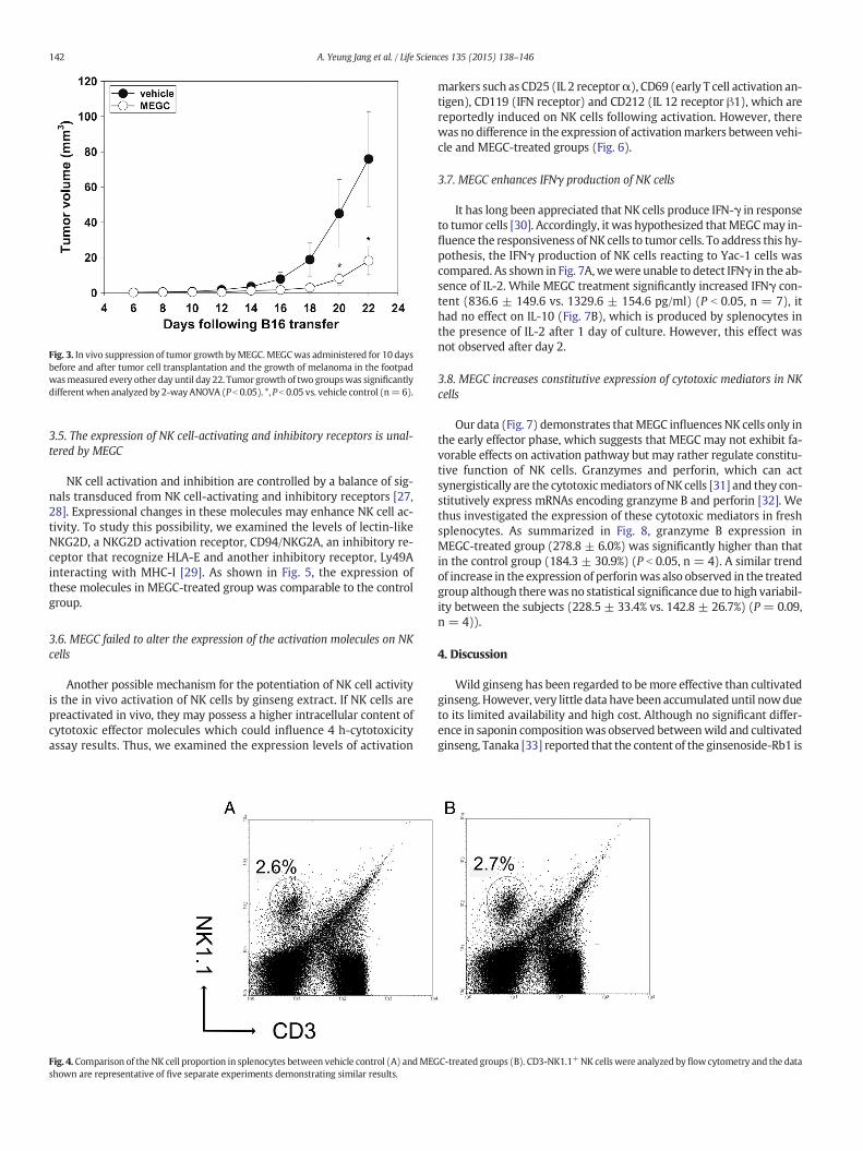
Fig. 3. In vivo suppression of tumor growth byMEGC.MEGCwas administered for 10 daysbefore and after tumor cell transplantation and the growth of melanoma in the footpadwasmeasured every other day until day 22. Tumor growth of two groupswas significantlydifferentwhen analyzed by 2-wayANOVA (P b 0.05). *, P b 0.05 vs. vehicle control (n=6).
142 A. Yeung Jang et al. / Life Sciences 135 (2015) 138–146
3.5. The expression of NK cell-activating and inhibitory receptors is unal-tered by MEGC
NK cell activation and inhibition are controlled by a balance of sig-nals transduced from NK cell-activating and inhibitory receptors [27,28]. Expressional changes in these molecules may enhance NK cell ac-tivity. To study this possibility, we examined the levels of lectin-likeNKG2D, a NKG2D activation receptor, CD94/NKG2A, an inhibitory re-ceptor that recognize HLA-E and another inhibitory receptor, Ly49Ainteracting with MHC-I [29]. As shown in Fig. 5, the expression ofthese molecules in MEGC-treated group was comparable to the controlgroup.
3.6. MEGC failed to alter the expression of the activation molecules on NKcells
Another possible mechanism for the potentiation of NK cell activityis the in vivo activation of NK cells by ginseng extract. If NK cells arepreactivated in vivo, they may possess a higher intracellular content ofcytotoxic effector molecules which could influence 4 h-cytotoxicityassay results. Thus, we examined the expression levels of activation
Fig. 4. Comparison of the NK cell proportion in splenocytes between vehicle control (A) andMEGshown are representative of five separate experiments demonstrating similar results.
markers such as CD25 (IL 2 receptorα), CD69 (early T cell activation an-tigen), CD119 (IFN receptor) and CD212 (IL 12 receptor β1), which arereportedly induced on NK cells following activation. However, therewas no difference in the expression of activationmarkers between vehi-cle and MEGC-treated groups (Fig. 6).
3.7. MEGC enhances IFNγ production of NK cells
It has long been appreciated that NK cells produce IFN-γ in responseto tumor cells [30]. Accordingly, it was hypothesized thatMEGCmay in-fluence the responsiveness of NK cells to tumor cells. To address this hy-pothesis, the IFNγ production of NK cells reacting to Yac-1 cells wascompared. As shown in Fig. 7A,wewere unable to detect IFNγ in the ab-sence of IL-2. While MEGC treatment significantly increased IFNγ con-tent (836.6 ± 149.6 vs. 1329.6 ± 154.6 pg/ml) (P b 0.05, n = 7), ithad no effect on IL-10 (Fig. 7B), which is produced by splenocytes inthe presence of IL-2 after 1 day of culture. However, this effect wasnot observed after day 2.
3.8. MEGC increases constitutive expression of cytotoxic mediators in NKcells
Our data (Fig. 7) demonstrates thatMEGC influences NK cells only inthe early effector phase, which suggests that MEGC may not exhibit fa-vorable effects on activation pathway but may rather regulate constitu-tive function of NK cells. Granzymes and perforin, which can actsynergistically are the cytotoxicmediators of NK cells [31] and they con-stitutively express mRNAs encoding granzyme B and perforin [32]. Wethus investigated the expression of these cytotoxic mediators in freshsplenocytes. As summarized in Fig. 8, granzyme B expression inMEGC-treated group (278.8 ± 6.0%) was significantly higher than thatin the control group (184.3 ± 30.9%) (P b 0.05, n = 4). A similar trendof increase in the expression of perforinwas also observed in the treatedgroup although therewas no statistical significance due to high variabil-ity between the subjects (228.5 ± 33.4% vs. 142.8 ± 26.7%) (P = 0.09,n = 4)).
4. Discussion
Wild ginseng has been regarded to bemore effective than cultivatedginseng. However, very little data have been accumulated until nowdueto its limited availability and high cost. Although no significant differ-ence in saponin compositionwas observed betweenwild and cultivatedginseng, Tanaka [33] reported that the content of the ginsenoside-Rb1 is
C-treated groups (B). CD3-NK1.1+NK cellswere analyzed byflowcytometry and the data
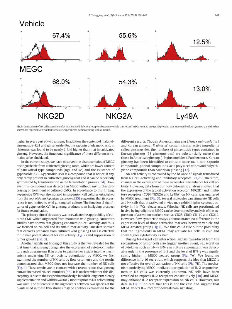
Fig. 5. Comparison ofNK cell expression of activation and inhibitory receptors between vehicle control andMEGC-treated groups. Expressionwas analyzed byflowcytometry and the datashown are representative of four separate experiments demonstrating similar results.
143A. Yeung Jang et al. / Life Sciences 135 (2015) 138–146
higher in every part ofwild ginseng. In addition, the content ofmalonyl–ginsenoside–Rb1 and ginsenoside–Ro, the saponin of oleanolic acid, inrhizomes was found to be nearly 2-fold higher than that in cultivatedginseng. However, the functional significance of these differences re-mains to be elucidated.
In the current study, we have observed the characteristics of MEGCdistinguishable from cultivated ginseng roots, which are lower contentof panaxatriol type compounds (Rg1 and Re) and the existence ofgypenoside XVII. Gypenoside XVII is a compound that is not or, if any,only rarely present in cultivated ginseng root and it can be reportedlysynthesized by transformation in the fermentation process [34]. How-ever, this compound was detected in MEGC without any further pro-cessing or treatment of cultured CMCs. In accordance to this finding,gypenoside XVII was also isolated in suspension cell cultures establishedfrom the root of Panax japonicus var. repens [35], suggesting that its occur-rence is not limited to wild ginseng cell culture. The function al signifi-cance of gypenoside XVII in ginseng products is an intriguing prospectfor future examination.
The primary aimof this studywas to evaluate the applicability of cul-tured CMC which originated from mountain wild ginseng. Numerousstudies have shown that ginseng enhances NK cell activity, and thus,we focused on NK cell and its anti-tumor activity. Our data showedthat extracts prepared from cultured wild ginseng CMCs is effectivefor in vivo potentiation of NK cell activity (Fig. 2) and suppression oftumor growth (Fig. 3).
Another significant finding of this study is that we revealed for thefirst time that ginseng upregulates the expression of cytotoxic media-tors such as granzyme B. In order to gain further insight into the mech-anisms underlying NK cell activity potentiation by MEGC, we firstexamined the number of NK cells by flow cytometry and the resultsdemonstrated that MEGC does not increase the number of NK cells(Fig. 4). These results are in contrast with a recent report that ginsengextract increased NK cell numbers [36]. It is unclear whether this dis-crepancy is due to their experimental design in which long term dietarysupplementation andwithdrawal for 2months prior to NK cell countingwas used. The difference in the ingredients between two species of theplants used in these two studies may be another explanation for the
different results. Though American ginseng (Panax quinquefolius)and Korean ginseng (P. ginseng) contain similar active ingredientscalled ginsenosides, the numbers of ginsenoside types contained inKorean ginseng (38 ginsenosides) are substantially more thanthose in American ginseng (19 ginsenosides). Furthermore, Koreanginseng has been identified to contain more main non-saponincompounds, phenol compounds, acid polysaccharides and polyeth-ylene compounds than American ginseng [37].
NK cell activity is controlled by the balance of signals transducedfrom NK cell-activating and inhibitory receptors [27,28]. Therefore,changes in the expression of these molecules may enhance NK cell ac-tivity. However, data from our flow cytometric analysis showed thatthe expression of the typical activation receptor (NKG2D) and inhibi-tory receptors (CD94/NKG2A and Ly49A) on NK cells was unalteredby MEGC treatment (Fig. 5). Several molecules can stimulate NK cellsand NK cells that preactivated in vivo may exhibit higher cytotoxic ac-tivity in 4 h-51Cr release assay. Whether NK cells are prestimulatedin vivo by ingredients inMEGC can be determined by analysis of the ex-pression of activation markers such as CD25, CD69, CD119 and CD212.However, flow cytometric analysis demonstrated no difference in theexpression level of those activation markers, between vehicle andMEGC-treated group (Fig. 6). We thus could rule out the possibilitythat the ingredients in MEGC may activate NK cells in vivo andshow higher cytotoxicity ex vivo.
During NK–target cell interaction, signals transduced from therecognition of tumor cells also trigger another event, i.e., secretionof cytokines such as IFN-γ. IFN-γ in culture supernatant was detect-able only in the presence of IL-2 and the level of IFN-γ was signifi-cantly higher in MEGC-treated group (Fig. 7A). We found nodifference in IL-10 secretion, which supports the idea that MEGC isnot effective for overall activation of NK cells (Fig. 7B). The mecha-nism underlying MEGC-mediated upregulation of IL-2 responsive-ness in NK cells was currently unknown. NK cells have beenrevealed to express IL-2 receptors constitutively [38] and MEGCmay enhance IL-2 receptor expression on NK cells. However, ourdata in Fig. 6 indicate that this is not the case and suggest thatMEGC affects IL-2 receptor downstream signaling.

Fig. 6.Comparison of NK cell expression of activationmarkers between vehicle control andMEGC-treated groups. Expressionwas analyzed by flow cytometry and the data shown are representative of four separate experiments demonstrating similarresults.
144A.Yeung
Jangetal./Life
Sciences135
(2015)138
–146
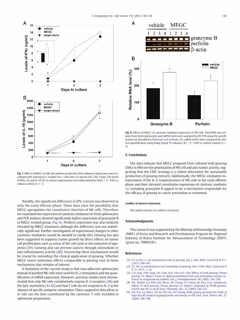
Fig. 7. Effects ofMEGC onNK cell cytokine production. Non-adherent splenocyteswere in-cubated withmitomycin C-treated Yac-1 cells and 12.5 ng/ml of IL-2 for 3 days. The levelsof IFNγ (A) and IL-10 (B) in culture supernatants were determined by ELISA. *, P b 0.05 vs.vehicle control (n = 7).
Fig. 8. Effects of MEGC on cytotoxic mediator expression in NK cells. Total RNA was iso-lated from fresh splenocytes andmRNA levels were analyzed by RT-PCR using the specificprimers as described in Materials and methods (A). mRNA levels were compared by rela-tive quantification using Image Quant TL software (B). *, P b 0.05 vs. vehicle control (n =4).
145A. Yeung Jang et al. / Life Sciences 135 (2015) 138–146
Notably, the significant difference in IFN-γ levels was observed inonly the early effector phase. These data raise the possibility thatMEGC upregulates the constitutive function of NK cells. Therefore,we examined the expression of cytotoxicmediators in fresh splenocytesand PCR analysis showed significantly higher expression of granzyme Bin MEGC-treated group (Fig. 8). Perforin expression was also similarlyelevated by MEGC treatment although the difference was not statisti-cally significant. Further investigation of expressional changes in othercytotoxic mediators would be needed to clarify this. Ginseng has alsobeen suggested to suppress tumor growth by direct effects on tumorcell proliferation such as arrest of the cell cycle or the induction of apo-ptosis [39]. Ginseng also can prevent cancers through antioxidant oranti-inflammatory activity [40]. Uncovering these mechanisms wouldbe crucial for extending the clinical application of ginseng. WhetherMEGC exerts antitumor effects comparable to ginseng root in thosemechanisms also remains of interest.
A limitation of the current study is that non-adherent splenocytesinstead of purified NK cells were used for IL-2 stimulation and the quan-tification of mRNA expression. However, previous studies have demon-strated that only NK cells constitutively express IL-2 receptors [38] andthe lytic machinery [41,42] and that T cells do not respond to IL-2 in theabsence of specific antigenic stimulation. These supportive data allowusto rule out the bias contributed by the cytotoxic T cells included insplenocyte preparation.
5. Conclusions
Our data indicate that MEGC prepared from cultured wild ginsengCMCs is effective for potentiation of NK cell and anti-tumor activity, sug-gesting that the CMC strategy is a robust alternative for sustainableproduction of ginseng extracts. Additionally, the MEGC-mediated en-hancement of the IL-2 responsiveness of NK cells in the early effectorphase and their elevated constitutive expression of cytotoxic mediator(s) including granzyme B appear to be a mechanism responsible forthe efficacy of ginseng in cancer prevention or treatment.
Conflict of interest statement
The authors declare no conflicts of interest.
Acknowledgments
This researchwas supported by theMinistry of Knowledge Economy(MKE) of Korea and Research and Development Program for RegionalIndustry of Korea Institute for Advancement of Technology (KIAT)(grant no. 70005547).
References
[1] C.P. Li, R.C. Li, An introductory note to ginseng, Am. J. Chin. Med. (Gard City N Y) 1(1973) 249–261.
[2] S.Y. Hu, A contribution to our knowledge of ginseng, Am. J. Chin. Med. (Gard City NY) 5 (1977) 1–23.
[3] C.H. Jung, H.M. Seog, I.W. Choi, H.D. Choi, H.Y. Cho, Effects of wild ginseng (Panaxginseng C.A. Meyer) leaves on lipid peroxidation levels and antioxidant enzyme ac-tivities in streptozotocin diabetic rats, J. Ethnopharmacol. 98 (2005) 245–250.
[4] M.L. Mollah, G.S. Kim, H.K. Moon, S.K. Chung, Y.P. Cheon, J.K. Kim, et al., Antiobesityeffects of wild ginseng (Panax ginseng C.A. Meyer) mediated by PPAR-gamma,GLUT4 and LPL in ob/ob mice, Phytother. Res. 23 (2009) 220–225.
[5] S.N. Yun, S.J. Moon, S.K. Ko, B.O. Im, S.H. Chung, Wild ginseng prevents the onset ofhigh-fat diet induced hyperglycemia and obesity in ICR mice, Arch. Pharm. Res. 27(2004) 790–796.

146 A. Yeung Jang et al. / Life Sciences 135 (2015) 138–146
[6] A.C. Cabral de Oliveira, A.C. Perez, G. Merino, J.G. Prieto, A.I. Alvarez, Protective ef-fects of Panax ginseng on muscle injury and inflammation after eccentric exercise,Comp Biochem Physiol C Toxicol Pharmacol. 130 (2001) 369–377.
[7] L.J. Hofseth, M.J. Wargovich, Inflammation, cancer, and targets of ginseng, J. Nutr.137 (2007) 183S–185S.
[8] A. Takeda, N. Katoh, M. Yonezawa, Restoration of radiation injury by ginseng. III. Ra-dioprotective effect of thermostable fraction of ginseng extract on mice, rats andguinea pigs, J Radiat Res (Tokyo) 23 (1982) 150–167.
[9] S.I. Gum, S.J. Jo, S.H. Ahn, S.G. Kim, J.T. Kim, H.M. Shin, et al., The potent protective ef-fect ofwild ginseng (Panax ginseng C.A.Meyer) against benzo[alpha]pyrene-inducedtoxicity through metabolic regulation of CYP1A1 and GSTs, J. Ethnopharmacol. 112(2007) 568–576.
[10] P.H. Chang, The effect of ginseng (Panax ginseng C. A. Mey) on organism reactivity,Yao Xue Xue Bao 13 (1966) 106–111.
[11] G.N. Predy, V. Goel, R. Lovlin, A. Donner, L. Stitt, T.K. Basu, Efficacy of an extract ofnorth American ginseng containing poly-furanosyl-pyranosyl-saccharides forpreventing upper respiratory tract infections: a randomized controlled trial, Can.Med. Assoc. J. 173 (2005) 1043–1048.
[12] H. Sung, S.M. Kang, M.S. Lee, T.G. Kim, Y.K. Cho, Korean red ginseng slows depletionof CD4 T cells in human immunodeficiency virus type 1-infected patients, Clin.Diagn. Lab. Immunol. 12 (2005) 497–501.
[13] H. Sung, Y.S. Jung, Y.K. Cho, Beneficial effects of a combination of Korean red ginsengand highly active antiretroviral therapy in human immunodeficiency virus type 1-infected patients, Clin. Vaccine Immunol. 16 (2009) 1127–1131.
[14] J.D. Park, Recent studies on the chemical constituents of Korean Ginseng (Panax gin-seng C. A. Meyer), Korean J. Ginseng Sci 20 (1996) 389–415.
[15] T.K. Yun, Y.S. Lee, Y.H. Lee, S.I. Kim, H.Y. Yun, Anticarcinogenic effect of Panax ginsengC.A. Meyer and identification of active compounds, J. Korean Med. Sci. 16 Suppl(2001) S6–S18.
[16] T. Konoshima, M. Takasaki, E. Ichiishi, T. Murakami, H. Tokuda, H. Nishino, et al.,Cancer chemopreventive activity of majonoside-R2 from Vietnamese ginseng,Panax vietnamensis, Cancer Lett. 147 (1999) 11–16.
[17] H.R. Shin, J.Y. Kim, T.K. Yun, G. Morgan, H. Vainio, The cancer-preventive potential ofPanax ginseng: a review of human and experimental evidence, Cancer Causes Con-trol 11 (2000) 565–576.
[18] M. Hao, W.Wang, Y. Zhao, R. Zhang, H. Wang, Pharmacokinetics and tissue distribu-tion of 25-hydroxyprotopanaxadiol, an anti-cancer compound isolated from Panaxginseng, in athymic mice bearing xenografts of human pancreatic tumors, Eur. J.Drug Metab. Pharmacokinet. 35 (2011) 109–113.
[19] Y.H. Jie, S. Cammisuli, M. Baggiolini, Immunomodulatory effects of Panax ginsengC.A. Meyer in the mouse, Agents Actions 15 (1984) 386–391.
[20] Y.S. Yun, H.S. Moon, Y.R. Oh, S.K. Jo, Y.J. Kim, T.K. Yun, Effect of red ginseng onnatural killer cell activity in mice with lung adenoma induced by urethan andbenzo(a)pyrene, Cancer Detection and Prevention. Supplement: Official Publi-cation of the International Society for Preventive Oncology, Inc. 1 (1987)301–309.
[21] Y.S. Kim, E.J. Hahn, H.N.Murthy, K.Y. Paek, Adventitious root growth and ginsenosideaccumulation in Panax ginseng cultures as affected bymethyl jasmonate, Biotechnol.Lett. 26 (2004) 1619–1622.
[22] E.K. Lee, Y.W. Jin, J.H. Park, Y.M. Yoo, S.M. Hong, R. Amir, et al., Cultured cambial mer-istematic cells as a source of plant natural products, Nat. Biotechnol. 28 (2010)1213–1217.
[23] T. Murashige, F. Skoog, A revised medium for rapid growth and bio assays with to-bacco tissue cultures, Physiol. Plant. 15 (1962) 473–497.
[24] K.T. Brunner, J. Mauel, J.C. Cerottini, B. Chapuis, Quantitative assay of the lytic actionof immune lymphoid cells on 51-Cr-labelled allogeneic target cells in vitro; inhibi-tion by isoantibody and by drugs, Immunology 14 (1968) 181–196.
[25] L. Fang, V.C. Lee, E. Cha, H. Zhang, S.T. Hwang, CCR7 regulates B16murinemelanomacell tumorigenesis in skin, J. Leukoc. Biol. 84 (2008) 965–972.
[26] A.P. Makrigiannis, D.W. Hoskin, Inhibition of CTL induction by rapamycin: IL-2 res-cues granzyme B and perforin expression but only partially restores cytotoxic activ-ity, J. Immunol. 159 (1997) 4700–4707.
[27] C. Bottino, L. Moretta, D. Pende, M. Vitale, A. Moretta, Learning how to discriminatebetween friends and enemies, a lesson from natural killer cells, Mol. Immunol. 41(2004) 569–575.
[28] L. Zamai, C. Ponti, P. Mirandola, G. Gobbi, S. Papa, L. Galeotti, et al., NK cells and can-cer, J. Immunol. 178 (2007) 4011–4016.
[29] W.M. Yokoyama, S. Kim, A.R. French, The dynamic life of natural killer cells, Annu.Rev. Immunol. 22 (2004) 405–429.
[30] G. Scala, J.Y. Djeu, P. Allavena, T. Kasahara, J.R. Ortaldo, R.B. Herberman, J.J.Oppenheim, Cytokine secretion and noncytotoxic functions of human large granularlymphocytes, in: E. Lotzova, R.B. Herberman (Eds.), Immunobiology of Natural KillerCells, Vol. II, CRC Press, Boca Raton 1986, pp. 133–144.
[31] C.J. Froelich, V.M. Dixit, X. Yang, Lymphocyte granule-mediated apoptosis: mattersof viral mimicry and deadly proteases, Immunol. Today 19 (1998) 30–36.
[32] T.A. Fehniger, S.F. Cai, X. Cao, A.J. Bredemeyer, R.M. Presti, A.R. French, et al., Acqui-sition ofmurine NK cell cytotoxicity requires the translation of a pre-existing pool ofgranzyme B and perforin mRNAs, Immunity 26 (2007) 798–811.
[33] O. Tanaka, Solubilizing properties of ginseng saponins, Korea–Japan Panax ginsengSymposium. Seoul, Korea 1987, pp. 67–74.
[34] L.Q. Cheng, J.R. Na, M.K. Kim,M.H. Bang, D.C. Yang, Microbial conversion of ginsenosideRb1 tominor ginsenoside F2 and gypenoside XVII by INTRASPORANGIUM sp. GS603 iso-lated from soil, J. Microbiol. Biotechnol. 17 (2007) 1937–1943.
[35] D.V. Kochkin, G.P. Zaitsev, V.V. Kachala, A.O. Chizhov, E.V. Demidova, M.V. Titova, et al.,The occurrence of gypenosideXVII in suspension cell culture of ginseng Panax japonicusvar. repens. Doklady, Biochemistry and Biophysics 442 (2012) 42–45.
[36] S.C. Miller, L. Ti, J. Shan, Dietary supplementation with an extract of north Americanginseng in adult and juvenile mice increases natural killer cells, Immunol. Invest. 41(2012) 157–170.
[37] K.-t. Choi, Botanical characteristics, pharmacological effects andmedicinal componentsof Korean Panax ginseng C A Meyer, Acta Pharmacol. Sin. 29 (2008) 1109–1118.
[38] M.A. Caligiuri, A. Zmuidzinas, T.J. Manley, H. Levine, K.A. Smith, J. Ritz, Functionalconsequences of interleukin 2 receptor expression on resting human lymphocytes.Identification of a novel natural killer cell subset with high affinity receptors, J.Exp. Med. 171 (1990) 1509–1526.
[39] M.L. King, L.L. Murphy, Role of cyclin inhibitor protein p21 in the inhibition ofHCT116 human colon cancer cell proliferation by American ginseng (Panaxquinquefolius) and its constituents, Phytomedicine 17 (2010) 261–268.
[40] Y. Jin, V.S. Kotakadi, L. Ying, A.B. Hofseth, X. Cui, P.A. Wood, et al., American ginsengsuppresses inflammation and DNA damage associated with mouse colitis, Carcino-genesis 29 (2008) 2351–2359.
[41] G. Trinchieri, Biology of natural killer cells, Adv. Immunol. 47 (1989) 187–376.[42] L. Moretta, C. Bottino, D. Pende, M.C. Mingari, R. Biassoni, A. Moretta, Human natural
killer cells: their origin, receptors and function, Eur. J. Immunol. 32 (2002) 1205–1211.