poultry anatomy
DESCRIPTION
Poultry AnatomyTRANSCRIPT

Poultry Anatomy 6-6-08
Poultry Anatomy Objectives After a student completed this module, he or she will be able to accomplish the following
1. On a real specimen or drawing of a young chicken, identify and locate the following
anatomical structures:
(a) External Anatomy:
(1) Uropygial gland/ oil gland/ oil sack/ preen gland
(2) Feather follicle
(3) Different feather tracts
(4) Comb
(5) Wattles
(6) Shank
(7) Hock joint
(8) Vent opening
(b) Internal Anatomy:
(1) Esophagus/ gullet/ goozle
(2) Crop/ ingluvies/ craw
(3) Proventriculus/ true stomach/ glandular stomach
(4) Ventriculus/ gizzard/ muscular stomach
(5) Small intestine
(6) Duodenum
(7) Pancreas (located in the loop of the duodenum)
(8) Large intestine/ rectum/ colon
(9) Cecum (a pair of ceca present)
(10) Cloaca/ Vent (anus)
(11) Mesentery vessels
(12) Heart
(13) Coronary band
(14) Pericardial sac
(15) Spleen
(16) Liver
(17) Gall bladder
Poultry Slaughter Inspection Training Page 1

Poultry Anatomy 6-6-08
(18) Kidneys
(19) Thymus
(20) Bursa of fabricius
(21) Trachea/ windpipe
(22) Lungs
(23) Syrinx
(24) Air sacs:
(a) Interclavicular
(b) Thoracic
(c) Abdominal
(25) Reproductive system:
(a) Ovary/oviduct
(b) Testicles
(c) Skeletal anatomy:
(1) Vertebral column consists of:
(a) Cervical vertebrae
(b) Thoracic vertebrae
(c) Lumbar vertebrae
(d) Sacral vertebrae
(e) Coccygeal vertebrae
(2) Pectoral girdle consists of:
(a) Clavicle/ wishbone/ pulleybone/ furculum
(b) Coracoid
(c) Scapula/ shoulder blade
(3) Wing consists of:
(a) Humerus
(b) Radius
(c) Ulna
(d) Wing tip
(4) Pelvic girdle consists of:
(a) Ilium
(b) Ischium
(c) Pubis
(d) synsacrum
Poultry Slaughter Inspection Training Page 2

Poultry Anatomy 6-6-08
(5) Legs consist of:
(a) Femur
(b) Tibia
(c) Fibula
(d) shank
(6) Other skeletal bones:
(a) Sternum/ keel/ breast bone
(b) Vertebral ribs
(c) Sternal ribs
2. Identify at least one general difference between avian species and mammals in the
following categories:
a. Lymphatic system
b. Respiratory system
c. Skeletal system
d. Urinary system
3. Identify three organs (glands or tissues) in the avian species that are major sites of
lymphatic tissue. 4. Identify the number of air sacs found in young chickens and indicate which are fused
or paired. 5. Name the air sac in young chickens that is observed on postmortem inspection and
has communication with bone. 6. Name three body systems in young chickens that terminate through the cloaca. 7. Name three organs or tissues that are present in the young bird or embryo that either
fail to develop or diminish in size as the bird matures. 8. Identify the anatomical location of diverticula from the interclavicular air sac. 9. Describe why inflammation of the interclavicular air sac is significant as compared to
inflammation of the abdominal air sacs. 10. Identify two reasons the skin of poultry may be different colors when observed at the
postmortem inspection station. 11. Name the organs used for giblets. 12. Name the bone that is located in the thigh.
Poultry Slaughter Inspection Training Page 3
Poultry Anatomy 6-6-08
13. Name the two bones that are located in the drumstick. 14. Identify three major areas where fat is commonly stored in the live bird. 15. Define “debeaking” and identify the reason this practice is followed by many poultry
growers. 16. Identify at least two proposed functions of air sacs in live poultry. 17. Identify at least two reasons why the color of fat might differ between lots of young
chickens presented for postmortem inspection. 18. Identify the anatomical location of poultry kidneys. 19. Give the main function of each major organ or portion of the digestive tract. 20. Identify the only skin gland of significance in the chicken.
Poultry Slaughter Inspection Training Page 4

Poultry Anatomy 6-6-08
Poultry Anatomy Introduction This module is designed to fill any voids you may have on poultry anatomy, and to familiarize you with both the technical and common terms for different anatomical features of poultry. The species selected to serve as our example throughout this module is the young chicken. External Anatomy The comb and wattles are external structures on the head of poultry and are largely ornamental. The size and color are associated with gonad development and secretion of the sex hormones. The red color can be attributed to the high vascularity of the dermis covering these structures. The comb and waddles have different characteristics in male vs. female birds, and among different poultry species. Snoods and whiskers (beards) are external structures only found in turkeys. Feathers cover almost the entire surface of the bird. They grow from follicles, which are organized into tracts. Feather tracts are named according to the structure they are associated with. For example, the femoral feather tract is on the thigh, which contains the femur. Most birds lose their feathers or mold once a year and replace their feathers by growing new ones. Generally speaking, young chickens come to slaughter before going through a complete molt. The skin of chickens is thinner and more delicate than that of mammals. In addition, the color of the skin varies. Some of the factors contributing to this variation of species, age, diet and breed. The skin of young chickens at postmortem inspection may also be affected by the scalder temperature. The uropygial gland, preen gland, oil gland, or oil bag is considered the only significant gland of the skin. It is located dorsally and near the tip of the tail. In the chicken there are two lobes that drain through a median nipple-like papilla. The function of the gland is somewhat uncertain but preening chickens take oil from this gland and apply it to their feathers.
Poultry Slaughter Inspection Training Page 5

Poultry Anatomy 6-6-08
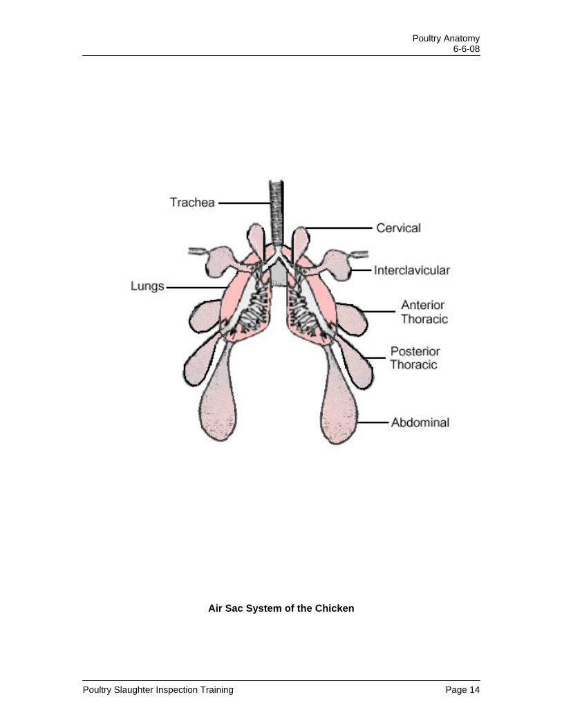
Internal Anatomy Respiratory System: The respiratory system of birds is more complex than the mammalian counterpart. For our purposes, the system in the bird is comprised of the trachea, syrinx, lungs, and air sacs. The trachea, or windpipe, is the structure through which air enters the bird, and has cartilaginous rings along its length. The syrinx is located where the trachea bifurcates (splits into two separate branches), and is similar to the larynx (voice box) of mammals. Air passes through the trachea and at the terminal portions of the trachea the air sac structures bud out. These are very thin, colorless membranes that, when inflated with air, resemble tiny balloons inside the body cavity. The function of the lungs is to facilitate gas exchange, as it is in mammals. However, because avians have air sacs, air flows through avian lungs on both inspiration and expiration. As well as functioning in respiration, air sacs may also regulate intrabody pressure and body temperature. The number of air sacs varies in different species. There is a chart in this module that lists the air sacs of each species of poultry. The most anterior air sac is the cervical. It lies, as the name implies, in the neck area and is not observed during postmortem inspection. It is the only air sac in the young chicken not observed during postmortem inspection. The next air sac, moving in a caudal direction, is the interclavicular air sac. Some information to remember about this particular air sac is that it lies between the clavicles (as the name implies), is the most anterior air sac observed on postmortem inspection, and has communication with other tissues, including bone, through diverticula (tiny fingerlike projections). The diverticula from the interclavicular air sac have the following communications: (1) into the breastbone, (2) into the bones of the shoulder girdle, and (3) around the shoulder joint. The thoracic air sacs are next and lie in the rib cage area. In chickens and ducks there are two pairs- anterior and posterior- whereas the turkey only has one pair of thoracic air sacs. The abdominal air sacs are paired and are located in the abdominal part of the body cavity. Because of their location, these air sacs are typically destroyed or displaced during the evisceration process. Digestive System: The digestive tract of the bird begins with the mouth, which does not contain lips or teeth. As in mammals, the mouth is connected to the esophagus, also called the goozle or gullet. In chickens the distal end of the esophagus has a specialized area for the storage of feed called the, crop, craw, or ingluvies. The crop is located at the base of the neck as it is viewed externally.
Poultry Slaughter Inspection Training Page 6

Poultry Anatomy 6-6-08
Following the crop is another short section of esophagus ending in the stomach. In birds the stomach consists of two parts. The proventriculus, also called the glandular stomach or true stomach, is located caudal to the crop. It secretes hydrochloric acid and pepsin, which are used to aid in protein digestion. The ventriculus, also called the gizzard or the muscular stomach, is caudal to the proventriculus, and is much larger and more muscular in appearance when compared to the proventriculus. The major function of the ventriculus is to grind the food. This grinding action prepares the food for digestion. Birds frequently deposit substantial amounts of fat around the gizzard. The ventriculus empties into the small intestine, which consists of the duodenum, the jejunum, and the ileum. The duodenal portion is the most cranial, and is significant because the pancreas is located in the duodenal loop. The secretions of the pancreas contain enzymes, which enter the duodenum through the pancreatic ducts. This organ is seldom involved in pathology. The large intestine consists of a pair of cecae and a short straight intestine, called the colon or rectum. This section of large intestine is similar to the rectum of mammals. The cloaca is the termination of the digestive system. This portion of the digestive system represents a common passage for digestive, urinary, and reproductive systems. The cloaca opens externally in what is called the vent. The bursa of Fabricius is located as a diverticulum in the dorsal wall of the cloaca. This bursa contains lymphatic tissue and has a function related to immunity and antibody production. It regresses in size and disappears as the bird matures. On postmortem inspection, the bursa of Fabricius is called the “rosebud”. When it is intact, it appears as a small sac on the side of the cloaca. When it has been opened during the evisceration process, it appears like a rosebud, which is the common name for this structure because it has several small folds in its mucosal surface. Birds that are healthy and well-nourished will usually deposit substantial amount of fat throughout their tissues. The abdominal area and vent flaps are major fat depots, as are the areas surrounding the gizzard and the coronary band of the heart. There may be considerable variation in the color of poultry fat. Diet, age, health status, and breed are all factors that can influence this color The normal liver is a single organ which has two lobes. The color varies somewhat depending on the fat content. Each lobe of the liver is drained by a bile duct. The right lobe is drained by the hepatocystic duct and the left lobe is drained by the hepatoenteric duct. The duct from the right lobe is enlarged to form the gallbladder. Both ducts enter the small intestine together. Circulatory System: The heart of the chicken is four-chambered, like those of mammals, and beats at a rate of 250 beats per minute for larger breeds and up to around 350 beats per minute for smaller breeds. In contrast, the heart rate for human beings is typically around 80 beats per minute. The deep body temperature of a chicken is around 107 degrees F, versus that of mammals which is typically between 98 and 102 degrees F. Avian red blood cells, or erythrocytes, are nucleated, whereas mammalian red blood cells are not.
Poultry Slaughter Inspection Training Page 7
Poultry Anatomy 6-6-08
Some points to remember about the heart are that the heart’s coronary band (around the top portion) has a normal fat structure that may show changes in quantity and appearance when a systemic disease occurs. Other points to remember are that the pericardial sac is the thin membrane that encloses the heart, and that they heart normally has a small deposit of fat at the tip as well as around the coronary band. Lymphatic System: The lymphatic system of chickens does not contain lymph nodes and in general is poorly developed when compared with mammals. There are several organs which contain lymphatic tissue- the bursa of Fabricius, the spleen, and the thymus. The thymus gland consists of about five pairs of pale pink, flattened, irregularly shaped lobes strung out along both sides of the neck, just superficial to the jugular veins. The thymus decreases in size as the bird matures. The spleen is a small, round, soft organ similar in color to the liver. The normal spleen is about ¾ inch in diameter, located near the ventriculus (gizzard) in the body cavity. Histologically, it is composed of red and white pulp. The functions of the spleen include phagocytosis of worn-out erythrocytes in red pulp, lymphocyte production in white pulp, and antibody production in both the red and white pulp. Urinary System: The urinary system of the chicken does not contain a urinary bladder. There are two tri-lobed kidneys, one on each side of the ventral surface of the vertebral column. This pair of kidneys is embedded in the deep bony crypts of the pelvic and synsacral area of the skeleton. Ureters carry the urinary waste to the cloaca. The uric acid is discharged into the cloaca and excreted with the feces. The white pasty material in chicken droppings is considered to be urinary system excretion. Birds excrete their nitrogen waste as uric acid, whereas mammals excrete it in the form of urea. Reproductive System: The female reproductive system consists of the left ovary and oviduct. Although present in the embryo, the right ovary and oviduct fail to develop. The oviduct terminates in the cloaca. The male reproductive system consists of two testicles, which secrete semen through a vas deferens. The vas deferens terminates in the cloaca. The chicken has a rudimentary penis. Skeletal System: The chicken’s beak is composed of hard keratinized epidermal tissue. This rostral structure forms part of the upper and lower jaws. The beak functions much like the lips and teeth of mammals. Debeaking is the removal of approximately one-half of the upper and lower level. In some cases only the upper beak is removed. Debeaking has been used in the poultry industry to prevent cannibalism.
Poultry Slaughter Inspection Training Page 8

Poultry Anatomy 6-6-08
Some bones of the avian species are considered pneumatic as a result of diverticula from the air sacs. These air sac diverticula result in a direct connection between the respiratory system and the skeletal system of avians. The vertebral column is divided into cervical, thoracic, lumbar, sacral, and coccygeal areas:
1. The cervical vertebrae are the neck bones. 2. The thoracic vertebrae are those in the thoracic, or chest, area. 3. The lumbar vertebrae are those in the abdominal area. 4. The sacral vertebrae are those in the pelvic area. 5. The coccygeal vertebrae are those in the tail area.
The bones of the pectoral girdle are the clavicle, coracoid, and scapula:
1. The clavicle, also called the wishbone, pulleybone, or furculum, lies at the base of the neck. It is a fused bone. The intraclavicular air sacs are located between the two branches of the clavicle.
2. The coracoid bones lie on either side of the ribcage, and attach the shoulders to the breast bone. They lie just caudal to the clavicle, and are thick bones when compared to the clavicle.
3. The scapula, or shoulder blade, is a long thin bone which runs along the top of each side of the ribcage.
The bones of the wing are the humerus, radius, ulna, and wingtip:
1. The humerus is the upper wing bone. This bone has the same name as the upper front leg bone in mammals, and the upper arm bone in humans.
2. The radius is the small straight lower wing bone. This bone has the same name as the larger lower front leg bone in mammals, and the lower arm bone in humans.
3. The wingtip bone is the bone at the very end of the wing. It is frequently fractured during the slaughter process.
The pelvic girdle is composed of the synsacrum, ilium, ischium, and pubis (there is not a pubic symphysis in the chicken skeleton, probably an adaptation for egg laying):
1. The lumbar, sacral, and first six caudal (coccygeal) vertebrae are fused into an immobile dorsal bony structure referred to as the synsacrum.
2. The ilium is part of the pelvic girdle, and is a flat bone on each side of the anterior half of the synsacrum.
3. The ischium is part of the pelvic girdle, and is a flat bone on each side of the posterior half of the synsacrum.
4. The pubis, also known as the pin bone, is a long thin bone that runs along the ventral side of the ischium.
The leg of the chicken is composed of the femur, tibia, fibula, and shank:
1. The femur is the upper leg bone, which is located in the thigh of the chicken, as in mammals.
Poultry Slaughter Inspection Training Page 9

Poultry Anatomy 6-6-08
2. The tibia is the major lower leg bone, which is located in the drumstick of the
chicken, as in the lower leg of mammals. 3. The fibula is a very small bone in the lower leg, or drumstick, of chickens. It
is much smaller than it is in mammals. 4. The shank is the portion of the leg below the hock joint. It is normally
removed during the slaughter process along with the paw. 5. The hock joint is located between the drumstick and the shank. It is
normally exposed for postmortem inspection. The sternum, also called the keel or breast bone, is a single large bone on the ventral surface of the body. Ribs are divided into two types. The vertebral ribs are those that originate from the vertebral column. The sternal ribs are those that originate from the sternum (keel, breast bone).
Poultry Slaughter Inspection Training Page 10

Poultry Anatomy 6-6-08
Definitions Abdominal: pertaining to the abdomen, or belly. Anterior: situated in front of or in the front part of the body. Caudal: denoting a position more toward the cauda or tail. Cervical: pertaining to the neck. Coronary band: the area around the top (large) end of the heart. Cranial: pertaining to the anterior end of the body. Dermis: connective tissue underlying the skin. Diverticulum: a fingerlike projection of a pouch or sac. Dorsal: pertaining to the back. Epidermal: pertaining to the skin. Gonad: a sex organ, such as an ovary or a testicle, which produces the gametes (egg or sperm). Histologically: pertaining to the microscopic structure of a tissue. Keratinized: made hard by the deposition of keratin, an insoluble protein. Lateral: denoting a position farther from the median plane or midline of the body or of a structure. Lymphatic: pertaining to the lymph or immune system. Medial: pertaining to the middle; closer to the median plane or to the midline of a body or structure. Mesentery: a membranous fold attaching various organs to the body wall. Nucleated: having a cell nucleus. Papilla: a small nipple-shaped projection, elevation, or structure. Pectoral: pertaining to the breast or chest. Pelvic: pertaining to the pelvis, or hip region. Pneumatic: pertaining to air or respiration. Posterior: situated in back of or in the back part of the body.
Poultry Slaughter Inspection Training Page 11

Poultry Anatomy 6-6-08
Regress: subsiding, or returning to a former or earlier state. Rudimentary: poorly developed and not functional. Symphysis: a type of cartilaginous joint in which the apposed bony surfaces are firmly united by a plate of fibrocartilage. Thoracic: pertaining to the thorax, or chest. Ventral: pertaining to the belly.
Poultry Slaughter Inspection Training Page 12
Poultry Anatomy 6-6-08
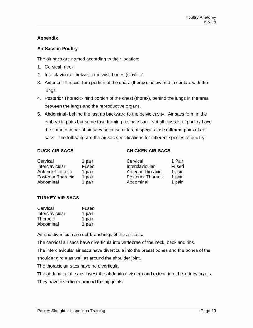
Appendix Air Sacs in Poultry The air sacs are named according to their location:
1. Cervical- neck
2. Interclavicular- between the wish bones (clavicle)
3. Anterior Thoracic- fore portion of the chest (thorax), below and in contact with the
lungs.
4. Posterior Thoracic- hind portion of the chest (thorax), behind the lungs in the area
between the lungs and the reproductive organs.
5. Abdominal- behind the last rib backward to the pelvic cavity. Air sacs form in the
embryo in pairs but some fuse forming a single sac. Not all classes of poultry have
the same number of air sacs because different species fuse different pairs of air
sacs. The following are the air sac specifications for different species of poultry:
DUCK AIR SACS CHICKEN AIR SACS Cervical 1 pair Cervical 1 Pair Interclavicular Fused Interclavicular Fused Anterior Thoracic 1 pair Anterior Thoracic 1 pair Posterior Thoracic 1 pair Posterior Thoracic 1 pair Abdominal 1 pair Abdominal 1 pair TURKEY AIR SACS
Cervical Fused Interclavicular 1 pair Thoracic 1 pair Abdominal 1 pair
Air sac diverticula are out-branchings of the air sacs.
The cervical air sacs have diverticula into vertebrae of the neck, back and ribs.
The interclavicular air sacs have diverticula into the breast bones and the bones of the
shoulder girdle as well as around the shoulder joint.
The thoracic air sacs have no diverticula.
The abdominal air sacs invest the abdominal viscera and extend into the kidney crypts.
They have diverticula around the hip joints.
Poultry Slaughter Inspection Training Page 13
Poultry Anatomy 6-6-08
Air Sac System of the Chicken
Poultry Slaughter Inspection Training Page 14

Poultry Anatomy 6-6-08
A Comparison of Scientific Terms to Common Poultry Plant Terms:
Scientific Term Plant Term Uropygial gland Oil gland, preen gland, oil bag Stifle joint knee joint Esophagus gullet, goozle Ingluvies crop, craw Proventriculus stomach Ventriculus gizzard Cloacal bursa (Bursa of Fabricius) rosebud, flower Ceca blind guts Trachea windpipe Syrinx voice box Lungs lights Pericardium heart sac Tibia and fibula drumstick Femur thigh bone, thigh Pubis pin bone Clavicles (fused) wishbone Sternum keel, breast bone Fused metacarpals wing tip Radius and ulna wing portion Humerus peg leg
Poultry Slaughter Inspection Training Page 15
Poultry Anatomy 6-6-08
WORKSHOP 1. List two general differences between avian species and mammals with respect to the
following body systems:
a. Lymphatic System b. Respiratory System c. Skeletal System d. Urinary System e. Reproductive System
2. List three organs (glands or tissues) in the avian species that contain lymphatic
tissue: 3. List the air sacs found in young chickens and indicate which are fused or paired: 4. Name the air sac in young chickens that is observed on postmortem inspection and
has communication with bone:
Poultry Slaughter Inspection Training Page 16

Poultry Anatomy 6-6-08
5. List three body systems in young chickens that terminate through the cloaca: 6. List three organs or tissues that are present in the young bird or embryo that either
fail to develop or become absent in the mature bird: 7. Give the anatomical location of diverticula from the interclavicular air sac: 8. Describe why inflammation of the interclavicular air sacs is significant in the
slaughter and processing of poultry carcasses as compared to inflammation of the abdominal air sacs:
9. List two reasons the skin of poultry may be different colors when observed at the
postmortem inspection station: 10. List the bone that is located in the thigh: 11. List the organs used for giblets: 12. List the two bones that are located in the drumstick:
13. List three major areas where fat is commonly deposited in the live bird:
Poultry Slaughter Inspection Training Page 17

Poultry Anatomy 6-6-08
14. Match the anatomical part with the letter from Figure 1 on the next page. Some
letters may be used more than once.
Uropygial gland _____________________ Wing tip ____________________ Sternum _____________________ Shank _____________________ Hock joint _____________________ Femoral feather tract _____________________ Wattle _____________________ Crural feather tract _____________________ Beak _____________________ Pectoral feather tract _____________________ Oil gland _____________________ Breast _____________________ Comb _____________________ Thigh _____________________ Preen gland _____________________ Drumstick _____________________ Oil bag _____________________ Keel _____________________
Poultry Slaughter Inspection Training Page 18

Poultry Anatomy 6-6-08
Figure 1
Poultry Slaughter Inspection Training Page 19
Poultry Anatomy 6-6-08
15. Match the anatomical part with the letter from Figure 2 on the next page. Some
letters may be used more than once.
Tibia _____________________ Breast bone ____________________ Coracoid _____________________ Pulley bone _____________________ Femur _____________________ Pelvic girdle _____________________ Cervical vertebrae _____________________ Clavicle _____________________ Humerus _____________________ Sternum _____________________ Synsacrum _____________________ Furculum _____________________ Neck _____________________ Hock joint _____________________
Radius _____________________ Keel _____________________ Thigh bone _____________________ Wishbone _____________________
Poultry Slaughter Inspection Training Page 20

Poultry Anatomy 6-6-08
Figure 2
Poultry Slaughter Inspection Training Page 21

Poultry Anatomy 6-6-08
16. Match the anatomical part with the letter from Figure 3 on the next page. Some
letters may be used more than once.
Proventriculus _____________________ Blind gut _____________________ Goozle _____________________ Ventriculus _____________________
Cloacal bursa _____________________ Crop _____________________ Rectum _____________________ Muscular stomach _____________________ Esophagus _____________________ Glandular Stomach _____________________ Craw _____________________ Cloaca _____________________ Pancreas _____________________ Colon _____________________ Gizzard _____________________ Gullet _____________________ Cecum _____________________ True stomach _____________________ Ingluvies _____________________ Liver _____________________ Bursa of Fabricius _____________________
Poultry Slaughter Inspection Training Page 22

Poultry Anatomy 6-6-08
Figure 3
Poultry Slaughter Inspection Training Page 23

Poultry Anatomy 6-6-08
17. Describe what is meant by “debeaking” and the reason such practice is followed by
many poultry producers. 18. List two functions of air sacs in live poultry. 19. List three reasons why the color of fat might differ between lots of young chickens
presented for postmortem inspection:
Poultry Slaughter Inspection Training Page 24