powerpoint presentation materials to accompany genetics: analysis and principles robert j. brooker...
TRANSCRIPT

PowerPoint Presentation Materialsto accompany
Genetics: Analysis and PrinciplesRobert J. Brooker
Copyright ©The McGraw-Hill Companies, Inc. Permission required for reproduction or display
CHAPTER 10
CHROMOSOME ORGANIZATION AND MOLECULAR STRUCTURE
INTRODUCTION
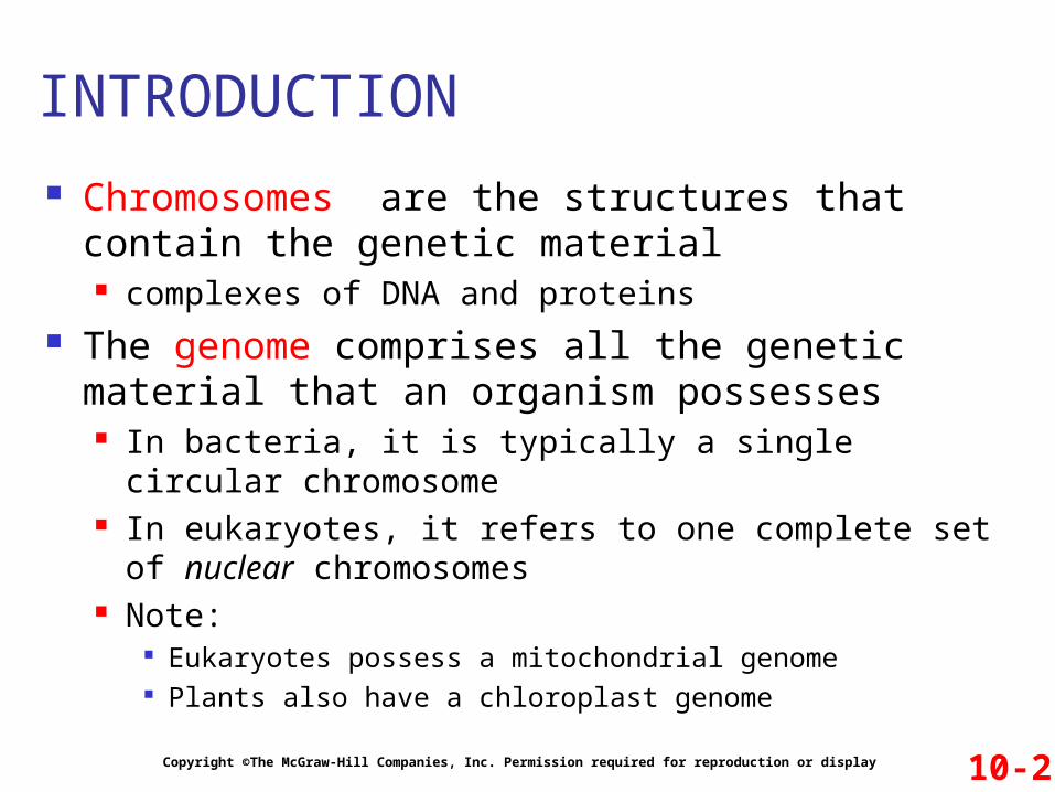
Chromosomes are the structures that contain the genetic material complexes of DNA and proteins
The genome comprises all the genetic material that an organism possesses In bacteria, it is typically a single circular chromosome In eukaryotes, it refers to one complete set of nuclear
chromosomes Note:
Eukaryotes possess a mitochondrial genome Plants also have a chloroplast genome
10-2Copyright ©The McGraw-Hill Companies, Inc. Permission required for reproduction or display
INTRODUCTION
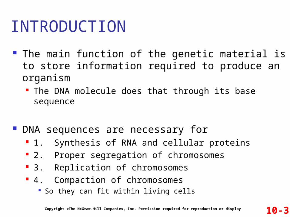
The main function of the genetic material is to store information required to produce an organism The DNA molecule does that through its base sequence
DNA sequences are necessary for 1. Synthesis of RNA and cellular proteins 2. Proper segregation of chromosomes 3. Replication of chromosomes 4. Compaction of chromosomes
So they can fit within living cells
10-3Copyright ©The McGraw-Hill Companies, Inc. Permission required for reproduction or display
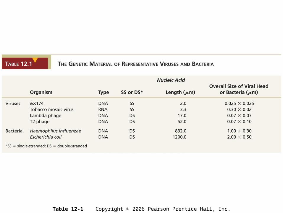
Table 12-1 Copyright © 2006 Pearson Prentice Hall, Inc.

Figure 12-2 Copyright © 2006 Pearson Prentice Hall, Inc.

Figure 12-3 Copyright © 2006 Pearson Prentice Hall, Inc.
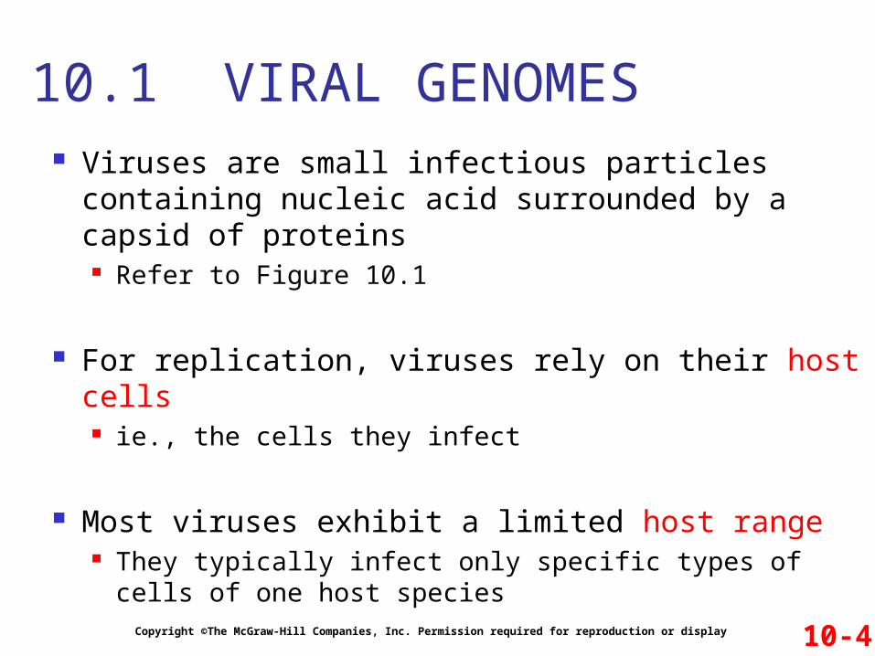
Viruses are small infectious particles containing nucleic acid surrounded by a capsid of proteins Refer to Figure 10.1
For replication, viruses rely on their host cells ie., the cells they infect
Most viruses exhibit a limited host range They typically infect only specific types of cells of one
host species
Copyright ©The McGraw-Hill Companies, Inc. Permission required for reproduction or display
10.1 VIRAL GENOMES
10-4
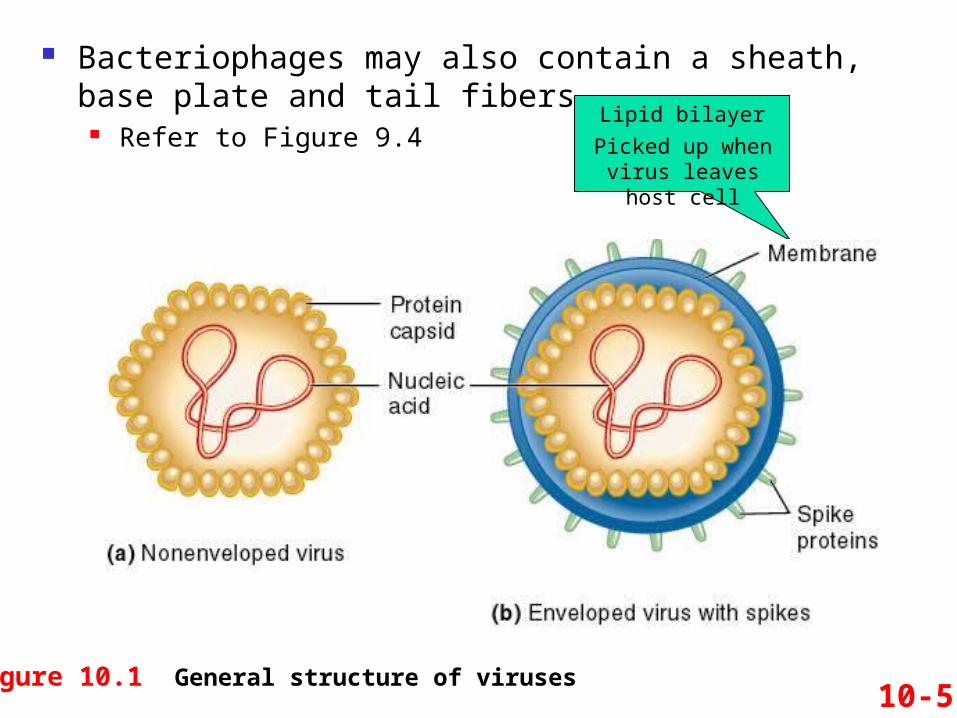
Figure 10.1 General structure of viruses10-5
Bacteriophages may also contain a sheath, base plate and tail fibers
Refer to Figure 9.4Lipid bilayer
Picked up when virus leaves host cell
The genome can be DNA or RNA Single-stranded or double-stranded Circular or linear
Viral genomes vary in size from a few thousand to more than a hundred thousand nucleotides
Copyright ©The McGraw-Hill Companies, Inc. Permission required for reproduction or display
Viral Genomes
10-6

Copyright ©The McGraw-Hill Companies, Inc. Permission required for reproduction or display 10-7

During an infection process, mature viral particles need to be assembled
Copyright ©The McGraw-Hill Companies, Inc. Permission required for reproduction or display 10-8
Viruses with a simple structure may self-assemble Genetic material
and capsid proteins spontaneously bind to each other
Example: Tobacco mosaic virus Figure 10.2
Capsid protein
Capsid composed of 2,130 identical protein subunits

The bacterial chromosome is found in a region called the nucleoid Refer to Figure 10.3
The nucleoid is not membrane-bounded
Copyright ©The McGraw-Hill Companies, Inc. Permission required for reproduction or display
10.2 BACTERIAL CHROMOSOMES
10-10

Fig. 10.3
Bacterial chromosomal DNA is usually a circular molecule that is a few million nucleotides in length Escherichia coli ~ 4.6 million base pairs Haemophilus influenzae ~ 1.8 million base pairs
A typical bacterial chromosome contains a few thousand different genes Structural gene sequences (encoding proteins) account
for the majority of bacterial DNA The nontranscribed DNA between adjacent genes are
termed intergenic regions
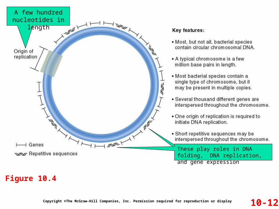
Figure 10.4 summarizes the key features of bacterial chromosomes
Copyright ©The McGraw-Hill Companies, Inc. Permission required for reproduction or display 10-11
Copyright ©The McGraw-Hill Companies, Inc. Permission required for reproduction or display 10-12
Figure 10.4
A few hundred nucleotides in length
These play roles in DNA folding, DNA replication, and gene expression

To fit within the bacterial cell, the chromosomal DNA must be compacted about a 1000-fold This involves the formation of loop domains
Copyright ©The McGraw-Hill Companies, Inc. Permission required for reproduction or display 10-13
Figure 10.5
The number of loops varies according to the size of the bacterial chromosome and the species
E. coli has 50-100 with 40,000 to 80,000 bp of DNA in each
The looped structure compacts the chromosome about 10-fold
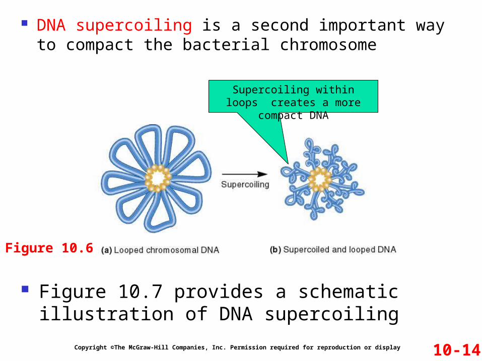
DNA supercoiling is a second important way to compact the bacterial chromosome
Copyright ©The McGraw-Hill Companies, Inc. Permission required for reproduction or display 10-14
Figure 10.6
Figure 10.7 provides a schematic illustration of DNA supercoiling
Supercoiling within loops creates a more compact DNA

10-15
Figure 10.7Plates preventing DNA
ends from rotating freely
Fewer turnsMore turns
Both overwinding and underwinding can
induce supercoiling
These three DNA conformations are topoisomers of each other
These two DNA conformations do not occur in living cells

Figure 12-4 Copyright © 2006 Pearson Prentice Hall, Inc.
The chromosomal DNA in bacteria is negatively supercoiled In E. coli, there is one negative supercoil per 40 turns of
the double helix
Negative supercoiling has two major effects 1. Helps in the compaction of the chromosome
Refer to Figure 10.6 2. Creates tension that may be released by DNA
strand separation Refer to Figure 10.8
Copyright ©The McGraw-Hill Companies, Inc. Permission required for reproduction or display
Chromosome Function Is Influenced by DNA Supercoiling
10-16

Copyright ©The McGraw-Hill Companies, Inc. Permission required for reproduction or display 10-17Figure 10.8
This enhances DNA replication and transcription

The control of supercoiling in bacteria is accomplished by two main enzymes
1. DNA gyrase (also termed DNA topoisomerase II) Introduces negative supercoils using energy from ATP
Refer to Figure 10.9 It can also relax positive supercoils when they occur
2. DNA topoisomerase I Relaxes negative supercoils
The competing action of these two enzymes governs the overall supercoiling of bacterial DNA
Copyright ©The McGraw-Hill Companies, Inc. Permission required for reproduction or display 10-18

Eukaryotic species contain one or more sets of chromosomes
Each set is composed of several different linear chromosomes
The total amount of DNA in eukaryotic species is typically greater than that in bacterial cells
Chromosomes in eukaryotes are located in the nucleus To fit in there, they must be highly compacted
This is accomplished by the binding of many proteins The DNA-protein complex is termed chromatin
Copyright ©The McGraw-Hill Companies, Inc. Permission required for reproduction or display
10.3 EUKARYOTIC CHROMOSOMES
10-21

Eukaryotic genomes vary substantially in size Refer to Figure 10.10a
In many cases, this variation is not related to complexity of the species For example, there is a two fold difference in the size of
the genome in two closely related salamander species Refer to Figure 10.10b
The difference in the size of the genome is not because of extra genes
Rather, the accumulation of repetitive DNA sequences These do not encode proteins
Copyright ©The McGraw-Hill Companies, Inc. Permission required for reproduction or display 10-22
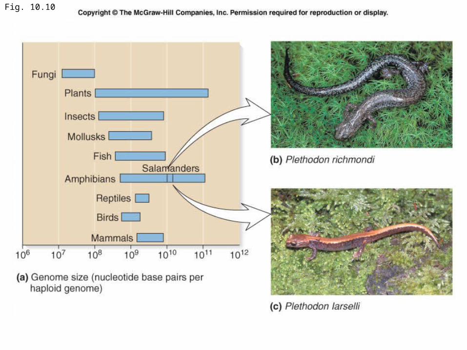
Fig. 10.10

A eukaryotic chromosome contains a long, linear DNA molecule Refer to Figure 10.11
Three types of DNA sequences are required for chromosomal replication and segregation Origins of replication Centromeres Telomeres
Copyright ©The McGraw-Hill Companies, Inc. Permission required for reproduction or display
Organization of Eukaryotic Chromosomes
10-24

10-25Figure 10.11

Genes are located between the centromeric and telomeric regions along the entire chromosome A single chromosome usually has a few hundred to
several thousand genes
In lower eukaryotes (such as yeast) Genes are relatively small
They contain primarily the sequences encoding the polypeptides ie: Very few introns are present
In higher eukaryotes (such as mammals) Genes are long
They tend to have many introns
Copyright ©The McGraw-Hill Companies, Inc. Permission required for reproduction or display 10-26
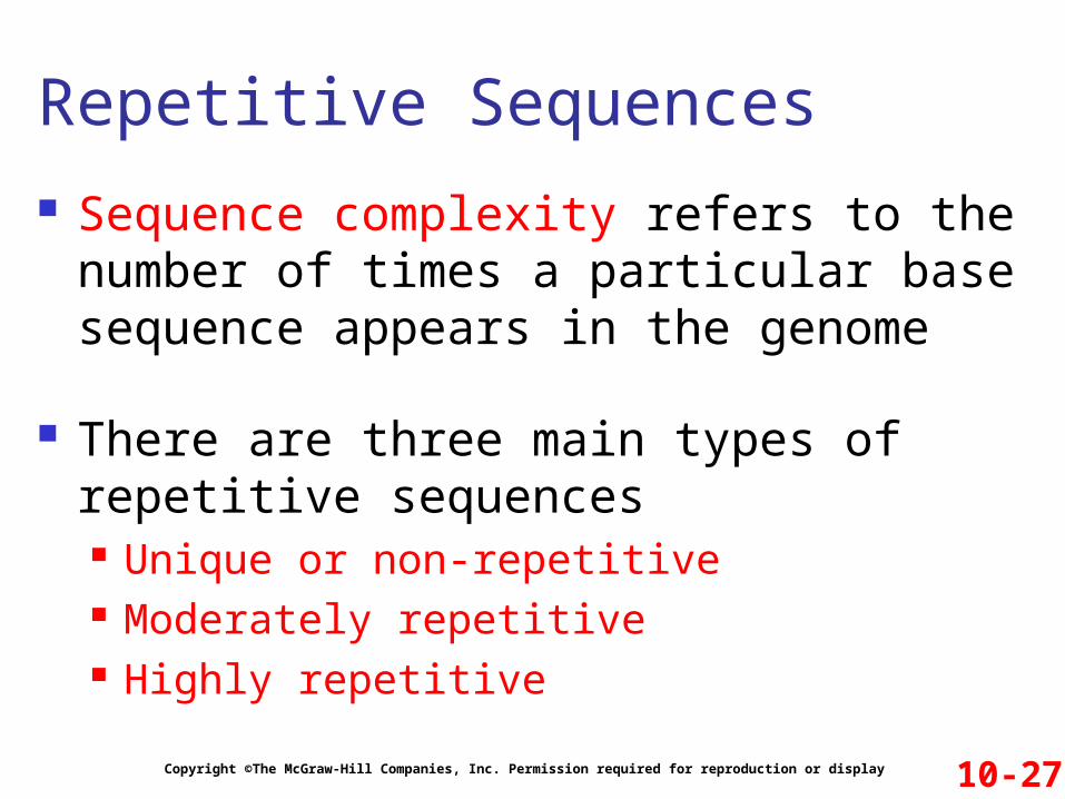
Sequence complexity refers to the number of times a particular base sequence appears in the genome
There are three main types of repetitive sequences Unique or non-repetitive Moderately repetitive Highly repetitive
Copyright ©The McGraw-Hill Companies, Inc. Permission required for reproduction or display
Repetitive Sequences
10-27
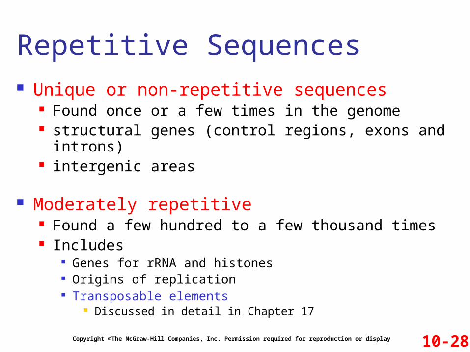
Unique or non-repetitive sequences Found once or a few times in the genome structural genes (control regions, exons and introns) intergenic areas
Moderately repetitive Found a few hundred to a few thousand times Includes
Genes for rRNA and histones Origins of replication Transposable elements
Discussed in detail in Chapter 17
Copyright ©The McGraw-Hill Companies, Inc. Permission required for reproduction or display
Repetitive Sequences
10-28
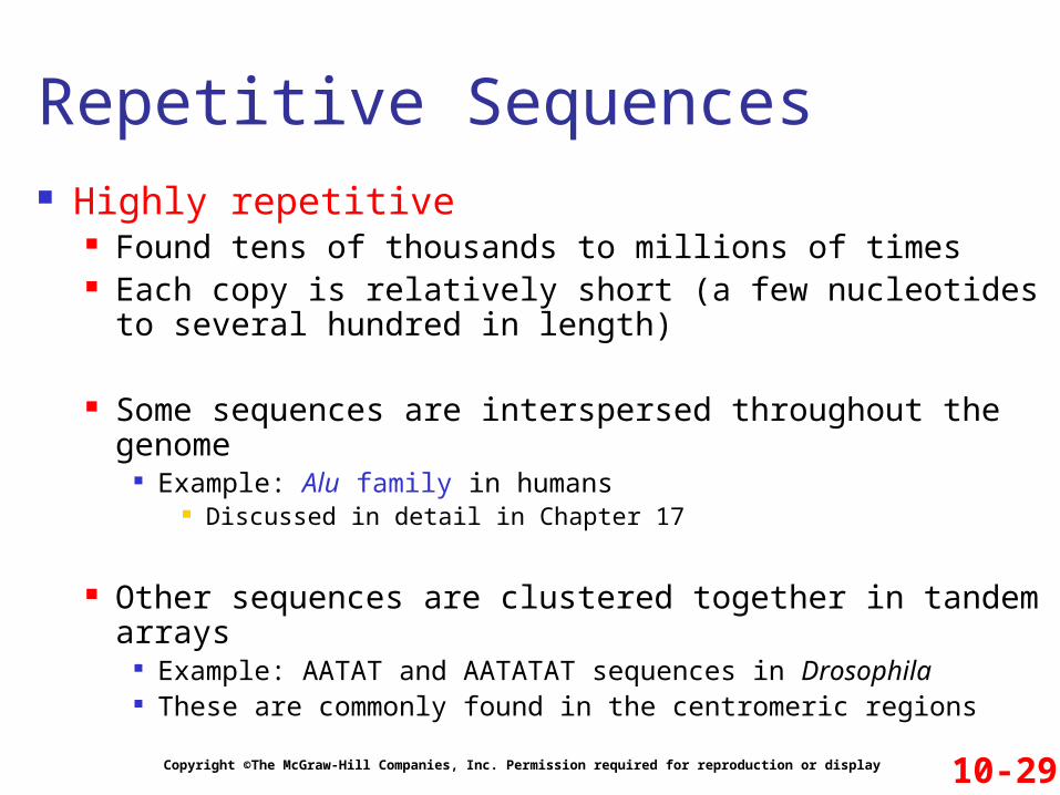
Highly repetitive Found tens of thousands to millions of times Each copy is relatively short (a few nucleotides to several
hundred in length)
Some sequences are interspersed throughout the genome Example: Alu family in humans
Discussed in detail in Chapter 17
Other sequences are clustered together in tandem arrays Example: AATAT and AATATAT sequences in Drosophila These are commonly found in the centromeric regions
Copyright ©The McGraw-Hill Companies, Inc. Permission required for reproduction or display
Repetitive Sequences
10-29
10-38Copyright ©The McGraw-Hill Companies, Inc. Permission required for reproduction or display
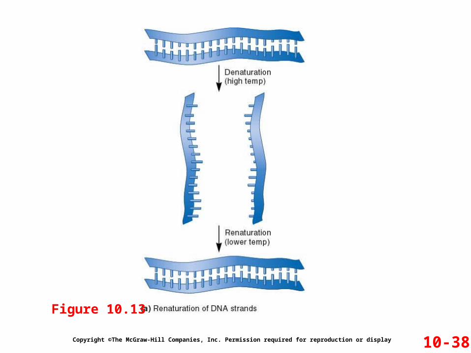
Figure 10.13

Figure 10-21 Copyright © 2006 Pearson Prentice Hall, Inc.

Effect of base composition on DNA melting temperature
• Hyperchromic shift during DNA denaturation is used to determine the melting temperature (Tm) (Figure 10.20). Melting
temperature is a method for estimating the base composition of DNA.
•G:C base pairs more stable than A:T base pairs
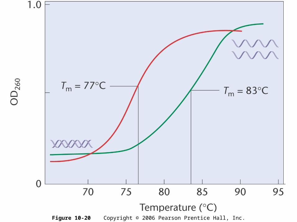
Figure 10-20 Copyright © 2006 Pearson Prentice Hall, Inc.
Copyright ©The McGraw-Hill Companies, Inc. Permission required for reproduction or display
If stretched end to end, a single set of human chromosomes will be over 1 meter long! Yet the cell’s nucleus is only 2 to 4 m in diameter
The compaction of linear DNA involves interactions between DNA and various proteins
Eukaryotic Chromatin Compaction
10-43

Copyright ©The McGraw-Hill Companies, Inc. Permission required for reproduction or display
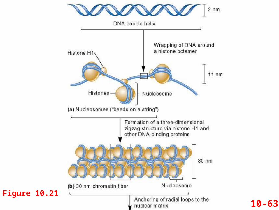
The repeating structural unit within eukaryotic chromatin is the nucleosome
It is composed of double-stranded DNA wrapped around an octamer of histone proteins An octamer is composed two copies each of four different
histones 146 bp of DNA make 1.65 negative superhelical turns
around the octamer
Refer to Figure 10.14a
Nucleosomes
10-44

Copyright ©The McGraw-Hill Companies, Inc. Permission required for reproduction or display 10-45
Overall structure of connected nucleosomes resembles “beads on a string” This structure shortens the DNA length about seven-fold
Figure 10.14
Vary in length between 20 to 100 bp, depending on species and cell type
Diameter of the nucleosome
Copyright ©The McGraw-Hill Companies, Inc. Permission required for reproduction or display
Histone proteins are basic They contain many positively-charged amino acids
Lysine and arginine These bind with the phosphates along the DNA backbone
There are five types of histones H2A, H2B, H3 and H4 are the core histones
Two of each make up the octamer
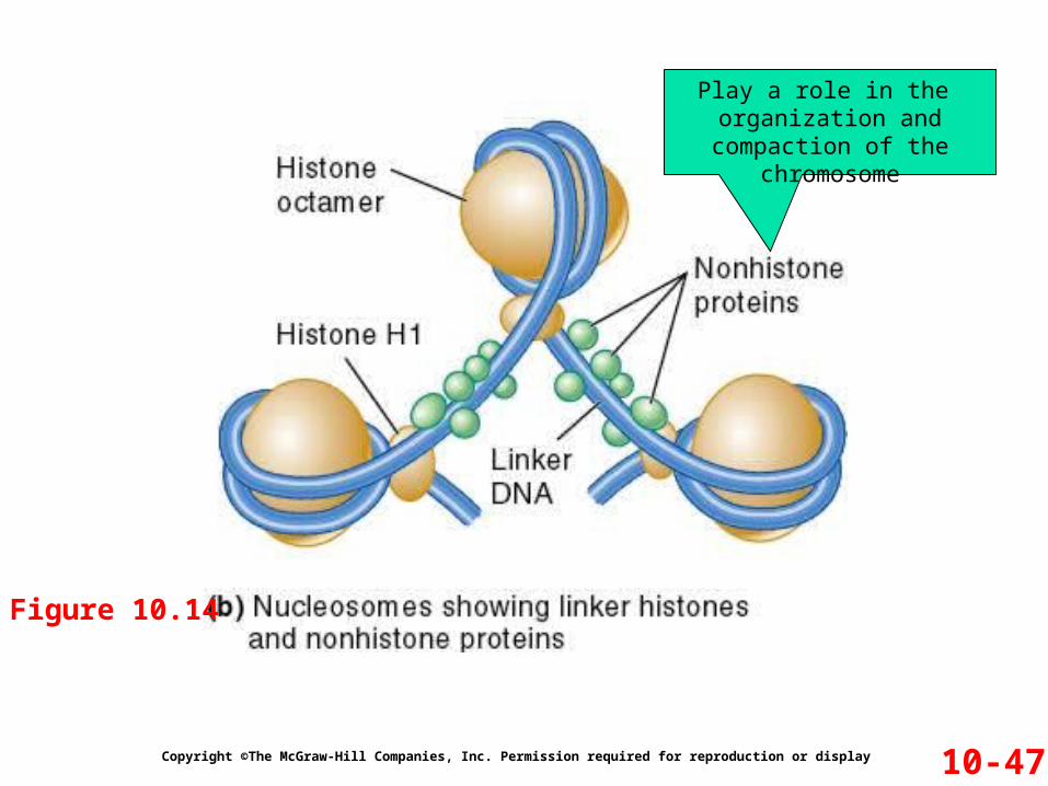
H1 is the linker histone Binds to linker DNA Also binds to nucleosomes
But not as tightly as are the core histones
Refer to Figure 10.14b
10-46
Copyright ©The McGraw-Hill Companies, Inc. Permission required for reproduction or display 10-47
Figure 10.14
Play a role in the organization and compaction
of the chromosome
The model of nucleosome structure was proposed in 1974 by Roger Kornberg
Kornberg based his proposal on various observations about chromatin Biochemical experiments X-ray diffraction studies Electron microscopy images
Copyright ©The McGraw-Hill Companies, Inc. Permission required for reproduction or display
Experiment 10B: Nucleosome Structure Revealed
10-48

Figure 12-8a Copyright © 2006 Pearson Prentice Hall, Inc.

Markus Noll tests Kornberg’s model
He did this via the following procedure Digest DNA with the enzyme DNase I Accurately measure the molecular weight of the
resulting DNA fragments
The rationale is that the linker DNA is more accessible than the “core DNA” to the DNase I
Copyright ©The McGraw-Hill Companies, Inc. Permission required for reproduction or display
Experiment 10B: Nucleosome Structure Revealed
10-49
The Hypothesis The experiment tests the beads-on-a-string model
of chromatin structure It the model is correct, DNase I should cut in the linker
region Thereby producing DNA pieces that are about 200 bp long
Copyright ©The McGraw-Hill Companies, Inc. Permission required for reproduction or display
Testing the Hypothesis
Refer to Figure 10.15
10-50

10-51Figure 10.15
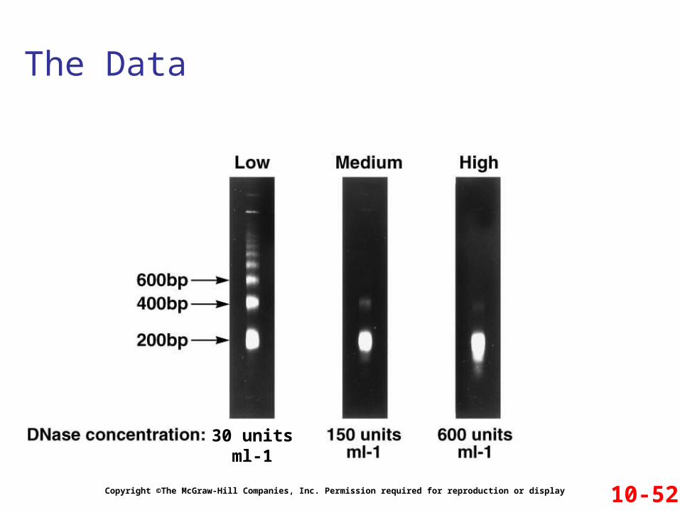
The Data
Copyright ©The McGraw-Hill Companies, Inc. Permission required for reproduction or display 10-52
30 unitsml-1

Interpreting the Data
Copyright ©The McGraw-Hill Companies, Inc. Permission required for reproduction or display 10-53
30 unitsml-1
All chromosomal DNA digested into fragments that are ~ 200 bp in length
These longer pieces were all in multiples of 200 bp
At low concentrations, DNase I did not cut at all
the linker DNA
This fragment contains two nucleosomes
And this, three

Copyright ©The McGraw-Hill Companies, Inc. Permission required for reproduction or display
Nucleosomes associate with each other to form a more compact structure termed the 30 nm fiber
Histone H1 plays a role in this compaction At moderate salt concentrations, H1 is removed
The result is the classic beads-on-a-string morphology At low salt concentrations, H1 remains bound
Beads associate together into a more compact morphology
Refer to Figure 10.16
Nucleosomes Join to Form a 30 nm Fiber
10-54
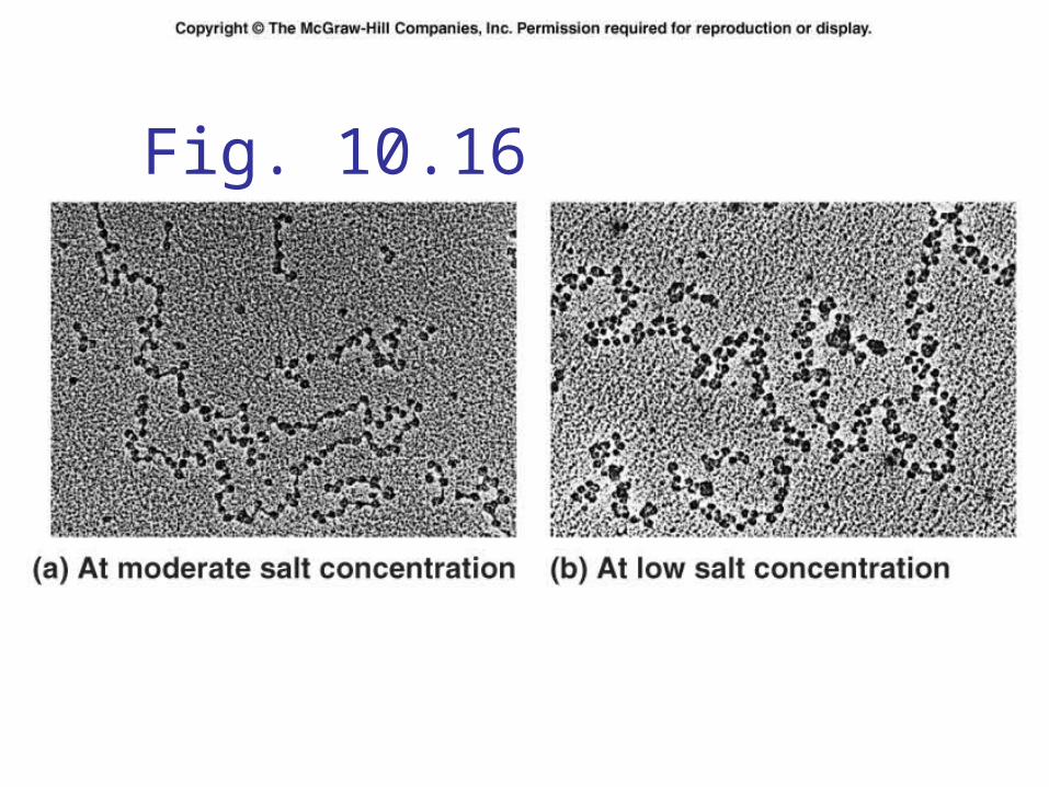
Fig. 10.16

Copyright ©The McGraw-Hill Companies, Inc. Permission required for reproduction or display
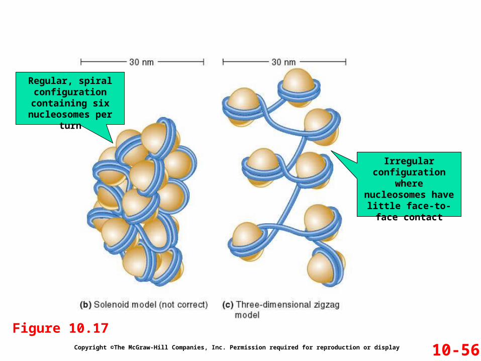
The 30 nm fiber shortens the total length of DNA another seven-fold
Its structure has proven difficult to determine The DNA conformation may be substantially altered
when extracted from living cells
Two models have been proposed Solenoid model Three-dimensional zigzag model
Refer to Figure 10.17b and c
10-55
Copyright ©The McGraw-Hill Companies, Inc. Permission required for reproduction or display 10-56
Figure 10.17
Regular, spiral configuration containing six
nucleosomes per turn
Irregular configuration where nucleosomes have little face-to-face
contact

Copyright ©The McGraw-Hill Companies, Inc. Permission required for reproduction or display
The two events we have discussed so far have shortened the DNA about 50-fold
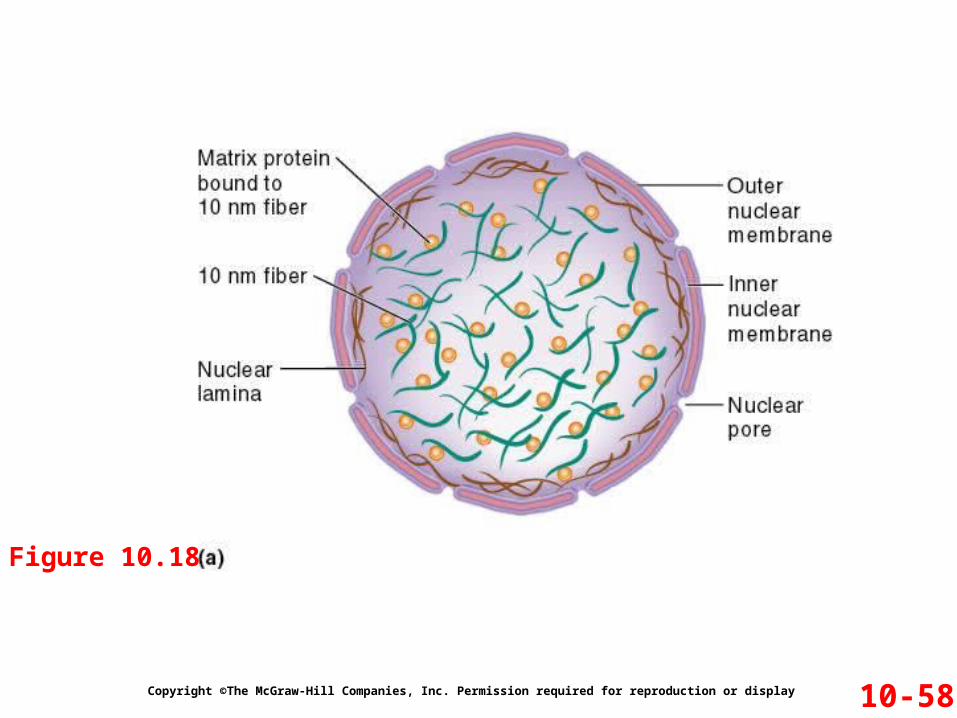
A third level of compaction involves interaction between the 30 nm fiber and the nuclear matrix
The nuclear matrix is composed of two parts Nuclear lamina Internal matrix proteins
10 nm fiber and associated proteins
Further Compaction of the Chromosome
10-57
Copyright ©The McGraw-Hill Companies, Inc. Permission required for reproduction or display 10-58
Figure 10.18
Copyright ©The McGraw-Hill Companies, Inc. Permission required for reproduction or display 10-59
Figure 10.18
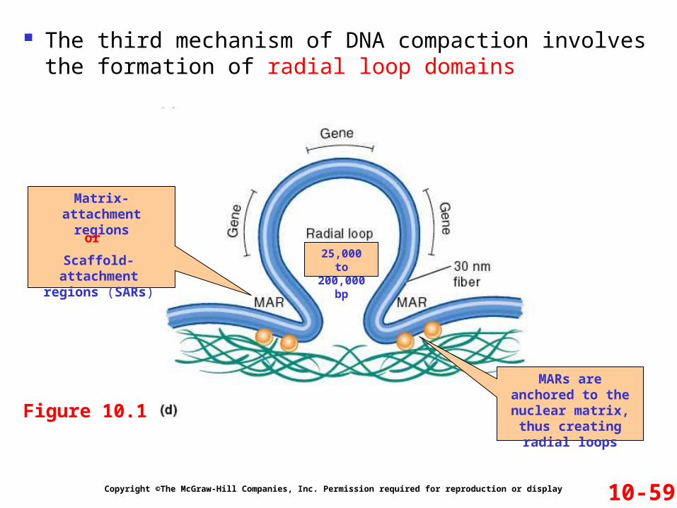
The third mechanism of DNA compaction involves the formation of radial loop domains
Matrix-attachment regions
Scaffold-attachment regions (SARs)
or
MARs are anchored to the nuclear
matrix, thus creating radial loops
25,000 to 200,000 bp
Copyright ©The McGraw-Hill Companies, Inc. Permission required for reproduction or display
The attachment of radial loops to the nuclear matrix is important in two ways 1. It plays a role in gene regulation
Discussed in Chapter 15
2. It serves to organize the chromosomes within the nucleus
Each chromosome in the nucleus is located in a discrete and nonoverlapping chromosome territory
Refer to Figure 10.19
Further Compaction of the Chromosome
10-60

Fig. 10.19

Euchromatin Less condensed regions of chromosomes Transcriptionally active Regions where 30 nm fiber forms radial loop domains
Heterochromatin Tightly compacted regions of chromosomes Transcriptionally inactive (in general) Radial loop domains compacted even further
Heterochromatin vs Euchromatin
10-61
10-63Figure 10.21

10-64Figure 10.21
Compaction level in euchromatin
Compaction level in heterochromatin
During interphase most chromosomal
regions are euchromatic
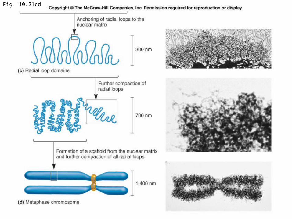
Fig. 10.21cd

Copyright ©The McGraw-Hill Companies, Inc. Permission required for reproduction or display
As cells enter M phase, the level of compaction changes dramatically By the end of prophase, sister chromatids are entirely
heterochromatic Two parallel chromatids have an overall diameter of
1,400 nm
These highly condensed metaphase chromosomes undergo little gene transcription
Metaphase Chromosomes
10-65
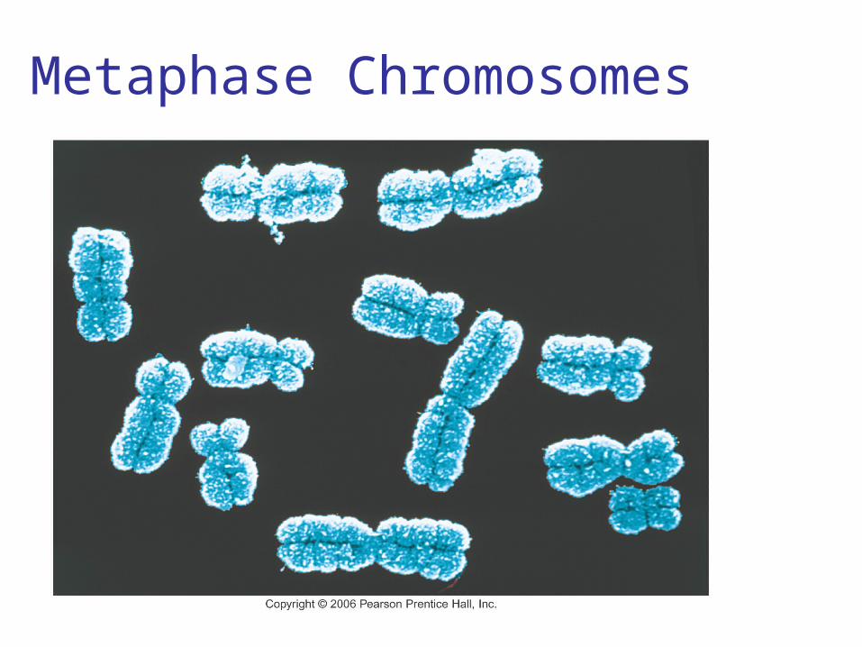
Metaphase Chromosomes

Fig. 10.22a

Fig. 10.22b