practical alpaca reproduction - nunoaproject.org
TRANSCRIPT

Practical Alpaca
Reproduction
Stephen R. Purdy, DVM
North American Camelid Studies Program
Providing education and support for farmers,
veterinarians, and students in the US and Peruwww.nunoaproject.org

Learning Objectives:
• To describe the normal anatomy of the male and female alpaca and llama.
• To describe the physiology associated with alpaca and llama reproduction.
• To describe reproductive behavior in camelids.
• To introduce ultrasound pregnancy detection in camelids

Female Reproductive
Tract Anatomy
• Ovaries
– produce gametes and hormones that act on other parts of the reproductive tract
• Uterus
– receives the semenduring breeding, and protects and nourishes the developing embryo and fetus during pregnancy

• Cervix– a barrier that secretes mucus during receptivity
and produces a mucus plug seal during pregnancy
• Vagina– the copulatory organ and produces lubricating
mucus during receptivity• Oviducts
– provide optimum environment for fertilizationand pre-attachment development of the embryo


Cervix
• External os protrudes slightly into the vagina
• 2 - 3 non continuous rings within
• Dilation = abnormal

Cervical appearance


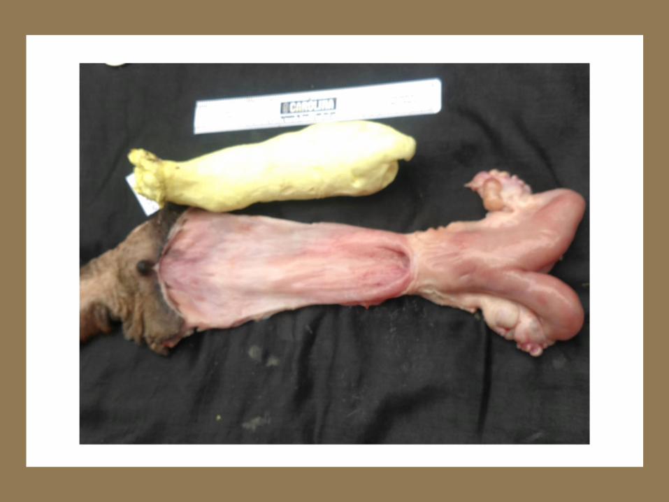
Uterus and Ovary with Follicle
Ovarian
Follicle
Uterine
Body
Uterine
Horn
Oviduct

Vulva
Anus
Vulva

Major effects of
estrogen on the camelid
reproductive tract
– Increased blood flow
– Relaxation of the cervix
– Increased white blood cells to counteract the contamination which occurs at breeding
– Increased secretion of mucus
– uterine gland growth
– Increased uterine muscular tone
– Behavioral receptivity

•All types (maturation levels) of follicles are present in the ovary at all times.• Corpora lutea (CLs) may or may not be present
depending on whether an ovulation has occurred.
Ovarian Physiology
• Follicular Phase– Follicles are fluid filled structures within the
ovaries that contain the eggs (ova)– Camelids do not have estrous cycles as in
other mammals– Camelids are termed “induced ovulators”
• breeding causes ovulation= rupture of the follicle and release of the egg
• does not mean always receptive!

Follicular Growth
– Several small (< 3mm) follicles are present at all times
– Some follicles grow and regress
– Interrupted by ovulation after copulation or exogenous hormone stimulation
– follicles are capable of ovulation at 6 mm diameter.
– If no ovulation occurs the follicle regresses, shrinking back down in size, or disappears within 2 days (unpublished, 2011)
– Follicular sizes do not necessarily correlate with sexual receptivity


Ovarian Follicles

Ovarian Follicles

Ovarian Follicles

Anovulatory Follicles
• > 15 mm diameter are abnormal
• Grow up to 30 mm in diameter.
• No effect on sexual receptivity
• Resolve without treatment through rupture

Luteal phase of the ovarian cycle
• Occurs after ovulation
• Lasts for months if the female is pregnant
• Ovulatory Corpus Luteum (CL) is responsible for progesterone production throughout pregnancy
• 6-15 mm follicles burst releasing the ovum or egg approximately 24 hours after breeding
• Follicles develop during pregnancy

Corpus Hemorrhagicum
• Transitory blood filled structure for ~ 48 – 96 hrs. after ovulation
• Can be visualized with transrectal ultrasound at approximately 48 hours after breeding
• Replaced by the “corpus luteum” (CL) which grows and secretes progesterone.


Corpus luteum, embryonic vesicle, ovarian follicle

Retained CL
• CL remains functioning without a pregnancy = non-receptive behavior
• treatment with PGF2α
• suggest 200 µg of cloprostenol sodium (0.8 ml of Estrumate® for alpacas or llamas) SQ, two days in a row
• May be over diagnosed

Return to Receptivity
• If no pregnancy is established expect in approximately 12 to 14 days
– a longer period of time if the animal experienced early embryonic death
– varies with each ovulation
– not necessarily consistent for a specific female

Pregnancy Termination
• Two doses of 200 µg of cloprostenol sodium (0.8 ml of Estrumate® for alpacas or llamas) SQ daily in a row is most effective after 60 days of pregnancy
• One dose may be sufficient before 60 days
• Check (behavior test) for return to receptivity within 7 days
• Recheck pregnancy status with ultrasound if animal does not become receptive.

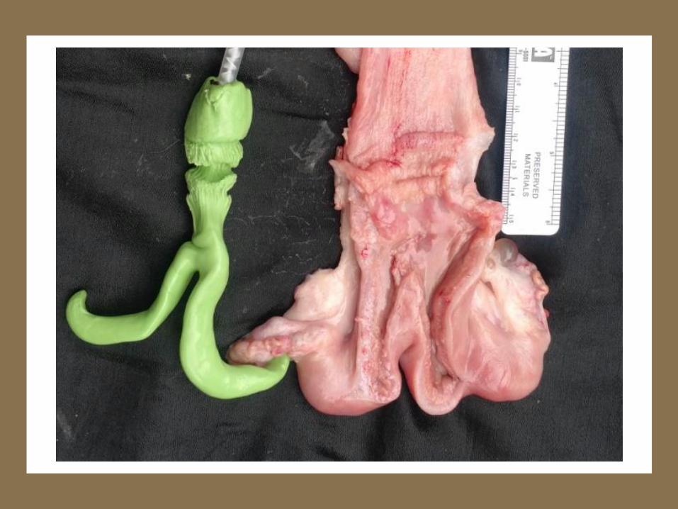
Male Reproductive
Anatomy
• Two testicles located in the scrotum– much smaller than those
of other species of comparable body size• Alpacas
– length 3.5 to 5.0 cm
– thickness 2.0 to 3.0 cm
• measure with ultrasound or calipers
– larger testicle ? = greater sperm production
– size does not vary with photoperiod length

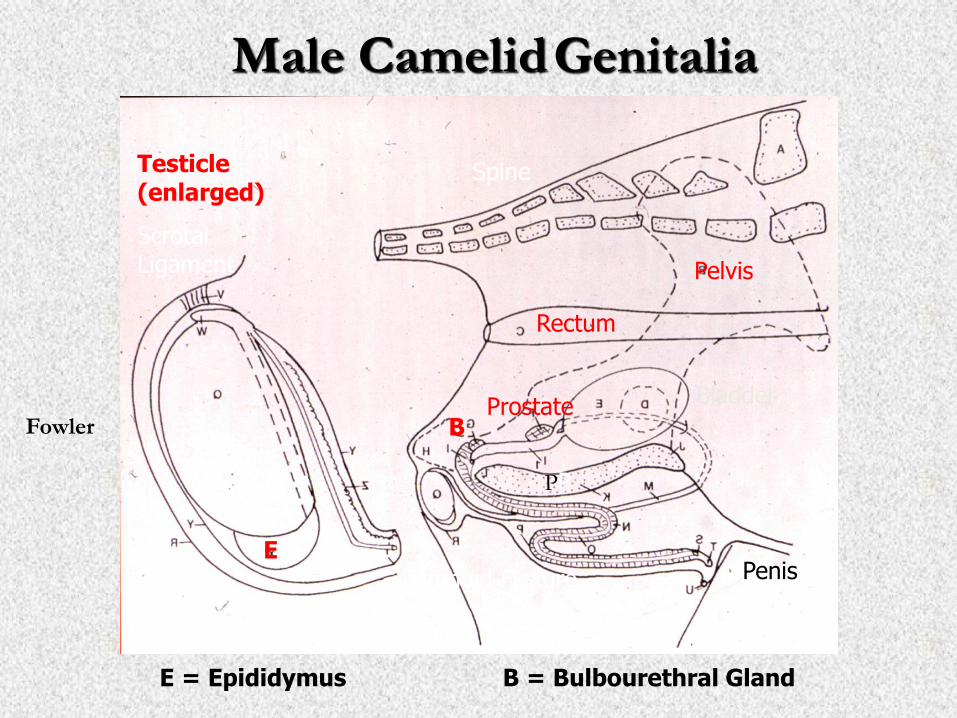
Testicle(enlarged)
Bladder
Penis
Rectum
Male CamelidGenitalia
Fowler
Pelvis
Scrotal Ligament
PP
Prostate
Sigmoid Flexure
Spine
B
B = Bulbourethral Gland
Testes
E = Epididymus
E

Male Tract
(ventral view)
• Retractor penis muscles (RPM)
• Prostate gland (PG)
• Glans penis (GP)
PGGP

Penis is fibroelastic with a sigmoid flexure when relaxed
– Males urinate backwards between the legs since the tip of the sheath points caudally and the penis is not extended during urination
– Mature male alpacas partially extend the penis out of its sheath in the standing position when mounting receptive females
– The penis has a corkscrew appendage at its tip that is used to dilate the cervix and enter the uterus during breeding
– Cartilaginous process on the end of the penis- function unknown


Glans Penis corkscrew appendage - auxiliary process

Tail
Head
Testis
Testisand
Epididymis

Reproductive Behavior in
Alpacas

Sexual receptivity in the female
• Extremely variable!
• In the presence of the male:
– Immediate acceptance= receptive (R)
– Female stands when mounted by the male, but does not drop= recheck later= not receptive (NR)
– Refusal= not receptive (NR)
• Acceptance = female dropping for breeding upon approach of, or mounting by, the male
• Refusal = standing, running away, spitting, vocalizing

Behavior Testing

Home Schooling
45


Receptive Behavior
Some females run with others and do not show receptivity unless cornered
Females demonstrate preferences for different males on the same day
Males and females get used to each other if housed in close proximity and will stop showing aggressive breeding and receptivity behavior
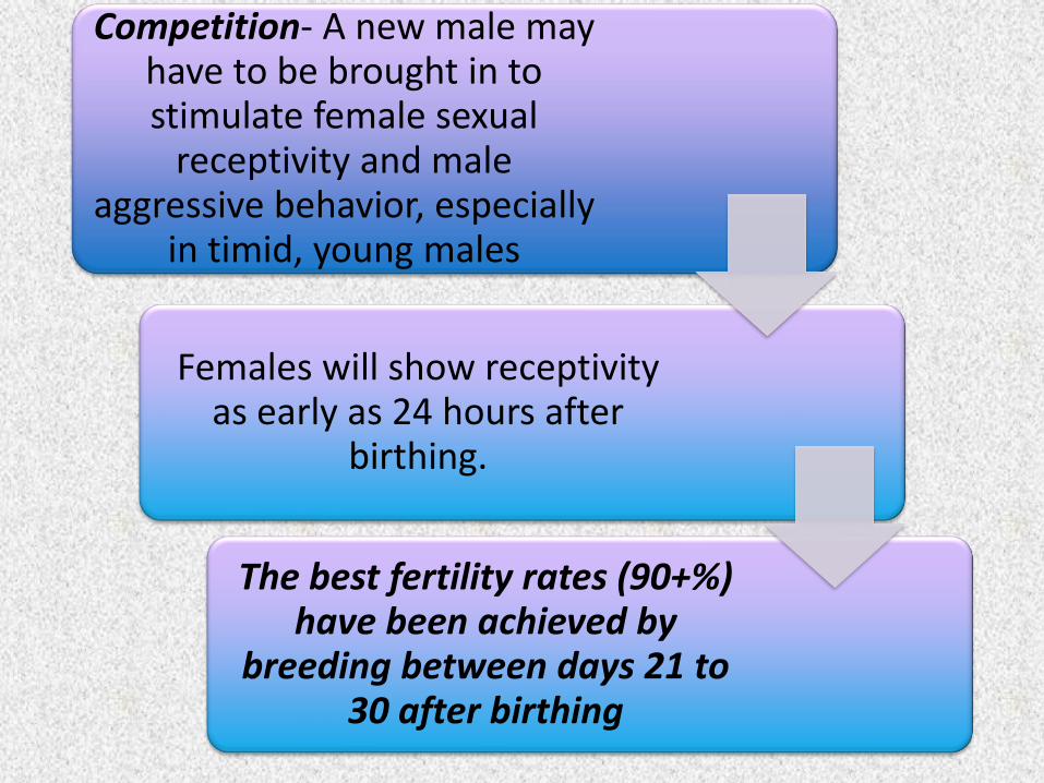
Competition- A new male may have to be brought in to stimulate female sexual
receptivity and male aggressive behavior, especially
in timid, young males
Females will show receptivity as early as 24 hours after
birthing.
The best fertility rates (90+%) have been achieved by
breeding between days 21 to 30 after birthing


Male Breeding Behavior
Most males will be interested in breeding at
1+ years old if allowed but it is best to start at 2 to 3
years of age to achieve the best fertility.
Penile adhesions prevent full extension of the penis
through the cervix
May have low libido especially if they have not
seen breedingsSmall males do not work well with large females-organ mismatch due to requirement for cervical
penetration, and breeding position


Breeding ActBreeding - 10 to 45 minutes (average
20) in the down position and noisy.
• The male moves forward to achieve the correct penile penetration into the uterus.
Produce a dribble ejaculate of small volume [0.5 to 7.5 ml collected
(alpacas) in an artificial vagina] over the course of the breeding.
Hot, humid weather reduces libido

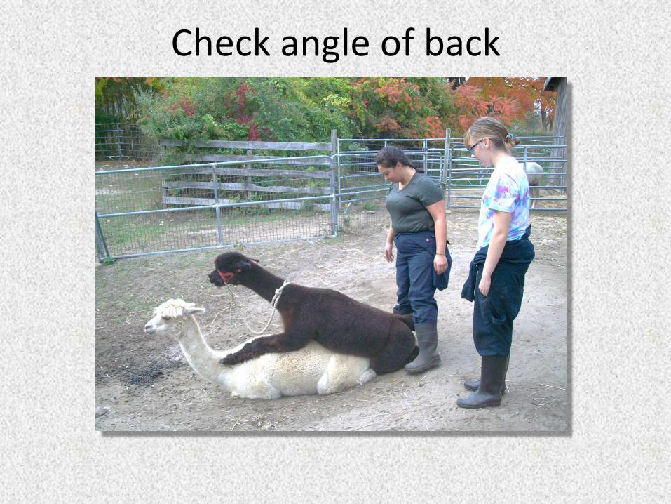
Check angle of back


Success!

Semen Deposition
• Average 20 minute breeding
• Look for the hemorrhage

Males usually more aggressive in smaller pens than on pasture
Males may breed the same female multiple times per day
Males may stop breeding to fight with others if they are too close together
Lower pecking order males may not BT well if the dominant male is nearby
Immature males (2-3 yrs. old) need time and training

Let the males and females figure it out

Pregnancy Detection
in Camelids
• The highest incidence of early embryonic death (EED) occurs in the first 30 to 50 days. ~ 20% incidence– Highest in first 30 days– A natural occurrence and a
way of eliminating abnormal embryos.
– Advantageous to lose bad embryos early and to quickly return to receptivity.
• Weekly refusal during behavior testing is presumptive of pregnancy.
• Elevated progesterone above normal baseline levels (approx. 2 ng/ml) indicate a functional CL, but are notspecific for pregnancy.

Ultrasound Pregnancy Detection
• an easy technique to master• easily performed with a 5 MHz or 7.5 MHz
linear probe• Embryonic age and viability can be evaluated
quickly by size of the pregnancy and presence of a heartbeat after 25 days
• Fetal movement is usually evident after 40 days of pregnancy
• The procedure is well tolerated by most alpacas and llamas

Small Intestine
LargeIntestine

Uterine horn (between the + signs) as imaged by transrectal ultrasound with a 7.5 MHz linear probe.
(urinary bladder appears in the left lower corner)

Transrectal Ultrasound of 25 day Camelid Pregnancy
A
B
A = embryonic fluid
B = embryo proper
[10 mm vertical divisions]

Early Embryonic Death
eliminate bad embryos >> female has a
chance to become
pregnant again within a short
time
10 pregnancies in 5 multiparous
females (Brunsden, Purdy, et al, 2011)
4 EED = 40%
1 before 12 days; the others at
30,31, and 41 days
7 pregnancies in 8 females- 7
multiparous and 1 primiparous (Purdy
et al, 2013)
4 EED = 57%
All in the first 30
days

Embryonic and Fetal Development
• Twin conceptions
–Occur as the result of double ovulations
– Identify at 15 to 30 days of pregnancy
–15% occurrence in a sample of 40 breedings; 1 set of twins persisted to 45 days (Purdy et al, 2011, 2013)
–40 % in our herd over 4 years

Twin Births
• Twin birthing rate is very low
– Most often one embryo will continue to increase in size while the other decreases
– Follow with ultrasound to monitor natural reduction to a singleton pregnancy
• 700 births in 2 northeast herds over 10-20 years
– 1 set of live twins
– No aborted twins
• Should we worry about diagnosing twins?

2 year results- 10
females
3- 11 years old
Bred spring and fall semesters with young males
48 pregnancies
• EED = 19%
• Twins = 40%

Twin Embryos

Internal Ultrasound
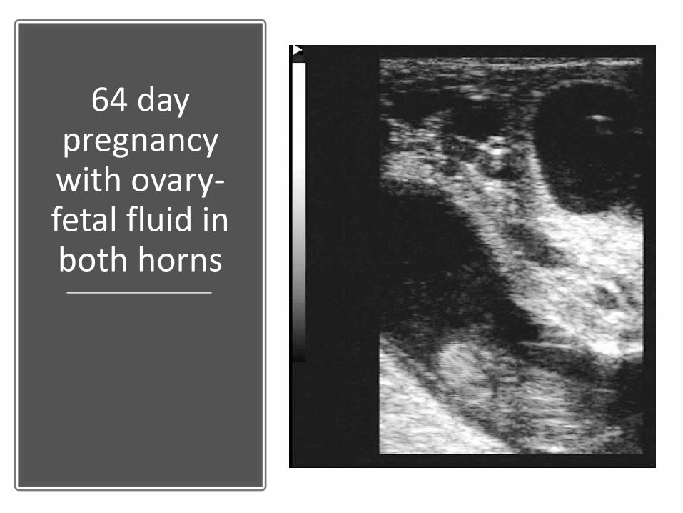
64 day pregnancy with ovary-fetal fluid in both horns

Transabdominal Ultrasound
• performed on left side of the caudal abdomen adjacent to the udder between 40 - 80 days
• on the right side 80 days until term
– Abdominal anatomical restrictions (stomach) are responsible for this shift in location.
• After a negative ultrasound
– Transrectal ultrasound to identify both uterine horns
– behavior test
– vaginal exam


78 day fetus shows
beginning of ossification of the skull
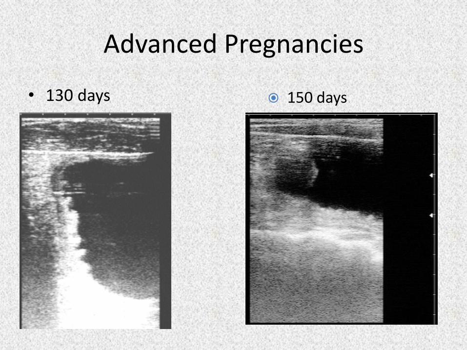
Advanced Pregnancies
• 130 days 150 days

210 days Later

Thank You and
Questions??