practical neurology back pain wendy blount, dvm. some rules about back pain completely unilateral...
TRANSCRIPT

Practical NeurologyBack Pain
Wendy Blount, DVM
Some rules about back pain
• Completely unilateral neuro signs rarely arise from the spinal cord– Usually bilateral– Though may be more pronounced on one
side– Monoparesis – think peripheral nerve
disease first
• The first neuro deficit is:– Conscious proprioreception– Then voluntary motor– Then superficial pain– Then deep pain

Does this dog’s back hurt?
Things that can look like Back Pain• Referred abdominal pain
– Abnormalities on abdominal x-rays, barium series, ultrasound or bloodwork
• Muscle pain– CPK high, Confirm with muscle biopsy– Immune mediated polymyositis– Beagle Pain Syndrome
• Orthopedic pain– Bilateral knees and hips– Complete musculoskeletal exam & x-rays
• Neuro exam normal on imitators

Does this dog’s back hurt?
Back Pain can look like something else• Limb lameness
– Root signature - limping on one leg– Extension of the limb does indeed hurt
• Abdominal pain– Pressure put on back when palpation
abdomen
• Constipation– Dogs with lumbosacral pain don’t want to
squat to defecate

Does this dog’s back hurt?
Back Pain can look like something else• Lethargy
– Can be confused with reluctance to move
• Orchitis, Epididymitis– Appear as if back hurts

DDx for back/neck pain
• Intervertebral Disc Disease• Wobbler Syndrome• Congenital spinal malformations• Neoplasia• Discospondylitis • Meningitis• Spinal arthritis & spondylosis• Trauma• Forebrain mass

DDx for back/neck pain
Uncommon Causes of Back Pain• Extradural synovial cysts, arachoid
cysts, dermoid cysts• Myelodysplasia
– Meningocoeles/Myelomeningocoeles– Syringomyelia/Hydromyelia– Spinal dysraphism
• Spina bifida– Failure of dorsal laminae to fuse– Associated spinal cord malformations

DDx for back/neck pain
Uncommon Causes of Back Pain• Multiple Cartilagenous exostoses
– Nodules of cartilage/bone proliferate from growth plates
• Hypervitaminosis A– Cats fed primarily liver– Vertebral exostoses– Prognosis poor
• Methionine deficiency– Hunting dogs fed primarily tripe (Europe)– T3-L3 progressive myelinopathy– Prognosis good with proper diet

DDx for back/neck pain
Uncommon Causes of Back Pain• Calcinosis Circumscripta• Dural Ossification• Disseminated idiopathic skeletal
hyperostosis (DISH)– Periarticular ossification throughout the
body

DDx for back/neck pain
Causes of Progressive Rear end Weakness without Pain
• LMN Reflexes– Degenerative Myelopathy– Hypothyroidism Polyneuropathy– Botulism– Coonhound paralysis– Tick paralysis– End stage myasthenia gravis
• UMN Reflexes– Rottweiler Leukoencaphalomyelopathy– Hereditary Ataxia of Jack Russell Terriers– Afghan Hound Myelopathy

Intervertebral Disc Disease
Type I Disc Disease• Annulus around the disk weakens• Disc material acutely extrudes• Acute pain• +/- neuro deficits• Small dogs
Type II Disc Disease• Annulus gradually thickens• Insidious weakness• Neuro deficits > pain• Large dogs

Intervertebral Disc Disease
Presentation• Uncommon in cats
• Upper cervical extrusion (Type I)– “The Screaming Chihuahua”– C2-3 most common– Severe neck pain– Mild neuro deficits– Nose down posture with arched back– Neck muscle fasciculations– Thoracic limb root signature

Intervertebral Disc Disease
Presentation• Type I TL Disc Disease
– Acute presentation– Usually T11-L5– Rarely T2-T10
• Intercapital ligament– Neuro deficits more common than with
upper cervical type I
• Type II Disc Disease– Progressive weakness with some back pain– Larger dogs

Intervertebral Disc Disease
Diagnosis• History and signalment• Physical Exam
– CP deficits tell you there is neuro disease– Neuro exam localizes the lesion
• CBC, panel, lytes, UA – normal– Urine culture if urine retention
• Radiographs• Referral – myelogram, CSF tap, CT/MRI

Intervertebral Disc Disease
Modified Frankel Scale• Grade 0 – paraplegia, no deep pain• Grade 1 – paraplegia, no superficial
pain• Grade 2 – paraplegia with normal pain
sensation• Grade 3 – nonambulatory paraparesis
– Some voluntary motor– Can’t bear weight without support
• Grade 4 – nonambulatory paraparesis– Can stand but not walk
• Grade 5 – ambulatory paraparesis

Intervertebral Disc Disease
Radiographs• Under sedation – GUARD THE SPINE!!
– Positioning is everything (esp. traction)– Patient comfort– Slightest movement causes blurring
• Survey radiographs can identify the site of disc herniation in 50-60% of cases
• Radiographic signs of disc disease:– Narrowing or wedging of disc space– Decreased size of intervertebral foramen– Reduced space between articular facets– Mineralized disc material in vertebral canal
or intervertebral foramen

Intervertebral Disc Disease
When is it Surgical?• Emergency surgery
– Rapidly deteriorating neurologic function• Do twice daily neurologic exams
– Non-ambulatory (can’t walk without assistance)
• Scheduled Surgery – chronic severe pain– Moderate to severe neuro deficits that fail to
improve
Intervertebral Disc Disease
Emergency Treatment• Confinement• IV fluid therapy
– Mediates ischemia
• Analgesia– Tramadol 3-5 mg/kg PO TID– NSAIDs– Opiates if needed

Intervertebral Disc Disease
Emergency Treatment• Glucocorticoids
– High dose SoluMedrol widely used– Also dexamethasone– Little evidence that it changes outcome in
dogs who proceed to surgery– Serious side effects are possible
• 33% have GI side effects to MPSS• Dexamethasone can increase risk of
colon perforation– Clinical experience tells us that it does help
non-surgical cases– Use in moderation NOT WITH NSAIDs
• 0.1 mg/kg SID-QOD

Intervertebral Disc Disease
Emergency Treatment• Free Radical Scavengers
– Fewer side effects than glucocorticoids– But no proven benefits– DMSO– Tirilazad– Polyethylene glycol– Poloxamer 188– Solcoseryl– Naloxone– Crocetin– TRH– Mannitol was associated with harm in feline
experimental model

Intervertebral Disc Disease
Long Term Treatment• CAGE REST!!!! (how long?)
– At least 2 weeks– Some recommend 4-6 weeks– Crate size – can change positions but not
walk around– Activity limited to leash walks– Gradually back to normal activity over 2-6
weeks after cage rest finished
• Monitor for progressive neuro signs– Weakness, paralysis– Difficulty urinating
• Analgesics
Intervertebral Disc Disease
Long Term Treatment• +/- Antiinflammatories (dose??)
– Prednisone – 0.5 mg/kg PO BID x 5-7 d, then SID x 7d, then QOD 7 doses
– NSAIDs (not both!!!)– DO NOT give anti-inflammatories without
cage rest!!
• Muscle Relaxants– Methocarbamol 15-20 mg/kg PO TID
• Acupuncture• Glucosamine/chondroitin

Intervertebral Disc Disease
Prognosis• Very few outcome studies on medically
managed dogs• No deep pain
– 40-50% will walk again with medical treatment
– 60-80% will walk again with surgery– 33% of those that walk again will have
intermittent incontinence– Recovery of deep pain within 2 weeks
carries a good prognosis
• Length of time between loss of deep pain and surgery – Surgery sooner is better than later– 48 hour rule – no longer widely accepted

Intervertebral Disc Disease
Prognosis• Non-ambulatory with pain sensation
– 80-95% success with surgery
• Mean time from surgery to ambulation– 10-13 days for small dogs– Much longer for large dogs
• Mean 7 weeks to ambulation• 62% walking in 4 weeks• 92% walking within 12 weeks• Longer for older, heavier patients
• Back pain alone without neuro deficits– 24 of 25 of dogs improved with surgery– No studies I am aware of on medical Tx

Intervertebral Disc Disease
Prognosis• More acute paralysis carries worse
prognosis– Those that go from walking to paralyzed in
less than one hour don’t do as well– Those who go down gradually (1-2 days)
have better prognosis
• Respiratory compromise– Prognosis same with a ventilator – Prognosis grave without ventilator
• Dogs non-ambulatory from type II disease over weeks to months have worse prognosis than type I

Intervertebral Disc Disease
Prognosis• 20% of dogs who have back surgery
will have another episode of back pain with neuro deficits– Most do not require surgery– Re-operate rate is <10%– 40% recurrence when treated medically
• Dogs with 5 or more mineralized discs at surgery have 50% recurrence rate

Lucky
• 17 year old male cocker spaniel with:– Hypothyroidism (Soloxine)– Glaucoma & prostheses– Cognitive Dysfunction Disorder– Hip Dysplasia (Rimadyl PRN, glucosamine)– Carcinoma L ear canal – debulked twice
• HPI - Started showing behavioral changes a few weeks ago– Episodes of panic – DDx
• Pain• Cerebral Disease
– Cognitive Dysfunction– Brain Tumor (ear tumor met??)– Infectious, Inflammatory, Metabolic
• Hypertension

Lucky
• Review of record shows BUN creeping up over past year (40-50)
• PE & Neuro Exam– Can’t assess vision ;-)– Short stride rear legs– CP deficits worse on L– Hip pain bilateral– Very brisk bilateral patellar reflexes
• Lesion – forebrain, cervical, TL, LS• CBC – normal• GHP/lytes – BUN 54• UA – SG 1.017, culture negative

Lucky
• Dx Plan – Episodes of Panic– Look for pain
• No new pain found on PE• Abdominal US - normal
– Look for metastasis• Chest x-rays and Abd US normal
– Blood Pressure 220/110– CSF tap/MRI discussed– Spinal films – cervical and TL normal
• No sedation• IVDDz L6-7 L7-S1, LS instability, severe
hip dysplasia

Lucky
• Tx Plan – New Problems– CRF
• K/D diet• Fish oil
– CCD• Antioxidants and fish oil
– LS Instability – no new treatment– Hypertension
• hydralazine & rechecks of BUN and BP
• Despite controlling hypertension, episodes of panic continued– Referring vet tried short course of
decreasing pred in case of brain tumor
Lucky
A few weeks later….• CC – acute collapse – lifeless and pale• PE – very pale mucous membranes,
weak pulses, can’t do neuro exam• CBC – HCT 11%, retics 8% (>100,000)• GHP & lytes – BUN 280, creat 7, phos
11, albumin 1.4, globulins 1.6• UA – SG 1.017, sediment quiet, protein
negative• Fecal – no evidence of blood• 1 drop blood + 1 drop saline – no
autoagglutination
Lucky
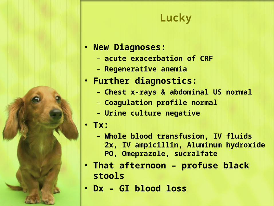
• New Diagnoses:– acute exacerbation of CRF– Regenerative anemia
• Further diagnostics:– Chest x-rays & abdominal US normal– Coagulation profile normal– Urine culture negative
• Tx:– Whole blood transfusion, IV fluids 2x, IV
ampicillin, Aluminum hydroxide PO, Omeprazole, sucralfate
• That afternoon – profuse black stools• Dx – GI blood loss

Lucky
Three days later…• Lucky needs another transfusion• He is still passing melena• Surgery/endoscopy to resect/cauterize
the ulcer declined• Barium PO
Over the next week…• BUN falls to 100ish, creat 4ish, phos
normal• Bleeding stops, PCV low 30’s• Remains anorectic
Lucky goes home…

Lucky
• Owner force feeds for 2 weeks• Lucky starts eating• Lucky lives a happy life again
6 months later…• Lucky starts having seizures, and is
euthanized• No Necropsy
Don’t give Pred and NSAIDs together, especially when there is CRF

Lucky
Things that could have avoided this problem…
• Don’t do this on purpose• Tech review medications at the
beginning of each visit• Always get updated records when
seeing a client that also uses another vet
• Always give drug handouts listing side effects when new drugs are prescribed

Intervertebral Disc Disease
Progressive Myelomalacia• 5-10% of dog who lose deep pain• Hemorrhagic necrosis and softening of
the spinal cord• Ascends and descends through the
spinal cord (first sign?)• HINT: cranial migration of panniculus• Flaccid abdominal muscles• Migrating flaccid paralysis• Eventual respiratory paralysis• Grave prognosis

Intervertebral Disc Disease
Spinal Walking• dogs can begin walking reflexively, with
no spinal cord recovery• Ambulation with no deep pain• Toes are subject to injury from
dragging• Usually remain incontinent

Intervertebral Disc Disease
Post-Operative Care• Physical Therapy – 5 Steps
– Step One – TID until weight bearing• Cold pack incision 10 minutes TID
– Until incision cool to touch• Passive range of motion exercises• Massage affected limb muscles
– Step Two – TID until limb motion• Standing exercises• Neuromuscular stimulation
– Step Three – BID until walking• Weight shifting exercises• Assisted walking• Swimming, underwater treadmill

Intervertebral Disc Disease
Post-Operative Care• Physical Therapy – 5 Steps
– Step Four - BID• Sit to stand exercises• Balance and coordination exercises• Walks of increasing length
– Step 5 - SID• Increased intensity walking and
swimming• It can take 6 months to get to 100%
recovery

Intervertebral Disc Disease
Post-Operative Care• Bladder management
– UMN bladder (drugs?)• Alpha blockers to relax the sphincter
– Phenoxybenzamine 5-15 mg PO SID-BID
– Prazosin 1 mg/30 lbs PO SID-TID• Skeletal muscle relaxants
– Diazepam– Dantrolene
• Bethanechol only if bladder flaccid– 2.5-25 mg PO TID– 3 days after phenoxybenzamine
• Express or catheterize TID-QID

Intervertebral Disc Disease
Post-Operative Care• Bladder management
– LMN bladder• Bethanechol• Alpha blocker if needed• Express or catheterize TID-QID (which?)
– Intermittent catheterization carries no more risk for UTI than manual expression
– Indwelling catheter only if no other option• Large female with bladder difficult to
express• Aggressive dog• To manage urine scalding

Intervertebral Disc Disease
Post-Operative Care• Bladder management
– Monitor for UTI• UA once monthly until urinating on own• Then q4-6 months until spinal cord
disease resolves• Urine culture q6months

Intervertebral Disc Disease
Post-Operative Care• Analgesia• Preventing pressure sores
– Padded beds (where?)– DogLeggs.com– Sling– Turn every 4 hours– Avoid urine leakage, keep skin dry
• Watch for neurologic deterioration

Wobbler Syndrome
Aka Caudal Cervical Spondylomyelopathy
Aka Cervical Vertebral Instability• Presentation
– Middle aged to older large dogs– Onset & progression usually chronic
• Occasionally acutely down– Cervical Myelopathy (neuro exam?)
• Sensory ataxia, Postural deficits• Low neck carriage• Mild to moderate neck pain• UMN all 4 – pelvic worse• May have UMN bladder

Wobbler Syndrome
Diagnosis• Usually depends on
myelography/CT/MRI with stress– Flexion, extension – make lesions worse
• Perform with caution– linear traction - relieve lesions
Treatment• Medical therapy may or may not work• Condition is usually progressive• Surgery may or may not work

Wobbler Syndrome
Prognosis• Generally good with surgery and
intensive care– But not as good as type I disc– More like type II– Better if ambulatory– Worse if more than once disc space– 71% get worse for 2 days after surgery
• Time to ambulation can be prolonged– 2.5 months to ambulation– 3.6 months to optimal results
• Stabilizing and distracting one disc space may aggravate another– “domino effect”– Recurrence 20-30%

Congenital Spinal Malformation
Hemivertebrae• wedge shaped
Butterfly vertebrae• Central vertebral body fails to form
Block vertebrae• Fusion of two or more vertebrae
Stenotic vertebral canal
Transitional vertebrae• vertebrae of one spinal segment take
on characteristics of another• Lumbarization of S1 & vice versa

Congenital Spinal Malformation
Presentation• Puppy to middle age• Hemivertebrae in “Screwtail breeds”
– Bulldogs– Boston terriers
• Some malformations are incidental findings
• Much like Type II Disc Disease or Wobbler– Usually progressive– Occasional acute decompensation

Congenital Spinal Malformation
Treatment• Medical treatment if pain only or
ambulatory with mild to moderate neuro deficits
• Surgery if non-ambulatory• Because of abnormal anatomy of
hemivertebrae, some surgeons think that surgery carries increased risk of destabilization
• Some surgeons won’t cut as long as there is voluntary motor, unless medical therapy has failed for a really long time

Zoey
• Sig – 3 year old SF Pomeranian• Comes in for dental• Pre-A exam and bloodwork NSF• Dental and anesthetic recovery go fine• Between afternoon appointments, you
tech takes you aside to let you know that Zoey can’t walk
• Neuro exam– Mentation & CN normal– all 4 limbs inc tone with hyperreflexia, rear
worse– No deep pain (lesion?)

Zoey

Zoey
• Diagnostic Plan– Go to the restroom to vomit & have diarrhea– Take upper & lower cervical films
• Lower cervical film appears normal

Congenital Spinal Malformation
Atlantoaxial instability
Presentation• Toy breeds• Neck pain to tetraplegia• UMN reflexes all 4 legs, worse rear
Diagnosis• Survey radiography
– Increased space between C1 and C2– Hypoplastic or absent dens– Dens not attached to floor of C1
• DO NOT perform flexed view• Confirm with CT/MRI

Congenital Spinal Malformation
Atlantoaxial instability
Treatment• Medical treatment if just pain or
ambulatory with mild to moderate neuro deficits
• Surgical stabilization if prolonged neuro deficits that don’t respond or non-ambulatory

Congenital Spinal Malformation
Atlantoaxial instability
Prognosis• Fair to good for mild to moderate neuro
deficits• Guarded if tetraplegic
– 13% do not survive surgery• Respiratory arrest• Dysphagia & aspiration pneumonia
Take care with Toy Breed necks during anesthesia
Especially if history of neck pain

Petunia
• Sig – 10 year old brown tabby cat, outdoor
• CC – can no longer jump up to reach food bowl, seems wobbly
• PE and Neuro– Hyperreflexive femoral and ischiatic reflexes– She bites you hard when you palpate TL
spine
• DDx• Dx Plan – TL films normal

Petunia
• Owner declines referral, but approves lumbar CSF tap– Increased microprotein, normal cell counts– Culture negative
• Dx – likely neoplasia– LSA most likely
• Tx– Prednisone 10 mg daily– Declines chemo or oncology referral
• Asymptomatic for one month– Then symptoms return– euthanized– Necropsy confirms SC lymphoma

Neoplasia
Primary Spinal Cord Neoplasia• Glioma• Meningioma• Nerve sheath tumors
– Hemangiopericytoma– Schwannoma– Blastoma - rare
• lymphoma
Metastatic Spinal Cord Neoplasia• Lymphoma• Carcinoma (mammary, prostate)• Melanoma
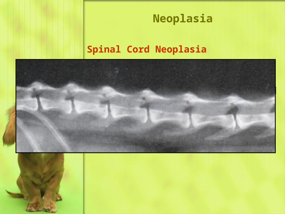
Neoplasia
Spinal Cord Neoplasia• Dx
– Radiographs usually normal• Unless tumor is mineralized• Or invades bone• Or is a nerve sheath rumor,
enlarging the IV foramen

Suzy
• Sig – 10 year old SF Chiuahua mix• CC – coughing again• Hx – chronic bronchitis
– PDA coil placed 10 years ago
• PE – TL spinal pain• Neuro – CP deficits rear legs• Dx plan
– CBC, GHP, lytes, UA – normal– TL spine radiographs
• DDx – osteomyelitis, neoplasia– Thoracic radiographs
• Large Solitary lung mass• PDA coil

Suzy
• Dx Plan– US guided aspirate of lung mass– Cytology and culture– Squamous cell carcinoma– No growth
The same symptoms can develop a new cause
Unless the owner tells you not to, always take 2 views

Neoplasia
Primary Vertebral neoplasia• Osteosarcoma• Chrondrosarcoma• Myeloma (plasma cell tumor)• Fibrosarcoma• hemangiosarcoma
Metastatic Vertebral Neoplasia• Distant metastasis
– Carcinoma (prostate, mammary, lung)
• Local invasion– Bladder carcinoma– Anal sac tumor

Neoplasia
Presentation• Usually middle aged to older
– Young dogs or cats• Lymphoma (median age 2-3 year)• Blastoma (6 months to 3 years)
– GSD– Labrador Retrievers
• Onset usually progressive– Lymphoma sometimes acute
• Severe pain precedes motor deficits for cord tumors
• Neuro deficits come earlier for vertebral tumors

Neoplasia
Diagnosis• Signalment
– Cats with severe TL pain progressing to neuro deficits - LSA
• Hyperglobulinemia and proteinuria with myeloma
• Bony tumors seen on survey rads• CSF tap
– Very rarely see neoplastic cells– Increased protein without increased cells
• SC often tumors require advanced imaging– Myelogram, epidurogram, CT, MRI

Neoplasia
Treatment• Anti-inflammatories for cord edema
– Prednisone 0.5 mg/kg PO BID
• Analgesics– Opiates or Tramadol
• Chemotherapy for LSA or myeloma– Palliative piroxicam for carcinomas
• Decompressive surgery• Palliative radiation

Neoplasia
Prognosis
• Grave for bony neoplasia• Poor for cord neoplasias treated
supportively– Short term can be good
• Days to weeks to months– Grave long term
• Long term remissions sometimes possible with surgery– Prognosis may not be determined
without histopath

Belle
Sig – 3 year old female Pit Bull Terrier
CC – laying around, eating fine, owner has $100
PE & neuro exam – mid-thoracic pain
DDx –
Dx Plan – lateral radiograph thoracic spine without sedation - normal
Tx Plan – • Dermaxx SID x 7 days and cage rest x 2
weeks

Belle
3 day follow-up call – back to normal, still doing cage rest
10 days after first visit – laying around again refuses to move, won’t eat
PE & neuro – pain at same spot is worse
DDx –
Dx Plan – T spine films with sedation
Dx – discospondylitis
Radiographs can be normal early in the course of discospondylitis

Discospondylitis
Infection if the Intervertebral discs & vertebral end plates
• Bacterial– Staphylococcus spp.– Brucella canis– Many others
• Less commonly Fungal• L7-S1 most common• If ambulatory, prognosis good for all
but Brucella– relapsing, chronic discospondylitis
• Diagnosis – radiographs, urine culture, Brucella serology, CSF culture, LS aspiration cytology & culture

Belle
• Tx Plan – – Baytril 5 mg/kg PO BID x 3 weeks
• Follow-up call in 2 weeks – Belle back to normal
• 3 months later – Belle won’t move again• PE & Neuro – Temp 104F, LS pain• DDx• Dx Plan – lumbosacral radiograph with
sedation, Brucella titer, urine culture• Tx Plan – OHE, Streptomycin and
tetracycline x 30 days, then recheck spinal rads

Marti – “Doc’s Spicy Martini”
• Sig – 4 month old female golden retriever
• Stiffness, sore, was fine yesterday• PE & Neuro – neck pain – rest of neuro
exam normal, possible muscular pain, possible joint pain
• CBC – grans 20600/ul, monos 2000/ul, HCT 30%
• GHP/Lytes – phos 8.1• UA – USG 1.003• DDx
– myositis, polyarthritis, meningitis, unnoticed trauma, neoplasia

Marti – “Doc’s Spicy Martini”
• Dx Plan– Cervical rads with sedation – normal– CPK – normal
• DDx – Meningitis, polyarthritis, neoplasia
• Rickettsial disease• immune mediated• Bacterial• Fungal• Neospora/Toxoplasma• Lymphoma• (Hepatozoon)

Marti – “Doc’s Spicy Martini”
• Tx Plan– Doxycycline 10 mg/kg divided BID x 3 weeks– Clindamycin 15 mg/kg PO BID x 3 weeks– Tramadol 3 mg/lg qhrs PRN for pain– Deramaxx 1 mg/lb PO SID
3 days later….• Marti is laterally recumbent & unwilling
to move, but neuro exam normal, Temp 103.5F– Immobility due to pain, neck pain suspected– Joint pain can not be ruled out
• CBC, GHP, lytes, UA – no change

Marti – “Doc’s Spicy Martini”
• Dx Plan– CSF Tap
• Grossly normal• Culture negative• Cytology – neutrophilic pleocytosis,
hypersegmented segs, increased protein– Joint Taps of stifles and elbows– Urine culture – negative– Hepatozoon PCR – negative– Tick Panel – RMSF, Lyme, Ehrlichia – neg– Toxoplasma/Neospora Titers – negative
(Dx?)
• Diagnosis – Steroid Responsive Meningitis-Arteritis

Marti – “Doc’s Spicy Martini”
• Tx Plan– Prednisone 1 mg/lb (30 mg) PO divided BID x 2
weeks– Prednisone 10 mg PO BID x 4 weeks– Prednisone 10 mg PO SID x 4 weeks– Prednisone 10 mg PO QOD x 4 weeks– Prednisone 5 mg PO QOD x 2 weeks– If only partial response to 1 mg/lb divided
BID, go to 1 mg/lb PO BID x 1-2 weeks– Wean off pred very slowly over 3-4 months– If any relapse of symptoms, inc. to previous
dose, repeat interval and try again to reduce– 50% will need lifelong pred at some dose, or
intermittently– If incomplete response to pred, can try
Imuran or other immunosuppresives

Immune Mediated Meningitis
Similar CSF results• Culture negative• Neutrophilic pleocytosis• Elevated protein
All respond to immunosuppression
Different histopath on necropsy
Steroid Responsive Meningitis-Arteritis (SRMA)
Aka Aseptic Meningitis• Nova Scotia Duck Tolling Retrievers
(“Tollers”)

Immune Mediated Meningitis
Necrotizing vasculitis• Prognosis not as good as SRMA• Bernese Mt Dog, Beagle, GSPPyogranulomatous ME• Rapidly progressive, neck pain, brain
stem lesions, seizures, vomiting• PointersAseptic meeningitis/polyarthritis of AkitasGranulomatous Meningioencephalitis
(GME)• Lesions throughout the CNS• Focal or mutlifocal• Prognosis varies• Particular appearance on MRI

Rose
• Sig – 5 year old spayed female Labrador Retriever
• CC – Rear limb paralysis actue yesterday, referred for back surgery
• Neuro Exam– No spinal pain detected– Cutaneous trunci stops R T8 T11 L– CP – 0 RR, 1 LR– Voluntary motor – 0 RR, 1 LR– Patellar Reflex – 4 R 3 L– Ischiatic Groove Reflex – 3R 3L
• Lesion?– Mid-thoracic lateralized right

Rose
• DDx - • Dx Plan
– TL Spinal Films – normal– Myelogram - normal– CBC – normal– GHP/lytes – glucose 1500– UA – glucosuria, no ketones
• Referring Vet Record says Rose was given 10cc Dexamethasone SP and 1 cc Banamine
• cPLI – strong positive• Abdominal US – edematous pancreas

Rose
• Dx – – Pancreatitis with possible Diabetes Mellitus– Fibrocartilagenous Embolism
• Tx Plan– Tx pancreatitis – IV fluids, pain meds, NPO x
24 hours, antiemetics, low dose heparin– Rose developed GI ulcers and sterile bloody
urine over the next 2 days• Pepcid, Carafate
– Insulin for 2 days, then no longer needed– Began physical therapy immediately– Rose walked out of the clinic 10 days later

Glucocorticoid Doses
Immunosuppressive
Anti-inflammatory
Anti-pruritic
Physiologic Replacement
Duration of action
Pred
1-2 mg/lb/day
0.5 mg/lb/day
0.25 mg/lb/day
0.1 mg/lb/day
12-36 hours
Dex
0.2 mg/lb/day
0.1 mg/lb/day
0.05 mg/lb/day
0.02 mg/lb/day
36 hours +

Fibrocartilagenous Embolism (FCE)
• Fibrocartilage from nucleus pulposis of the disc plugs up blood supply to or from the spinal cord
• Presentation– 80% Large to giant dogs– Also schnauzers– Young to middle aged– Peracute to acute onset– Progresses and peaks in 6-24 hours– Cry out in pain during exercise– May show some pain on presentation, but
quickly non-painful within 24 hours– Neuro lesions depend on location
• Usually lateralized

Fibrocartilagenous Embolism (FCE)
• Diagnosis– Neuro exam localizes to spinal cord, usually
lateralized– Often no significant pain– Rads, CSF analysis and myelogram normal
• Treatment– Anti-inflammatory glucocorticoids,
decreasing– Physical therapy– No exercise restriction needed

Fibrocartilagenous Embolism (FCE)
• Prognosis– Variable – depends on ischemic damage– Good if ambulatory within 2 weeks– Poor Px related to
• lack of deep pain• severe LMN damage• lack of PT provided

Porsche
• Sig – 1 year old spayed female Boxer• CC – Hit by Car yesterday, doesn’t want
to move her head or neck, screams when you touch her
• Neuro Exam– Severe upper neck pain– CP deficits all 4 limbs, worse rear– Increased muscle tone all 4 limbs– Patellar Reflex – 3 R 3 L– Ischiatic Groove Reflex – 3R 3L– Can walk reasonably well
• Lesion?– Upper cervical

Porsche
• Dx Plan– Upper cervical radiographs with sedation– Fx C1 and C2
• Tx Plan– Cage rest for 3 weeks– Deramaxx 50 mg PI SID PRN for pain– Porsche healed well within 1 month

Spinal Trauma
• When to do surgery?– Acute worsening of neurologic signs– Moderate to severe displacement of
spinal fragments– Severe neurologic function or pain
• Non-ambulatory• Especially no deep pain
– Evidence of spinal cord compression on myelogram, CT, MRI

Sonny
• Sig – 12 year old CM Golden Retriever• Med Hx – Hypothyroidism (soloxine),
Hyperlipidemia• CC – Rear end weakness, severe, onset
over 1-2 months• PE & Neuro –
– Crepitus palpable in the hips– No spinal pain– Patellar & ischiatic reflexes – 1 R 1 L– CP deficits all 4 limbs, rear worse
• DDx – LMN disease caudal SC– Degenerative myelopathy– Hypothyroidism– Hip arthritis

Sonny
• Dx Plan– LS spinal films– VD pelvis
• Review of the Record– Thyroxine dose was increased from 0.3 to
0.6 PO BID last year when hyperlipidemia began, and T4 was 0.4
– T4 after 4 weeks of increased dose was in normal range
– For the past 6 months, T4 has been refilled at 0.3 mg PO BID
• Dx– Vertebral Spondylosis– Clinical Hypothyroidism– Hip Dysplasia

Sonny
• Tx Plan– Increase thyroxine to 0.6 mg PO BID– Recheck T4 and neuro exam on month– Glycoflex III per label instructions– Deramaxx 50 mg PO SID PRN for pain
• Rear end weakness much improved within 3-4 weeks
• Neuro exam normal in 3 months

DDx Multifocal CNS Disease
Degenerative• CNS atrophy of old age• Lysosomal Storage Disease• Various Leukodystrophies• Various abiotrophies
Anomalous• Dandy Walker Syndrome
– Cerebellar hypoplasia, hydrocephalus
Neoplastic• LSA• Metastatic neoplasia (prostate CA,
ammary CA, melanoma, etc.)

DDx Multifocal CNS Disease
Nutritional• Thiamine deficiency
Infectious• Bacterial – many, including Lyme• Fungal – esp. Cryptococcus neoformans and
Cocciodioides immitus• Viral – FIP, CDV• Rickettsial – RMSF, Ehrlichia• Protozoal – Toxoplasma gondii, Neospora
caninum• Algal – Prototheca spp.• Parasitic – Dirofilaria, Cuterebra, Bayliascaris• Prion – Feline Spongioform Encephalitis

DDx Multifocal CNS Disease
Inflammatory
Vascular• Ischemic encephalopathy