practical neurology (cerebellar ataxia)
TRANSCRIPT

Practical Neurology 2012;12:14–24. doi:10.1136/practneurol-2011-000108
REVIEW
14
AbstractThe clinical management of cerebellar ataxia is challenging, mainly because ataxia is a symptom of many neurological diseases. Many types of ataxia disorders are genetic and some are extremely rare. Here, the authors suggest a diagnostic approach to ataxia developed around a case of sporadic, late-onset, slowly progressive ataxia. Clinical information such as age of onset, rate of progression, family history and certain non-cerebellar features can narrow the differential diagnosis. Brain MRI is almost obligatory and may reveal valuable diagnostic clues. Having ruled out structural lesions, the two other most common diagnoses are infl ammatory and degenerative (including genetic) disorders. Although only a minority of underlying diseases are treatable, there are still many options for supportive care.
IntroductionThe clinical management of cerebel-lar ataxia is challenging for several reasons. First, there is an extensive differential diagnosis, with each condition requiring different inves-tigations. Second, with the study of genetic ataxias progressing rapidly, neurologists cannot easily keep them-selves sufficiently updated to select which genes, if any, to test. Third, even ‘common’ causes of cerebellar ataxia are quite uncommon, making it difficult to build expertise in these dis-orders. Last, although most cerebel-lar ataxias are untreatable, clinicians are wrong to assume that they can do little for the patient’s well-being. Based on a case history, we suggest a guide to clinical decision-making for patients with ataxia, focusing mainly on sporadic, late-onset (after the age of 30 years) cerebellar ataxia.
History and examination: things you get for freeAn old-fashioned comprehensive clinical evaluation is important. The causes of cerebellar ataxia are grouped into genetic, acquired and non-genetic neurodegenerative disorders (table 1). A careful history and physical exami-nation allows the clinician to rank these causes in order of suspicion, sometimes with quite a narrow dif-ferential. The four important points in the history are: speed of onset/pro-gression, age at onset, family history and some aspects in the general medi-cal history.
Speed of onset/progression
Ataxia presenting abruptly obviously suggests a stroke. Ataxia developing over hours might suggest Wernicke’s encephalopathy, although other fea-tures are common. Onset over days might suggest Miller Fisher syn-drome or para-infectious cerebellitis,
▶ Additional data are published online only. To view the fi les, please visit the journal online (http://pn.bmj.com/content/12.1.toc).
Department of Neurology & Donders Institute for Brain, Cognition and Behaviour, Radboud University Nijmegen Medical Centre, Nijmegen, The Netherlands
Correspondence to Dr Bart P C van de Warrenburg, Department of Neurology, Radboud University Nijmegen Medical Centre, Reinier Postlaan 4, Nijmegen, 6525 GC, The Netherlands; [email protected]
Received 5 September 2011Accepted 1 December 2011
A practical approach to late-onset cerebellar ataxia: putting the disorder with lack of order into order
Judith van Gaalen, Bart P C van de Warrenburg
Case
A 62-year-old previously well man presented with a 3-year history of gradually worsening unsteadiness and shaking of his hands on ac-tion. His speech and swallowing were normal, but there was some urinary urgency. There was no family history. He drank 3–4 glasses of wine a day. On examination, there was titubation, a bilateral terminal tremor on fi nger–nose test-ing, dysmetria during fi nger-chasing, abnormal heel-to-shin testing, mild gait ataxia and clearly disturbed tandem gait, and brisk tendon re-fl exes with bilateral extensor plantar responses. Investigations showed normal serum vitamin levels and thyroid function; an MR scan of the brain showed cerebellar atrophy, mainly of the vermis.
04_practneurol-2011-000108.indd 1404_practneurol-2011-000108.indd 14 1/14/2012 3:23:24 PM1/14/2012 3:23:24 PM

Practical Neurology 2012;12:14–24. doi:10.1136/practneurol-2011-000108
REVIEW
15
encephalopathy with antithyroid antibodies (SREAT; previously referred to as Hashimoto’s encephalopathy) and antiglutamic acid decar-boxylase (anti-GAD)-associated cerebellar ataxia.
particularly in children. Subacute onset and pro-gression (weeks to months) suggest only a few con-ditions: paraneoplastic cerebellar degeneration, Creutzfeldt–Jakob disease, steroid-responsive
Table 1 Overview of the genetic and non-genetic causes of cerebellar ataxia, with clinical clues from history or examination, and suggestions for initial investigations
Cause Clinical clues Suggestions for investigation
Genetic ataxiasDominant
Autosomal dominant cerebellar ataxias Family history, slowly progressive ataxia, some genotype-specifi c features (online supplementary table S1)
Mutation analysis of SCA genes
Dentato-rubro-pallidoluysian atrophy Dementia, chorea, myoclonus, mainly in the Japanese Mutation analysis of ATN1 gene
Alexander disease (Pseudo)bulbar signs, spasticity, hyperrefl exia Mutation analysis of GFAP gene
Recessive
Autosomal recessive cerebellar ataxias Family history, systemic features, often neuropathy (see also online supplementary table S2)
Mutation analysis of relevant recessive genes, lysosomal enzymes, α-fetoprotein, metabolic investigation
Other
‘Mitochondrial’ disorders Family history (diabetes mellitus, deafness), multisystem involvement
Mutation analysis of polymerase γ and mitochondrial DNA
Muscle biopsy
Fragile X-associated tremor/ataxia syndrome
Action tremor, behavioural changes, autonomic dysfunction; family history (learning disability)
Mutation analysis of FMR1 gene, typical MRI (fi gure 4)
Non-genetic ataxiasDegenerative
Multiple system atrophy Autonomic failure, Parkinsonism MRI (fi gure 8), nuclear imaging, autonomic testing, see also online supplementary table S1
Idiopathic late onset cerebellar ataxia Mostly pure, slowly progressive ataxia No specifi c test, other causes excluded
Sporadic Creutzfeldt–Jakob disease Rapid progression, myoclonus, cognitive disturbances EEG, CSF 14-3-3 protein/tau, typical MRI (fi gure 9)
Inflammatory
Gluten ataxia Gastro-intestinal symptoms Antigliadin/-tissue transglutaminase/-endomysium antibodies
Hashimoto encephalopathy Subacute, seizures, myoclonus Anti-TPO antibodies
Paraneoplastic cerebellar degeneration Rapid progression, history of malignancy Neuronal antibodies, tumour screen
Anti-GAD ataxia Subacute, type 1 diabetes mellitus Anti-GAD antibodies
(Para)infectious
Miller Fisher syndrome Subacute, ophthalmoplegia, arefl exia Anti-GQ1b antibodies, EMG
Epstein–Barr virus Subacute, preceding infection CSF, serologic testing
HIV Known HIV status CSF, search for opportunistic infections
Structural lesions
Stroke, tumour, multiple sclerosis, etc Focal neurological signs MRI, other investigations depend on imaging fi ndings
Toxic
Alcohol Medical history, peripheral neuropathy Liver function
Drugs (eg, lithium, phenytoin) Medical history Screening drug level, stop offending drug
Metabolic
Acquired vitamin defi ciencies
Vitamin E Diarrhoea Vitamin E level, coeliac screen, serum amylase
Vitamin B1 Confusion, nystagmus, ophthalmoplegia Vitamin B1, typical MRI
Hypothyroidism Fatigue, weight gain, constipation Thyroid stimulating hormone, free T4
Hypoparathyroidism Cataract, extrapyramidal symptoms Parathyroid hormone, calcium, phosphate, typical CT scan (fi gure 2)
Other
Superfi cial siderosis Sensorineural deafness Typical MRI (fi gure 7), audiogram, xanthochromic CSF
CSF, cerebrospinal fl uid; EMG, electromyogram; GAD, glutamic acid decarboxylase; SCA, spinocerebellar ataxia; TPO, thyroperoxidase.
04_practneurol-2011-000108.indd 1504_practneurol-2011-000108.indd 15 1/14/2012 3:23:25 PM1/14/2012 3:23:25 PM

Practical Neurology 2012;12:14–24. doi:10.1136/practneurol-2011-000108
REVIEW
16
loss may suggest an underlying tumour, weight gain can suggest hypothyroidism, diarrhoea can point to coeliac disease, postural dizziness may indicate MSA and previous—even mild—head trauma may suggest superficial siderosis. A recent malignancy history clearly suggests pareneoplas-tic cerebellar degeneration. Patients with HIV or AIDS may develop ataxias from opportunis-tic infections, cerebellar lymphoma or cerebellar degeneration.1 Check and recheck the alcohol intake and ask for past and present medication.
Examination
The clinical examination confirms that the ataxia is indeed cerebellar. However, its added value is to identify other relevant neurological and non-neurological features.
Valuable non-neurological abnormalities ■
include telangiectasias, scoliosis and orthostatic hypotension.A severe ■ saccadic eye movement disorder (oculomotor apraxia) immediately points to a very limited number of conditions, such as ataxia telangiectasia and ataxia with oculomotor apraxia type 2.Ataxia with ■ chorea requires substantially different investigation from ataxia with spasticity; if there is concomitant chorea one should for example consider ataxia telangiectasia or SCA-17, while spasticity combined with ataxia fits with diagnoses such as autosomal recessive spastic ataxia of Charlevoix–Saguenay or late onset Friedreich’s ataxia.
Insidious onset with progression over years does not suggest any particular diagnosis but mostly rules out the above-mentioned diseases.
Age at onset
Age at onset is very important. For example, a slowly progressive ataxia starting before the age of 25 years may well be genetic or metabolic. Some disorders rarely appear before a certain age, for example, multiple system atrophy (MSA) always manifests after the age of 30 years and the fragile X tremor/ataxia syndrome (FXTAS) also develops after the age of 45 years.
Family history
A family history of cerebellar ataxia clearly suggests a genetic cause. It is worth investing time in drawing up a full pedigree, as the mode of inheritance deter-mines which mutations to request for investigation. The absence of a family history does not rule out a genetic cause. This is particularly true for recessive disease, as smaller families often have no affected siblings. Consanguinity strongly suggests recessive disease and clinicians should ask for information about this. When going through the family history for ataxia, check also for other movement disorders: for example, spinocerebellar ataxia type 3 (SCA-3) can present with Parkinsonism rather than ataxia, mitochondrial disorders may present with diabetes mellitus and deafness, and FXTAS may present with learning disability and premature ovarian failure.
General medical conditions
A detailed medical history is important since dia-gnostic clues are possible in any domain. Weight
Figure 1 Spinocerebellar ataxia type 2. Cerebellar and pontine atrophy on a sagittal T2-weighted MRI.
Figure 2 Hypoparathyroidism. Cerebellar calcifi cation on CT brain scan.
04_practneurol-2011-000108.indd 1604_practneurol-2011-000108.indd 16 1/14/2012 3:23:25 PM1/14/2012 3:23:25 PM

Practical Neurology 2012;12:14–24. doi:10.1136/practneurol-2011-000108
REVIEW
17
Previous intracranial bleeding ■ may leave superficial siderosis with haemosiderin appearing as a black rim around the posterior fossa structures on gradient-echo or susceptibility-weighted MRI (figure 7).
Is alcohol the culprit?Our patient drank 3–4 glasses of wine a day. There is often doubt as to whether ataxia might be alcohol-related. It is easier if there is a his-tory of true alcohol abuse, but more commonly the patient drinks just slightly more than aver-age. There are no good data on the duration and quantity of alcohol consumption that cause long-term ataxia. Despite the lack of epidemio-logical data, alcoholic cerebellar degeneration is one of the most common sporadic ataxias, especially in middle-aged men. Nevertheless, we recommend a search for other causes of ataxia in patients who report drinking only moderately.
In patients with alcohol-related ataxia, the symptoms affect gait and lower limbs more than arms and speech, often also with signs of periph-eral neuropathy. The ataxia can stabilise or even improve with stopping alcohol, but worsen in those who continue.4 5 Brain imaging typically shows vermis atrophy.
Alcohol is directly toxic to the cerebellum, causing degeneration of the anterior superior vermis and hemispheres.6 This is worsened by associated vitamin B1 deficiency, resulting from both a poor diet and a direct toxic effect on vitamin B1 metabolism. Alcohol impairs thia-mine uptake from the gastro-intestinal tract, reduces thiamine-dependent enzyme activity and depletes liver thiamine stores.7 Severe vitamin B1 deficiency causes Wernicke’s encephalopathy,
The presence or absence of ■ peripheral neuropathy (later corroborated by nerve conduction studies) is important in early onset cases (see online supplementary table S3).
Always do a brain MRIThe investigation of patients with cerebellar ataxia should always include—in fact begin with—an MRI brain scan. The only exception might be the patient whose family has a known genetic defect. The MRI first allows demonstration or exclusion of a structural lesion, such as cerebellar tumour or abscess. If there is no such lesion, one should meticulously search the images for other specific abnormalities (see figures).
Cerebellar atrophy ■ usually indicates a degenerative disease (which could be genetic) or to later stages of paraneoplastic cerebellar degeneration (figure 1).Tadpole sign ■ in adult-onset Alexander disease has very pronounced atrophy of the medulla and cervical cord; with the preserved pons, the sagittal image looks like a tadpole (figure 5).Chiari type I malformation ■ can lead to cerebellar symptoms, cranial neuropathies and headache, usually in adolescence or adulthood (figure 3).2
Cerebellar white matter high-signal changes ■ could indicate polymerase γ (POLG) mutations, Langerhans cell histiocytosis or cerebrotendinous xanthomatosis. FXTAS often gives changes in the middle cerebellar peducles (figures 6 and 4, respectively).The ‘hot cross bun’ sign ■ in the pons occurs in MSA from pontocerebellar fibre degeneration; a ‘putaminal rim’ results from a high signal line extending laterally from a slightly darkened putamen (figure 8).Creutzfeldt–Jakob disease ■ may give thalamic changes (pulvinar sign) or abnormal cerebral cortex on diffusion-weighted MRI (figure 9).3
Figure 3 Chiari malformation. Low-hanging, elongated cerebellar tonsils displaced below the level of the foramen magnum on a sagittal T1-weighted MRI.
Figure 4 Fragile X tremor/ataxia syndrome. Medial cerebellar peduncle high signal changes on axial T2-image.
04_practneurol-2011-000108.indd 1704_practneurol-2011-000108.indd 17 1/14/2012 3:23:25 PM1/14/2012 3:23:25 PM

Practical Neurology 2012;12:14–24. doi:10.1136/practneurol-2011-000108
REVIEW
18
Subacute onset1. SREAT—formerly Hashimoto’s encephalopathy—
gives ataxia progressing over weeks, with cognitive disturbance, myoclonus, seizures and ataxia. Patients have high serum thyroperoxidase antibody levels, although thyroid function is normal in half of the cases. The mean age at onset is 45–55 years, and it is five times more common in women than in men. Patients often have other autoimmune disorders, for example, type 1 diabetes mellitus and Sjögren’s syndrome. It is readily treatable and improves dramatically with corticosteroids; in fact, the steroid response is a prerequisite for the diagnosis. The sooner treatment is started, the better the outcome.12
2. Creutzfeldt–Jakob disease is another rapidly progressive disorder with cerebellar ataxia. Sooner or later, patients develop a plethora of other neurological signs, including dementia, myoclonus and Parkinsonism. It is difficult to make the diagnosis very early. There are some suggestive MRI features (see above). Cerebrospinal fluid (CSF) often shows 14-3-3 protein and increased tau levels. The EEG typically shows periodic synchronous biphasic or triphasic sharp wave complexes.13 14 Patients usually die within a year.
3. Paraneoplastic cerebellar degeneration also progresses over weeks. When suspected, patients
presenting the classical triad: encephalopathy, oculomotor dysfunction and gait ataxia. It is a neurological emergency, requiring urgent vita-min B1 replacement.8
What about the other acquired causes? Should I really do a screen for malignancy?We will not address the structural lesions further, as these will be apparent on the ‘obligatory’ initial MRI brain scan. However, there are some other acquired causes, which we discuss in order of their speed of onset.
Acute onset
(Para)infectious cerebellopathy is more com-mon in children than in adults. Typically, there is a prodromal infectious phase, often caused (in adults) by the Epstein–Barr virus, followed by subacute cerebellar symptoms. The prognosis is usually good.9
In Miller Fisher syndrome, a variant of acute inflammatory demyelinating polyradiculoneu-ropathy (Guillain–Barré syndrome), the triad of ophthalmoplegia, areflexia and ataxia develops over 1–2 weeks. The cause is molecular mimicry relating to antecedent infections, for example, Campylobacter jejuni. Serum anti-GQ1b antibod-ies are commonly elevated.10 The treatment is with intravenous immunoglobulins or plasma exchange; the outcome is similar with either, with a trend towards a faster recovery after plasma exchange.11
Figure 5 Alexander disease. Sagittal fl uid attenuated inversion recovery images showing atrophy of cerebellum, brainstem (except pons) and spinal cord, and high signal changes within the cerebellar and brainstem white matter.
Figure 6 Polymerase γ. Symmetric, high signal changes in the cerebellar white matter dorsally from the dentate nucleus on axial T2-weighted MRI.
04_practneurol-2011-000108.indd 1804_practneurol-2011-000108.indd 18 1/14/2012 3:23:26 PM1/14/2012 3:23:26 PM
Practical Neurology 2012;12:14–24. doi:10.1136/practneurol-2011-000108
REVIEW
19
4. Ataxia associated with anti-GAD antibodies also has an inflammatory cause. It is more common in patients with type 1 diabetes mellitus and in women. There is no proven best treatment, but corticosteroids and immunoglobulins occasionally improve the symptoms.17
Chronic onset1. Coeliac disease may be complicated by neurological
features including peripheral neuropathy and cerebellar ataxia. The ataxia is caused by inflammatory changes in the cerebellum. However, there is debate whether the so-called ‘gluten ataxia’ exists. This is a sporadic, otherwise unexplained cerebellar ataxia where the patient has coeliac-associated autoantibodies (antigliadin, antiendomysium and antitissue transglutaminase). There is a broad range of age at onset and usually no other specific signs. Most patients have no bowel symptoms or abnormalities on intestinal biopsies, and so no features of definite coeliac disease. Some authors claim that gluten ataxia could be the most common sporadic ataxia. The controversy is mainly due to these antibodies being also found in both hereditary ataxias and healthy controls.18 Rather than causes, these antibodies might be a non-specific marker of another neuroinflammatory disease or an epiphenomenon triggered by the degeneration.19 It is important to settle this issue since some patients appear to improve on a gluten-free diet. Although
need additional imaging to identify an underlying malignancy, for example, mammography, testicular or ovarian ultrasound, CT scan of chest and abdomen, or whole-body positron-emission tomography scan. However, paraneoplastic syndromes can develop in the early stages of a malignancy, and so it might be difficult to demonstrate the tumour. If the initial screening is negative, repeat screening is advisable every 6 months for 4 years.15 Brain MRI is often normal in the first weeks, but cerebellar atrophy develops over the subsequent months. The top four malignancies associated with paraneoplastic cere-bellar degeneration are small cell lung cancer, breast cancer, ovarian tumour and Hodgkin’s lymphoma. Like other paraneoplastic syndromes, these conditions are probably caused by cross-reacting humoral or cellular immune responses. Antineuronal antibodies can be tested for, but their low sensitivity means that a negative result does not exclude the diagnosis. Treating the underlying tumour, combined with immunomodulatory treatment, might improve or stabilise the symptoms, but for most people the prognosis is poor; up to 80% of patients never again walk unaided.16 Previous reports suggested that the interval between onset of ataxia and tumour detection may be up to 4 years; there is thus uncertainty whether to perform a tumour screen and antineuronal antibody testing in all patients with ataxia starting from the last 4 years. The clinical context is paramount here, with the more rapid progression more likely associated with underlying malignancy.
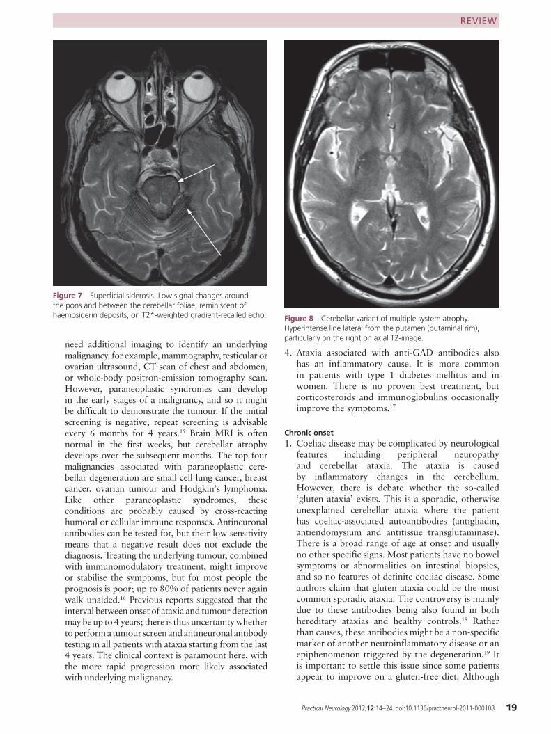
Figure 7 Superfi cial siderosis. Low signal changes around the pons and between the cerebellar foliae, reminiscent of haemosiderin deposits, on T2*-weighted gradient-recalled echo. Figure 8 Cerebellar variant of multiple system atrophy.
Hyperintense line lateral from the putamen (putaminal rim), particularly on the right on axial T2-image.
04_practneurol-2011-000108.indd 1904_practneurol-2011-000108.indd 19 1/14/2012 3:23:27 PM1/14/2012 3:23:27 PM

Practical Neurology 2012;12:14–24. doi:10.1136/practneurol-2011-000108
REVIEW
20
5. Low-grade infections or other inflammatory causes should be searched for by CSF examination if progressive adult-onset ataxia remains unexplained after the above investigations.
Beware of MSAThe Parkinsonian variant of MSA is more common than the cerebellar variant worldwide, apart from Japan. MSA is an important cause of sporadic cer-ebellar ataxia, although the diagnosis is usually not made at the first visit. The onset peaks at between 50 and 70 years of age, although MSA can start as early as the fourth decade. The disease course is the giveaway, as other features soon appear, dev-astating quality of life and independence. Median survival is less than 10 years. Autonomic failure is prominent, with urinary incontinence, erectile dysfunction in men and postural hypotension; in retrospect, its onset often precedes the ataxia. Other features include Parkinsonism, pyramidal
we screen for these antibodies in patients with sporadic cerebellar ataxia, the yield in our unit has been extremely low. If positive, we openly discuss the controversy of this diagnosis with the patient, and jointly decide whether or not to try a gluten-free diet for about 6 months.
2. Superficial siderosis is a rare disorder characterised by cerebellar ataxia, with sensorineural deafness, pyramidal signs and dementia.20 The age of onset age ranges between 14 and 77 years. Neuronal damage results from the deposition of free iron, ferritin and haemosiderin following repeated subarachnoid bleeding from either vascular malformations or previous brain surgery or trauma.21 The typical MRI pattern is of a black rim around posterior fossa structures (see figure 7). CSF shows xanthochromia with siderophages. Removal of the bleeding source is the only effective treatment; iron chelation and CSF shunting do not help.22
3. Medication-induced ataxia may occur with several drugs, including antiepileptics (particularly phenytoin, carbamazepine and gabapentin), metronidazole, amiodarone, some anticancer drugs and pregabalin.
4. Metabolic causes of cerebellar ataxia are worth excluding by measuring serum vitamin levels (E and B), liver enzymes and (para)thyroid function.23 24
Figure 9 Creutzfeldt–Jakob disease. High signal changes on axial fl uid attenuated inversion recovery image with the pulvinar (arrow), head of caudate and putamen.
Case follow-up
Besides ataxia, our patient reported urinary urgency and had pyramidal features. These also occur in many cerebellar dis-orders due to spinal cord involvement. The cerebellar atrophy and slow progression suggested a degenerative process. Rou-tine blood tests were normal, including the gluten sensitivity screen. Alcohol excess seemed an unlikely cause.
Figure 10 Mitochondrial encephalomyopathy, lactate acidosis and stroke-like episodes. Recent cortical infarction on axial fl uid attenuated inversion recovery image.
04_practneurol-2011-000108.indd 2004_practneurol-2011-000108.indd 20 1/14/2012 3:23:27 PM1/14/2012 3:23:27 PM

Practical Neurology 2012;12:14–24. doi:10.1136/practneurol-2011-000108
REVIEW
21
Idiopathic late-onset cerebellar ataxia is the diagnosis of exclusion, having excluded relevant causes in a patient with cerebellar ataxia (includ-ing genetic ones, see below). One can debate where early-onset cerebellar ataxia ends and idi-opathic late onset cerebellar ataxia begins; there-fore, some prefer the term ‘sporadic adult-onset ataxia’. This is clearly an aetiologically heteroge-neous group and an important reason for long-term follow-up is to identify ‘conversion’ to MSA that may occur later.26
Could this be genetic?A negative family history, even done properly, does not exclude a genetic cause. Patients with sporadic ataxia may particularly have recessive disorders, but also occasionally dominant, X linked and mitochondrial diseases.
There have been recent major advances in the molecular genetics of ataxia. Online supplemen-tary tables S2 and S3 provide an overview of the known dominant and recessive genes, and there are excellent reviews for more details on these genetic ataxia variants.27 28 Below, we only briefly touch upon the genetic ataxias, focusing mainly on those presenting at later ages.
Dominant inheritance
The term ‘spinocerebellar ataxias’ (SCAs) is used synonymously with dominant ataxias. However, other ataxias with a dominant inheritance include dentatorubropallidoluysian atrophy and the epi-sodic ataxias; there are also conditions where ataxia is one feature, such as Alexander disease, hereditary prion disease and neuroferritinopathy.
There are already over 30 different SCA types and SCA-36 is the most recent. The most common SCA subtypes are those caused by an expanded coding repeat, but there are non-coding expansions and conventional mutations in some of the other subtypes.27 Age at onset is typically 30–40 years, but some SCA types occur much later (particularly SCA-6) and there are infantile cases (SCA-2 and SCA-7). The clinical picture ranges from isolated cerebellar ataxia to complex neurological multi-system diseases. Non-cerebellar features such as pyramidal features, peripheral neuropathy and movement disorders are very common, and are even the presenting or dominating feature. The clinical overlap between SCAs29 makes it impossi-ble to predict the underlying genotype accurately. Thus, when there is a dominant family history, we have to test for many SCA genes. There are some exceptions (see online supplementary table S2);
signs, antecollis, Pisa syndrome, facial dystonia, cold hands and feet, rapid-eye movement sleep behaviour disorder, inspiratory sighs, nocturnal stridor, a high-pitched quivery voice and poly-mini-myoclonus.
Online supplementary table S1 gives the revised consensus criteria. Clinically, we can only diag-nose probable MSA: definite MSA requires neuropathological confirmation of α-synuclein deposits in glial cells.25 Probable and possible MSA differ in the severity of their autonomic disturbance. Features supporting possible MSA (besides ataxia) are stridor, pyramidal signs and basal ganglia involvement (inferred either clini-cally from Parkinsonism or radiologically by structural or functional imaging). MR brain scan-ning may show not only the cerebellar and brain-stem atrophy but also the ‘hot cross bun’ sign or a ‘putaminal rim’ (figure 8).
Table 2 Symptom treatment options
Symptom Drugs
Ataxia Low dose benzodiazepinesRiluzoleBuspironeAmantadineAcetazolamide5-Hydroxytryptophan
Parkinsonian features L-dopaDopamine agonists
Cerebellar tremor PropranololPrimidoneClonazepam
Nystagmus BaclofenGabapentinClonazepam3,4-diaminopyridine
Dystonia Botulinum toxin injectionsAnticholinergics
Muscle cramps MagnesiumBenzodiazepinesQuinineMexiletine
Spasticity BaclofenTizanidineBenzodiazepinesBotulinum toxin injections
Urinary urgency Spasmolytic agentsAdrenergic α- receptor blockers
Restless legs Dopamine agonistsL-dopaOpioidsClonazepam
04_practneurol-2011-000108.indd 2104_practneurol-2011-000108.indd 21 1/14/2012 3:23:28 PM1/14/2012 3:23:28 PM

Practical Neurology 2012;12:14–24. doi:10.1136/practneurol-2011-000108
REVIEW
22
X linked inheritance
There are few X linked forms of inherited ataxia, of which the FXTAS is the most important. This syndrome is caused by premutations (55–200 CGG repeats in the FMR1 gene, the same gene that causes fragile X when there is a full mutation (>200 repeats)). It predominantly affects men aged over 45 years and leads to action tremor, cerebel-lar ataxia and behavioural changes, and sometimes Parkinsonism, peripheral neuropathy, cognitive decline and autonomic dysfunction. Screening for this mutation in cohorts of (mainly male) patients with sporadic, unexplained ataxia identifies muta-tion rates of 0–4%. Additional clues are a family history of learning disability in boys or premature ovarian failure in women; MR brain scan shows T2-hyperintensities in the middle cerebellar pedun-cles (the MCP sign) in 50–60% of FXTAS patients. There have been reports of female FXTAS patients.
Mitochondrial inheritance
Mitochondrial disorders are caused by mutations in the mitochondrial DNA itself or in the nuclear genes controlling mitochondrial function. They gener-ally have multisystem involvement, including cen-tral and peripheral nervous system, eyes, heart and endocrine glands. Cerebellar ataxia is common in many mitochondrial diseases, particularly in mito-chondrial encephalomyopathy, lactate acidosis and stroke-like episodes (MELAS; figure 10), myoclonic epilepsy and mitochondrial myopathy with ragged-red fibres (MERFF), neurogenic muscle weakness, ataxia and retinitis pigmentosa (NARP) and mito-chondrial disease due to POLG mutations.37
Often, other features or a family history makes one suspect a mitochondrial disorder, which then means in-depth investigations that often include a muscle biopsy.38
POLG is a nuclear gene coding for polymerase γ, which is involved in the maintenance of mitochon-drial DNA. The POLG phenotype is very varied and includes (among others) Alpers’ syndrome, chronic progressive external ophthalmoplegia, neuropathy, epilepsy and some movement disorders including myoclonus, Parkinsonism and cerebellar ataxia. Its ataxia–neuropathy phenotype seems to be associ-ated with specific recessive mutations in this gene. Acronyms referring to this clinical constellation are mitochondrial recessive ataxia syndrome (MIRAS) and sensory ataxia neuropathy dysarthria and oph-thalmoplegia (SANDO).39 POLG syndromes can manifest after the age of 40 years,40 and they may turn out to be the second most common recessive ataxia.41
for example, if there is severe visual loss, SCA-7 is top of the list and if there is chorea, consider SCA-17.30 31 The patient’s ethnic origin is also helpful; for example, SCA-12 occurs mainly in India and SCA-10 in Latin America. SCA-3, although the most frequent subtype worldwide, is rare in the UK and Italy.
Recessive inheritance
Recessive disorders tend to have an early age at onset (below 20 years) and a complex, multisystem phenotype. However, occasional recessive ataxias present (much) later and/or are purely cerebellar.
Several childhood conditions can present later, including Friedreich’s ataxia, ataxia with oculo-motor apraxia types 1 and 2, ataxia telangiecta-sia, autosomal recessive cerebellar ataxia type 1, Tay–Sachs disease, Sandhoff ’s disease, cerebro-tendinous xanthomatosis and Refsum’s disease. These recessive ataxias often show an additional peripheral neuropathy.32
Friedreich’s ataxia is the best known and most prevalent recessive ataxia. The classical pheno-type starts before the age of 20 years with pro-gressive cerebellar and sensory ataxia, absent deep tendon reflexes and extensor plantar responses. Additional features include cardiomyopathy, dia-betes mellitus, scoliosis and foot deformities.33 It is caused by intronic GAA repeat expansions in the FXN gene. About 25% of FXN mutation carriers have an atypical phenotype, such as late onset, for example up to 64 years. In such very late-onset cases, there is often both ataxia and spastic paraparesis (spastic ataxia).34
Ataxia telangiectasia also may have atypical forms. The cerebellar ataxia can manifest after the age of 30 years, but often there is already a pre-vious (unexplained) extrapyramidal syndrome, comprising chorea, dystonia or a resting tremor. Conjunctival telangiectasias and oculomotor apraxia can develop. There may be a raised serum α-fetoprotein level. It is important to diagnose ataxia telangiectasia promptly because patients may develop mainly haematological malignan-cies, even in those with atypical forms.35
Adult-onset ataxia with oculomotor apraxia type 1 has a different phenotype from the more common, juvenile onset form. Here, oculomotor apraxia and chorea can be absent, with the core phenotype being only cerebellar ataxia and axonal neuropathy. A mutation in the APTX gene is diag-nostic, but other laboratory findings include low serum albumin, high cholesterol and high serum creatine kinase.36
04_practneurol-2011-000108.indd 2204_practneurol-2011-000108.indd 22 1/14/2012 3:23:28 PM1/14/2012 3:23:28 PM

Practical Neurology 2012;12:14–24. doi:10.1136/practneurol-2011-000108
REVIEW
23
complications. Some medications, for example, amantadine or riluzole, might help ataxia, but there are no good quality studies examining this benefit.42–44 There are also medications for some other disease manifestations, for example, nystagmus, spasticity, Parkinsonism and urinary urgency (see table 2).
Physical interventions
There is increasing evidence for physiotherapy benefiting patients with ataxia.45 Treatment should focus on gait and balance training and on prevention of falls, as these are very common in ataxia patients.46 A speech therapist can help patients with dysarthria and swallowing difficul-ties to slow down articulation, to assume a certain posture during swallowing and to suggest types of food that are more easily swallowed. An occupa-tional therapist can advise on walking aids and on adjustments to be made at home.47
A neurologist who is not familiar with genetic testing or counselling should refer the patient to a clinical geneticist if suspecting a genetic ataxia. Requesting, interpreting and discussing DNA test-ing is a delicate issue, with many potential caveats within the whole process.
Acknowledgements We are grateful to Mark Wardle, Cardiff, UK, for refereeing this paper.
Provenance and peer review Commissioned; externally peer reviewed.
References 1. Tagliati M, Simpson D, Morgello S, et al. Cerebellar
degeneration associated with human immunodeficiency virus infection. Neurology 1998;50:244–51.
2. Nohria V, Oakes WJ. Chiari I malformation: a review of 43 patients. Pediatr Neurosurg 1990;16:222–7.
3. Viau M, Boulanger Y. Characterization of ataxias with magnetic resonance imaging and spectroscopy. Parkinsonism Relat Disord 2004;10:335–51.
Genetic screening
For patients with sporadic, pure cerebellar ataxia developing after the age of 45 years, screen for SCA-6, Friedreich’s ataxia and FXTAS. If the family history is uncertain, consider analysing the other relatively common SCA genes. Other fea-tures may suggest the need to extend the genetic testing, for example, SPG-7 if there is spasticity or SCA-17 if ataxia is combined with chorea.
Patients with sporadic ataxia developing in their 30s need a more extensive investigation to also detect recessive disorders with a later onset age. With this onset age group, it is important to identify non-cerebellar signs which may re-focus investigations, for example, cataract in cer-ebrotendinous xanthomatosis or demyelinating neuropathy in Refsum’s diseases. If there is no distinctive phenotype, this could largely be cov-ered by adding specific blood tests (see table 1) and a metabolic screen.
Neat diagnosis, now what?Specifi c interventions
There are interventions and treatments for some acquired and even genetic ataxias, which can improve or stabilise ataxia, or prevent further complications.
These include:in toxic ataxia, stopping alcohol or the offending ■
drug;in gluten ataxia and Refsum’s disease, making ■
specific dietary restrictions;in autosomal recessive ataxia with vitamin E ■
deficiency, starting vitamin E supplements;in patients with SREAT or ataxia associated with ■
anti-GAD antibodies, starting corticosteroids;in paraneoplastic cerebellar degeneration, ■
treating the underlying tumour and starting immunomodulatory drugs;in cerebrotendinous xanthomatosis, giving bile acid ■
replacement;in Friedreich’s ataxia, prescribing idebenone to ■
reduce cardiac hypertrophy.32
Symptom management
For most other causes of cerebellar ataxia, there is no specific treatment. Treatment of symp-toms is therefore very important, as well as offering supportive care and trying to prevent
Case follow-up
In our patient, mutation analysis of the CACNA1A gene was positive, with 22 CAG repeats on the expanded allele. The fi nal diagnosis was therefore SCA-6.
Careful clinical evaluation is essential to guide investigations; ■
these should be targeted fi rst at structural lesions and treatable conditions.Many treatable causes are infl ammatory and progressive; do ■
not blame alcohol too quickly.Many ataxias turn out to be degenerative: in chronic cases, ■
consider genetic testing even without a family history, and follow-up in non-genetic cases looking for multiple system atrophy.Having excluded a treatable disorder, follow-up the patient to ■
identify treatable symptoms and refer appropriately to allied healthcare workers.
Key points
04_practneurol-2011-000108.indd 2304_practneurol-2011-000108.indd 23 1/14/2012 3:23:28 PM1/14/2012 3:23:28 PM

Practical Neurology 2012;12:14–24. doi:10.1136/practneurol-2011-000108
REVIEW
24
27. Durr A. Autosomal dominant cerebellar ataxias: polyglutamine expansions and beyond. Lancet Neurol 2010;9:885–94.
28. Vermeer S, van de Warrenburg BP, Willemsen MA, et al. Autosomal recessive cerebellar ataxias: the current state of affairs. J Med Genet 2011;48:651–9.
29. Schöls L, Bauer P, Schmidt T, et al. Autosomal dominant cerebellar ataxias: clinical features, genetics, and pathogenesis. Lancet Neurol 2004;3:291–304.
30. Payami H, Nutt J, Gancher S, et al. SCA2 may present as levodopa-responsive parkinsonism. Mov Disord 2003;18:425–9.
31. Wild EJ, Mudanohwo EE, Sweeney MG, et al. Huntington’s disease phenocopies are clinically and genetically heterogeneous. Mov Disord 2008;23:716–20.
32. Fogel BL, Perlman S. Clinical features and molecular genetics of autosomal recessive cerebellar ataxias. Lancet Neurol 2007;6:245–57.
33. Pandolfo M. Friedreich ataxia. Arch Neurol 2008;65: 1296–303.
34. Bidichandani SI, Garcia CA, Patel PI, et al. Very late-onset Friedreich ataxia despite large GAA triplet repeat expansions. Arch Neurol 2000;57:246–51.
35. Taylor AM, Metcalfe JA, Thick J, et al. Leukemia and lymphoma in ataxia telangiectasia. Blood 1996;87:423–38.
36. Criscuolo C, Mancini P, Menchise V, et al. Very late onset in ataxia oculomotor apraxia type I. Ann Neurol 2005;57:777.
37. Finsterer J. Ataxias with autosomal, X-chromosomal or maternal inheritance. Can J Neurol Sci 2009;36:409–28.
38. Chinnery PF. Could it be mitochondrial? When and how to investigate. Pract Neurol 2006;6:90–101.
39. Finsterer J. Mitochondrial ataxias. Can J Neurol Sci 2009;36:543–53.
40. Schicks J, Synofzik M, Schulte C, et al. POLG, but not PEO1, is a frequent cause of cerebellar ataxia in Central Europe. Mov Disord 2010;25:2678–82.
41. Schulte C, Synofzik M, Gasser T, et al. Ataxia with ophthalmoplegia or sensory neuropathy is frequently caused by POLG mutations. Neurology 2009;73:898–900.
42. Trujillo-Martín MM, Serrano-Aguilar P, Monton-Alvarez F, et al. Effectiveness and safety of treatments for degenerative ataxias: a systematic review. Mov Disord 2009;24:1111–24.
43. Ristori G, Romano S, Visconti A, et al. Riluzole in cerebellar ataxia: a randomized, double-blind, placebo-controlled pilot trial. Neurology 2010;74:839–45.
44. Fogel BL, Perlman S. An approach to the patient with late-onset cerebellar ataxia. Nat Clin Pract Neurol 2006;2:629–35; quiz 1 p following 635.
45. Ilg W, Synofzik M, Brötz D, et al. Intensive coordinative training improves motor performance in degenerative cerebellar disease. Neurology 2009;73:1823–30.
46. Fonteyn EM, Schmitz-Hübsch T, Verstappen CC, et al. Falls in spinocerebellar ataxias: results of the EuroSCA Fall Study. Cerebellum 2010;9:232–9.
47. Morton SM, Bastian AJ. Can rehabilitation help ataxia? Neurology 2009;73:1818–19.
4. Zahr NM, Kaufman KL, Harper CG. Clinical and pathological features of alcohol-related brain damage. Nat Rev Neurol 2011;7:284–94.
5. Diener HC, Dichgans J, Bacher M, et al. Improvement of ataxia in alcoholic cerebellar atrophy through alcohol abstinence. J Neurol 1984;231:258–62.
6. Yokota O, Tsuchiya K, Terada S, et al. Frequency and clinicopathological characteristics of alcoholic cerebellar degeneration in Japan: a cross-sectional study of 1,509 postmortems. Acta Neuropathol 2006;112:43–51.
7. Butterworth RF. Pathophysiology of alcoholic brain damage: synergistic effects of ethanol, thiamine deficiency and alcoholic liver disease. Metab Brain Dis 1995;10:1–8.
8. Galvin R, Bråthen G, Ivashynka A, et al. EFNS guidelines for diagnosis, therapy and prevention of Wernicke encephalopathy. Eur J Neurol 2010;17:1408–18.
9. Klockgether T, Döller G, Wüllner U, et al. Cerebellar encephalitis in adults. J Neurol 1993;240:17–20.
10. Lo YL. Clinical and immunological spectrum of the Miller Fisher syndrome. Muscle Nerve 2007;36:615–27.
11. Schabet M. Miller Fisher syndrome. Pract Neurol 2009;9:289–91.12. Aquino RT, Mutarelli EG. Hashimoto’s encephalopathy. Arq
Neuropsiquiatr 2009;67(3A):724–5.13. Johnson RT. Prion diseases. Lancet Neurol 2005;4:635–42.14. Wieser HG, Schindler K, Zumsteg D. EEG in Creutzfeldt-Jakob
disease. Clin Neurophysiol 2006;117:935–51.15. Titulaer MJ, Soffietti R, Dalmau J, et al. Screening for tumours
in paraneoplastic syndromes: report of an EFNS task force. Eur J Neurol 2011;18:19–e3.
16. Shams’ili S, Grefkens J, de Leeuw B, et al. Paraneoplastic cerebellar degeneration associated with antineuronal antibodies: analysis of 50 patients. Brain 2003;126(Pt 6):1409–18.
17. Honnorat J, Saiz A, Giometto B, et al. Cerebellar ataxia with anti-glutamic acid decarboxylase antibodies: study of 14 patients. Arch Neurol 2001;58:225–30.
18. Abele M, Schöls L, Schwartz S, et al. Prevalence of antigliadin antibodies in ataxia patients. Neurology 2003;60:1674–5.
19. Hadjivassiliou M, Grünewald R, Sharrack B, et al. Gluten ataxia in perspective: epidemiology, genetic susceptibility and clinical characteristics. Brain 2003;126(Pt 3):685–91.
20. Posti JP, Juvela S, Parkkola R, et al. Three cases of superficial siderosis of the central nervous system and review of the literature. Acta Neurochir (Wien) 2011;153:2067–73.
21. Koeppen AH, Michael SC, Li D, et al. The pathology of superficial siderosis of the central nervous system. Acta Neuropathol 2008;116:371–82.
22. Vernooij MW, Ikram MA, Hofman A, et al. Superficial siderosis in the general population. Neurology 2009;73:202–5.
23. Klockgether T. Sporadic ataxia with adult onset: classification and diagnostic criteria. Lancet Neurol 2010;9:94–104.
24. Abele M, Bürk K, Schöls L, et al. The aetiology of sporadic adult-onset ataxia. Brain 2002;125(Pt 5):961–8.
25. Gilman S, Wenning GK, Low PA, et al. Second consensus statement on the diagnosis of multiple system atrophy. Neurology 2008;71:670–6.
26. Klockgether T, Schroth G, Diener HC, et al. Idiopathic cerebellar ataxia of late onset: natural history and MRI morphology. J Neurol Neurosurg Psychiatr 1990;53:297–305.
04_practneurol-2011-000108.indd 2404_practneurol-2011-000108.indd 24 1/14/2012 3:23:28 PM1/14/2012 3:23:28 PM