practice brain quiz and csf lecture.ppt
TRANSCRIPT
4/9/2010
1
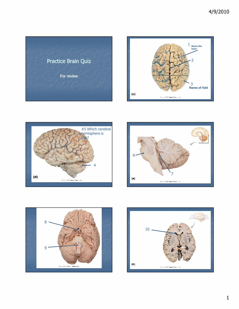
Practice Brain QuizPractice Brain Quiz
For reviewFor review
1
2
3
Name the layer
Name of fold
4
#5 Which cerebral hemisphere is this?
6
7
8
9
10

4/9/2010
2
Answers to QuizAnswers to Quiz
1) Pia mater1) Pia mater
2) Longitudinal Fissure2) Longitudinal Fissure
3) Gyri3) Gyri
4) Cerebellum4) Cerebellum
5) Left cerebral hemisphere5) Left cerebral hemisphere
6) Pons6) Pons
7) 47) 4thth ventricleventricle
8) Optic chiasma8) Optic chiasma
9) Medulla oblongata9) Medulla oblongata
10) Lateral Ventricles10) Lateral Ventricles
Practice Brain QuizPractice Brain Quiz

4/9/2010
3
Protection of the BrainProtection of the Brain
�� Bone (skull)Bone (skull)
�� Membranes (meninges)Membranes (meninges)
�� Watery cushion (cerebrospinal fluid)Watery cushion (cerebrospinal fluid)
�� BloodBlood--brain barrierbrain barrier
Meninges Meninges -- Layers of the BrainLayers of the Brain
Dura materDura mater
Arachnoid Arachnoid
Pia materPia mater
Figure 12.24
Skin of scalpPeriosteum
Falx cerebri(in longitudinalfissure only)
Blood vesselArachnoid villusPia materArachnoid mater
Duramater Meningeal
Periosteal
Bone of skull
Superiorsagittal sinus
Subduralspace
Subarachnoidspace
Dura MaterDura Mater
-- Strongest meninxStrongest meninx
-- Two layers of fibrous connective tissue Two layers of fibrous connective tissue (around the brain) separate to form dural (around the brain) separate to form dural sinusessinuses
Arachnoid MaterArachnoid Mater
-- Middle layer with weblike extensionsMiddle layer with weblike extensions
-- Separated from the dura mater by the Separated from the dura mater by the subdural spacesubdural space
-- Subarachnoid space contains CSF and Subarachnoid space contains CSF and blood vesselsblood vessels
-- Arachnoid villi protrude into the superior Arachnoid villi protrude into the superior sagittal sinus and permit CSF reabsorptionsagittal sinus and permit CSF reabsorption
Pia MaterPia Mater
Layer of delicate vascularized connective Layer of delicate vascularized connective tissue that clings tightly to the braintissue that clings tightly to the brain

4/9/2010
4
Figure 12.24
Skin of scalpPeriosteum
Falx cerebri(in longitudinalfissure only)
Blood vesselArachnoid villusPia materArachnoid mater
Duramater Meningeal
Periosteal
Bone of skull
Superiorsagittal sinus
Subduralspace
Subarachnoidspace
Cerebrospinal Fluid (CSF)Cerebrospinal Fluid (CSF)
Functions:Functions:
> Gives buoyancy to the CNS organs> Gives buoyancy to the CNS organs
> Protects the CNS from blows and other > Protects the CNS from blows and other traumatrauma
> Nourishes the brain and carries chemical > Nourishes the brain and carries chemical signals signals
Figure 12.26b
Ependymalcells
Capillary
Connectivetissue ofpia mater
Wastes andunnecessarysolutes absorbed
Sectionof choroid
plexus
(b) CSF formation by choroid plexuses
Cavity ofventricle
CSF forms as a filtratecontaining glucose, oxygen, vitamins, and ions(Na+, Cl–, Mg2+, etc.)
Choroid PlexusesChoroid Plexuses
�� Produce CSF at a constant rate Produce CSF at a constant rate
�� Hang from the roof of each ventricleHang from the roof of each ventricle
�� Clusters of capillaries enclosed by pia Clusters of capillaries enclosed by pia mater and a layer of ependymal cellsmater and a layer of ependymal cells
�� Ependymal cells use ion pumps to control Ependymal cells use ion pumps to control the composition of the CSF and help the composition of the CSF and help cleanse CSF by removing wastescleanse CSF by removing wastes