pre-lab exercises manuals/2018... · right intertransversarii muscles. 10 these muscles located...
TRANSCRIPT

1

2
PRE-LAB EXERCISES
Before coming to lab, get familiar with a few muscle groups we’ll be exploring during lab. Go to the Views section in Human Anatomy Atlas. Under Systems, scroll down to the Muscular System views. Select View 9. Inhalation and find the following muscles. When you select a muscle, note the book icon in the content box. Selecting this icon allows you to read the muscle’s definition.
1. External intercostals
2. Pectoralis minor
Define the following terms:
1. Flexion
2. Extension
3. Elevation
4. Depression
Right external intercostal muscles
Right pectoralis minor muscle

3
IN-LAB EXERCISES
Use the following modules to guide your exploration of the thoracic and abdominal regions of the muscular system. As you explore the modules, locate the muscles on any charts, models, or specimen available. These muscles are located on the thorax, abdomen, and back, and serve to protect the cavities they enclose as well as provide movement.
These muscle groups will have different jobs depending on where they are located. Those muscles on the chest wall around the ribs play roles in changing the size of the thoracic cavity for inspirations and expirations. Muscles located along the spine are involved in movement of the back, and muscles lining the abdomen help to protect the organs underneath while also allowing for movement of the trunk.
The long names of some of these muscles can be daunting, but they are often very descriptive. You can find origins, insertions, actions, and/or locations of these muscles simply in the names. When reviewing the action of a muscle, it will be helpful to think about where the muscle is located and where the insertion is. Muscle physiology requires that a muscle will “pull” instead of “push” during contraction, and the insertion is the part that will move. Imagine that the muscle is pulling on the bone or tissue it is attached to at the insertion.
Access 3D views and animated muscle actions in Human Anatomy Atlas, which will be especially helpful to visualize muscle actions. When you select a structure in Atlas, you’ll see options to read the definition and hear the pronunciation in the content box. When you select a muscle, be sure to select the blue pin icon in the content box. This will give you the option to view origins and insertions as visible pins on the muscle (select Attachments), view the blood supply, and/or the nerve supply.
In the modules below, identify the following:
• Muscle location
• Origin(s) and insertion(s)
• Muscle action
• Nerve supply

4
A. Inspiratory Muscles
Go to Systems: Muscular System Views and select 9. Inhalation.
Go to Muscle Actions and view Muscle Action: Ribs elevation.
Right serratus anterior muscles
Right pectoralis minor muscle
Right scalene muscles
Diaphragm
Right external intercostal muscles
Right serratus posterior superior
muscles

5
These muscles are responsible for inspiration during pulmonary ventilation. Although the diaphragm and, to a lesser extent, the external intercostals are primarily responsible for inspiration, additional accessory respiratory muscles can contract to assist in a more forceful inspiration.
Anatomically, the diaphragm marks the division between the thoracic and abdominal cavities. Observe the openings in the diaphragm that allow the passage of the esophagus and major blood vessels.
Insertion
Inspiratory Muscles
OriginMuscle
Diaphragm
External intercostal
Pectoralis minor
Serratus anterior
Serratus posterior superior
Scalenes
Action Innervation

6
B. Expiratory Muscles
Go to Systems: Muscular System Views and select 10. Exhalation.
Go to Muscle Actions and view Muscle Action: Ribs depression.
Right serratus posterior inferior muscles
Right transversus thoracis muscles
Left transversus abdominis muscle
Left internal oblique muscle
Left rectus abdominis muscle
Left internal intercostal muscles

7
These muscles are responsible for expiration during pulmonary ventilation. In a normal, quiet exhalation, the relaxation of the diaphragm and external intercostals are responsible for air departing the lungs. However, accessory respiratory muscles may be used in a more forceful exhalation.
It can be easy to confuse the external and internal intercostals. The external intercostals are so named because they are superficial to the internal intercostals. It will also be helpful to pay attention to the direction of the fibers in these two muscles since they run in opposite directions.
Insertion
Expiratory Muscles
OriginMuscle
Internal intercostal
Transversus thoracis
Serratus posterior inferior
Rectus abdominis
Internal oblique
Transversus abdominis
Action Innervation
TIME TO PRACTICE!GO TO THE MUSCULAR SYSTEM QUIZZES MENU AND COMPLETE QUIZ 20. INTERCOSTALS.

8
C. Back Muscles
Go to Systems: Muscular System Views and select 14. Upper Back and 15. Lower Back.
Go to Muscle Actions and view Muscle Actions: Spine flexion, Spine extension, Spine lateral flexion, Spine rotation.
Left quadratus lumborum muscle
Left longissimus muscle
Left iliocostalis muscle
Left semispinalis thoracis muscle
Left semispinalis capitis muscle
Right splenius capitis muscle
Right splenius cervicis muscle
Right spinalis muscle

9
View 15. Lower Back
Left semispinalis cervicis muscle
Left multifidus muscle
Right rotatores muscles
Right intertransversarii muscles
Right intertransversarii muscles

10
These muscles located along the vertebral column function to support and extend the neck and/or back. The spinalis, longissimus, and iliocostalis are part of the erector spinae group, which lie parallel to the spine and extend the back. The transversospinales group, composed of the semispinalis, multifidus, and rotatores muscles, are so named because of their position between the transverse and spinous processes on the vertebrae.
Some of these muscles are deep to other muscles listed, so be sure to use the Hide function on the superficial muscles on one side of each of the views to reveal the deeper muscles.
Insertion
Back Muscles
OriginMuscle
Splenius capitis
Splenius cervicis
Semispinalis capitis
Semispinalis cervicis
Semispinalis thoracis
Spinalis
Longissimus
Action Innervation

11
Insertion
Back Muscles (continued)
OriginMuscle
Rotatores
Iliocostalis
Interspinales
Multifidus
Intertransversarii
Quadratus lumborum
Action Innervation
TIME TO PRACTICE!GO TO THE MUSCULAR SYSTEM QUIZZES MENU AND COMPLETE QUIZ 21. POSTERIOR THORAX.

12
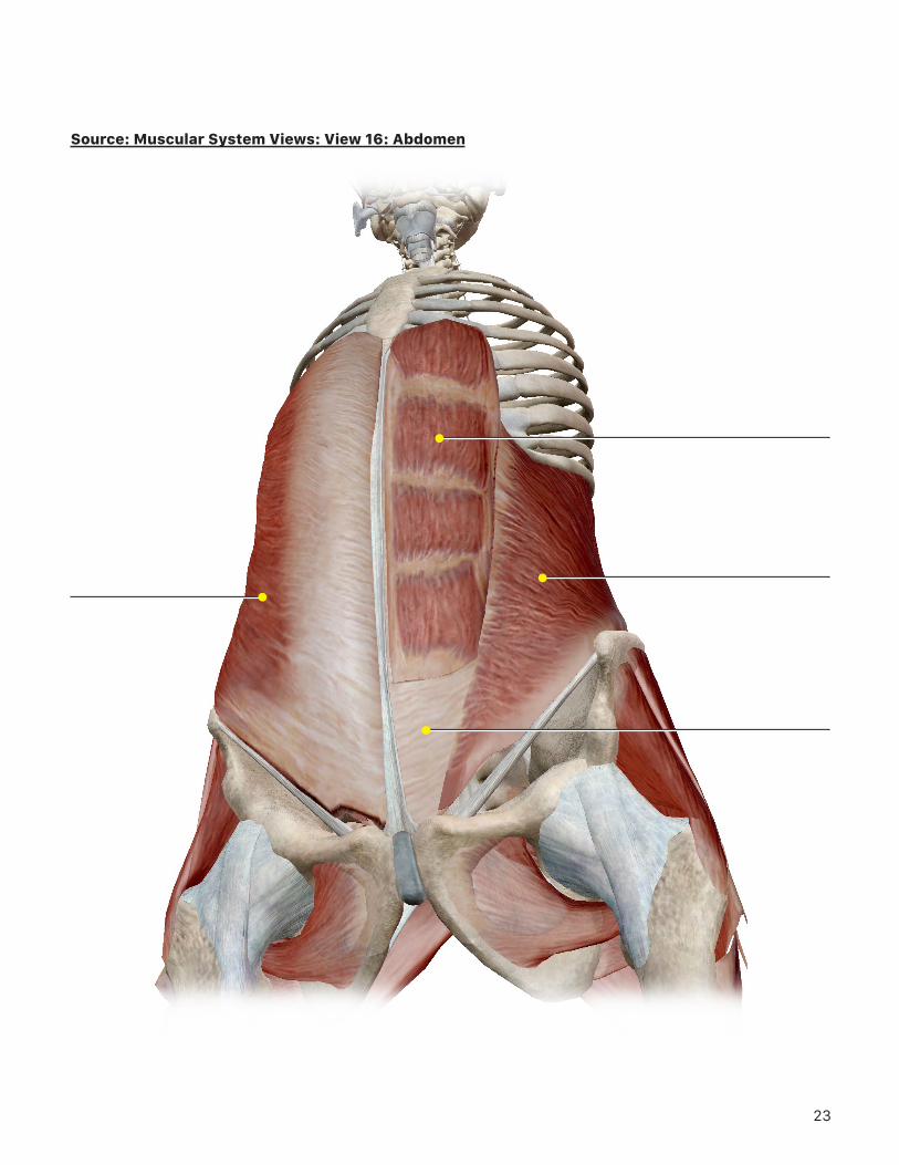
D. Abdomen
Go to Systems: Muscular System Views and select 16. Abdomen.Then, go to Cross Sections and view Cross Sections Abdomen (Axial): 4. Abdomen (L02-L03).Finally, go to Muscle Actions and view Muscle Action: Spine rotation.
Right external oblique muscle
Left rectus abdominis muscle
Left internal oblique muscle
Left transversus abdominis muscle

13
Left external oblique muscle
Left transversus abdominis muscle
Left internal oblique muscle
Left rectus abdominis muscle

14
The abdominal wall is composed of four muscles whose fibers run in different directions. These muscle layers protect the underlying organs, assist in forced respirations, and cause rotation of the trunk when contracted.
When viewing the cross section, note how the abdominal muscles overlap each other, especially on the anterior side.
Insertion
Abdomen
OriginMuscle
Rectus abdominis
External oblique
Internal oblique
Transversus abdominis
Action Innervation
TIME TO PRACTICE!GO TO THE MUSCULAR SYSTEM QUIZZES MENU AND COMPLETE QUIZ 22. ABDOMEN.

15
PUTTING IT ALL TOGETHER
1. Based on what you’ve learned about the muscles in this exercise, what do you think the following terms mean?
a. External
b. Internal
c. Oblique
d. Rectus
e. Capitis
f. Spinalis
2. Which muscles are used when performing the following actions?
a. Rowing a boat
b. Standing erect
c. Twisting your torso (as when swinging a baseball bat)
d. Taking a bow after a performance
16
e. Inhaling deeply
3. Sometimes acid that regurgitates from the stomach can irritate the phrenic nerve, causing it to fire spontaneously. What effect do you think this would have?

17

18
Source: Muscular System Views: View 9: Inhalation

19
Source: Muscular System Views: View 9: Inhalation

20
Source: Muscular System Views: View 10: Exhalation

21
Source: Muscular System Views: View 14: Upper Back

22
Source: Muscular System Views: View 15: Lower Back
23
Source: Muscular System Views: View 16: Abdomen

24
Source: Cross Sections: Abdomen (Axial): View 4: Abdomen (L02-L03)