pre-transfusion testing - uclapathology.ucla.edu/workfiles/education/transfusion medicine/3-4... ·...
TRANSCRIPT
Pre-Transfusion Testing
Alyssa Ziman, MD
UCLA Health System July 2012
Pre-Transfusion Testing
• Purpose
– Avoid risks to donor and recipient
– Meet product specifications
• Consist of both donor and patient testing.
– Donor testing and residual risk of infection
– Patient testing
Donor Pre-Transfusion Evaluation
• Donor medical history and risk factor assessment
• Infectious disease testing
• ABO and Rh typing
• Test for antibodies to red cell antigens
• Capturing post donation information
Donor Infectious Disease Testing
• Hepatitis B, sAg and anti-core antibody
• Hepatitis C antibody
• HIV 1 and 2 antibodies
• HTLV 1 and 2 antibodies
• Serologic Test for Syphilis
• Nucleic Acid Testing (NAT) for HIV, HBV, HCV and WNV
• Detection of Bacteria in platelet products
• CMV antibody for select recipients
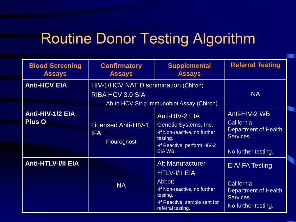
Routine Donor Testing Algorithm
Blood Screening
Assays
Confirmatory
Assays
Supplemental
Assays
Referral Testing
Anti-HCV EIA HIV-1/HCV NAT Discrimination (Chiron)
RIBA HCV 3.0 SIA
Ab to HCV Strip Immunoblot Assay (Chiron)
NA
Anti-HIV-1/2 EIA
Plus O Licensed Anti-HIV-1
IFA
Flourognost
Anti-HIV-2 EIA
Genetic Systems, Inc.
•If Non-reactive, no further
testing.
•If Reactive, perform HIV-2
EIA WB.
Anti-HIV-2 WB
California
Department of Health
Services
No further testing. Anti-HTLV-I/II EIA
NA
Alt Manufacturer
HTLV-I/II EIA
Abbott
•If Non-reactive, no further
testing.
•If Reactive, sample sent for
referral testing.
EIA/IFA Testing
California
Department of Health
Services
No further testing.
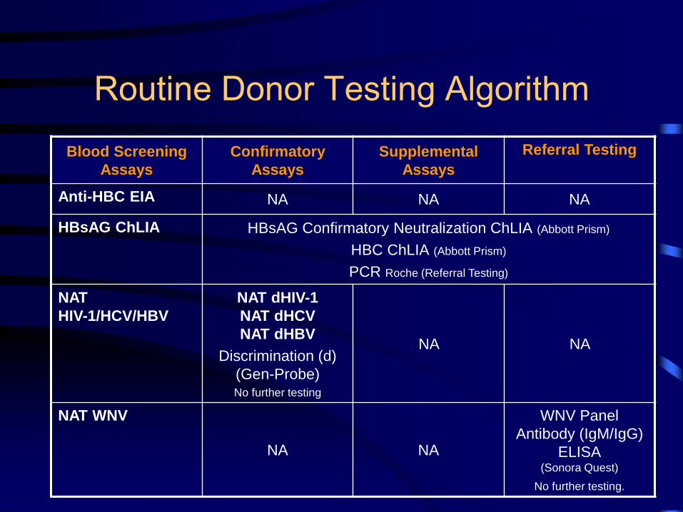
Routine Donor Testing Algorithm
Blood Screening
Assays
Confirmatory
Assays
Supplemental
Assays
Referral Testing
Anti-HBC EIA NA NA NA
HBsAG ChLIA HBsAG Confirmatory Neutralization ChLIA (Abbott Prism)
HBC ChLIA (Abbott Prism)
PCR Roche (Referral Testing)
NAT
HIV-1/HCV/HBV
NAT dHIV-1
NAT dHCV
NAT dHBV
Discrimination (d)
(Gen-Probe) No further testing
NA NA
NAT WNV
NA NA
WNV Panel
Antibody (IgM/IgG)
ELISA (Sonora Quest)
No further testing.
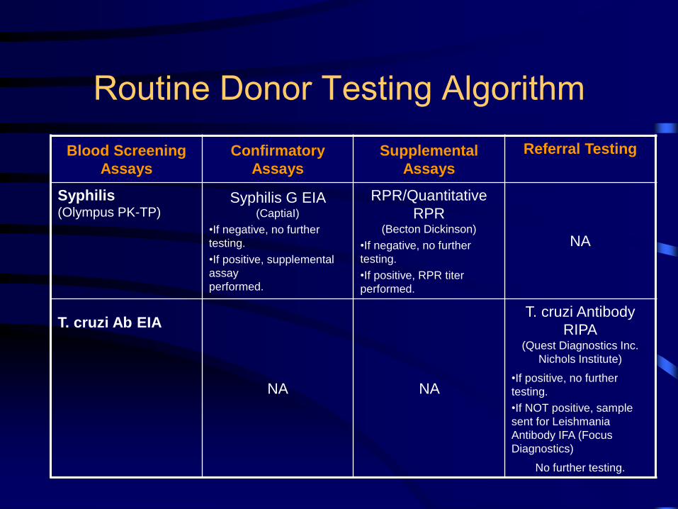
Routine Donor Testing Algorithm
Blood Screening
Assays
Confirmatory
Assays
Supplemental
Assays
Referral Testing
Syphilis (Olympus PK-TP)
Syphilis G EIA (CaptiaI)
•If negative, no further
testing.
•If positive, supplemental
assay
performed.
RPR/Quantitative
RPR (Becton Dickinson)
•If negative, no further
testing.
•If positive, RPR titer
performed.
NA
T. cruzi Ab EIA
NA NA
T. cruzi Antibody
RIPA (Quest Diagnostics Inc.
Nichols Institute)
•If positive, no further
testing.
•If NOT positive, sample
sent for Leishmania
Antibody IFA (Focus
Diagnostics)
No further testing.
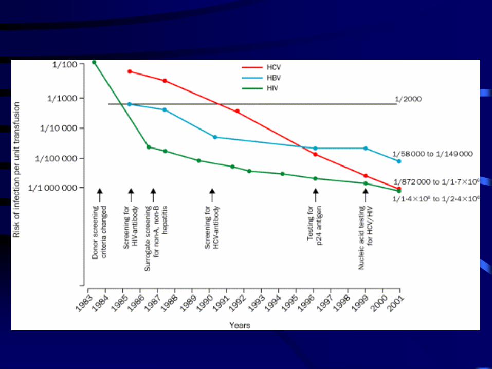

Estimates of Known Viral Infectious
Disease Risks of Transfusion
Virus Risk per Unit
Transfusion
Transmission
Rate
Window
Period
(mean)
HIV-1 1:2,135,000 90% 9 - 11 days
HCV 1:1,935,000 90% 7 - 10 days
HBV 1:600,000 70% 30 - 35 days
HTLV 1:3,000,000 30% 51 days
WNV 1:350,000 Unknown 11- 13 days
Parvo B19 1:40,000 to 3,000 Low -
Hepatitis A/E 1:1,000,000 Low -
Transfusion 2002;42:975-979.
N Engl J Med 2005;353:451-9.
N Engl J Med 2011;364:236-47.
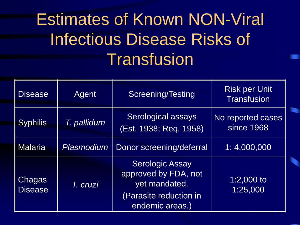
Estimates of Known NON-Viral
Infectious Disease Risks of
Transfusion
Disease Agent Screening/Testing Risk per Unit
Transfusion
Syphilis T. pallidum Serological assays
(Est. 1938; Req. 1958)
No reported cases
since 1968
Malaria Plasmodium Donor screening/deferral 1: 4,000,000
Chagas
Disease T. cruzi
Serologic Assay
approved by FDA, not
yet mandated.
(Parasite reduction in
endemic areas.)
1:2,000 to
1:25,000
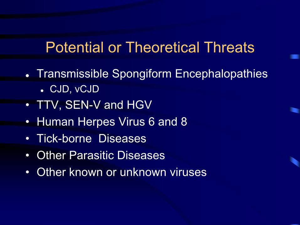
Potential or Theoretical Threats
Transmissible Spongiform Encephalopathies
CJD, vCJD
• TTV, SEN-V and HGV
• Human Herpes Virus 6 and 8
• Tick-borne Diseases
• Other Parasitic Diseases
• Other known or unknown viruses
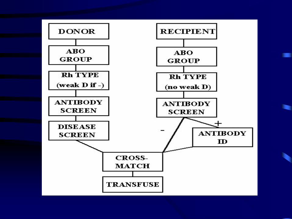
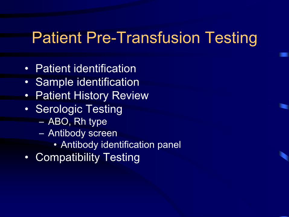
Patient Pre-Transfusion Testing
• Patient identification
• Sample identification
• Patient History Review
• Serologic Testing – ABO, Rh type
– Antibody screen
• Antibody identification panel
• Compatibility Testing
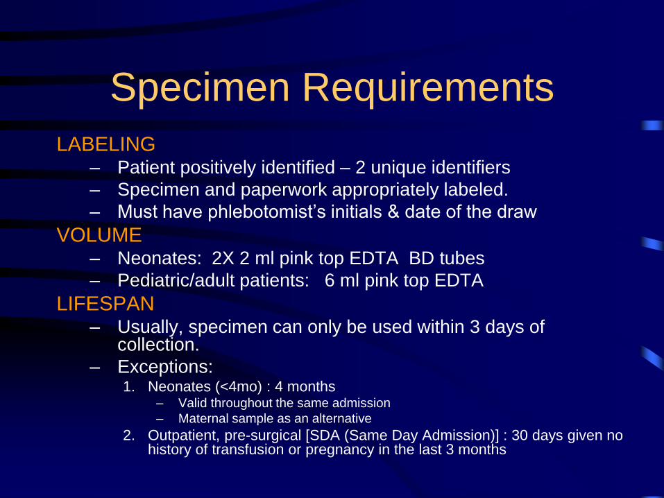
Specimen Requirements
LABELING – Patient positively identified – 2 unique identifiers
– Specimen and paperwork appropriately labeled.
– Must have phlebotomist’s initials & date of the draw
VOLUME – Neonates: 2X 2 ml pink top EDTA BD tubes
– Pediatric/adult patients: 6 ml pink top EDTA
LIFESPAN – Usually, specimen can only be used within 3 days of
collection.
– Exceptions: 1. Neonates (<4mo) : 4 months
– Valid throughout the same admission
– Maternal sample as an alternative
2. Outpatient, pre-surgical [SDA (Same Day Admission)] : 30 days given no history of transfusion or pregnancy in the last 3 months

Common Blood Bank Orders HOLD CLOT: RBC need is unknown. Specimen is held for 3
days “just in case”. No testing done unless requested.
TYPE AND SCREEN: RBC need is possible. Specimen typed
for ABO-Rh and screened for RBC antibodies.
TYPE AND CROSSMATCH: RBC need likely or definite. T&S
performed. Requested # RBCs crossmatched and reserved.
– If pt >4mo until the specimen used for testing is 3 days old.
– If pt <4mo, reserved for the remainder of the admission.
CHECK TYPE: 2nd separately drawn specimen to confirm
ABO-Rh.
KEEP AHEAD ORDERS: Blood Bank ensures that a specified
# of RBCs or FP reserved for the patient at all times.

Turn-Around-Times
ROUTINE – TYPE AND SCREEN
• ABO-Rh: 15 minutes
• Antibody Screen: 60 minutes
– CROSSMATCH • ~ 15 minutes.
• Longer if patient has antibodies.
STAT – Uncrossmatched O-Neg RBCs: 10 minutes
– STAT blood type: 5 minutes
– Uncrossmatched type specific RBCs: 15 minutes
– STAT type and screen: 60 minutes
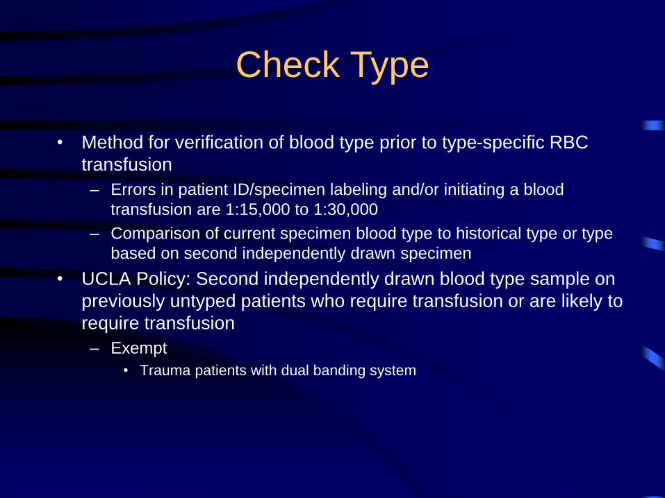
• Method for verification of blood type prior to type-specific RBC
transfusion
– Errors in patient ID/specimen labeling and/or initiating a blood
transfusion are 1:15,000 to 1:30,000
– Comparison of current specimen blood type to historical type or type
based on second independently drawn specimen
• UCLA Policy: Second independently drawn blood type sample on
previously untyped patients who require transfusion or are likely to
require transfusion
– Exempt
• Trauma patients with dual banding system
Check Type
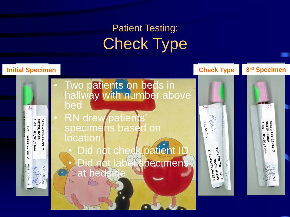
Patient Testing:
Check Type
• Two patients on beds in hallway with number above bed
• RN drew patients’ specimens based on location
• Did not check patient ID
• Did not label specimens at bedside
3rd Specimen Check Type Initial Specimen
02
8/
w1
11
-2
2-3
3 7
SM
IT
H, R
OS
A
F 6
5 0
1/
01
/1
94
4
w1
11
-22
-33
7
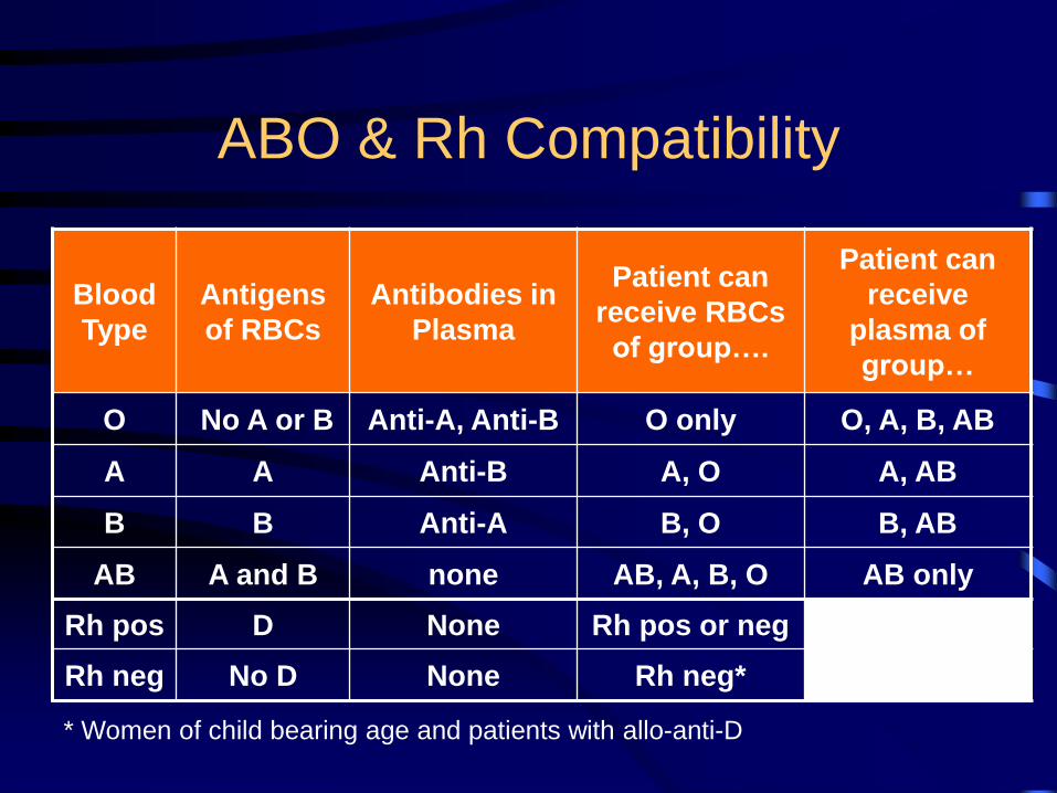
ABO & Rh Compatibility
Blood
Type
Antigens
of RBCs
Antibodies in
Plasma
Patient can
receive RBCs
of group….
Patient can
receive
plasma of
group…
O No A or B Anti-A, Anti-B O only O, A, B, AB
A A Anti-B A, O A, AB
B B Anti-A B, O B, AB
AB A and B none AB, A, B, O AB only
Rh pos D None Rh pos or neg
Rh neg No D None Rh neg*
* Women of child bearing age and patients with allo-anti-D
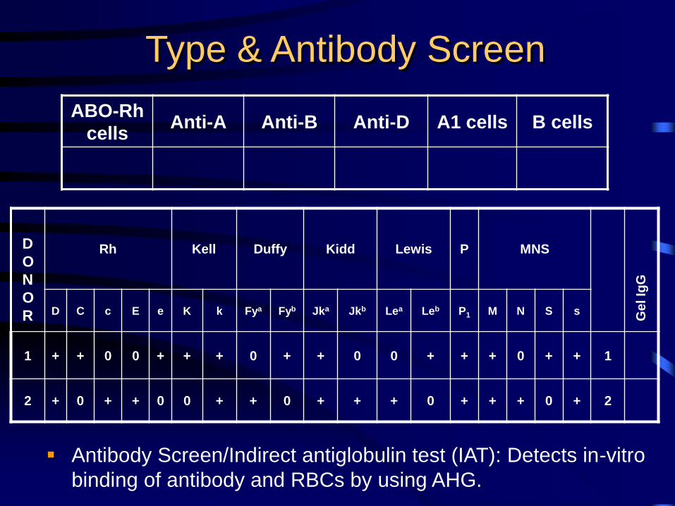
Type & Antibody Screen
ABO-Rh
cells Anti-A Anti-B Anti-D A1 cells B cells
Rh Kell Duffy Kidd Lewis P MNS
D C c E e K k Fya Fyb Jka Jkb Lea Leb P1 M N S s
1 + + 0 0 + + + 0 + + 0 0 + + + 0 + + 1
2 + 0 + + 0 0 + + 0 + + + 0 + + + 0 + 2
D
O
N
O
R Gel Ig
G
Antibody Screen/Indirect antiglobulin test (IAT): Detects in-vitro
binding of antibody and RBCs by using AHG.
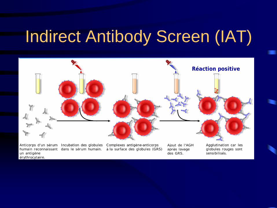
Indirect Antibody Screen (IAT)
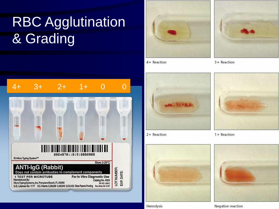
RBC Agglutination
& Grading
4+ 3+ 2+ 1+ 0 0
Application of the IAT
• RBC antibody screen and identification
• Weak D testing
• Antibody titration
• IgG Crossmatching
• Typing of erythrocytes antigens
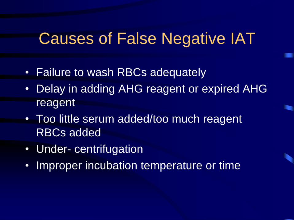
Causes of False Negative IAT
• Failure to wash RBCs adequately
• Delay in adding AHG reagent or expired AHG
reagent
• Too little serum added/too much reagent
RBCs added
• Under- centrifugation
• Improper incubation temperature or time
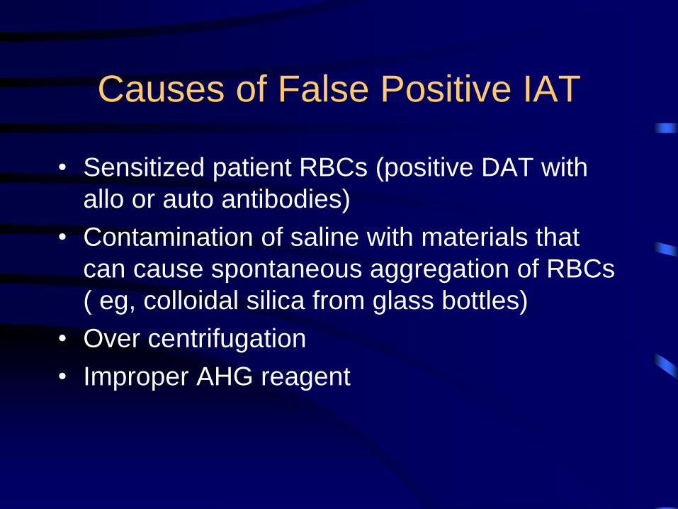
Causes of False Positive IAT
• Sensitized patient RBCs (positive DAT with
allo or auto antibodies)
• Contamination of saline with materials that
can cause spontaneous aggregation of RBCs
( eg, colloidal silica from glass bottles)
• Over centrifugation
• Improper AHG reagent
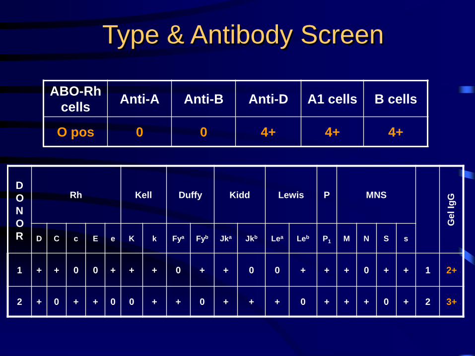
Type & Antibody Screen
ABO-Rh
cells Anti-A Anti-B Anti-D A1 cells B cells
O pos 0 0 4+ 4+ 4+
Rh Kell Duffy Kidd Lewis P MNS
D C c E e K k Fya Fyb Jka Jkb Lea Leb P1 M N S s
1 + + 0 0 + + + 0 + + 0 0 + + + 0 + + 1 2+
2 + 0 + + 0 0 + + 0 + + + 0 + + + 0 + 2 3+
D
O
N
O
R
Gel Ig
G
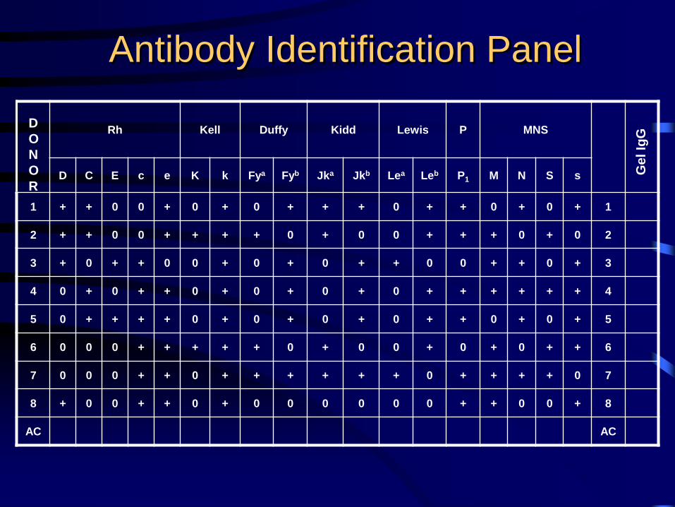
Antibody Identification Panel
Rh Kell Duffy Kidd Lewis P MNS
D C E c e K k Fya Fyb Jka Jkb Lea Leb P1 M N S s
1 + + 0 0 + 0 + 0 + + + 0 + + 0 + 0 + 1
2 + + 0 0 + + + + 0 + 0 0 + + + 0 + 0 2
3 + 0 + + 0 0 + 0 + 0 + + 0 0 + + 0 + 3
4 0 + 0 + + 0 + 0 + 0 + 0 + + + + + + 4
5 0 + + + + 0 + 0 + 0 + 0 + + 0 + 0 + 5
6 0 0 0 + + + + + 0 + 0 0 + 0 + 0 + + 6
7 0 0 0 + + 0 + + + + + + 0 + + + + 0 7
8 + 0 0 + + 0 + 0 0 0 0 0 0 + + 0 0 + 8
AC AC
D
O
N
O
R
Gel Ig
G
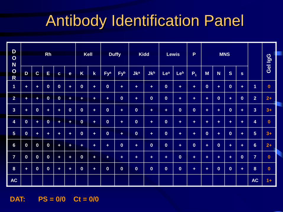
Antibody Identification Panel
Rh Kell Duffy Kidd Lewis P MNS
D C E c e K k Fya Fyb Jka Jkb Lea Leb P1 M N S s
1 + + 0 0 + 0 + 0 + + + 0 + + 0 + 0 + 1 0
2 + + 0 0 + + + + 0 + 0 0 + + + 0 + 0 2 2+
3 + 0 + + 0 0 + 0 + 0 + + 0 0 + + 0 + 3 3+
4 0 + 0 + + 0 + 0 + 0 + 0 + + + + + + 4 0
5 0 + + + + 0 + 0 + 0 + 0 + + 0 + 0 + 5 3+
6 0 0 0 + + + + + 0 + 0 0 + 0 + 0 + + 6 2+
7 0 0 0 + + 0 + + + + + + 0 + + + + 0 7 0
8 + 0 0 + + 0 + 0 0 0 0 0 0 + + 0 0 + 8 0
AC AC 1+
D
O
N
O
R
Gel Ig
G
DAT: PS = 0/0 Ct = 0/0
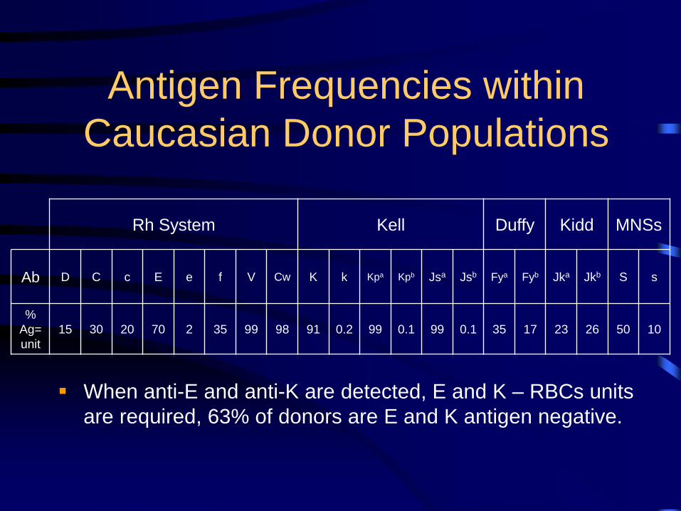
Antigen Frequencies within
Caucasian Donor Populations
Rh System Kell Duffy Kidd MNSs
Ab D C c E e f V Cw K k Kpa Kpb Jsa Jsb Fya Fyb Jka Jkb S s
%
Ag=
unit
15 30 20 70 2 35 99 98 91 0.2 99 0.1 99 0.1 35 17 23 26 50 10
When anti-E and anti-K are detected, E and K – RBCs units
are required, 63% of donors are E and K antigen negative.
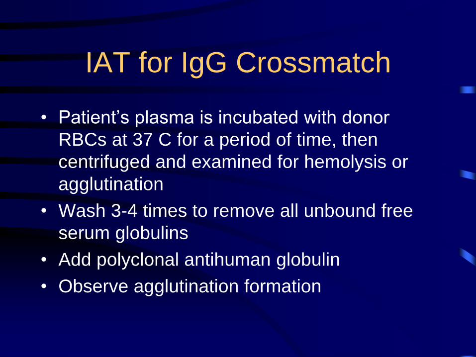
IAT for IgG Crossmatch
• Patient’s plasma is incubated with donor
RBCs at 37 C for a period of time, then
centrifuged and examined for hemolysis or
agglutination
• Wash 3-4 times to remove all unbound free
serum globulins
• Add polyclonal antihuman globulin
• Observe agglutination formation
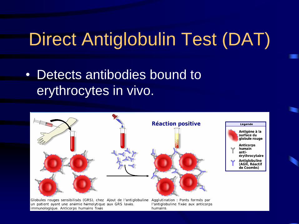
Direct Antiglobulin Test (DAT)
• Detects antibodies bound to
erythrocytes in vivo.
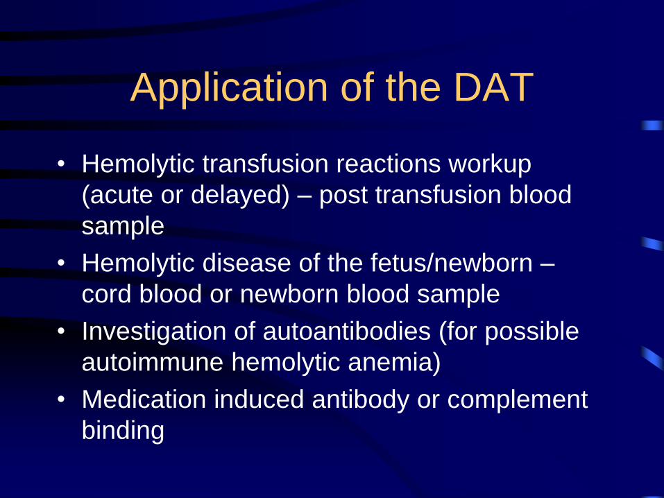
Application of the DAT
• Hemolytic transfusion reactions workup
(acute or delayed) – post transfusion blood
sample
• Hemolytic disease of the fetus/newborn –
cord blood or newborn blood sample
• Investigation of autoantibodies (for possible
autoimmune hemolytic anemia)
• Medication induced antibody or complement
binding
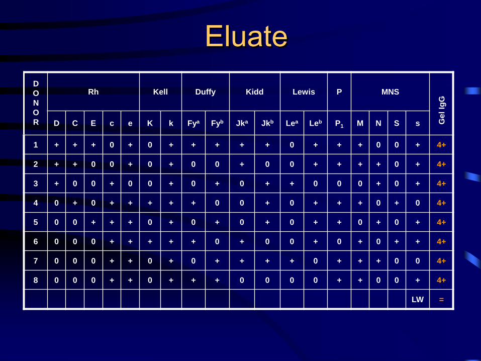
Eluate
Rh Kell Duffy Kidd Lewis P MNS
D C E c e K k Fya Fyb Jka Jkb Lea Leb P1 M N S s
1 + + + 0 + 0 + + + + + 0 + + + 0 0 + 4+
2 + + 0 0 + 0 + 0 0 + 0 0 + + + + 0 + 4+
3 + 0 0 + 0 0 + 0 + 0 + + 0 0 0 + 0 + 4+
4 0 + 0 + + + + + 0 0 + 0 + + + 0 + 0 4+
5 0 0 + + + 0 + 0 + 0 + 0 + + 0 + 0 + 4+
6 0 0 0 + + + + + 0 + 0 0 + 0 + 0 + + 4+
7 0 0 0 + + 0 + 0 + + + + 0 + + + 0 0 4+
8 0 0 0 + + 0 + + + 0 0 0 0 + + 0 0 + 4+
LW =
D
O
N
O
R Ge
l Ig
G
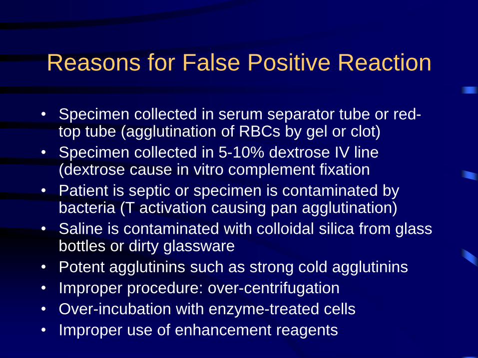
Reasons for False Positive Reaction
• Specimen collected in serum separator tube or red-top tube (agglutination of RBCs by gel or clot)
• Specimen collected in 5-10% dextrose IV line (dextrose cause in vitro complement fixation
• Patient is septic or specimen is contaminated by bacteria (T activation causing pan agglutination)
• Saline is contaminated with colloidal silica from glass bottles or dirty glassware
• Potent agglutinins such as strong cold agglutinins
• Improper procedure: over-centrifugation
• Over-incubation with enzyme-treated cells
• Improper use of enhancement reagents
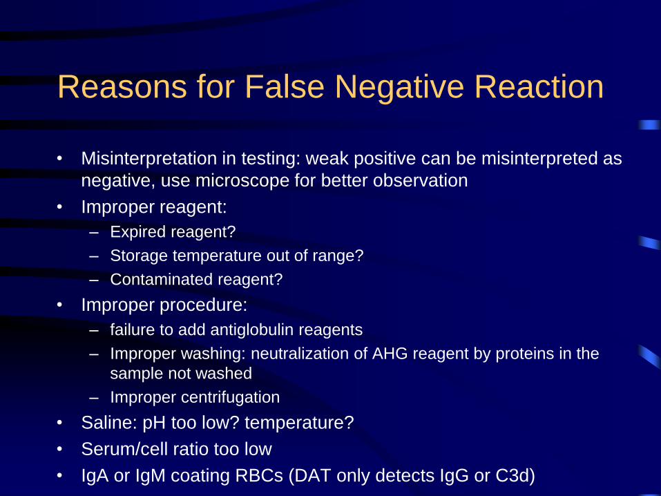
Reasons for False Negative Reaction
• Misinterpretation in testing: weak positive can be misinterpreted as
negative, use microscope for better observation
• Improper reagent:
– Expired reagent?
– Storage temperature out of range?
– Contaminated reagent?
• Improper procedure:
– failure to add antiglobulin reagents
– Improper washing: neutralization of AHG reagent by proteins in the
sample not washed
– Improper centrifugation
• Saline: pH too low? temperature?
• Serum/cell ratio too low
• IgA or IgM coating RBCs (DAT only detects IgG or C3d)
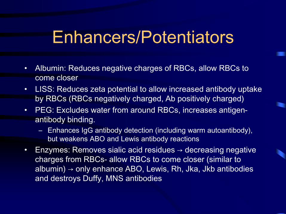
Enhancers/Potentiators
• Albumin: Reduces negative charges of RBCs, allow RBCs to
come closer
• LISS: Reduces zeta potential to allow increased antibody uptake
by RBCs (RBCs negatively charged, Ab positively charged)
• PEG: Excludes water from around RBCs, increases antigen-
antibody binding.
– Enhances IgG antibody detection (including warm autoantibody),
but weakens ABO and Lewis antibody reactions
• Enzymes: Removes sialic acid residues → decreasing negative
charges from RBCs- allow RBCs to come closer (similar to
albumin) → only enhance ABO, Lewis, Rh, Jka, Jkb antibodies
and destroys Duffy, MNS antibodies
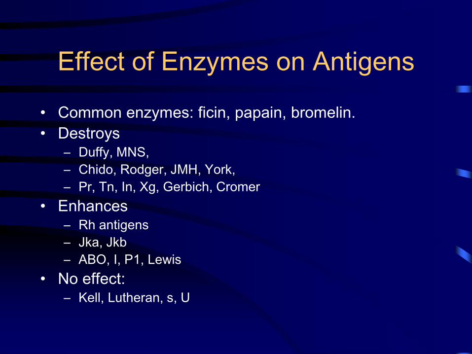
Effect of Enzymes on Antigens
• Common enzymes: ficin, papain, bromelin.
• Destroys – Duffy, MNS,
– Chido, Rodger, JMH, York,
– Pr, Tn, In, Xg, Gerbich, Cromer
• Enhances – Rh antigens
– Jka, Jkb
– ABO, I, P1, Lewis
• No effect: – Kell, Lutheran, s, U
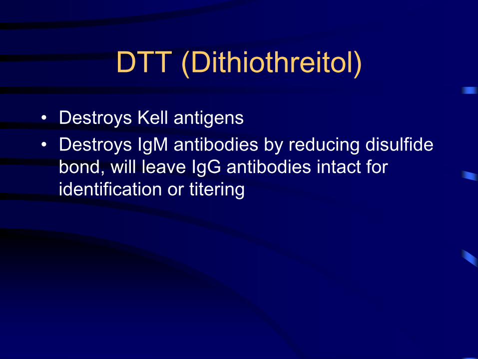
DTT (Dithiothreitol)
• Destroys Kell antigens
• Destroys IgM antibodies by reducing disulfide
bond, will leave IgG antibodies intact for
identification or titering
Rules to live by……
• Don’t panic
• Follow routine procedures
• Be consistent and systematic
• Document all tests and results
• Never hesitate to ask for help
• Identify and use your resources
• Consider what is “safest” for the patient!