pregnancies aneuploidies and sub-chromosomal copy number
TRANSCRIPT

Page 1/13
A Retrospective Study of Noninvasive Prenatal Testing for ChromosomeAneuploidies and Sub-Chromosomal Copy Number Variations in 24359 SinglePregnanciesYuefang Liu
huaian maternal and child hospitalLongfei Cheng
huaian maternal and child hospitalYuan Peng
huaian maternal and child hospitalZhe Liang
huaian maternal and child hospitalPan Qiong ( [email protected] )
huaian maternal and child health care hospital https://orcid.org/0000-0001-8151-0923
Research
Keywords: Noninvasive prenatal testing (NIPT), Sub-chromosomal copy number variations (CNVs), Positive predictive value(PPV)
Posted Date: November 13th, 2020
DOI: https://doi.org/10.21203/rs.3.rs-104459/v1
License: This work is licensed under a Creative Commons Attribution 4.0 International License. Read Full License

Page 2/13
AbstractBackground: With the development of whole-genome sequencing, small sub-chromosomal deletions and duplications could be found by non-invasive prenataltesting(NIPT). This study aimed to review the e�ciency of NIPT as a screening test for aneuploidies and sub-chromosomal copy number variations (CNVs) in24359 single pregnancies.
Methods: A total of 24359 single pregnancies with different clinical indications were retrospectively analyzed. The positive predictive value PPV ofchromosome aneuploidies and subchromosomal CNVs were analyzed. Pathogenicity of abnormal NIPT results were assessed according to American Collegeof Medical Genetics and Genomics(ACMG).
Results: A total of 442 pregnancies (442/24359,1.9%) were with abnormal NIPT results. PPV for trisomy 21(T21), trisomy 18 (T18), trisomy 13 (T13), and sexchromosome aneuploidies (SCAs) was 84.8%, 54.2%, 11.1% an 40.5% respectively. The PPV for sub-chromosomal CNVs was 59.0% (46/78). The PPV forCNVs ≤5 Mb was 68.9% (31/45), for CNVs within 5-10 Mb was 83.3%(5/6) and for CNVs ≥10 Mb was 37.1% (10/27) respectively. The clinical information,prenatal diagnosis results and follow-up results of 46 true positive cases, 6 cases with sub-chromosomal CNVs inconsistent with NIPT and 1 false negativecase were also described in detail.
Conclusions: Our data have potential signi�cance in demonstrating the signi�cance of NIPT not only for common whole chromosome aneuploidies but alsofor sub-chromosomal CNVs. Besides, the clinical information, prenatal diagnosis results and follow-up results of 52 cases with sub-chromosomal CNVs and 1false negative case would provide important guidance for genetic counseling.
IntroductionThe discovery of cell-free fetal DNA (cffDNA) in maternal plasma by Lo et al in 1997 has opened up new approaches for NIPT[1]. At present, NIPT has beengradually applied as a �rst-tier aneuploidy screening strategy in clinical practice [2–3]. Previous large-scale clinical studies have revealed high accuracy ofNIPT in screening on trisomy 21, 18 and 13, with sensitivity and speci�city higher than 95% [4–5].
Genomic structural changes, such as copy number variations (CNVs), are also always associated with human disease. Recently, further development andexpansion use of NIPT has focused on microduplication/microdeletion syndromes(MMSs)[6]. The most common microdeletion is at 22q11.2, and recentreports indicate that the clinical incidence rate may exceed 1/1000 in the prenatal population[7]. The 22q11.2 microdeletion syndrome causing DiGeorgesyndrome has a very broad clinical phenotype that can include congenital heart disease, palatal, gastrointestinal, genitourinary anomalies, immunode�ciency,endocrine disturbance, developmental delay, cognitive de�cits and psychiatric illness[8]. In addition to 22q11.2 microduplication/microdeletion syndromes,other newly described CNVs like the distal 1q21.1 microdeletions/microduplications, 15q11.2 deletion, 16p13.11 deletion and 16p11.2 deletion/duplication arealso identi�ed as disease-causing CNVs[9–12]. Therefore, it is very important to evaluate the accuracy of NIPT for CNVs, which could help identify high-riskpregnancies and offer the possibility of a con�rmatory invasive diagnostic test. However, there are many challenges for NIPT in clinical practice especially lowpositive predictive values (PPV). In the present study, we retrospectively analyzed 24359 single pregnancies including 125 cases of sub-chromosomal CNVs toassess the e�ciency of NIPT technology on the detection of sub-chromosomal CNVs.
Results
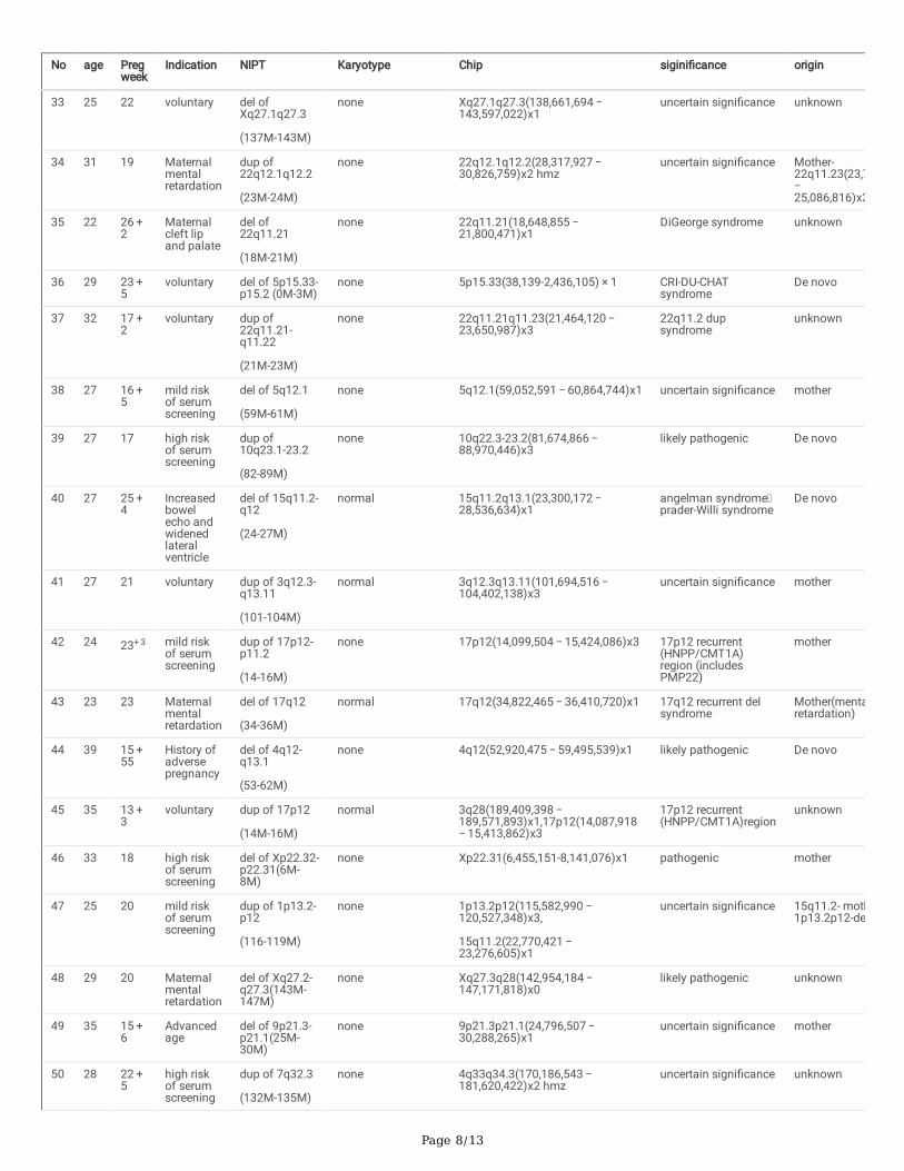
An overview of clinical dataAmong the 24367 cases undergoing NIPT, 8 cases were not eligible for the next analysis due to the low concentration of fetal DNA, so the remaining 24359cases were under analyzed in the present study. The maternal age for the 24359 pregnancies ranged from 16 to 51 years old. The group aged 25 to 29 yearswas the majority (8084, 33.2%). Pregnant women older than 35 years were 3966(16.3%). The gestational age ranged from 9 to 34 weeks, and 56.9% had agestational age from 17 to 20 weeks (see Table 1). The positive rate from younger group to older group were 1.4%(85/6097), 1.8%(144/8084), 1.5%(95/6212),2.6%(92/3472) and 5.3%(26/494) respectively. (Fig. 1). Older group (≥ 41) has the highest positive rate.

Page 3/13
Table 1Maternal age and gestational age of 24359 blood sampling
Maternal age at NIPT (years) Number Percent (100%)
≤ 24 6097 25.0%
25–29 8084 33.2%
30–34 6212 25.5%
35–40 3472 14.3%
≥ 41 494 2.0%
Advanced maternal age (≥ 35 years old) 3966 16.3%
Gestational age at NIPT (weeks)
≤ 8 0 0%
9–12 680 2.8%
13–16 6525 26.8%
17–20 13867 56.9%
21–24 2612 10.7%
25–28 639 2.6%
≥ 29 36 0.1%
Range (weeks) 12–34 /
Figure 1 The positive rate of NIPT for aneuploidy and subchromosomal CNV increases with maternal age and the number of positive cases in the �ve agegroups.
There were 442 pregnant cases with chromosome aneuploidies and submicroscopic anomalies, whose basic information and clinical reasons for NIPT werecollected (Table 2 and Table 3). We found that 61(13.8%,61/442 ) cases were with advanced maternal age more than 35 years (included), and 26(5.8%,26/442)pregnant cases had ultrasound abnormalities. 123(27.8%, 123/442) cases were with high risk of serum biochemistry screening. The number of thevoluntary group were 210(47.5%, 210/442). Other reasons included poor fertility history and maternal chromosomal abnormality or mental retardation.
Table 2Clinical information of 442 pregnancy cases with chromosome
aneuploidies and subchromosomal CNVsMaternal age at NIPT (years) Number Percent (100%)
≤ 24 77 17.4%
25–29 148 33.9%
30–34 95 21.5%
35–40 97 21.9%
≥ 41 25 5.7%
Advanced maternal age (≥ 35 years old) 122 27.6%
Gestational age at NIPT (weeks)
≤ 8 0 0%
9–12 13 2.9%
13–16 116 26.2%
17–20 252 57.0%
21–24 46 10.4%
25–28 13 2.9%
≥ 29 2 0.5%
Range (weeks) 12–30+ 1

Page 4/13
Table 3Reasons of 442 pregnant cases with chromosome aneuploidies and sub-
chromosomal CNVsReasons Number Percent (100%)
Advanced age 61 13.8%
Ultrasound abnormalities 26 5.9%
Abnormal serum biochemistry screening 123 27.8%
Poor fertility histroy 2 0.5%
Voluntary detection 210 47.5%
NIPT results for T21, T18, T13 and sex chromosome aneuploidies
In 442 pregnancies with abnormal NIPT, there were 12 cases of T13, 35 cases of T18, 90 cases of T21, and 106 cases of sex chromosome abnormalities(SCAs) (Table 4). Among them, there were 102 true-positive cases, 76 false-positive cases, and 65 unveri�ed cases that chose to continue gestation or toterminate the pregnancy. For the 102 true-positive cases, there were 56 cases of T21, 1 case of T13, 13 cases of T18, and 32 cases of SCAs (Table 4).
Table 4PPV of common chromosome aneuploidie
Common chromosome aneuploidie Cases Unveri�ed prenatal
diagnosis
True
Positive
False
positive
PPV
T21 90 24 66 56 10 84.8%
T18 35 11 24 13 11 54.2%
T13 12 3 9 1 8 11.1%
SCAs 106 27 79 32 47 40.5%
Total 243 65 178 102 76 57.3%
Nipt Results For Other Chromosome AneuploidieIn 442 pregnancies with abnormal NIPT results, there were 74 cases of other chromosome aneuploidie including 39 cases of T7, 9 cases of T20, 12 case ofT16, 3 cases of T10, 6 cases of T8, 3 cases of T9 and 2 cases of T14. Only 43 cases chosen amniocentesis for further diagnosis and result showed that therewas only one true positive cases in T20. Therefore, the PPV for other chromosome aneuploidie in our study was 1.3%.
Table 5Different PPVs according to pregnancies characteristics
Indications PPV of T21 PPV of T18 PPV of T13 PPV of SCAs PPV of CNVs
Fetal structural abnormalities by ultrasound 0 100%(1/1) 0 0 0
Abnormal soft index of ultrasound 50.0%(1/2) 0(0/1) 0 16.7%(1/6) 60.0%(3/5)
Abnormal serological screening 75.0%(24/32) 55.6%(5/9) 20.0%(1/5) 36.7%(11/30) 62.1%(18/29)
Advanced maternal age (≥ 35 years) 80.0%(24/30) 60.0%(6/10) 0%(0/2) 50.0%(11/22) 46.2%(6/13)
Other 0 0 0 25.0%(1/4) 71.4%(5/7)
No clinical indications 25.0%(1/4) 33.3%(1/3) 0(0/2) 37.5%(6/16) 53.6%(15/28)
Nipt Results For CnvsIn addition, NIPT could also identify positive cases of sub-chromosomal CNVs. In 442 pregnancies with abnormal NIPT, there were 125 (28.3%,125/442) casesof sub-chromosomal CNVs. After genetic counseling about the clinical signi�cance of these sub-chromosomal CNVs, 78 cases, including 27 cases with sub-chromosomal CNV ≥ 10M, 45 cases with CNV ≤ 5 M and 6 cases with CNV within 5–10 Mb, chosen to perform amniotic �uid puncture for further prenataldiagnosis. The PPV for sub-chromosomal CNVs screened by NIPT were 59% (46/78). The PPV for CNVs ≤ 5 Mb was 68.9% (31/45), for CNVs within 5–10 Mbwas 83.3%(5/6) and for CNVs ≥ 10 Mb was 37.1% (10/27) respectively. Remaining 32 false positive cases included 6 cases with inconsistent CNV and 26cases with no abnormality. Among 52 abnormal cases, 31 cases were correlated to microdeletion or microduplication syndromes suggesting that NIPT maybe an important method to �nd potential birth defect. Moreover, we compared fetal free DNA concentration between 46 true positive cases and 32 falsepositive cases. The result showed that true positive cases had higher fetal fraction than false positive cases (p = 0.013) (Fig. 2). The clinical information,prenatal diagnosis results and follow-up results of 52 abnormal cases are shown in the Table 6 and Table 7 (sorted by CNV size)

Page 5/13
Table 6The clinical information, prenatal diagnosis results and follow-up results of 15 cases with CNVs ≥ 10M.
No age Preg-week
Indication NIPT Karyotype Chip sigini�cance origin Fo
1 38 26 + 0
advancedage
dup of3q24-q29(146M-194M)
46,XX,der(8)t(3;8)(q24;p23)
3q24q29(144,804,358 − 197,851,444)x3,8p23.3p23.1(158,048 − 6,982,257)x1
Pathogenic Te
2 26 19 voluntary dup of13q14.11-q21.32
(46-65M)
46,XN,dup(13)(q14q21)
13q14.11q21.32(43,450,607 − 66,726,903)x3
Pathogenic farther Te
3 23 17 + 2
voluntary dup of
18p11.32-p11.21
(1M-11M),
normal 15q11.2(22,754,322 − 23,222,284)x1 Uncertainsigni�cance
unknown un
4 24 17 + 2
voluntary dupof4q24.3(1(19M-144M)
normal 4p16.3p16.1(71,566-9,371,116) × 1 Pathogenic De novo Te
5 30 20 + 6
voluntary del ofXq23-q28
(110M-153M)
46,X,del(X)(q23) Xp22.31(6,386,248-8,141,017)x3;Xq23q28(110,798,069–152,651,757)x1
Uncertainsigni�cance
unknown Nofe
6 31 17 + 6
voluntary dup of19q13.2-q13.43
(42M-57M)
normal 11p14.1p13(27,473,981 − 33,896,715)x2 hmz
Uncertainsigni�cance
unknown un
7 30 17 + 6
High riskof serumscreening
del of5p15.33-p13.3
(1M-29M)
46,XN,del(5)(p14.2p15.3)
5p15.33p14.2(38,139 − 23,389,253)x1 Pathogenic unknown ab
8 33 18 High riskof serumscreening
del of5p15.33-p15.2
(15M)
46,XN,del(5)(p15.1) 5p15.33p15.1(113,576 − 16,275,896)x1 Pathogenic(cri-du-chatsyndrome)
De novo Te
9 22 19 + 4
High riskof serumscreening
del of5q14.3
(48M )
none 5q14.3q23.2(82,812,442 − 121,787,549) × 1
Pathogenic unknown Te
10 24 22 voluntary dup of2q33.1-q37.3 (199M-241M)
46,XN,dup(2)(q33.1q37.1)
none Pathogenic unknown Te
11 25 13 + 4
voluntary del of11q12.1-q13.2(56M-67M)
normal 16q23.1(77,909,692 − 78,568,430)x3 Pathogenic unknown no
12 38 16 voluntary dup of3q27.1-q29
(183-196M)
none 4.8M del of5q35.2-5q35.3,
13.1M dup of3q27.2-3q29
5q35 delsyndrome
De novo Fecom
13 24 19 High riskof serumscreening
dup of16q13.3-13.11
(1-26M),
dup of16q12.2-24.3
(49-88M)
normal 16p12.2(21,966,869 − 22,662,193)x1 Uncertainsigni�cance
mother no
14 27 19 + 4
High riskof serumscreening
dup of10p15.3-p12.2
(1M-23M),
del of
12q13.33-q13.32
(1M-5M)
46,XN,der(12)t(10;12)(p12.31;p13.31)
12p13.33p13.31(173,786-6,437,099)x1,10p15.3p12.31(100,047 − 20,255,943)x3
Pathogenic unknown Te

Page 6/13
No age Preg-week
Indication NIPT Karyotype Chip sigini�cance origin Fo
15 24 18 + 2
High riskof serumscreening
del of
8p23.2-23.3
(1M-4M),
dup of
21q21.3-22.3
(28M-46M)
46,XN,der(8)t(8;21)(p23.2;q21.3)
8p23.3p23.2(158,048 − 4,896,398)x1,21q21.3q22.3(27,985,829 − 48,093,361)x3
Pathogenic unknown Te

Page 7/13
Table 7The clinical information, prenatal diagnosis results and follow-up results of 37 cases with CNV < 10M and 1 false negative c
No age Pregweek
Indication NIPT Karyotype Chip sigini�cance origin
16 29 18 + 5
Abnormalultrasound
del of 5p14.1-p13.3(26M-29M)
normal 5p14.1(26,067,126 − 28,837,434)x1,17p13.3(716,837-1,201,192)x3
Uncertain signi�cance unknown
17 24 16 + 1
voluntary dup of 16p12.2
(22M-23M)
normal 16p12.2(21,841,353 − 22,431,031)x3
Uncertain signi�cance unknown
18 27 17 + 0
voluntary del of 15q13.1-q14
(30M-34M)
normal 15q13.2q13.3(30,955,149 − 32,513,176)x1
15q13.3 del syndrome mother
19 28 17 + 2
voluntary dup ofXq21.33-q22.3
(98M-107M)
normal Xq21.33q22.3(97,776,700 − 107,811,504)x3
PathogenicPelizaeus-Merzbacher disease
mother
20 29 17 + 2
voluntary del of17p13.3-p13.2
(0.1M-6M)
normal 17p13.3p13.2(4,888-4,818,558)x1 miller-diekerlissencephalysyndrome
unknown
21 29 14 + 6
voluntary dup of 8p23.3-p23.2
(1M-5M)
normal 8p23.2(3,687,399-5,950,104)x4 uncertain signi�cance unknown
22 31 19 + 4
voluntary del of 17p13.3
(0.1M-3M)
normal 17p13.3(1,330,366-3,059,811)x1;17q12(34,824,845 − 36,339,294)x3
miller-diekerlissencephalysyndrome
unknown
23 34 21 + 0
voluntary del of16p13.12-p12.3(15M-17M)
normal 16p13.12p13.11(14,780,640 − 16,458,424)x1
16p13.11 recurrentregion (includesMYH11)
unknown
24 28 16 + 6
High riskof serumscreening
dup of22q11.21
(18.9M-22M,)
46,XN,inv(9)p11q13 3p26.3(1,536,945-2,579,649)x3;22q11.21(18,844,632 − 21,462,353)x3
22q11.2 dupsyndrome
unknown
25 33 19 + 3
High riskof serumscreening
del of Xp11.23-p11.22
(48-53M)
normal Xp11.23p11.22(48,735,882 − 53,521,570)x1
uncertain signi�cance unknown
26 24 18 + 6
High riskof serumscreening
dup of 21q21.1
(17M-18M)
normal 21q21.1(17,775,056 − 19,154,417)x3
uncertain signi�cance unknown
27 32 17 + 0
mild riskof serumscreening
dup of15q11.2-q13.1((23M-28M,)
normal 15q11.2q13.1(22,764,491 − 29,071,810)x3
15q11-q13 dupsyndrome
unknown
28 25 17 + 0
mild riskof serumscreening
del of 4 q34.1-q34.3
(182M-183M)
normal 4q34.3q35.1(182,542,070–183,305,274)x1
uncertain signi�cance unknown
29 25 17 + 3
mild riskof serumscreening
dup of22q11.21(19M-20M)
normal 22q11.21(18,648,855 − 21,800,471)x3
22q11.2 dupsyndrome
unknown
30 31 20 + 0
mild riskof serumscreening
dup of22q11.21-q11.23
(22M-24M)
normal 22q11.21q11.23(21,059,669 − 24,629,406)x3
22q11.2 recurrentregion (distal type I, D-E/F)
unknown
31 22 17 + 5
mild riskof serumscreening
dup of17q12
(35M-37M)
normal 17q12(34,822,465 − 36,243,365)x3 17q12 dup syndrome mother
32 27 24 + 5
Wideningof lateralventricle
dup of13q12.11-q12.12
(22M-24M)
none 13q12.12(23,554,650 − 24,826,638)x3
uncertain signi�cance unknown
Page 8/13
No age Pregweek
Indication NIPT Karyotype Chip sigini�cance origin
33 25 22 voluntary del ofXq27.1q27.3
(137M-143M)
none Xq27.1q27.3(138,661,694 − 143,597,022)x1
uncertain signi�cance unknown
34 31 19 Maternalmentalretardation
dup of22q12.1q12.2
(23M-24M)
none 22q12.1q12.2(28,317,927 − 30,826,759)x2 hmz
uncertain signi�cance Mother-22q11.23(23,7− 25,086,816)x3
35 22 26 + 2
Maternalcleft lipand palate
del of22q11.21
(18M-21M)
none 22q11.21(18,648,855 − 21,800,471)x1
DiGeorge syndrome unknown
36 29 23 + 5
voluntary del of 5p15.33-p15.2 (0M-3M)
none 5p15.33(38,139-2,436,105) × 1 CRI-DU-CHATsyndrome
De novo
37 32 17 + 2
voluntary dup of22q11.21-q11.22
(21M-23M)
none 22q11.21q11.23(21,464,120 − 23,650,987)x3
22q11.2 dupsyndrome
unknown
38 27 16 + 5
mild riskof serumscreening
del of 5q12.1
(59M-61M)
none 5q12.1(59,052,591 − 60,864,744)x1 uncertain signi�cance mother
39 27 17 high riskof serumscreening
dup of10q23.1-23.2
(82-89M)
none 10q22.3-23.2(81,674,866 − 88,970,446)x3
likely pathogenic De novo
40 27 25 + 4
Increasedbowelecho andwidenedlateralventricle
del of 15q11.2-q12
(24-27M)
normal 15q11.2q13.1(23,300,172 − 28,536,634)x1
angelman syndromeprader-Willi syndrome
De novo
41 27 21 voluntary dup of 3q12.3-q13.11
(101-104M)
normal 3q12.3q13.11(101,694,516 − 104,402,138)x3
uncertain signi�cance mother
42 24 23+ 3 mild riskof serumscreening
dup of 17p12-p11.2
(14-16M)
none 17p12(14,099,504 − 15,424,086)x3 17p12 recurrent(HNPP/CMT1A)region (includesPMP22)
mother
43 23 23 Maternalmentalretardation
del of 17q12
(34-36M)
normal 17q12(34,822,465 − 36,410,720)x1 17q12 recurrent delsyndrome
Mother(mentaretardation)
44 39 15 + 55
History ofadversepregnancy
del of 4q12-q13.1
(53-62M)
none 4q12(52,920,475 − 59,495,539)x1 likely pathogenic De novo
45 35 13 + 3
voluntary dup of 17p12
(14M-16M)
normal 3q28(189,409,398 − 189,571,893)x1,17p12(14,087,918 − 15,413,862)x3
17p12 recurrent(HNPP/CMT1A)region
unknown
46 33 18 high riskof serumscreening
del of Xp22.32-p22.31(6M-8M)
none Xp22.31(6,455,151-8,141,076)x1 pathogenic mother
47 25 20 mild riskof serumscreening
dup of 1p13.2-p12
(116-119M)
none 1p13.2p12(115,582,990 − 120,527,348)x3,
15q11.2(22,770,421 − 23,276,605)x1
uncertain signi�cance 15q11.2- moth1p13.2p12-de
48 29 20 Maternalmentalretardation
del of Xq27.2-q27.3(143M-147M)
none Xq27.3q28(142,954,184 − 147,171,818)x0
likely pathogenic unknown
49 35 15 + 6
Advancedage
del of 9p21.3-p21.1(25M-30M)
none 9p21.3p21.1(24,796,507 − 30,288,265)x1
uncertain signi�cance mother
50 28 22 + 5
high riskof serumscreening
dup of 7q32.3
(132M-135M)
none 4q33q34.3(170,186,543 − 181,620,422)x2 hmz
uncertain signi�cance unknown

Page 9/13
No age Pregweek
Indication NIPT Karyotype Chip sigini�cance origin
51 35 22 voluntary del of Xq24q25
(117-122M)
none Xq24q25(117,865,893 − 122,724,000)x1
uncertain signi�cance unknown
52 30 12 voluntary dup of 8p23.2
(3-5M)
none 8p23.2(3,687,399-5,950,104)x4 uncertain signi�cance unknown
53 28 20 + 3
High riskof serumscreening
low risk 46,XX,del(4)(p14) none pathogenic De novo
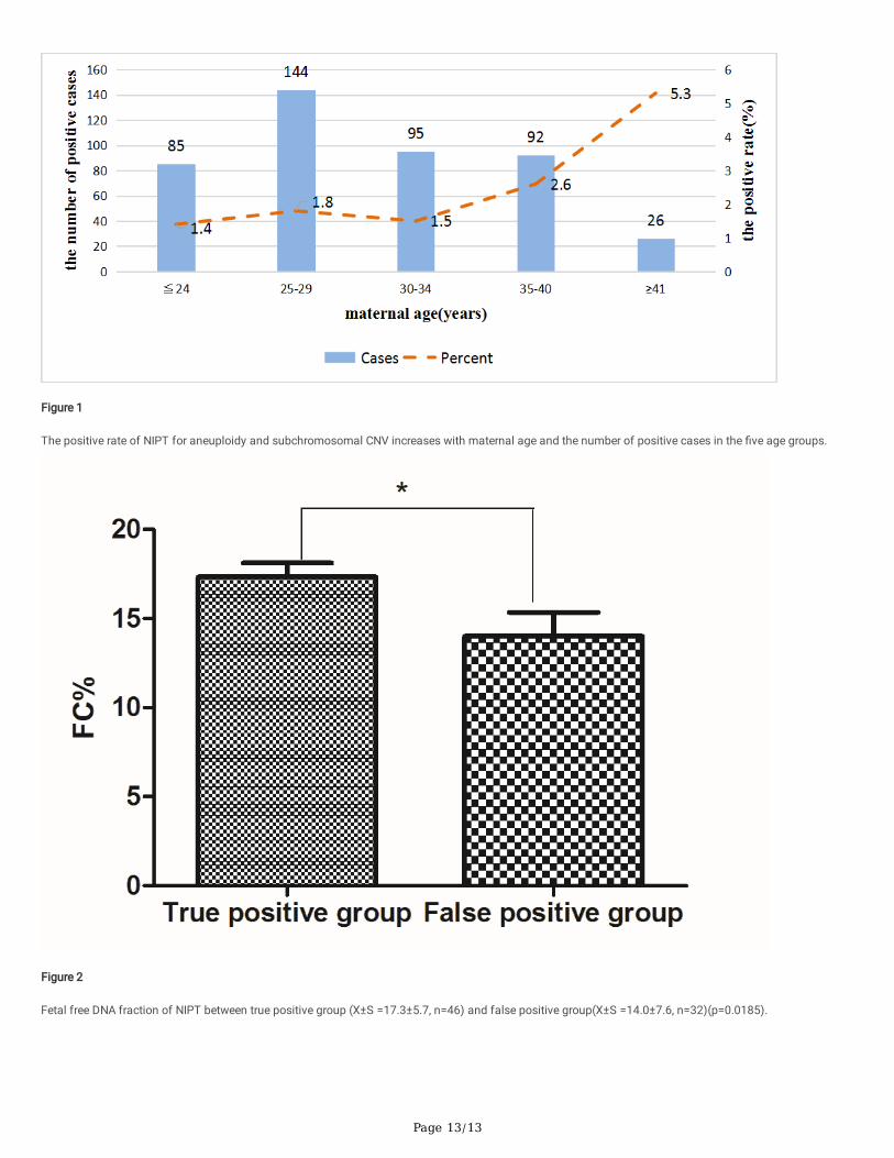
Figure 2 Fetal free DNA fraction of NIPT between true positive group (X ± S = 17.3 ± 5.7, n = 46) and false positive group(X ± S = 14.0 ± 7.6, n = 32)(p = 0.0185).
Different Ppv According To Pregnancies CharacteristicsWe also analyzed different PPV according to pregnancies characteristics, Different PPVs of NIPT according to pregnancies indications are shown in Table 5.The total PPV of T21 was 84.8%, the PPV of T21 fetuses in women of advanced maternal age was 80.0% and in abnormal serological screening group was75.0%. Similarly, the PPV of T18 and SCAs in advanced maternal age group were also the highest. It is worth noting that PPVs of CNVs in pregnancies withcleft lip, mental retardation or history of bad pregnancy was the highest.
Five Cases With Distal 22q11.2 Microdeletions And MicroduplicationsIn our study, the PPV of 22q11.2 microdeletions and microduplications is 100% (5/5). There were four fetuses with 22q11.2 duplication syndrome and onewith 22q11.2 deletion syndrome (Table 8). Case 24, 29 and 37 were all clinically healthy after birth but case 30 was with ventricular septal defect, aorticabnormalities and 1 bright spot in the left ventricle subsequently detected by ultrasound. Case 35 was con�rmed to have a 22q11.21 deletion syndrome withhoarseness, atrial septal defect, patent ductus arteriosus and myocardial injury after birth. However, it was not clear that whether these mutations were denovo or inherited from parents. Case 34 was con�rmed to have the loss of heterozygosity in 22q12.1q12.2 whose clinical phenotypes were unavailable. Fetalmother were also advised to have a array comparative genomic hybridization because of her mental retardation. CMA analysis showed that 22q11.23microduplication may contribute to her mental retardation.
Table 8The clinical information, prenatal diagnosis results and follow-up results of six cases distal 22q11.2 microdeletions and microduplications.
No age Pregnantweek
22q Dup/del bp start; stop
(NCBI37/hg19)
signi�cance origin Follow-up
24 28 16 + 6 duplication 3p26.3(1,536,945-2,579,649)x3;22q11.21(18,844,632 − 21,462,353)x3
22q11.2 dupsyndrome
unknown normal
29 25 17 + 3 duplication 22q11.21(18,648,855 − 21,800,471)x3
22q11.2 dupsyndrome
unknown normal
30 31 20 duplication 22q11.21q11.23(21,059,669 − 24,629,406)x3
22q11.2recurrent region(distal type I, D-E/F)
unknown congenital heartmalformation,ventricularseptal defect, aorticabnormalities
34 31 loss ofheterozygosity
22q12.1q12.2(28,317,927 − 30,826,759)x2 hmz
uncertainsigni�cance
mother with22q11.23(23,700,639 − 25,086,816)x3
unknown
35 22 26 + 2 deletion 22q11.21(18,648,855 − 21,800,471)x1
DiGeorgesyndrome
unknown hoarseness, congenitalheart disease
37 32 17 + 2 duplication 22q11.21q11.23(21,464,120 − 23,650,987)x3
22q11.2 dupsyndrome
unknown normal
One Case Of Xp22.31 MicrodeletionCase 46
was detected to have a 1.6 Mb microdeletion in Xp22.31 by NIPT, which often causes ichthyosis (X-linked recessive genetic disease). Most female carriers ofXp22.31 microdeletion have a normal phenotype, a few female carriers may show abnormal symptoms due to inactivation of X chromosomes and all malecarriers show ichthyosis. In our study, case 46
chosen to perform prenatal diagnosis for further con�rmation and the result showed that the fetus was female and this mutation is inherited from her normalmother.

Page 10/13
One Case Of 5.4 mb Microdeletion In 9p21.3-p21.1Case 49
was detected to have a 5.4 Mb microdeletion in 9p21.3-p21.1(24,796,507 − 30,288,265) by NIPT, which contains 8 OMIM genes such as TEK and C9orf72.Heterozygous mutations of TEK gene often causes venous malformations, multiple cutaneous and mucosal (autosomal dominant genetic disease). C9orf72is related with frontotemporal dementia and/or amyotrophic lateral sclerosis 1(autosomal dominant genetic disease). However, case 49
was healthy and this 5.4 Mb microdeletion was inherited from his/her healthy mother. Interestingly, our laboratory had previously detected a 4.2 Mbmicrodeletion in 9p21.2p21.1(26,210,360 − 30,492,812) by CMA from a fetus with NT = 1.3 cm and this 4.2 Mb microdeletion was de novo. These resultssuggested that 9p21.3-p21.1 microdeletion may have penetrance difference.
A false negative NIPT result for case 53 with 4p14 microdeletion
The NIPT result of case 53 with high risk of serum screening was normal. System structure screening (other hospital) in 24 week showed fetal growthretardation. The fetal was diagnosed as neonatal pneumonia, low weight, congenital heart disease and hyperbilirubinemia after birth. Karyotype detection ofperipheral blood showed 46, XX,del(4)(p4). Thus, case 53 was tested again by improved the experimental method with better cffDNA enrichment. The resultsshowed increased cffDNA fraction from 6.5–15.1% and a 34Mbp deletion in 4p16.3-p15.1 region, which is co-related with Wolf-Hirschhorn syndrome (WHS).
DiscussionIn this study, we are the �rst to reviewed the e�ciency of NIPT for screening common chromosome aneuploidies as well as sub-chromosomal CNV within acohort of 24359 single pregnancies in Huaian area. This NIPT technology uses a semiconductor sequencing platform with high enrichment of cffDNA(20%-40%) to reliably detect subchromosomal deletions/duplications. The PPV for T21, T18, T13 and SCAs was 84.8%, 54.2%, 11.1% an 40.5% respectively. Inseveral recent studies, the PPV of T21 was 65–94%, the PPV of T18 was 47–85%, and the PPV of T13 was 12–62%[13–15]. Our results are consistent withprevious studies. Interestingly, the PPV for CNVs was 59.0%, which is obviously higher than previous studies with 9–36% [16–18]. The reason for higher PPVof CNVs in our study may be related with our new enrichment strategy. The PPV for CNVs ≤ 5 Mb was 68.9% (31/45), for CNVs within 5–10 Mb was 83.3%(5/6) and for CNVs ≥ 10 Mb was 37.1% (10/27) respectively. However, previous reports demonstrated that PPV for CNVs ≥ 10 Mb was signi�cantly higherthan CNVs < 10 Mb [16–18]. Further analysis showed that there were only 27 cases with big CNVs (≥ 10 Mb) but 51 cases with small CNVs (< 10 Mb)suggesting that small CNVs occurred more frequently than big CNVs. Therefore, we speculate that frequent occurrence of small CNVs may be the potentialcause of higher PPV of small CNVs.
Further analysis about the different PPV of NIPT according to pregnancies indications was performed. The results showed that the PPV of NIPT was thehighest for T21 and was much lower for other aneuploidies. PPV of CNVs was close to T18 and much higher than T13. Advanced maternal age is a high riskfactor for T21 so PPV of T21 in advanced maternal age the highest. PPV of CNVs in advanced maternal age group was lower suggesting that advancedmaternal age was not signi�cantly related with CNVs. PPVs of CNVs in pregnancies with cleft lip, mental retardation or history of bad pregnancy was thehighest suggesting high risk factors for CNVs.
Among the 46 true positive cases and 7 abnormal false positive cases, 31 cases were correlated to microdeletion or microduplication syndromes with 6 casesinherited from parents, 3 cases de novo and other 22 cases unavailable. Early detection of pathogenic and potentially pathogenic CNVs by NIPT has goodbene�t in prenatal screening. 22q11.2 microduplication was the most frequent in our research. The phenotype of the �ve patients with 22q11.2microduplications were diverse, with symptoms ranging from being normal to mental retardation and congenital heart malformation. 22q11.2 microdeletionsis the second most common chromosomal abnormality secondary to Down syndrome [19]. However, the occurrence of 22q11.2 microduplications was morefrequent than 22q11.2 microdeletion in our study, which was contrary to previous research conclusion. The rare occurrence of 22q11.2 microduplication casesmay be explained by the absence of a de�ned phenotype and incomplete penetrance[20].
Studies have demonstrated that there is a small chance of a false negative result for NIPT[21]. In our study, there was a false negative case in 79 validatedNIPT. The most common factor associated with these false negative results is the low fetal fraction, which are often affected by maternal weight, gestationalage and extraction method[22–23]. In our research, extraction method for cffDNA enrichment was the main reason for the false negative cases. Therefore,improved extraction method for elevating fetal fraction were immediately used in December 2018, which may be the potential reason for improved the overallperformance of NIPT and higher PPV in this research. Faas BH et al in 2012 clari�ed that cell free fetal DNA in the maternal plasma originates fromcytotrophoblastic cells derived from trophoblast of the blastocyst[24]. The karyotypes of cytotrophoblast and fetus may be different due to fetus are derivedfrom the inner cell mass (ICM) of the blastocyst[21]. Other reasons for false negative results may be CPM and maternal mosaicism[25–27].
ConclusionsThis study demonstrated that the PPV for T21, T18, T13 and SCAs was 85%, 54%, 11% and 41% respectively. The PPV for CNVs was 59.0%. The PPV forCNVs ≤ 5 Mb was 68.9% (31/45), for CNVs within 5–10 Mb was 83.3%(5/6) and for CNVs ≥ 10 Mb was 37.1% (10/27) respectively. Our data have potentialsigni�cance in demonstrating the usefulness of NIPT not only for common whole chromosome aneuploidies but also for CNVs.
Materials And Methods
Patients

Page 11/13
From 2015 to July 2019, 24359 pregnant women opted for NIPT to screen fetal chromosome aneuploidies. Informed written consent was obtained from allpregnant women who agreed to receive NIPT. Pregnancies with high risks were divided into advanced maternal age, ultrasound abnormalities, poor fertilityhistory, positive serum screening, and other groups in this study.
NIPT sequencingMaternal peripheral blood (5 ml) was collected in an ethylenediaminetetraacetic acid (EDTA) tube. The blood sample was stored at 4˚C immediately aftercollection. Afterwards, cfDNA extraction, library construction, quality control, and pooling were performed according to the JingXin Fetal ChromosomeAneuploidy (T21, T18, T13) Testing Kits (CFDA registration permit No. 0153400300). Sequencing reads were �ltered and aligned to the human referencegenome (hg19). Combined GC correction and Z-score testing methods were used to identify fetal autosomal aneuploidies. A cut off value of Z-score > 3 wasused to determine whether the ratio of the chromosomes was increased. Here, each chromosome with an absolute value of the Z-score greater than 3 wasmarked with chromosome aneuploidies or microdeletions/ microduplications.
Chromosome karyotype analysisBanding cytogenetics was performed on G-banded metaphase chromosomes of cultured peripheral blood lymphocytes using routine techniques. Karyotypeswere interpreted according to the International System for Human Cytogenetic Nomenclature.
Chromosome karyotype analysisThe aCGH platform from CytoScanTM 750 k chip made by American Affymetrix was employed in this study. The standard operating procedures for genomicDNA digestion, ligation, ampli�cation, puri�cation, fragmentation, labeling, chip hybridization, washing and scanning, and data analysis were performed.Pathogenicity of genomic DNA fragments were determined with reference to the international public pathological CNVs database, international publicpathological CNVs database, international public benign CNVs database and human genetics cytogenetics microarray database.
AbbreviationsNIPT: Noninvasive prenatal testing; cffDNA: Cell-free DNA; CMA: Chromosomal microarray analysis; CNVs: Copy number variants; MMS:Microdeletion/microduplication syndromes; PPV: Positive predictive value; LCRs: Low copy repeats; NAHR: Non-allelic homologous recombination; CPM:Con�ned placental mosaicism; ICM: Inner cell mass.
DeclarationsEthics approval and consent to participate
This study was approved by the Ethics Committee of Huaian Maternal and Child Health Care Hospital.
Consent for publication
The authors declare that they have no competing interests and the patients in this case report had provided their consent for publication.
Availability of data and materials
The datasets used and/or analyzed during the current study are available from the corresponding author on reasonable request.
Competing interests
The authors declare that they have no competing interests.
Funding: Supported by the Maternal and Child Health project of Jiangsu Province (No. F201670 F201714) (Pan Qiong) the “333 Project” Foundation ofJiangsu Province (No. BRA2017250) (Pan Qiong).
Authors’contributions
All authors have materially participated in the study and manuscript preparation. YF Liu, LF Chen, Y Peng, Z Liang, X Jin and NN Yan collected all clinical data.YF Liu participated in the data analysis and drafted the manuscript. Q Pan designed the work and drafted and revised the manuscript. All authors haveapproved the �nal article.
Acknowledgements
We would like express our gratitude for �nancial support from Maternal and Child Health project of Jiangsu Province (No. F201670 F201714) and the “333Project” Foundation of Jiangsu Province (No. BRA2017250).
References1. Lo YM, Corbetta N, Chamberlain PF, Rai V, Sargent IL, Redman CW, et al. Presence of fetal DNA in maternal plasma and serum. Lancet.
1997;350(9076):485–7.
2. Filoche S, Lawton B, Beard A, Dowell A, Stone P. New screen on the block: non-invasive prenatal testing for fetal chromosomal abnormalities. J PrimHealth Care. 2017;9(4):248–53.

Page 12/13
3. McCullough RM, Almasri EA, Guan X, Geis JA, Hicks SC, Mazloom AR, et al. Non-invasive prenatal chromosomal aneuploidy testing–clinical experience:100,000 clinical samples. PLoS One. 2014;9(10):e109173.
4. Hu H, Liu H, Peng C, Deng T, Fu X, Chung C, et al. Clinical Experience of Non-Invasive Prenatal Chromosomal Aneuploidy Testing in 190,277 PatientSamples. Curr Mol Med. 2016;16(8):759–66.
5. Liang D, Lin Y, Qiao F, Li H, Wang Y, Zhang J, et al. Perinatal outcomes following cell-free DNA screening in > 32 000 women: Clinical follow-up data from asingle tertiary center. Prenat Diagn. 2018;38(10):755–64.
�. Oskarsdottir S, Vujic M, Fasth A. Incidence and prevalence of the 22q11 deletion syndrome: a population-based study in Western Sweden. Arch Dis Child.2004;89(2):148–51.
7. Grati FR, Molina GD, Ferreira JC, Dupont C, Alesi V, Gouas L, et al. Prevalence of recurrent pathogenic microdeletions and microduplications in over 9500pregnancies.2015; 35(8):801–809.
�. McDonald-McGinn DM, Sullivan KE, Marino B, Philip N, Swillen A, Vorstman JA, et al. 22q11.2 deletion syndrome. Nat Rev Dis Primers.2015.
9. Qiao Y, Badduke C, Tang F, Cowieson D, Martell S, Lewis SME, et al. Whole exome sequencing of families with 1q21.1 microdeletion or microduplication.Am J Med Genet A. 2017;173(7):1782–91.
10. Cox DM, Butler MG. The 15q11.2 BP1-BP2 microdeletion syndrome: a review. Int J Mol Sci. 2015;16(2):4068–82.
11. Smith AE, Jnah A, Newberry D. Chromosome 16p13.11 Microdeletion Syndrome in a Newborn: A Case Study. Neonatal Netw. 2018;37(5):303–9.
12. Rosenfeld JA, Coe BP, Eichler EE, Cuckle H, Shaffer LG. Estimates of penetrance for recurrent pathogenic copy-number variations. Genet Med.2013;15(6):478–81.
13. Neofytou MC, Tsangaras K, Kypri E, Loizides C, Ioannides M, Achilleos A, et al. Targeted capture enrichment assay for non-invasive prenatal testing oflarge and small size sub-chromosomal deletions and duplications. PLoS One. 2017;12(2):e0171319.
14. Yaron Y, Jani J, Schmid M, Oepkes D. Current Status of Testing for Microdeletion Syndromes and Rare Autosomal Trisomies Using Cell-Free DNATechnology. Obstet Gynecol. 2015;126(5):1095–9.
15. Sentilhes L, Salomon LJ, Vayssiere C. Cell-free DNA Analysis for Noninvasive Examination of Trisomy. N Engl J Med. 2015;373(26):2581–2.
1�. Chen Y, Yu Q, Mao X, Lei W, He M, Lu W. Noninvasive prenatal testing for chromosome aneuploidies and subchromosomalmicrodeletions/microduplications in a cohort of 42,910 single pregnancies with different clinical features. Hum Genomics. 2019;13(1):60.
17. Hu H, Wang L, Wu J, Zhou P, Fu J, Sun J, et al. Noninvasive prenatal testing for chromosome aneuploidies and subchromosomalmicrodeletions/microduplications in a cohort of 8141 single pregnancies. Hum Genomics. 2019;13(1):14.
1�. Liang D, Cram DS, Tan H, Linpeng S, Liu Y, Sun H, et al. Clinical utility of noninvasive prenatal screening for expanded chromosome disease syndromes.Genet Med. 2019;21(9):1998–2006.
19. McDonald-McGinn DM, Tonnesen MK, Laufer-Cahana A, Finucane B, Driscoll DA, Emanuel BS, et al. Phenotype of the 22q11.2 deletion in individualsidenti�ed through an affected relative: cast a wide FISHing net! Genet Med. 2001;3(1):23–9.
20. Wincent J, Bruno DL, van Bon BW, Bremer A, Stewart H, Bongers EM, et al. Sixteen New Cases Contributing to the Characterization of Patients with Distal22q11.2 Microduplications. Mol Syndromol. 2010;1(5):246–54.
21. Bianchi DW, Wilkins-Haug L. Integration of noninvasive DNA testing for aneuploidy into prenatal care: what has happened since the rubber met the road?Clin Chem. 2014;60(1):78–87.
22. Ashoor G, Syngelaki A, Poon LC, Rezende JC, Nicolaides KH. Fetal fraction in maternal plasma cell-free DNA at 11–13 weeks' gestation: relation tomaternal and fetal characteristics. Ultrasound Obstet Gynecol. 2013;41(1):26–32.
23. Jorgez CJ, Dang DD, Simpson JL, Lewis DE, Bischoff FZ. Quantity versus quality: optimal methods for cell-free DNA isolation from plasma of pregnantwomen. Genet Med. 2006;8(10):615–9.
24. Faas BH, de Ligt J, Janssen I, Eggink AJ, Wijnberger LD, van Vugt JM, et al. Non-invasive prenatal diagnosis of fetal aneuploidies using massively parallelsequencing-by-ligation and evidence that cell-free fetal DNA in the maternal plasma originates from cytotrophoblastic cells. Expert Opin Biol Ther.2012;12(Suppl 1):19–26.
25. Jiang F, Ren J, Chen F, Zhou Y, Xie J, Dan S, et al. Noninvasive Fetal Trisomy (NIFTY) test: an advanced noninvasive prenatal diagnosis methodology forfetal autosomal and sex chromosomal aneuploidies. BMC Med Genomics.2012; 5(57.
2�. Kalousek DK. Pathogenesis of chromosomal mosaicism and its effect on early human development. Am J Med Genet. 2000;91(1):39–45.
27. Gao Y, Stejskal D, Jiang F, Wang W. False-negative trisomy 18 non-invasive prenatal test result due to 48,XXX,+18 placental mosaicism. Ultrasound ObstetGynecol. 2014;43(4):477–8.
Figures
Page 13/13
Figure 1
The positive rate of NIPT for aneuploidy and subchromosomal CNV increases with maternal age and the number of positive cases in the �ve age groups.
Figure 2
Fetal free DNA fraction of NIPT between true positive group (X±S =17.3±5.7, n=46) and false positive group(X±S =14.0±7.6, n=32)(p=0.0185).