pregnancy cv exam during pregnancy - oregon acc · pathophysiology • defective antioxidant...
TRANSCRIPT

5/30/2012
1
©2011 MFMER | slide-1
Cardiovascular Disease and the Pregnant Patient
June 4, 2012Martha Grogan, MD, FACCACC 2012 Portland, Oregon
©2011 MFMER | slide-2
PregnancyHematologic Changes
Am J Physiol, 1983Am J Physiol, 1983
0000
Blood
volumes
(% control)
Blood
volumes
(% control)
1010
2020
3030
4040
5050
1010 2020 3030 4040
Gestational age (weeks)Gestational age (weeks)
PlasmaPlasma
Whole bloodWhole blood
ErythrocytesErythrocytes
©2011 MFMER | slide-3
Hemodynamic ChangesHemodynamic Changes
•> 40% ↑ blood volume
• ↓ SVR and PVR
• ↑ HR
•Mild ↓ in BP
•> 40% ↑ blood volume
• ↓ SVR and PVR
• ↑ HR
•Mild ↓ in BP
30-50%
↑ CO
30-50%
↑ CO
©2011 MFMER | slide-4
S4occasional
S4occasional
Systolic murmur 96%Systolic murmur 96%
Wide
loud split 1st 88%
Wide
loud split 1st 88%
MCMC TCTC A2A2 P2P2
Diastolic“flow”murmur 18%
Diastolic“flow”murmur 18%
S3loud 84%
S3loud 84%
CV Exam During PregnancyCV Exam During Pregnancy
©2011 MFMER | slide-5
CV Exam During PregnancyCV Exam During Pregnancy
• Brisk and full carotid upstroke
• JVP - normal or mildly ↑↑↑↑
• Displaced and enlarged apex
• Varicose veins and edema
• Normal exam can mimic heart disease
• Not normal
S4, loud SM, DM, fixed split S2
• Brisk and full carotid upstroke
• JVP - normal or mildly ↑↑↑↑
• Displaced and enlarged apex
• Varicose veins and edema
• Normal exam can mimic heart disease
• Not normal
S4, loud SM, DM, fixed split S2
©2011 MFMER | slide-6
Hemodynamic ChangesLabor and DeliveryHemodynamic ChangesLabor and Delivery
• CO ↑ 60-80%
• Volume changes
↑ Blood volume with uterine contraction
↑ Venous return
Volume loss during delivery
• CO ↑ 60-80%
• Volume changes
↑ Blood volume with uterine contraction
↑ Venous return
Volume loss during delivery

5/30/2012
2
©2011 MFMER | slide-7
Heart Disease and Pregnancy: Delivery
• Vaginal Delivery: Preferable in most cases
• Facilitate 2nd stage
• C-Section Indications:
• OB reasons
• Early labor still on warfarin
• Severe PH
• Fixed obstructive lesions
• Unstable aorta
©2011 MFMER | slide-8
Pregnancy “Contraindications”
• Severe Pulmonary Hypertension
• Severe obstructive lesions• AS, MS, PS, HCM, Coarct
• Ventricular dysfunction
• Class III or IV HF, EF <40%
• Dilated or unstable aorta
• Marfan with aorta ≥40 – 45 mm
• Severe cyanosis
©2011 MFMER | slide-9
CAREPREG Risk Stratification
Predictors
• CHF, arrhythmia, TIA or CVA
• NYHA > II or cyanosis
• Left Heart ObstructionMVA <2 cm2, AVA < 1.5
LVOT > 30 mmHg
• LV EF < 40%
Number of predictors
Siu SC et al. Prospective Multicenter Study of Pregancy Outcomes in Women with Heart Disease. Circ
2001; 104:515-521.
©2011 MFMER | slide-10
CARPREG Risk IndexPredictors of Cardiac Events
• Prior CHF, TIA, stroke or arrhythmia
• Baseline NYHA class >II or cyanosis
• Left heart obstruction
• MVA <2 cm2, AVA <1.5 cm2
• LVOT gradient >30 mmHg by Echo
• ↓ systemic vent function (EF <40%)
Risk index predicts CV event rate
©2011 MFMER | slide-11
Dilated Cardiomyopathy
What % of Patients will have an Affected Family Member
a. 5%
b. 10%
c. 15%
d. 25%
e. 50%
CP1081586-12
©2011 MFMER | slide-12
Familial Dilated Cardiomyopathy
• Mid 1980s - initial reports-
1-2% of pts with IDCM had familial disease
Echo screening changed thatJ

5/30/2012
3
©2011 MFMER | slide-13
Dilated Cardiomyopathy A heritable form of heart failureMichels, N Engl J Med 1992
Rationale for clinical screening of families
Impetus for human molecular genetics research
Frequency of familial DCM
• 6-8% by history
• 20-25% by echocardiography
• 35-50% by less stringent criteria
(LV dilation with borderline EF)
©2011 MFMER | slide-14
HFSA 2010 Practice GuidelineGenetic Evaluation—Clinical Screening
Clinical screening (includes echo) for cardiomyopathy in asymptomatic first-degree relatives is recommended
• Hypertrophic cardiomyopathy
• Dilated cardiomyopathy
• Arrhythmogenic RV cardiomyopathy
• Left ventricular noncompaction *
• Restrictive cardiomyopathy *
Lindenfeld J., et al, J Card Fail; 16e1-e194
* Level of evidence = B
©2011 MFMER | slide-15
27-Year-Old FemaleFamilial DCM
©2011 MFMER | slide-16
• Age 15 months
• Pulmonary edema, severely dilated LV, EF 15%
• WPW with SVT
• Index case for family
• Mom and sister: DCM and WPW
• Enrolled in GENES in DCM study (20 yrslater) - actin mutation
27 Year-Old Female – Familial DCM
©2011 MFMER | slide-17
• Competitive soccer – high school and college
• Age 22: inderal changed to coreg, continue dig, ACE-I
• MRI EF 47%, MUGA 44%, Echo 30-35 (est), 40% (calc)
27 Year-Old Female – Familial DCM
©2011 MFMER | slide-18
• Exercise test
• 10.2 min (87%)
• VO2 (79%) with plateau
• What do you recommend regarding pregnancy ?
• How high would you estimate her risk ?
27 Year-Old Female – Familial DCM

5/30/2012
4
©2011 MFMER | slide-19
• At 32 weeks pregnant: exercising 60 min, 4x/ per week
• Asymptomatic
• Echo: EF 35%, increase MR to grade ¾
• Premature Labor – uncomplicated delivery- 35 weeks
• Sister (DCM) uncomplicated preganancy-delivered 4 months later
27 Year-Old Female – Familial DCM
Their Mom J.
©2011 MFMER | slide-20
Peripartum Cardiomyopathy
• New diagnosis of HF due to LV dysfunction
• Last trimester → 6 mos postpartum
• Diagnosis of exclusion
• Incidence varies
US 1 in 3200 deliveries (1350/year)
South Africa 1 in 1000
Haiti 1 in 300
Elkayam JACC 2011
©2011 MFMER | slide-21
Peripartum Cardiomyopathy
• Standard HF Rx – O2, diuretic, iv NTG, inotrope
• Bromocriptine - blocks prolactin release
↑ EF in PPCM vs standard therapy, with AC - TE risk
• IV immune globulin – immune modulator
↑ EF in PPCM pt vs standard therapy
• Pentoxifylline – inhibits TNF-alpha production
↑ TNF-alpha in PPCM
• Anticoagulation if EF <35%, or with bromocriptine
• VAD or transplantation
©2011 MFMER | slide-22
Peripartum Cardiomyopathy
Pathophysiology
• Defective antioxidant defense mechanism
prolactin/bromocriptine
• Viral infection
• Autoimmune response
• Genetic susceptibility
©2011 MFMER | slide-23
Peripartum Cardiomyopathy
• Prognosis variable
• Major cause of preg related death in US
• Mortality 6 mos and 2 yr → 10 and 28%
• ↑ mortality with ↓ EF >6 mos postpartum
• ~50% improve in 6 months
• 20 – 40% normalize EF
©2012 MFMER | 3197755-24
ACC Survey: 44 pts with PPCM• Subsequent pregnancy
Gp 1 – 28 pre-preg normal LVEFGp 2 – 16 pre-preg↓ LVEF
• Pregnancies resulted in ↓ in mean LVEF
Gp 1 – 56±7% → 49±10%;Gp 2 – 36±9% → 32±11%
• CHF symptoms Gp 1 = 21%, Gp 2 = 44%
• Mortality rate Gp 1= 0%, Gp 2 = 19% (P=0.06)
• Subsequent preg – ↓ EF, clinical deterioration and death
• Advise against pregnancy – EF <25% at presentationor persistent ↓ EF
Elkayam et al: N Engl J Med, 2001

5/30/2012
5
©2011 MFMER | slide-25
FDA Classification of Drugs During Pregnancy
A Controlled studies show no risk
B No evidence of risk in humans, the chance of fetal harm is remote
C Risk not excluded. Adequate studies lacking. Chance of fetal harm but benefits outweigh risks
D Positive evidence of risk. Studies in humans show fetal risk. Potential benefit in pregnant women may outweigh risk
X Contraindicated
©2012 MFMER | 3197755-26
Cardiac Drugs in Pregnancy
• Most CV drugs cross placenta, secretedin breast milk
• Avoid when possible
• Use drugs with long safety record
• Prescribe lowest dose for shortest duration
• Avoid multi-drug regimens
• No drug is completely safe
©2012 MFMER | 3197755-27
Cardiac Drugs in Pregnancy
ACE Inhibitors – contraindicated in pregnancy
• 30% fetal morbidity with administration after week 14
• Fetal renal tubular dysplasia, neonatal renal failure
• Oligohydramnios, ↓ cranial ossification, IUGR
Cooper et al: N Engl J Med 2006
• 1st trimester ACE ↑ risk of congenital malformations
• ↑ CV and CNS malformations
• AT II blocker – contraindicated
• Safe during lactation
©2011 MFMER | slide-28
Heart Failure Medications - Pregnancy
• ACE-i/ARBs CONTRAINDICATED
• Metoprolol, Carvedilol– Class C, Atenolol D
• Thiazides – class B
• Loop diuretics – class C
Avoid Hypotension, Placental Hypoperfusion
• Nitrates – class C
• Hydralazine – class C
©2011 MFMER | slide-29
Aortic Disease and Pregnancy
©2011 MFMER | slide-30
Pregnancy and Marfan Syndrome
• Preexisting medial changes
• Changes with pregnancy
• Physiologic, hormonal
• Unpredictable maternal risk
• Dissection, rupture, IE, CHF
• Fetal risk
• 50% inheritance

5/30/2012
6
©2011 MFMER | slide-31
Pregnancy and Marfan Syndrome
ESC GUCH Guidelines 2010
• 50% chance inheritance risk → genetic counseling
• Aorta >45 mm - strongly discourage pregnancy, high
risk of dissection
• Aorta <40 mm rare problem; no safe diameter
• Aorta 40-45 mm, aortic growth and FH important
Even after aortic repair, Marfan patients remain at
risk for dissection of residual aorta
©2011 MFMER | slide-32
©2011 MFMER | slide-33
©2011 MFMER | slide-34
©2011 MFMER | slide-35
Valvular Heart Disease and Pregnancy
©2012 MFMER | 3197755-36
Pulmonary Edema
Mitral Stenosis in Pregnancy
↑↑↑↑ HR
↓↓↓↓ diastolic
filling
↓↓↓↓ SV
Reflex ↑↑↑↑ HR
Further ↑ in LA pressure

5/30/2012
7
©2012 MFMER | 3197755-37
Mitral StenosisManagement in Pregnancy
ββββ blockade, maintain NSR
anticoagulation, diuretics
Balloon valvotomy
Surgical valvotomy or MVR
©2012 MFMER | 3197755-38
↓↓↓↓ Placental perfusion
IUGR, preterm labor
Aortic Stenosis in Pregnancy
Unable to
augment CO
Preload and
hypotension
sensitive
CHF and ischemia
©2011 MFMER | slide-39
Anticoagulation in Pregnancy
©2011 MFMER | slide-40
©2011 MFMER | slide-41
©2011 MFMER | slide-42
Complex CHD in Pregnancy
5/30/2012
8
©2011 MFMER | slide-43
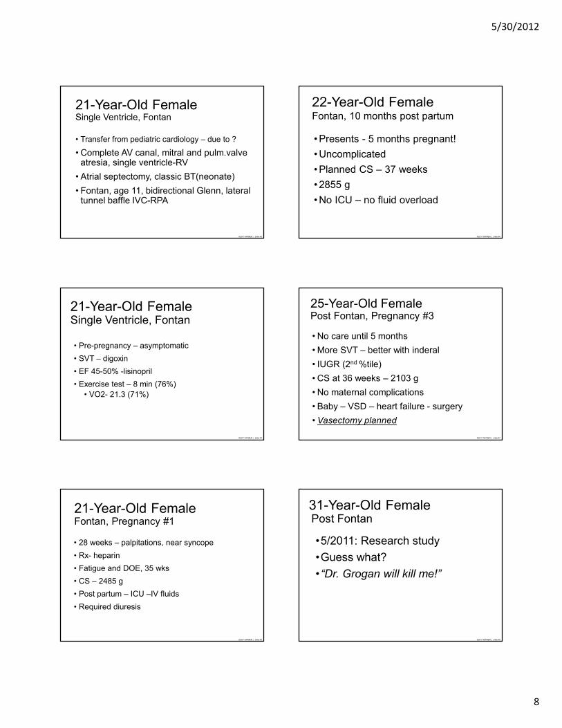
21-Year-Old FemaleSingle Ventricle, Fontan
• Transfer from pediatric cardiology – due to ?
• Complete AV canal, mitral and pulm.valve atresia, single ventricle-RV
• Atrial septectomy, classic BT(neonate)
• Fontan, age 11, bidirectional Glenn, lateral tunnel baffle IVC-RPA
©2011 MFMER | slide-44
21-Year-Old FemaleSingle Ventricle, Fontan
• Pre-pregnancy – asymptomatic
• SVT – digoxin
• EF 45-50% -lisinopril
• Exercise test – 8 min (76%)
• VO2- 21.3 (71%)
©2011 MFMER | slide-45
21-Year-Old FemaleFontan, Pregnancy #1
• 28 weeks – palpitations, near syncope
• Rx- heparin
• Fatigue and DOE, 35 wks
• CS – 2485 g
• Post partum – ICU –IV fluids
• Required diuresis
©2011 MFMER | slide-46
22-Year-Old FemaleFontan, 10 months post partum
• Presents - 5 months pregnant!
• Uncomplicated
• Planned CS – 37 weeks
• 2855 g
• No ICU – no fluid overload
©2011 MFMER | slide-47
25-Year-Old FemalePost Fontan, Pregnancy #3
• No care until 5 months
• More SVT – better with inderal
• IUGR (2nd %tile)
• CS at 36 weeks – 2103 g
• No maternal complications
• Baby – VSD – heart failure - surgery
• Vasectomy planned
©2011 MFMER | slide-48
31-Year-Old FemalePost Fontan
•5/2011: Research study
•Guess what?
•“Dr. Grogan will kill me!”

5/30/2012
9
©2011 MFMER | slide-49
Echo 2010
©2011 MFMER | slide-50
Echo 2010
©2011 MFMER | slide-51
Echo 2010
©2011 MFMER | slide-52
Pregnancy after Fontan
Contraindications
•EF <40%
•Class III-IV symptoms
•Cyanosis
•PLE
©2011 MFMER | slide-53
Pregnancy Post Fontan
• Atrial arrhythmias
• Ventricular Dysfunction
• Edema and Ascites
• Challenges of A/C mgmt.
• Spontaneous Abortion
• IUGR and Premature Birth
©2011 MFMER | slide-54

5/30/2012
10
©2011 MFMER | slide-55
Arrhythmias and Pregnancy
©2011 MFMER | slide-56
Arrhythmias and Pregnancy
• Palpitations common – often benign
• SVT most common
• Afib/flutter with CHD
• Hemodynamic instability: DC cardioversion
• Vagal maneuvers, adenosine
• Meds:1st line: Digoxin, metoprolol*
• Antiarrhythmic Rc – reserve for severe symptoms, recurrence
* Avoid atenolol- class D
©2011 MFMER | slide-57
Arrhythmias and Pregnancy
• VT – uncommon
• Antiarrhymic Rx: quinidine, procainamide, flecainide, sotolol
• Amiodarone – seldom used
• Ablation has been safely performed in pregnancy (atrial and ventricular arrhythmia)
• PPM and ICD can be performed
©2011 MFMER | slide-58
Pregnancy: MI, Radiation, Endocarditis
©2012 MFMER | 3197755-59
Acute MI in Pregnancy
• Exclude coronary anomaly and aortic dissection
• Coronary angio, aortic imaging
• PCI – bare metal stent
• CABG – limited data
• Thrombolysis
• Consider if cath/PCI not available
Roth: JACC 2008
©2011 MFMER | slide-60

5/30/2012
11
©2011 MFMER | slide-61
©2011 MFMER | slide-62
Cardiovascular Disease and PregnancySummary
• CVD complicates 1-2 % of pregnancies
• CHD most common in US
• Does not preclude successful outcome
• Increased risk: mother and fetus
• Individualized assessment
Preferably Prior to Conception