pregnancyandsmoothelin-likeprotein1(smtnl1)deletion ... · ·...
TRANSCRIPT
Pregnancy and Smoothelin-like Protein 1 (SMTNL1) DeletionPromote the Switching of Skeletal Muscle to a GlycolyticPhenotype in Human and Mice*□S
Received for publication, April 14, 2015, and in revised form, June 3, 2015 Published, JBC Papers in Press, June 5, 2015, DOI 10.1074/jbc.M115.658120
Beata Lontay‡§, Khaldon Bodoor‡1, Adrienn Sipos§, Douglas H. Weitzel‡, David Loiselle‡, Rachid Safi‡,Donghai Zheng¶, James Devente�, Robert C. Hickner¶**, Donald P. McDonnell‡, Thomas Ribar‡‡,and Timothy A. Haystead‡2
From the ‡Department of Pharmacology, Duke University Medical Center, Durham, North Carolina 27710, §Department of MedicalChemistry, Faculty of Medicine, University of Debrecen, Debrecen-4032, Hungary, ¶Departments of Kinesiology, East CarolinaUniversity, Greenville, North Carolina 27858, �Department of Obstetrics and Gynecology, East Carolina University, Greenville, NorthCarolina 27834, **Department of Biokinetics, Exercise, and Leisure Sciences, University of KwaZulu-Natal, Durban 4000,South Africa, and ‡‡Duke iPSC Shared Resource Facility, Duke University Medical Center, Durham, North Carolina 27710
Background: Pregnancy promotes physiological adaptations throughout the body mediated by the female sex hormones.Results: Pregnancy promotes switching of skeletal muscle to a glycolytic phenotype through the smoothelin-like protein 1transcriptional cofactor.Conclusion: Deletion of SMTNL1 is able to mimic the effect of pregnancy in mice.Significance: Novel mechanism to explain insulin resistance during pregnancy.
Pregnancy promotes physiological adaptations throughoutthe body, mediated by the female sex hormones progesteroneand estrogen. Changes in the metabolic properties of skeletalmuscle enable the female body to cope with the physiologicalchallenges of pregnancy and may also be linked to the devel-opment of insulin resistance. We conducted global microar-ray, proteomic, and metabolic analyses to study the role of theprogesterone receptor and its transcriptional regulator,smoothelin-like protein 1 (SMTNL1) in the adaptation ofskeletal muscle to pregnancy. We demonstrate that preg-nancy promotes fiber-type changes from an oxidative toglycolytic isoform in skeletal muscle. This phenomenon isregulated through an interaction between SMTNL1 and pro-gesterone receptor, which alters the expression of contractileand metabolic proteins. smtnl1�/� mice are metabolicallyless efficient and show impaired glucose tolerance. Preg-nancy antagonizes these effects by inducing metabolic activ-ity and increasing glucose tolerance. Our results suggest thatSMTNL1 has a role in mediating the actions of steroid hor-mones to promote fiber switching in skeletal muscle duringpregnancy. Our findings also bear on the management of ges-tational diabetes that develops as a complication of preg-nancy in �4% of women.
Skeletal muscle (SKM)3 shows plasticity and is able to adaptits contractile phenotype in response to physiological stress,including exercise training, hormonal shifts, aging, and patho-logical stress (1). Skeletal muscle plasticity has been studiedextensively in endurance exercise training in both humans andanimals and shown to promote the switching of SKM fiber con-tent to a more oxidative phenotype characterized by an increasein the relative numbers of type I slow-twitch and type2a fast-twitch fibers and a decrease in type2b fast-twitch glycolyticfibers (2, 3). Alternatively, intermittent bursting effort pro-motes transformation of type2a/b fibers to a more glycolytictype2b phenotype, with fewer mitochondria and increasedexpression of the MHC2b isoform as well as enzymes associ-ated with glycolysis. SKM is also the primary site for the caloricdisposal of glucose and long chain fatty acids, which is pro-foundly affected by the composition and the metabolic andenzymatic properties of its fibers. Switching of SKM to an oxi-dative phenotype is associated with a decreased susceptibility tothe development of insulin resistance. Exercise training alonecan reverse obesity-induced insulin resistance by increasing theoxidative capacity of muscle through the enhancement of mito-chondrial density and glucose transporter content (4, 5). Con-versely, reduced oxidative capacity of SKM is associated withpredisposition to the development of insulin resistance andincreased weight gain.
Pregnancy is also a physiological state that can promotemajor changes in SKM, mainly mediated by the female sex ste-* This work was supported, in whole or in part, by National Institutes of Health
Grant R01DK065954-05 (to T. A. H.). This work was also supported byGrants OTKA PD107898, TAMOP-4.2.2.A-11/1/KONV-2012-0025, andTAMOP 4.2.4.A/2-11-1-2012-0001, the University of Debrecen (RH/751/2015) (to B. L.), and by The King Hussein fellowship (to K. B.). The authorsdeclare that they have no conflicts of interest with the contents of thisarticle.
□S This article contains supplemental Table 1.1 A Duke University School of Medicine visiting scholar.2 To whom correspondence should be addressed: Duke University School of
Medicine, C118 LSRC Box 3813, Durham, NC 27710. Tel./Fax: 919-613-8606;E-mail: [email protected].
3 The abbreviations used are: SKM, skeletal muscle; smoothelin-like protein 1,SMTNL1; NP, non-pregnant; PR, progesterone receptor; ER�/�, estrogenreceptor �/�; IHC, immunohistochemistry; SQ-WB, semi quantitativeWestern blot analysis; MHC1, -2a, -2B, myosin heavy chain 1, 2a, 2b; IRS,insulin receptor substrate; CLAMS, comprehensive laboratory animal mon-itoring system; GLM, general linear models; RER, respiratory exchangeratio; Glut4, glucose transporter 4; IRS1, insulin receptor substrate 1; IP,immunoprecipitation; WN, WT non-pregnant; KN, KO-non-pregnant; KP,KO-pregnant.
THE JOURNAL OF BIOLOGICAL CHEMISTRY VOL. 290, NO. 29, pp. 17985–17998, July 17, 2015© 2015 by The American Society for Biochemistry and Molecular Biology, Inc. Published in the U.S.A.
JULY 17, 2015 • VOLUME 290 • NUMBER 29 JOURNAL OF BIOLOGICAL CHEMISTRY 17985
by guest on June 25, 2018http://w
ww
.jbc.org/D
ownloaded from
roid hormones estrogen and progesterone (6). Both progester-one receptors (PR) and estrogen receptors (ER) are expressed inSKM, but their function and relevance have yet to be defined(7). Some direct links between the effect of progesterone andestrogen on SKM come from studies on pregnancy-inducedinsulin resistance. The increased glucose requirements of thegravid uterus during pregnancy are thought to necessitatemajor adjustments in glucose production and utilization bymaternal SKM, adipose, and other tissues. This is the reasonwhy normal pregnancy is associated with the development ofinsulin resistance, which is thought of as a means by which themother can supply the developing fetus with sufficient glucosefor growth. However, in 2– 4% of all pregnancies, this conditionprogresses to a type II diabetic state known as gestational dia-betes and becomes a complication of pregnancy. At the mid-term of pregnancy both SKM and adipose tissues show insulinresistance (8), but the underlying molecular mechanisms thatpromote this response are unknown. These mechanisms havebeen speculated to be linked to increased circulating insulinlevels promoted by an increase in the number and mass of pan-creatic � cells induced by progesterone (9).
Smoothelin-like protein 1 (SMTNL1) plays a role in mediat-ing exercise-induced adaptations in both smooth and SKM thatare sex-dependent (10). For example, striated muscle in malesmntl�/� mice exhibit an endurance phenotype, whereasfemale null mice exhibit a more glycolytic phenotype (11). Preg-nancy was also found to induce the expression of SMTNL1 intissues such as uterine and vascular smooth muscle and sexhormone-related tissues. In uterine smooth muscle, SMTNL1plays a major role in pregnancy to promote adaptive responses,and this process is specifically mediated through interactions ofSMTNL1 with the steroid hormone receptor PR-B. In vitro andin vivo SMTNL1 selectively binds PR and does not bind othersteroid hormone receptors. This suggests that SMTNL1 is abifunctional co-regulator of PR-B signaling and thus provides amolecular mechanism whereby PR-B is targeted to alter geneexpression patterns to coordinately promote alterations inuterine smooth muscle function during pregnancy (12). Basedon these observations we have speculated that SMTNL1 couldfunction as a transcriptional regulator of SKM differentiationand plasticity; this putative role is evident by the nuclear local-ization of the protein upon phosphorylation at serine 301 byprotein kinase G/A and by the presence of a number of regula-tory transcription factor binding sites in the promoter region ofsmtnl1 (13). In this study we show that pregnancy inducesswitching of SKM to a glycolytic phenotype and this effect ismediated through SMTNL1. A range of proteomic, global genearray, and metabolic studies in wild type (WT) and smtnl1�/�
mice support this hypothesis. The finding that deletion ofSMTNL1 promotes fiber specific expression of both ER and PRsuggests that these events are likely to be mediated throughprogesterone or estrogen during pregnancy. We suggest that inSKM, these events are natural adaptations of normal pregnancyand potentially infer evolutionary advantages to the mother byincreasing her ability to store fat and her physical strength tocarry the developing fetus.
Experimental Procedures
Antibodies—Semi-quantitative Western blotting (SQ-WB)was performed with antibodies specific for PR (Abcam), ER�(Santa Cruz), SMTNL1, SMTNL1S301A (Proteintech Inc.),MYPT1 (D. J. Hartshorne, University of Arizona), tubulin(Sigma), anti-Glut4 (Santa Cruz Biotechnology), anti-IRS1(Cell Signaling Technology), anti-MHCI (14), anti-MHC2a,anti-MHC2b (14), and Developmental Studies HybridomaBank, University of Iowa). The specificity of MHC antibodieshas been established before (10, 14).
Mouse Colony Maintenance and Pregnancy Studies—Con-genic 129 SvEv smtnl1�/� mouse were created as described(10). Pregnancy and pseudo-pregnancy studies were conductedas described (11). For pregnancy studies 8-week-old mice weresacrificed at days 14 –17. Animal studies were approved by theDuke University Institutional Animal Care and Use Commit-tee. ERKO mice were housed, and tissue sections were dissectedat NIEHS/National Institutes of Health, Research TrianglePark, NC. Procedures involving humans were approved by theUniversity and Medical Center Institutional Review Board atEast Carolina University.
Intraperitoneal Glucose Tolerance Test—An intraperitonealglucose tolerance test was performed at 5– 6-week-old animalsusing smtnl�/� and smtnl�/� non-pregnant and 14-day preg-nant animals. After a 12-h fast, mice were injected intraperito-neally with glucose (2 mg/kg body weight). Blood glucose levelswere determined from tail vein blood at 0, 30, 60, and 120 minafter the glucose injection by Ascensia Breeze Blood GlucoseMonitoring System.
Microarray—RNeasy Lipid Tissue Mini kit (Qiagen) wasused to isolate total RNA from the plantarus skeletal muscle ofwild type and/or smtnl1�/� of pregnant (day 17) and/or non-pregnant mice according to the manufacturer’s protocol, andRNA were stored in liquid N2 at �80 °C until further pro-cessing. The quantity and quality of RNA were assessed using aNanoDrop ND-1000 spectrophotometer and an Agilent Bio-analyzer and samples with an RNA Integrity number (RIN) �7were only used for further analysis. For microarray hybridiza-tions, 100 ng of total RNA was amplified and labeled using theMessageAmp Premier Kit (Ambion). Equal amounts of labeledcRNA were hybridized to the Affymetrix Mouse Genome 4302.0 microarray (Affymetrix) according to the manufacturer’sprotocol. Partek Genomics Suite 6.4 (Partek Inc., St. Louis,MO) was used to perform data analysis. Robust multi-chipanalysis normalization was done on the entire data set. Multi-way analysis of variance and -fold change were performed toselect target genes that were differentially expressed betweenthe different comparisons (i.e. WN versus KN, KN versus KP,WP versus KP, WN versus WP). Top differentially expressedgenes were selected with a p value cut-off of 0.05 based on theanalysis of variance test and -fold change cutoff of �2. GeneOntology Enrichment analysis on the gene lists was performedwith �2 test and limited to functional groups with more thantwo genes. Hierarchical Clustering was performed on differen-tially expressed genes based on Average Linkage with Pearson’sDissimilarity. Additionally, the gene lists were analyzed usingthe GeneGo software for obtaining pathway maps, biological
Regulation of Skeletal Muscle Adaptation to Pregnancy
17986 JOURNAL OF BIOLOGICAL CHEMISTRY VOLUME 290 • NUMBER 29 • JULY 17, 2015
by guest on June 25, 2018http://w
ww
.jbc.org/D
ownloaded from

networks, and diseases relevant to the list. All microarrayexperimental results are available at the Duke Microarray facil-ity website.
Biochemical Assays and Semi-quantitative Western blotAnalysis—Tissue samples were frozen in liquid N2 at the time ofharvest, stored at �80 °C, and homogenized as described (10).Densitometry of the blots was performed, and scans were ana-lyzed by the Volume Analyze feature of the Molecular AnalystSoftware (Bio-Rad) and Image J. The density of the protein ofinterest was normalized to tubulin and plotted as relative num-bers except for the Western blot analysis of SMTNL1Ser-301
phosphorylation when density data were also normalized to theSMTNL1 expression. All IP procedures using anti-ER�, -PR-B,and SMTNL1 antibodies were carried out as described (15).
Immunohistochemistry and Fiber Typing—Immunohisto-chemistry and fiber typing of mouse tissues were performed asdescribed previously (10, 14). Human rectus abdominis sam-ples of premenopausal patients of hysterectomy (non-preg-nant) or C-sectioning (pregnant) and sections of vastus lateralisbiopsies of age-matched healthy women were treated similarly.Images taken on a Zeiss LSM 510 confocal laser scanningmicroscope were processed using LSM 5 Examiner softwareprogram. For detection of glycogen, Periodic Acid Schiff stain-ing (Sigma) was applied on mouse and human skeletal muscletissues following the instructions of manufacturer, and the den-sity was measured by Image J software and normalized to thedata of WT non-pregnant tissues. A representative set ofimages of n � 5–7 experiments is shown in the figures.
Proteomic Studies—Plantaris samples of WT and SMTNLnull mice in days 0 and 14 of pregnancy, for proteomic analysisas described in samples of WT and SMTNL null mice in days 0and 14 of pregnancy, were homogenized in a glass homogenizerin sample buffer (5 M urea, 4% CHAPS, 1 mM DTT) and centri-fuged at 15,000 � g for 15 min as described before (16). Sampleswere subjected parallel to multi-dimensional proteomic analy-sis using micro anion-exchange separation, one-dimensionalSDS-PAGE, and LC-MALDI TOF-TOF MS/MS analysis. Pro-teins were visualized with silver-staining, and gels were dried.Proteins showing increased recovery between each conditionwere excised for identification by MALDI-TOF TOF massspectrometry (supplemental Table S1). Changes in individualproteins were determined using a combination of densitometryand iTRAQ (17, 18) (supplemental Table S1).
Comprehensive Laboratory Animal Monitoring System(CLAMS)—Seven- to eight-week-old either pregnant or non-pregnant 129 WT and smtnl1�/� mice were fed a normal diet(5001 chow) and were individually housed for 1 week to accli-mate them to individual housing. Mice were then placed intoindividual CLAMS (Columbus Instruments, Columbus, OH)cages and acclimated for 24 h. Data collection consisted of 48-hfeeding/24-h fasting/24-h re-feeding phases. Mice were moni-tored over 4 days. File displays were collected every 20 min foroxygen consumption (VO2, volume of oxygen consumed;ml/kg/h), VCO2 (volume of carbon dioxide produced, ml/kg/h), RER (respiratory exchange ratio, the respiratory quotient ofVCO2/VO2, indicative of macronutrient utilization), heat(kcal/h), accumulated food (g), accumulated drink (g), XY totalactivity (all horizontal beam breaks in counts), XY and Z activ-
ity, and substrate utilization, estimated by the RER in fasting orfeeding conditions.
Statistical Analysis—Data were normalized by taking valuesobtained for WT males as 1 and by calculating a correspondingproportional value for WT females and both sexes of KO mice.Normalized data were analyzed by t tests (for two groups) or bygeneral linear models (GLM, for �2 groups). Parametric statis-tical tests were used if the assumptions of such tests were met;otherwise, we log-transformed data for analyses. In GLMs, wetested all possible interaction terms and report here the finalmodels obtained by excluding non-significant (p � 0.05) inter-actions. When any covariate or factor was significant in GLMs,we applied Tukey’s HSD procedure to test for pairwise differ-ences in group means. Tests were conducted in the R statisticalenvironment (R Development Core Team 2008).
Results
Pregnancy and SMTNL1 Regulate Glycolytic Fiber Switchingin Mice and Humans—To investigate the sex-related differ-ences in female smtnl1�/� mice, we conducted Western blotanalysis and fiber typing experiments in pregnant animals.Because exercise induces fiber transformation in non-pregnantanimals, pseudo-pregnant mice were also examined to discrim-inate between the physical effects of increased weight gain ofthe pregnant mother from the effects of hormonal regulation ofSKM. Pseudo-pregnant mice proceed with the normal hormo-nal changes observed in fully pregnant animals but do not expe-rience weight gain from the developing fetal mice in utero.Pregnancy and pseudo-pregnancy promote a 2.2-fold increaseof SMTNL1 expression within the mixed fiber plantaris musclecompared with non-pregnant females (tmax day 16 � 2) (Fig.1A). Fig. 1B shows that pregnancy induces the phosphorylation(tmax day 12 � 2) of SMTNL1 at Ser-301. Previously we dem-onstrated that in vivo SMTNL1 localization is highly regulated,and phosphorylation at Ser-301 promotes translocation fromthe cytosol to the nucleus (11), suggesting its potential role astranscriptional cofactor (12). Western analysis of MHC2a and-2b levels showed increased expression of MHC2b in responseto both pregnancy and SMTNL1 deletion with concomitantreduction in MHC2a expression (Fig. 1, C and D). Theseexpression changes were also mimicked in pseudo-pregnantmice, suggesting that these effects are regulated by the primarysex hormones rather than to physical stress arising from thedeveloping fetal mice (Fig. 1, C and D). Detailed fiber typing ofMHC isoform expression by immunohistochemistry (IHC) ofSKM from non-pregnant WT females showed that SMTNL1expression was confined only to type2a muscle fibers in murineplantaris muscle (Fig. 1E), which was also confirmed in humanSKM (Fig. 1G). By day 13 of pregnancy, SMTNL1 expressionwas greatly increased, which implied that pregnancy promotedincreased numbers of type2a fibers. However, IHC of MHC2aexpression showed a decline in the mean proportion of type2afibers (Fig. 1E). This phenomenon was more pronounced inSKM from pregnant female smtnl1�/� mice, which showed a�10% decline in type2a fibers relative to WT littermates (Fig. 1,E–I). Although SMTNL1 is not expressed in “bona fide” type2bfibers, co-staining experiments with anti-MHC2a and -MHC2b
Regulation of Skeletal Muscle Adaptation to Pregnancy
JULY 17, 2015 • VOLUME 290 • NUMBER 29 JOURNAL OF BIOLOGICAL CHEMISTRY 17987
by guest on June 25, 2018http://w
ww
.jbc.org/D
ownloaded from
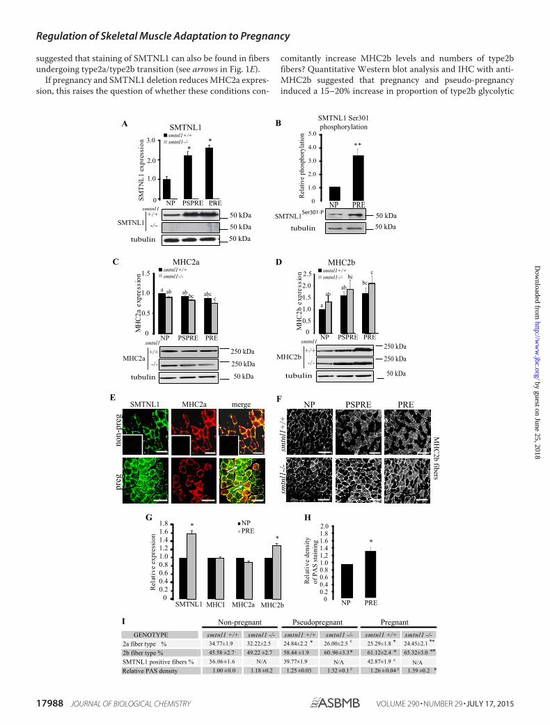
suggested that staining of SMTNL1 can also be found in fibersundergoing type2a/type2b transition (see arrows in Fig. 1E).
If pregnancy and SMTNL1 deletion reduces MHC2a expres-sion, this raises the question of whether these conditions con-
comitantly increase MHC2b levels and numbers of type2bfibers? Quantitative Western blot analysis and IHC with anti-MHC2b suggested that pregnancy and pseudo-pregnancyinduced a 15–20% increase in proportion of type2b glycolytic
E
F
sreb
ifb2
CH
M
H
NP PSPRE PRE
+/+1lntms
-/-1lntms
00.20.40.60.81.01.21.41.61.8
SMTNL1 MHCI MHC2a MHC2b
NPPRE
Rel
ativ
enoisserpxe
*
*
F
G
NP PRE
Rel
ativ
eytisned
gni niat sS
AP fo
00.20.40.60.81.01,21.41.61.82.0
*
gerp-nongerp
SMTNL1 mergeMHC2a
GENOTYPE smtnl1 +/+ smtnl1 -/- smtnl1 +/+ smtnl1 -/- smtnl1 +/+ smtnl1 -/-2a fiber type % 34.77±1.9 32.22±2.5 24.84±2.2 26.00±2.5 25.29±1.8 24.45±2.12b fiber type % 45.58 ±2.7 49.22 ±2.7 58.44 ±1.9 60 .96±3.3 61.12±2.4 65.32±3.0
tive fibers % 36.06±1.6 N/A 39.77±1.9 42.87±1.9Relative PAS density 1.00 ± 0.0 1.18 ±0.2 1.25 ±0.03 1.32 ±0.1 1.26 ±0.04 1.39 ±0.2
Non-pregnant Pseudopregnant Pregnant
# * ** *
****
N/A N/A#
*##
I
tubulin
+/+
-/-
smtnl1
tubulin
+/+
-/- MHC2b
smtnl1
MHC2a
+/+
-/-
smtnl1
SMTNL1
tubulin
50 kDa
250 kDa
250 kDa
50 kDa
250 kDa
250 kDa
50 kDa
50 kDa50 kDa
C
A BSMTNL1
noisserpxeb2
CH
M
smtnl1-/-smtnl1+/+
noisserpxea2
CH
M 00.5
1.0
1.5
2.0
2.5smtnl1-/-smtnl1+/+
0
0.5
1.0
1.5
a ab ab bc abcc
MHC2a MHC2b
a
abab
bcbc
c
SMTN
L1
expr
essi
on smtnl1-/-smtnl1+/+
3.0
2.0
1.0
0
**
NP PSPRE PRE
NP PSPRE PRE
NP PSPRE PRE
tubulin 50 kDa50 kDa
SMTNL1 Ser301 phosphorylation
Rela
tive p
hosp
hory
latio
n
D
NP PRE0
1.0
2.0
3.0
4.0
5.0
**
SMTNL1Ser301-P
SMTNL1 posi
Regulation of Skeletal Muscle Adaptation to Pregnancy
17988 JOURNAL OF BIOLOGICAL CHEMISTRY VOLUME 290 • NUMBER 29 • JULY 17, 2015
by guest on June 25, 2018http://w
ww
.jbc.org/D
ownloaded from

fibers (Fig. 1, F and I). IHC examination of the expression ofSMTNL1, MHC1, and MHC2a/2b in abdominis rectus muscleisolated from women undergoing hysterectomy (non-pregnantcontrol) or C-section (as pregnant) suggested that pregnancywas also likely to promote similar fiber switching in humans(Fig. 1G). Pregnancy or pseudo-pregnancy did not affect theexpression of MHCI, the marker of oxidative slow type 1 fibers.The decrease of protein expression of type2a marker MHC2awas only tendentious significant, whereas that of MHC2bincreased by 20%, indicating that fibers switched from oxidativeto the more glycolytic phenotype in human pregnant SKM. Theincreased type2b content in pregnancy is accompanied by a24% increase in glycogen content (Fig. 1H). These data suggestthat pregnancy promotes the transformation of SKM fiber typeto a more glycolytic phenotype in mice and humans and thatSMTNL1 may play a regulatory role in this process.
The Effects of SMTNL1 Deletion and Pregnancy on GlobalGene Expression in Skeletal Muscle—Given the observationsthat both SMTNL1 and pregnancy promote changes in the
mean total fiber composition of SKM, we carried out globalgene analysis to determine the extent of this switch in fourexperimental groups (smtnl1�/�, pregnant WT, and pregnantsmtnl1�/� groups were compared with non-pregnant WT). Atotal of 1384 genes were differentially expressed between thegroups. Specifically, the comparison of WT non-pregnant miceversus smtnl1�/� non-pregnant mice identified 195 genes,whereas the comparison of WT non-pregnant mice and WTpregnant mice resulted in 1261 genes expressed differently. Theexpression of 72 genes was related specifically to both preg-nancy and SMTNL1 deletion (Fig. 2A). Complete lists of genesfor all four comparisons are available at the Duke Microarrayfacility website. We also conducted Gene Ontology (GO) cate-gory enrichment analyses to determine the molecular functionof GO terms (Fig. 2A). Based on their biological functions, thesegenes play a role in cytoskeleton organization, calcium binding,regulation of metabolic processes and steroid synthesis,immune function, and growth regulation. Fifty percent of thegenes altered by SMTNL1 deletion were related to transcrip-
FIGURE 1. Pregnancy induced a fiber type switch to a glycolytic phenotype in SKM, and it is enhanced by SMTNL1 deletion. A, expression ofSMTNL1 in non-pregnant (NP), pregnant (PRE), and pseudopregnant (PSPRE) plantaris muscle by SQ-WB. B, phosphorylation of SMTNL1 Ser-301 innon-pregnant (NP) and pregnant (PRE) plantaris muscle. Means � SEM (n � 4 –5/group), t test, *, p 0.05. C and D, pregnancy reduces MHC2a expression(C) and increases expression of MHC2b (D) in SKM. Protein expression levels in non-pregnant, pregnant, and pseudopregnant by SQ-WB. n � 4 –13/group, data are presented as the mean � S.E., GLM with Tukey’s test. Different letters indicate significant differences, p 0.05. E, fiber typing by IHC andconfocal microscopy of plantaris muscle. SMTNL1 (green) is expressed in type2a fibers (red) in control SKM. SMTNL1 expression shows localization infibers different from type2a (arrows). Insets show secondary antibody controls. Scale bars � 20 �m. F, analysis of MHC2b expression (white) in plantarismuscle. Scale bar � 10 �m. G, SMTNL1, MHCI, MHC2a, and MHC2b expression in human SKM in pregnancy. H, quantitative Periodic Acid Schiff stainingfor glycogen content of human rectus abdominis sections. G and H, means � S.E. (n � 4 –5/group), t test. *, p 0.05. I, quantitative fiber counting ofplantaris muscle of non-pregnant, pregnant, and pseudopregnant (smtnl1�/� and smtnl1�/� animals. PAS, Periodic Acid Schiff. Data are the means �S.E. (n � 14 –15/group), t test. #, p 0.1; *, p 0.05; **, p 0.001.
FIGURE 2. The relationship between genes differentially regulated in the comparisons of pregnancy and SMTNL deletion and the ontology of therelated genes. A, Venn diagram; the numbers within the intersections of the circles indicate the common genes between the different groups (NP, non-pregnant;PRE, pregnant) compared to the smtnl1�/� non-pregnant data. The numbers outside the circles indicate the number of genes differentially regulated betweenthe two groups as indicated. The molecular function of the pregnancy and smtnl1�/� deletion-related genes are represented. B, the color code for the signalstrength is shown in the box at the bottom in which induced genes are indicated by red, and repressed genes are indicated by blue.
Regulation of Skeletal Muscle Adaptation to Pregnancy
JULY 17, 2015 • VOLUME 290 • NUMBER 29 JOURNAL OF BIOLOGICAL CHEMISTRY 17989
by guest on June 25, 2018http://w
ww
.jbc.org/D
ownloaded from

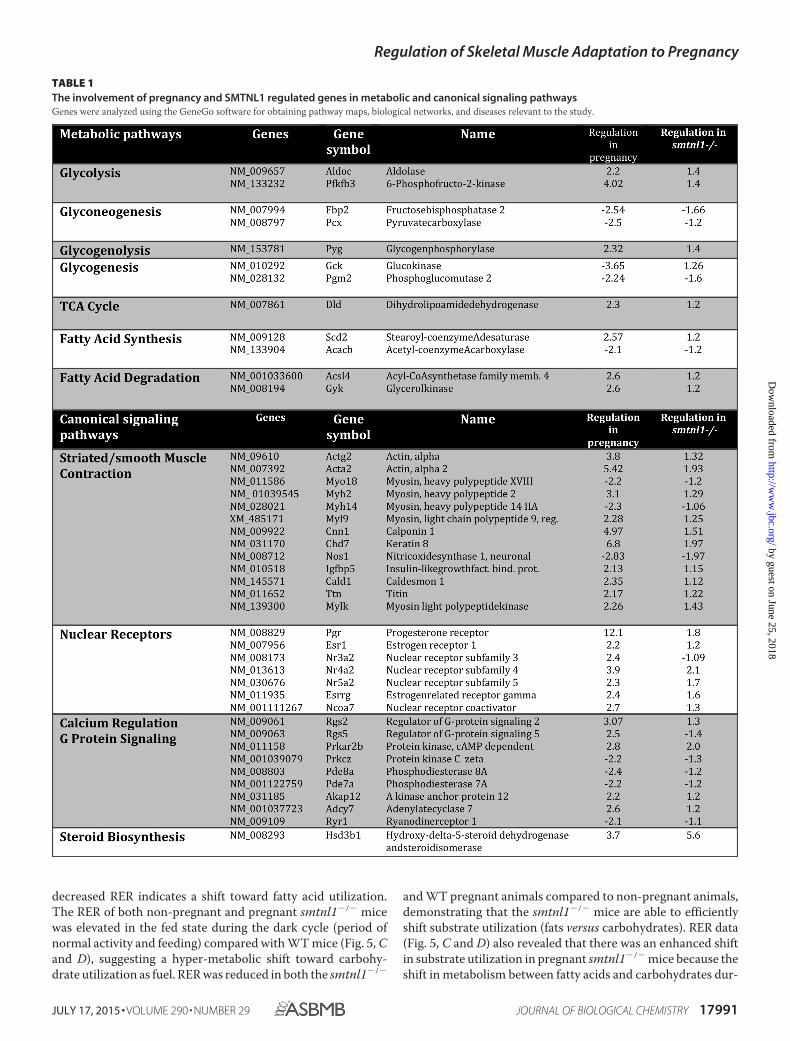
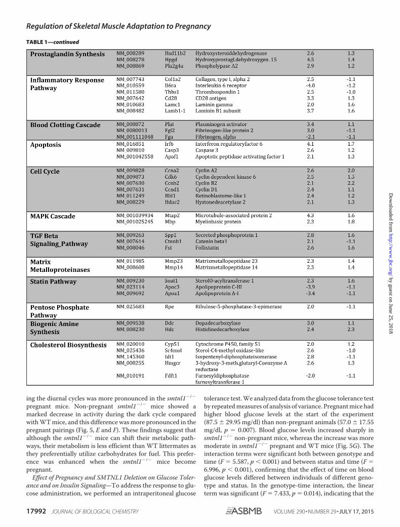
tional regulators, and the remaining 50% included structuralmolecules (23%) and enzyme regulators (15%) (Fig. 2A). Weperformed hierarchical clustering on all differentially expressedgenes using average linkage with Pearson’s dissimilarity andpresent the number of induced and repressed genes in a heatmap (Fig. 2B). Genes with significant changes in expressionwere assigned to different canonical pathways and subjected toGeneGo Analysis. Pregnancy was found to be largely relatedeither to metabolism such as glycolysis-glyconeogenesis, fattyacid synthesis, or contractile proteins or signaling pathwaysinvolving prostaglandin and steroid synthesis and nuclear hor-mone receptor signaling (Table 1). SMTNL1 deletion causedmostly the up-regulation of signaling pathways of nuclearreceptors and skeletal/smooth muscle contraction. The alteredcanonical pathways and the up- and down- regulated genes ofsignaling are listed in Table 1.
Proteomic Analysis Confirms Switching of SKM to a Glyco-lytic Phenotype in Response to Pregnancy and SMTNL1Deletion—Multidimensional proteomic analysis showed thatpregnancy promoted a striking and specific induction of severalglycolytic enzymes and contractile proteins, the expression ofwhich is specifically associated with type2b fibers (Fig. 3). Inparticular, 32% of the induced proteins are required for glycol-ysis (i.e. hexokinase, 6-phosphofructokinase, and aldolase),whereas others are associated with glycogen storage, e.g. glyco-gen phosphorylase and amylo-1,6-glucosidase. Additionally,�42% of the induced proteins were contractile proteins associ-ated with fast twitch fibers such as MHC2b, fast twitch forms oftroponins, fast twitch myosin light chains, and fast-type myo-sin-binding protein C, established markers of type2b fibers(supplemental Table S1 and Figshare/SMTNL1). Our pro-teomic data are consistent with the result of the microarrayanalysis and support the hypothesis that during pregnancyfemale mice adapt their skeletal muscle to a glycolytic pheno-type. The finding that these pregnancy-induced adaptations areless emphasized in SMTNL1-deleted tissues suggests that theprotein may regulate this process.
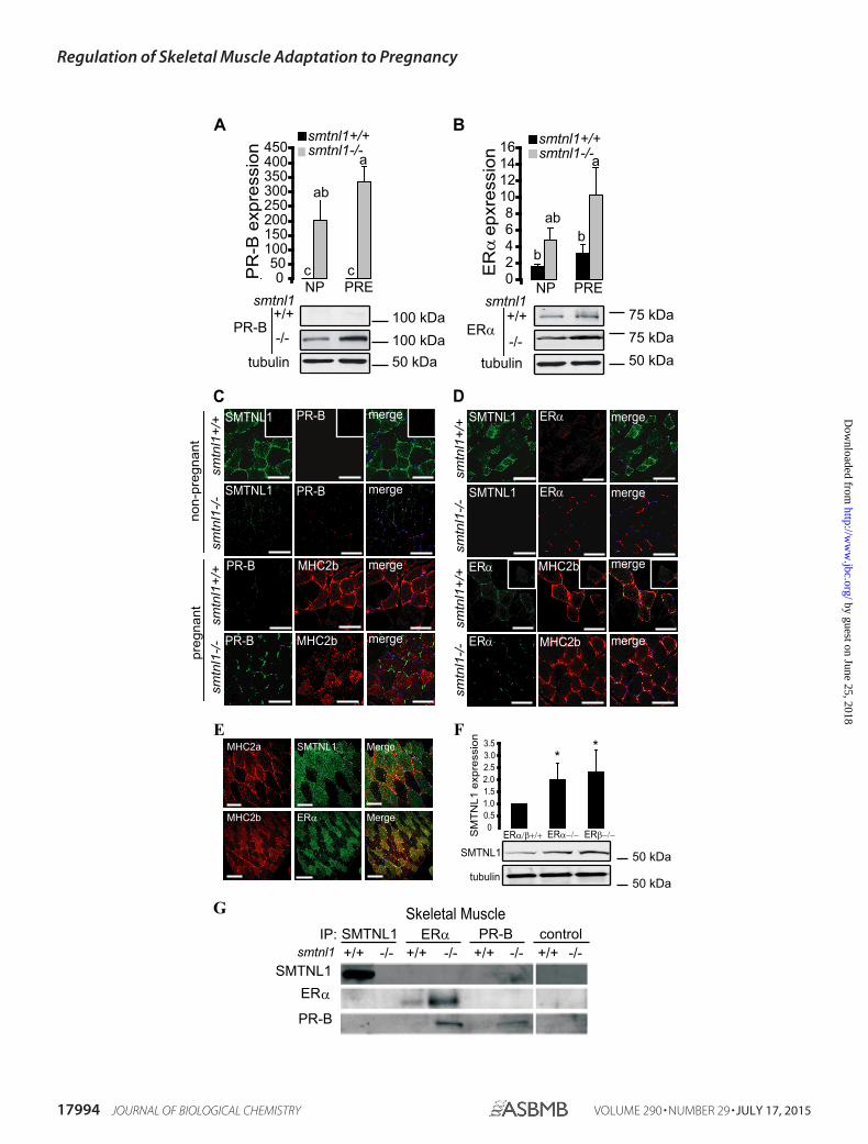
SMTNL1 Regulates the Expression of ER� and PR-B—It hasbeen established that SMTNL1 is expressed in smooth muscleas well as in reproductive tissues and that it is regulated throughpregnancy (12). This observation supports links between theprotein and steroid hormone action. Although both estrogenand progesterone clearly mediate adaptations in vascular anduterine smooth muscle, SKM is not generally recognized as atarget of these hormones. We, therefore, examined PR and ERexpression in SKM from smntl1�/� and WT mice. Westernblot analysis of WT plantaris muscles showed that pregnancyinduced a 1.5-fold increase in ER� expression by day 13.Remarkably, basal levels of ER� were induced 4 –5-fold overWT levels in SKM isolated from non-pregnant smtnl1�/� and10 –15-fold over WT levels in pregnant smtnl1�/� mice (Fig.4A). PR (A or B) was barely detectable in the muscle from eithernon-pregnant or pregnant WT mice using PR-B-specific anti-body, but the PR-B isoform showed a dramatic increase inexpression in SKM isolated from smtnl1�/� mice (Fig. 4A).In fiber typing experiments, PR-B expression was marginal innon-pregnant WT mice but showed discrete staining in allmuscle fibers, including both type2a and type2b fibers in mus-
cles isolated from pregnant animals (Fig. 4C). In contrast, ER�expression was confined only to type2b fibers in both non-preg-nant and pregnant SKM (Fig. 4D). Notably, the expression ofER� was greatly enhanced in type2b fibers in muscles isolatedfrom pregnant smtnl1�/� mice (Fig. 4D). A similar stainingpattern was also observed in human SKM (Fig. 2E). Moreover,the deletion of either ER� or ER� in mouse plantaris caused asignificant increase in the protein expression level of SMTNL1(Fig. 4F), suggesting the regulatory role of estrogen on SMTNL1gene expression. The discrete expression of ER� in type2bfibers-only implies a role for estrogen in mediating the fiberswitching process during pregnancy.
To investigate whether SMTNL1 mediates many of its cellu-lar effects on skeletal muscle adaptation through direct inter-actions with either ER or PR, we carried out co-immunopre-cipitation experiments from skeletal (Fig. 4G) muscle isolatedfrom WT and smtnl1�/� mice. SMTNL1 co-precipitated withnative PR-B but not ER�. In vivo interactions between theseproteins were confirmed by repeating the IP experiments withantibodies to each of these proteins and Western blotting theIPs with anti-SMTNL1. Besides using SKM, we applied uterinesmooth muscle extract as a positive control (data shown in ourprevious work in Bodoor et al. (12), as it expresses both ER� andPR under normal conditions). Smtnl1-deleted tissues did notpresent any interaction with SMTNL1 itself by IP. IP of PR-B orER� from skeletal and uterine smooth muscle extracts pre-pared from smtnl1�/� mice showed an induction of both pro-teins. Similarly, although IP with ER� antibody demonstratedthe presence of ER� and PR-B in the IP as expected, Westernanalysis with anti-SMTNL1 confirmed that it did not associatedirectly with ER� in vivo (Fig. 4G). Although our data do notsupport direct interactions of SMTNL1 with ER�, its increasedexpression observed after IP and Western analysis fromsmtnl1�/� mice may reflect that PR suppresses the expressionof ER� at the transcriptional level. In accordance with thismechanism, it was reported that both ER and PR mutually reg-ulate each others’ expression through transcriptionally con-trolled feedback mechanisms (19).
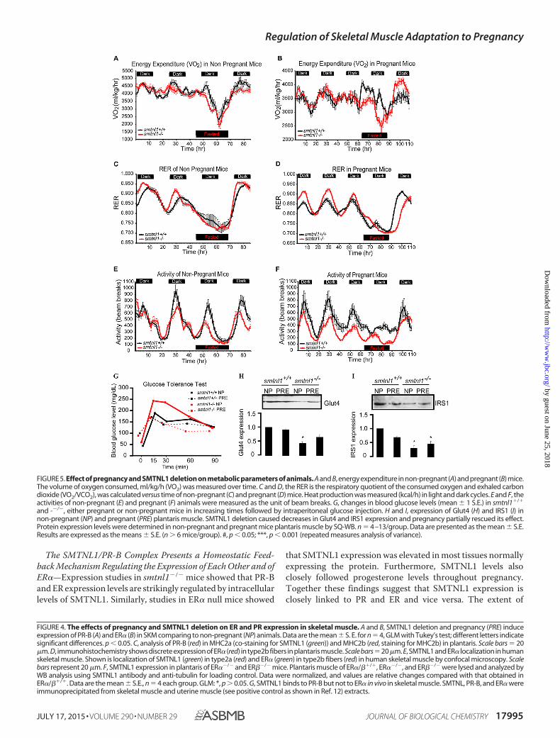
Pregnancy Induces Metabolic Activity and Reduces OxygenConsumption—One objective of this study was to determinewhether the effects of pregnancy and SMTNL1 deletion onSKM phenotype in mice resulted in any changes of energyexpenditure and/or feeding behavior. Pregnant or non-preg-nant WT and smtnl1�/� mice were housed in CLAMS appara-tus, and metabolic measurements taken daily (Fig. 5, A–F).During the study there was no significant difference in foodintake among the four experimental groups (pregnant and non-pregnant WT and smtnl1�/� mice; data not shown). Energyexpenditure (VO2), or the amount of energy an individual usesdaily to complete all regular body activities, of fed and fastednon-pregnant WT and smtnl1�/� mice was not significantlydifferent among groups (Fig. 5, A and B). VO2 however, wassignificantly lower in the fed pregnant mice than in the fednon-pregnant mice. Interestingly, in fasted pregnant mice, bothWT and smtnl1�/� animals showed a smaller decrease in VO2than did those fed, suggesting an overall rescuing effect of preg-nancy (Fig. 5, A and B). Normally, increases in the RER indicatesincreased utilization of carbohydrates for fuel. Conversely,
Regulation of Skeletal Muscle Adaptation to Pregnancy
17990 JOURNAL OF BIOLOGICAL CHEMISTRY VOLUME 290 • NUMBER 29 • JULY 17, 2015
by guest on June 25, 2018http://w
ww
.jbc.org/D
ownloaded from
decreased RER indicates a shift toward fatty acid utilization.The RER of both non-pregnant and pregnant smtnl1�/� micewas elevated in the fed state during the dark cycle (period ofnormal activity and feeding) compared with WT mice (Fig. 5, Cand D), suggesting a hyper-metabolic shift toward carbohy-drate utilization as fuel. RER was reduced in both the smtnl1�/�
and WT pregnant animals compared to non-pregnant animals,demonstrating that the smtnl1�/� mice are able to efficientlyshift substrate utilization (fats versus carbohydrates). RER data(Fig. 5, C and D) also revealed that there was an enhanced shiftin substrate utilization in pregnant smtnl1�/� mice because theshift in metabolism between fatty acids and carbohydrates dur-
TABLE 1The involvement of pregnancy and SMTNL1 regulated genes in metabolic and canonical signaling pathwaysGenes were analyzed using the GeneGo software for obtaining pathway maps, biological networks, and diseases relevant to the study.
Regulation of Skeletal Muscle Adaptation to Pregnancy
JULY 17, 2015 • VOLUME 290 • NUMBER 29 JOURNAL OF BIOLOGICAL CHEMISTRY 17991
by guest on June 25, 2018http://w
ww
.jbc.org/D
ownloaded from
ing the diurnal cycles was more pronounced in the smtnl1�/�
pregnant mice. Non-pregnant smtnl1�/� mice showed amarked decrease in activity during the dark cycle comparedwith WT mice, and this difference was more pronounced in thepregnant pairings (Fig. 5, E and F). These findings suggest thatalthough the smtnl1�/� mice can shift their metabolic path-ways, their metabolism is less efficient than WT littermates asthey preferentially utilize carbohydrates for fuel. This prefer-ence was enhanced when the smtnl1�/� mice becomepregnant.
Effect of Pregnancy and SMTNL1 Deletion on Glucose Toler-ance and on Insulin Signaling—To address the response to glu-cose administration, we performed an intraperitoneal glucose
tolerance test. We analyzed data from the glucose tolerance testby repeated measures of analysis of variance. Pregnant mice hadhigher blood glucose levels at the start of the experiment(87.5 � 29.95 mg/dl) than non-pregnant animals (57.0 � 17.55mg/dl, p � 0.007). Blood glucose levels increased sharply insmtnl1�/� non-pregnant mice, whereas the increase was moremoderate in smtnl1�/� pregnant and WT mice (Fig. 5G). Theinteraction terms were significant both between genotype andtime (F � 5.587, p 0.001) and between status and time (F �6.996, p 0.001), confirming that the effect of time on bloodglucose levels differed between individuals of different geno-type and status. In the genotype-time interaction, the linearterm was significant (F � 7.433, p � 0.014), indicating that the
TABLE 1—continued
Regulation of Skeletal Muscle Adaptation to Pregnancy
17992 JOURNAL OF BIOLOGICAL CHEMISTRY VOLUME 290 • NUMBER 29 • JULY 17, 2015
by guest on June 25, 2018http://w
ww
.jbc.org/D
ownloaded from

differences between KO and WT genotypes were similar ateach measurement (Fig. 5G). WT pregnant mice also hadslightly but not significantly lower blood glucose levels thanWT non-pregnant mice between 15 and 60 min. These resultsshow that smtnl1�/� non-pregnant mice have much lower glu-cose tolerance than smtnl1�/� pregnant or WT mice, indicat-ing that pregnancy increases glucose tolerance in KO mice tothe level of tolerance in WT mice. Impaired glucose tolerance isassociated with pronounced insulin resistance and at cellularlevel occurs in tandem with changes in key steps in the insulin-signaling cascade that regulates glucose uptake by SKM. Insulinreceptor substrate 1 (IRS1) and the insulin-sensitive glucosetransporter 4 (Glut4) are believed to be key elements of insulin-signaling in SKM (20) To verify the importance of pregnancyand SMTNL1 deletion in the development of insulin resistance,SQ-WB analysis was conducted on non-pregnant and pregnantplantaris tissues of WT and smtnl1�/� mice, determining theprotein expression of Glut4 and IRS1 (Fig. 5, H–I). SMTNL1deletion decreased the expression of Glut4 and IRS1 by 53 and58%, respectively. Pregnancy attenuated IRS1 expression sig-nificantly but had no effect on the Glut4 expression level in WTanimals. However, pregnant smtnl1�/� animals exhibited anincrease in both Glut4 and IRS1 expression in smtnl1�/� ani-mals. Our results suggest that the impaired glucose tolerance ofsmtnl1�/� is the consequence of decreased Glut4 and IRS1expression.
Discussion
Data presented herein suggest that pregnancy hormonessuch as progesterone and estrogen trigger the switching ofSKM to a glycolytic phenotype through their respectivenuclear receptors (Fig. 6). We hypothesize that SMTNL1plays a negative regulatory role on the transcriptional activ-ity of PR and ER in SKM and that protein kinase A- or G-me-diated signaling can influence this process specificallythrough the phosphorylation of SMTNL1 at Ser-301 (11).Functionally, therefore, SMTNL1 acts as a transcriptionalrepressor of PR in vivo, and the phosphorylation of SMTNL1specifically inhibits this function (12). Mechanistically thisimplies that the default function for SMTNL1 in non-preg-nant animals is to repress PR function from promoting preg-nancy-like adaptive responses in female SKM. Conversely,pregnancy (or adrenergic-induced signals) through Ser-301phosphorylation relieves SMTNL1 inhibition of PR to pro-mote appropriate changes in SKM gene expression resultingin switching to a more glycolytic phenotype. Intriguingly,protein kinase A-mediated signaling pathways have longbeen known to greatly affect both progesterone and estrogensignaling in vivo (21, 22). However, our study is the first tosuggest that cyclic nucleotide-mediated signaling pathwaysin SKM may also be related to the regulation of gene expres-sion by SMTNL1 acting through PR and ER.
FIGURE 3. Proteomic analysis shows that pregnancy and SMTNL1 deletion induces expression of glycolytic enzymes and contractile proteins associ-ated with Type2b fibers. Plantaris muscle extracts were subjected to proteomic analysis prepared from smtnl1�/� (WT), smtnl1�/�, pregnant (WP), smntl1�/�
(KO), and smtnl1�/� pregnant (KOP) animals. The data show -fold induction of proteins in WP, KO, and KOP samples compared with WT. TER ATPase, transitionalendoplasmic reticulum ATPase. See the related data in supplemental Table S1.
Regulation of Skeletal Muscle Adaptation to Pregnancy
JULY 17, 2015 • VOLUME 290 • NUMBER 29 JOURNAL OF BIOLOGICAL CHEMISTRY 17993
by guest on June 25, 2018http://w
ww
.jbc.org/D
ownloaded from
A
050
100150200250300350400450
NP PREc c
ab
a
+/+
-/-
tubulin
PR-B
smtnl102468
10121416 smtnl1-/-
smtnl1+/+
bb
ab
a
+/+
-/-tubulin
ERα
smtnl1NP PRE
noisserpxe B-
RP
noisserxpe αR
E
smtnl1-/-smtnl1+/+
50 kDa 75 kDa 75 kDa
50 kDa 100 kDa 100 kDa
00.51.01.52.02.53.03.5
ERα−/−
noisserpxe 1LN
TM
S
SMTNL1
tubulin
**
50 kDa
50 kDa
ERβ−/− ERα/β+/+
+/+1l ntms
SMTNL1
SMTNL1
PR-B
PR-B
PR-B
PR-B
MHC2b
MHC2b
merge
merge
merge
merge
CSMTNL1
SMTNL1
MHC2b
ERα
ERα
ERα
merge
merge
merge
mergeMHC2bERα
D
+/+1lntms
-/- 1lntms
-/-1lntms
+/+1lntms
+/+1lntms
-/ -1lntms
-/-1lntms
E
tnan ge rp-nontnang erp
SMTNL1MHC2a
MHC2b ERα Merge
Merge
D
F
G
SMTNL1ER
PR-B
SMTNL1 ERα PR-B control+/+ +/+ +/+ +/+-/- -/- -/- -/-
IP:smtnl1
Skeletal Muscle
α
B
Regulation of Skeletal Muscle Adaptation to Pregnancy
17994 JOURNAL OF BIOLOGICAL CHEMISTRY VOLUME 290 • NUMBER 29 • JULY 17, 2015
by guest on June 25, 2018http://w
ww
.jbc.org/D
ownloaded from
The SMTNL1/PR-B Complex Presents a Homeostatic Feed-back Mechanism Regulating the Expression of Each Other and ofER�—Expression studies in smtnl1�/� mice showed that PR-Band ER expression levels are strikingly regulated by intracellularlevels of SMTNL1. Similarly, studies in ER� null mice showed
that SMTNL1 expression was elevated in most tissues normallyexpressing the protein. Furthermore, SMTNL1 levels alsoclosely followed progesterone levels throughout pregnancy.Together these findings suggest that SMTNL1 expression isclosely linked to PR and ER and vice versa. The extent of
FIGURE 4. The effects of pregnancy and SMTNL1 deletion on ER and PR expression in skeletal muscle. A and B, SMTNL1 deletion and pregnancy (PRE) induceexpression of PR-B (A) and ER� (B) in SKM comparing to non-pregnant (NP) animals. Data are the mean � S. E. for n � 4, GLM with Tukey’s test; different letters indicatesignificant differences. p 0.05. C, analysis of PR-B (red) in MHC2a (co-staining for SMTNL1 (green)) and MHC2b (red, staining for MHC2b) in plantaris. Scale bars � 20�m. D, immunohistochemistry shows discrete expression of ER� (red) in type2b fibers in plantaris muscle. Scale bars�20�m. E, SMTNL1 and ER� localization in humanskeletal muscle. Shown is localization of SMTNL1 (green) in type2a (red) and ER� (green) in type2b fibers (red) in human skeletal muscle by confocal microscopy. Scalebars represent 20 �m. F, SMTNL1 expression in plantaris of ER��/� and ER��/� mice. Plantaris muscle of ER�/��/�, ER��/�, and ER��/� were lysed and analyzed byWB analysis using SMTNL1 antibody and anti-tubulin for loading control. Data were normalized, and values are relative changes compared with that obtained inER�/��/�. Data are the mean � S.E., n � 4 each group. GLM: *, p � 0.05. G, SMTNL1 binds to PR-B but not to ER� in vivo in skeletal muscle. SMTNL, PR-B, and ER� wereimmunoprecipitated from skeletal muscle and uterine muscle (see positive control as shown in Ref. 12) extracts.
FIGURE 5. Effect of pregnancy and SMTNL1 deletion on metabolic parameters of animals. A and B, energy expenditure in non-pregnant (A) and pregnant (B) mice.The volume of oxygen consumed, ml/kg/h (VO2) was measured over time. C and D, the RER is the respiratory quotient of the consumed oxygen and exhaled carbondioxide (VO2/VCO2), was calculated versus time of non-pregnant (C) and pregnant (D) mice. Heat production was measured (kcal/h) in light and dark cycles. E and F, theactivities of non-pregnant (E) and pregnant (F) animals were measured as the unit of beam breaks. G, changes in blood glucose levels (mean � 1 S.E.) in smtnl1�/�
and -�/�, either pregnant or non-pregnant mice in increasing times followed by intraperitoneal glucose injection. H and I, expression of Glut4 (H) and IRS1 (I) innon-pregnant (NP) and pregnant (PRE) plantaris muscle. SMTNL1 deletion caused decreases in Glut4 and IRS1 expression and pregnancy partially rescued its effect.Protein expression levels were determined in non-pregnant and pregnant mice plantaris muscle by SQ-WB. n � 4–13/group. Data are presented as the mean � S.E.Results are expressed as the means � S.E. (n � 6 mice/group). #, p 0.05; ***, p 0.001 (repeated measures analysis of variance).
Regulation of Skeletal Muscle Adaptation to Pregnancy
JULY 17, 2015 • VOLUME 290 • NUMBER 29 JOURNAL OF BIOLOGICAL CHEMISTRY 17995
by guest on June 25, 2018http://w
ww
.jbc.org/D
ownloaded from

SMTNL1 involvement in this feedback mechanism is most dra-matic in smtnl1�/� mice with the observation that the expres-sion of both PR and ER are greatly increased within specificmuscle fibers in response to SMTNL1 deletion. The findingthat ER� is specifically localized in type2b fibers suggests thatthese fibers are likely to be sensitive to estrogen when transi-tioned to glycolytic fibers. Interestingly, earlier work by otherssuggested that the expression of ER in SKM has a role in thedevelopment of muscle strength in humans (23, 24). Becauseprior work from our group showed that SMTNL1 only interactswith PR-B and-A and not with either isoform of ER (12), thealtered ER expression observed in smtnl1�/� mice most likelymust be mediated through PR via a homeostatic feedbackmechanism (25), as PR and ER are known to mutually regulatethe expression of each other. We, therefore, hypothesize thatthe differential localization and expression of SMTNL1, PR,and ER are key to the adaptation to pregnancy. We propose thatunder normal physiological conditions SMTNL1 is expressedin type2a fibers, where it represses the activity of PR. BecauseER shows type2b-specific localization, this provides fiber-selec-tive estrogen sensitivity. On one hand, circulating estrogenbound to ER might bind and repress the promoter of smtnl1,which provides an explanation to the low expression ofSMTNL1 in type2b fibers in normal conditions and theincreased SMTNL1 expression in ER�- or �-deleted animals(Fig. 4F). On the other hand, the increased number of type2bbut a slight decreased number of 2a fibers detected insmtnl1�/� SKM could be explained by the release of PR repres-sion by SMTNL1 in type2a fibers.
The Effects of Pregnancy on Metabolic Pathways Related toInsulin Resistance—Bioinformatics analysis of data from globalgene expression and multidimensional proteomic analyses
showed that pregnancy highly regulates sets of genes related tocanonical pathways that play a role in glycolysis, gluconeogen-esis, muscle contraction, and steroid biosynthesis as well asinflammatory responses, supporting our hypothesis of themanifestation of a glycolytic phenotype in pregnant SKM. Themain profile of genes induced in smtnl1�/� deletion was tran-scriptional regulation, suggesting a primary role of SMTNL1 ingene expression as genes such as PR and nuclear receptor sub-family 3– 4 are regulated. The majority of pathways regulated inpregnancy were related to the mediation of insulin resistance(26).Duringtheprogressionofnormalpregnancies,insulinresis-tance arises. Pronounced insulin resistance is associated withimpaired glucose tolerance and with a high risk of gestationaldiabetes; however, the underlying mechanisms are not very wellunderstood (27). Redundant control mechanisms operate tomaintain normal glucose homeostasis in SKM. Insulin activatesthe insulin receptor tyrosine kinase, which activates signalingpartners such as PKB/Akt and stimulates the translocation ofGLUT4 vesicles to the plasma. IRS1 protein expression wasincreased in smtnl1�/� mice; moreover, pregnancy itself had anegative effect on IRS expression. Importantly, IRS expressionis increased postpartum (20), and overexpression of the proteinin a spontaneous gestational diabetes is associated with thereversal of insulin resistance (28). Pregnancy and SMTNL1deletion are likely to regulate Glut4 expression through differ-ent pathways because smtnl1�/� tissues showed significantlylower levels of the Glut4 protein, an effect rescued by preg-nancy. Our results showed that ER and PR expression levels hada robust increase in pregnant and smtnl1�/� tissues. Moreover,ER expression was related to a more efficient metabolic state,and ER KO animals presented overt insulin resistance (29).Although these findings strongly suggest a better metabolic sta-tus of SMTNL1 knock-out mice, we found impaired metabo-lism in smtnl1�/� mice. This controversy may be explained bythe observation that circulating hormone levels influence theimprovement of insulin resistance (26) and that the levels ofcirculating progesterone and estrogen decreased significantlyduring pregnancy (12).
How Might Pregnancy-induced Glycolytic Fiber Switching inSkeletal Muscle Contribute to Insulin Resistance—In Fig. 6 wesuggest that pregnancy-induced fiber switching to a more gly-colytic phenotype is a coordinated physiological response thatmay be at the heart of increased weight gain and generalizedinsulin resistance associated with normal pregnancy (Fig. 6). Byvirtue of its mass, SKM dictates the overall caloric disposal ofboth glucose and free fatty acids. If the oxidative capacity ofSKM is increased through endurance exercise, one reduces therisk of developing insulin resistance, and exercise alone canreverse the symptoms of obesity-induced type II diabetes. Thisis because the primary sites of free fatty acid oxidation in mam-mals are oxidative type I and Type2a SKM fibers. Conversely, ifthe type2a content of SKM is reduced, the ability to oxidize freefatty acid is also reduced, thereby increasing storage of fattyacid (FA) as triglyceride in adipose tissue. When oxidativecapacity is reduced, the storage of FA as triglyceride is furtherexacerbated in the presence of high carbohydrate levels. This isbecause humans and most mammals derive much of theirstored triglyceride via pathways of de novo FA synthesis
FIGURE 6. Schematic showing the molecular mechanism by which SMTNL1regulates PR-B to coordinately promote skeletal muscle adaptations inresponse to pregnancy. In response to elevated steroid hormone, progester-one (S) levels SMTNL1 and PR-B enter the nucleus. In the non-pregnant state,SMTNL1 functions to repress PR-B activity; however, during pregnancy this inhi-bition is relieved and promotes activation of skeletal muscle-specific genes reg-ulating the expression of metabolic and contractile proteins. This coordinatedresponse adapts the mother’s physiological state to support the weight gainoriginating from developing fetus. Switching to a glycolytic phenotype in skele-tal muscle reduces the oxidative capacity of SKM, promoting increased storage offatty acid (FA) and inducing an insulin-resistant state resulting in increased circu-lating glucose. PKA/PKG, protein kinase A/G.
Regulation of Skeletal Muscle Adaptation to Pregnancy
17996 JOURNAL OF BIOLOGICAL CHEMISTRY VOLUME 290 • NUMBER 29 • JULY 17, 2015
by guest on June 25, 2018http://w
ww
.jbc.org/D
ownloaded from

through the metabolism of glucose in adipose tissue and liver(30). Excess fat and carbohydrate in the diet, therefore, providesan optimal environment for excessive weight gain, especially insedentary individuals. We hypothesize that switching to a gly-colytic phenotype in pregnancy, with a reduced oxidative fibercontent of SKM, therefore, provides a molecular basis toexplain increased weight gain and insulin resistance in preg-nant females. We suggest that this is a normal response to preg-nancy and infers the evolutionary advantage of enabling themother to increase circulating glucose levels for proper fetaldevelopment as well as store fat more efficiently to meet thecaloric demands of lactation. Our results also suggest thatswitching to a glycolytic phenotype infers an advantage byincreasing the mother’s physical strength. Type2b muscle fibersare associated with strength and resistance training such asseen with weight lifting. An increase in the mother’s physicalstrength would much better enable her to manage the increasedweight of the fetus in the latter stages of pregnancy. Impor-tantly, in �4% of all pregnancies, pregnancy-induced insulinresistance progresses to gestational diabetes. If pregnancy,therefore, promotes the transition to a more glycolytic pheno-type as an evolutionary benefit, then this may render somewomen even more susceptible to developing gestational diabe-tes, especially under the influence of high energy Western diets.
Finally, considering all our understanding of muscle physiologyand its importance to health and well being, one might ask whypregnancy-induced fiber switching has not already been reportedin the literature. A search of the current literature suggests that thismay be because the majority of muscle studies to date have beencarried out in males only. The experimental bias of studies towardmale over female physiology is likely attributable to the commonlyheld belief that the estrous cycle introduces hard-to-explain vari-ability in physiological processes in female animals. Indeed, instudies of female physiology, ovariectomized females are typicallyused to remove the oestrus-related aspects of physiology from theinquiry. As a result, many normal physiological responses relevantto female physiology get purposefully ignored. Although furtherinvestigation is certainly warranted, particularly non-invasivestudies of SKM in pregnant women, our findings could indicatethat important aspects of women’s health are being missed due toan experimental bias.
Author Contributions—B. L. designed, performed, and analyzed theexperiments shown in Figs. 1 and in part in Figs. 2, 3, 4, and 6 andwrote the paper. K. B. analyzed the experiments in Fig. 2. S. A. per-formed the experiments in Figs. 1 and 5. D. H. W. assisted in theCLAMS study. D. L. performed the proteomic analysis. D. P. M. andR. S. helped in ER and PR studies. D. Z. provided MHC-specific anti-bodies. J. D. and R. C. H. provided human biopsies and performedthe human study. T. R. performed CLAMS experiments. T. A. H.conceived and coordinated the study and wrote the paper.
Acknowledgments—We are grateful to Dr. Ferenc Erdodi (University ofDebrecen, Debrecen, Hungary) for revising the paper and Dr. KennetKorach (Reproductive and Developmental Toxicology Laboratory,Research Triangle Park, NC) for providing the ERKO mice as well as toDr. Szabolcs Lengyel (Hungarian National Academy of Science, EcologyResearch Center, Debrecen, Hungary) for helping in statistical analyses.
Note Added in Proof—Supplemental Table S1 was missing in theversion of this article that was published as a Paper in Press on April14, 2015. The supplemental table is now available.
References1. Halayko, A. J., and Solway, J. (2001) Molecular mechanisms of phenotypic
plasticity in smooth muscle cells. J. Appl Physiol. 90, 358 –3682. Booth, F. W., and Thomason, D. B. (1991) Molecular and cellular adapta-
tion of muscle in response to exercise: perspectives of various models.Physiol. Rev. 71, 541–585
3. Fitts, R. H. (2003) Effects of regular exercise training on skeletal musclecontractile function. Am. J. Phys. Med. Rehabil. 82, 320 –331
4. Carey, A. L., and Kingwell, B. A. (2009) Novel pharmacological approachesto combat obesity and insulin resistance: targeting skeletal muscle with“exercise mimetics.” Diabetologia 52, 2015–2026
5. Hamilton, M. T., and Booth, F. W. (2000) Skeletal muscle adaptation toexercise: a century of progress. J. Appl. Physiol. 88, 327–331
6. Edwards, D. P. (2005) Regulation of signal transduction pathways by es-trogen and progesterone. Annu. Rev. Physiol. 67, 335–376
7. Barros, R. P., Morani, A., Moriscot, A., and Machado, U. F. (2008) Insulinresistance of pregnancy involves estrogen-induced repression of muscleGLUT4. Mol. Cell. Endocrinol. 295, 24 –31
8. Sugaya, A., Sugiyama, T., Yanase, S., Shen, X. X., Minoura, H., and Toyoda,N. (2000) Expression of glucose transporter 4 mRNA in adipose tissue andskeletal muscle of ovariectomized rats treated with sex steroid hormones.Life Sci. 66, 641– 648
9. Branisteanu, D. D., and Mathieu, C. (2003) Progesterone in gestationaldiabetes mellitus: guilty or not guilty? Trends Endocrinol. Metab. 14,54 –56
10. Wooldridge, A. A., Fortner, C. N., Lontay, B., Akimoto, T., Neppl, R. L.,Facemire, C., Datto, M. B., Kwon, A., McCook, E., Li, P., Wang, S.,Thresher, R. J., Miller, S. E., Perriard, J. C., Gavin, T. P., Hickner, R. C.,Coffman, T. M., Somlyo, A. V., Yan, Z., and Haystead, T. A. (2008) Dele-tion of the protein kinase A/protein kinase G target SMTNL1 promotes anexercise-adapted phenotype in vascular smooth muscle. J. Biol. Chem.283, 11850 –11859
11. Lontay, B., Bodoor, K., Weitzel, D. H., Loiselle, D., Fortner, C., Lengyel, S.,Zheng, D., Devente, J., Hickner, R., and Haystead, T. A. (2010) Smooth-elin-like 1 protein regulates myosin phosphatase-targeting subunit 1 ex-pression during sexual development and pregnancy. J. Biol. Chem. 285,29357–29366
12. Bodoor, K., Lontay, B., Safi, R., Weitzel, D. H., Loiselle, D., Wei, Z.,Lengyel, S., McDonnell, D. P., and Haystead, T. A. (2011) Smoothelin-like1 protein is a bifunctional regulator of the progesterone receptor duringpregnancy. J. Biol. Chem. 286, 31839 –31851
13. Ulke-Lemee, A., Turner, S. R., Mughal, S. H., Borman, M. A., Winkfein,R. J., and MacDonald, J. A. (2011) Mapping and functional characteriza-tion of the murine smoothelin-like 1 promoter. BMC Mol. Biol. 12, 10
14. Akimoto, T., Ribar, T. J., Williams, R. S., and Yan, Z. (2004) Skeletal muscleadaptation in response to voluntary running in Ca2�/calmodulin-depen-dent protein kinase IV-deficient mice. Am. J. Physiol. Cell Physiol. 287,C1311–C1319
15. Lontay, B., Serfozo, Z., Gergely, P., Ito, M., Hartshorne, D. J., andErdodi, F. (2004) Localization of myosin phosphatase target subunit 1in rat brain and in primary cultures of neuronal cells. J. Comp. Neurol.478, 72– 87
16. Borman, M. A., MacDonald, J. A., and Haystead, T. A. (2004) Modulationof smooth muscle contractility by CHASM, a novel member of thesmoothelin family of proteins. FEBS Lett. 573, 207–213
17. Schmidt, C., and Urlaub, H. (2009) iTRAQ-labeling of in-gel digestedproteins for relative quantification. Methods Mol. Biol. 564, 207–226
18. Shadforth, I. P., Dunkley, T. P., Lilley, K. S., and Bessant, C. (2005) i-Tracker: for quantitative proteomics using iTRAQ. BMC Genomics 6, 145
19. Clarke, C. L., and Sutherland, R. L. (1990) Progestin regulation of cellularproliferation. Endocr. Rev. 11, 266 –301
20. Kirwan, J. P., Varastehpour, A., Jing, M., Presley, L., Shao, J., Friedman,J. E., and Catalano, P. M. (2004) Reversal of insulin resistance postpartumis linked to enhanced skeletal muscle insulin signaling. J. Clin. Endocrinol.
Regulation of Skeletal Muscle Adaptation to Pregnancy
JULY 17, 2015 • VOLUME 290 • NUMBER 29 JOURNAL OF BIOLOGICAL CHEMISTRY 17997
by guest on June 25, 2018http://w
ww
.jbc.org/D
ownloaded from
Metab. 89, 4678 – 468421. Coleman, K. M., Dutertre, M., El-Gharbawy, A., Rowan, B. G., Weigel,
N. L., and Smith, C. L. (2003) Mechanistic differences in the activation ofestrogen receptor-� (ER�)- and ER�-dependent gene expression bycAMP signaling pathway (s). J. Biol. Chem. 278, 12834 –12845
22. Rowan, B. G., Garrison, N., Weigel, N. L., and O’Malley, B. W. (2000)8-Bromo-cyclic AMP induces phosphorylation of two sites in SRC-1 thatfacilitate ligand-independent activation of the chicken progesterone re-ceptor and are critical for functional cooperation between SRC-1 andCREB binding protein. Mol. Cell. Biol. 20, 8720 – 8730
23. Onambele, N. G., Skelton, D. A., Bruce, S. A., and Woledge, R. C. (2001)Follow-up study of the benefits of hormone replacement therapy on iso-metric muscle strength of adductor pollicis in postmenopausal women.Clin. Sci. 100, 421– 422
24. Skelton, D. A., Phillips, S. K., Bruce, S. A., Naylor, C. H., and Woledge, R. C.(1999) Hormone replacement therapy increases isometric musclestrength of adductor pollicis in post-menopausal women. Clin. Sci. 96,357–364
25. Bradshaw, M. S., Tsai, S. Y., Leng, X. H., Dobson, A. D., Conneely, O. M.,
O’Malley, B. W., and Tsai, M. J. (1991) Studies on the mechanism offunctional cooperativity between progesterone and estrogen receptors.J. Biol. Chem. 266, 16684 –16690
26. Catalano, P. M. (2010) Obesity, insulin resistance, and pregnancy out-come. Reproduction 140, 365–371
27. Solomon, C. G., and Seely, E. W. (2001) Brief review: hypertension inpregnancy: a manifestation of the insulin resistance syndrome? Hyperten-sion 37, 232–239
28. Shao, J., Yamashita, H., Qiao, L., Draznin, B., and Friedman, J. E. (2002)Phosphatidylinositol 3-kinase redistribution is associated with skeletalmuscle insulin resistance in gestational diabetes mellitus. Diabetes 51,19 –29
29. Bryzgalova, G., Gao, H., Ahren, B., Zierath, J. R., Galuska, D., Steiler, T. L.,Dahlman-Wright, K., Nilsson, S., Gustafsson, J. A., Efendic, S., and Khan,A. (2006) Evidence that oestrogen receptor-� plays an important role inthe regulation of glucose homeostasis in mice: insulin sensitivity in theliver. Diabetologia 49, 588 –597
30. Towler, M. C., and Hardie, D. G. (2007) AMP-activated protein kinase inmetabolic control and insulin signaling. Circ. Res. 100, 328 –341
Regulation of Skeletal Muscle Adaptation to Pregnancy
17998 JOURNAL OF BIOLOGICAL CHEMISTRY VOLUME 290 • NUMBER 29 • JULY 17, 2015
by guest on June 25, 2018http://w
ww
.jbc.org/D
ownloaded from

Thomas Ribar and Timothy A. HaysteadMcDonnell,Rachid Safi, Donghai Zheng, James Devente, Robert C. Hickner, Donald P.
Beata Lontay, Khaldon Bodoor, Adrienn Sipos, Douglas H. Weitzel, David Loiselle,Switching of Skeletal Muscle to a Glycolytic Phenotype in Human and MicePregnancy and Smoothelin-like Protein 1 (SMTNL1) Deletion Promote the
doi: 10.1074/jbc.M115.658120 originally published online June 5, 20152015, 290:17985-17998.J. Biol. Chem.
10.1074/jbc.M115.658120Access the most updated version of this article at doi:
Alerts:
When a correction for this article is posted•
When this article is cited•
to choose from all of JBC's e-mail alertsClick here
Supplemental material:
http://www.jbc.org/content/suppl/2015/06/11/M115.658120.DC1
http://www.jbc.org/content/290/29/17985.full.html#ref-list-1
This article cites 30 references, 11 of which can be accessed free at
by guest on June 25, 2018http://w
ww
.jbc.org/D
ownloaded from