prenatal chromosome microarray - fetal genomics group
TRANSCRIPT
Dr Jenny Patterson MBChB MSc MRCP. ST4 Clinical Genetics.
Prenatal chromosome microarray
Prenatal chromosom
e microarray
Technique
Benefits/literature of clinical utility
Limitations
Pretest counselling considerations
Current Guidelines
Audit.
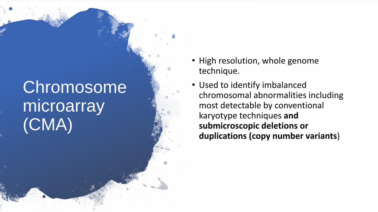
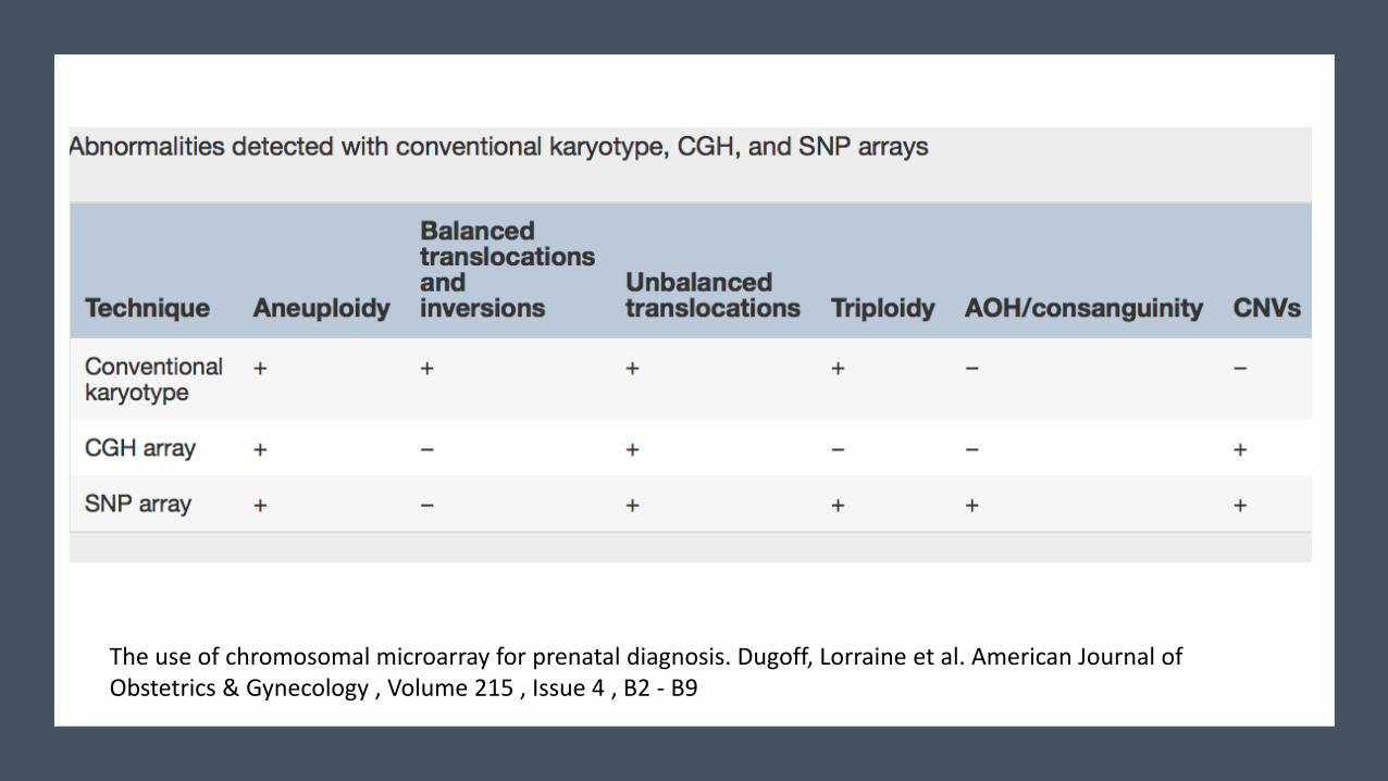
Chromosome microarray (CMA)
• High resolution, whole genome technique.
• Used to identify imbalanced chromosomal abnormalities including most detectable by conventional karyotype techniques and submicroscopic deletions or duplications (copy number variants)
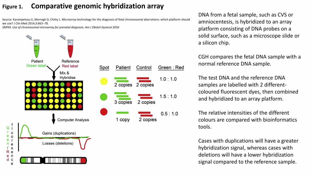
Figure 1
American Journal o Obstetrics & Gynecology 2016 215, B2-B9DOI: (10.1016/j.ajog.2016.07.016)
Figure 1. Comparative genomic hybridization array Source: Karampetsou E, Morrogh D, Chitty L. Microarray technology for the diagnosis of fetal chromosomal aberrations: which platform should we use? J Clin Med 2014;3:663−78. SMFM. Use of chromosomal microarray for prenatal diagnosis. Am J Obstet Gynecol 2016
DNA from a fetal sample, such as CVS or amniocentesis, is hybridized to an array platform consisting of DNA probes on a solid surface, such as a microscope slide or a silicon chip. CGH compares the fetal DNA sample with a normal reference DNA sample. The test DNA and the reference DNA samples are labelled with 2 different-coloured fluorescent dyes, then combined and hybridized to an array platform. The relative intensities of the different colours are compared with bioinformatics tools. Cases with duplications will have a greater hybridization signal, whereas cases with deletions will have a lower hybridization signal compared to the reference sample.
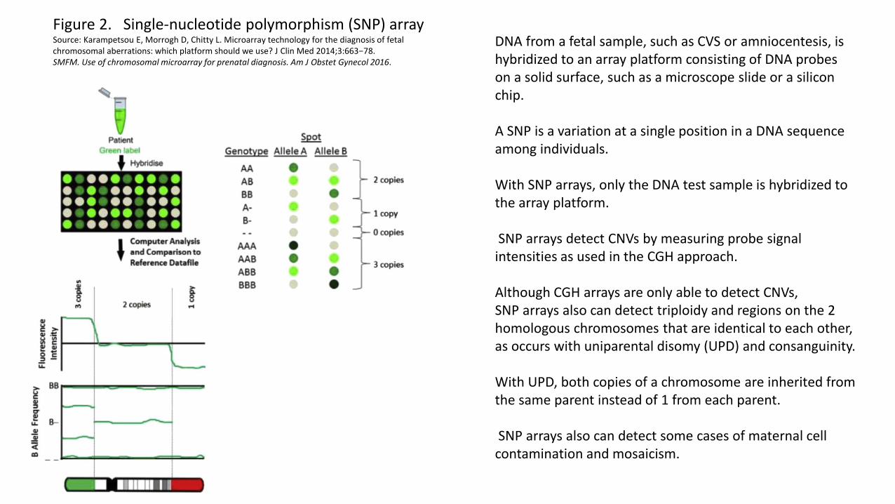
Figure 2. Single-nucleotide polymorphism (SNP) array Source: Karampetsou E, Morrogh D, Chitty L. Microarray technology for the diagnosis of fetal chromosomal aberrations: which platform should we use? J Clin Med 2014;3:663−78. SMFM. Use of chromosomal microarray for prenatal diagnosis. Am J Obstet Gynecol 2016.
DNA from a fetal sample, such as CVS or amniocentesis, is hybridized to an array platform consisting of DNA probes on a solid surface, such as a microscope slide or a silicon chip. A SNP is a variation at a single position in a DNA sequence among individuals. With SNP arrays, only the DNA test sample is hybridized to the array platform. SNP arrays detect CNVs by measuring probe signal intensities as used in the CGH approach. Although CGH arrays are only able to detect CNVs, SNP arrays also can detect triploidy and regions on the 2 homologous chromosomes that are identical to each other, as occurs with uniparental disomy (UPD) and consanguinity. With UPD, both copies of a chromosome are inherited from the same parent instead of 1 from each parent. SNP arrays also can detect some cases of maternal cell contamination and mosaicism.
The use of chromosomal microarray for prenatal diagnosis. Dugoff, Lorraine et al. American Journal of Obstetrics & Gynecology , Volume 215 , Issue 4 , B2 - B9
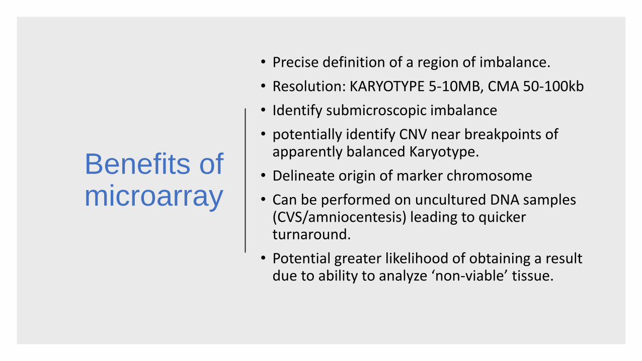
Benefits of microarray
• Precise definition of a region of imbalance.
• Resolution: KARYOTYPE 5-10MB, CMA 50-100kb
• Identify submicroscopic imbalance
• potentially identify CNV near breakpoints of apparently balanced Karyotype.
• Delineate origin of marker chromosome
• Can be performed on uncultured DNA samples (CVS/amniocentesis) leading to quicker turnaround.
• Potential greater likelihood of obtaining a result due to ability to analyze ‘non-viable’ tissue.
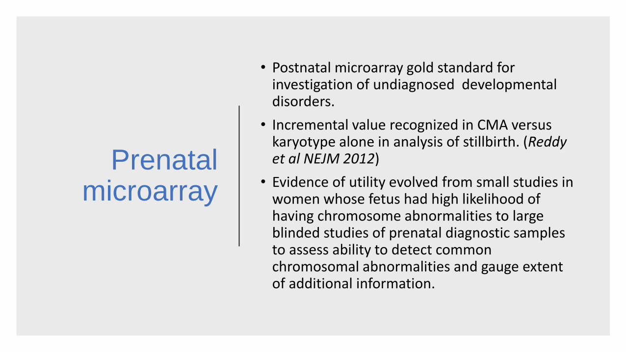
Prenatal microarray
• Postnatal microarray gold standard for investigation of undiagnosed developmental disorders.
• Incremental value recognized in CMA versus karyotype alone in analysis of stillbirth. (Reddy et al NEJM 2012)
• Evidence of utility evolved from small studies in women whose fetus had high likelihood of having chromosome abnormalities to large blinded studies of prenatal diagnostic samples to assess ability to detect common chromosomal abnormalities and gauge extent of additional information.
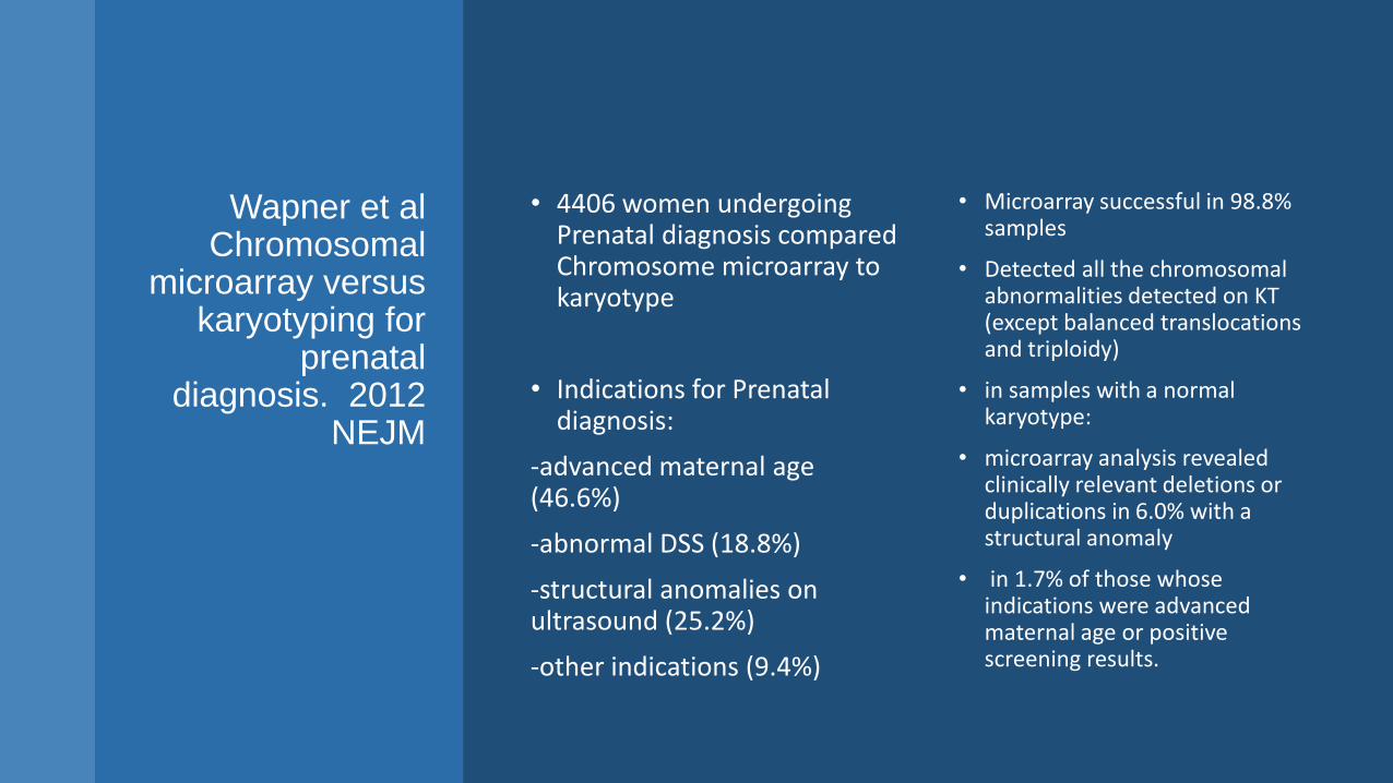
Wapner et al Chromosomal
microarray versus karyotyping for
prenatal diagnosis. 2012
NEJM
• 4406 women undergoing Prenatal diagnosis compared Chromosome microarray to karyotype
• Indications for Prenatal diagnosis:
-advanced maternal age (46.6%)
-abnormal DSS (18.8%)
-structural anomalies on ultrasound (25.2%)
-other indications (9.4%)
• Microarray successful in 98.8% samples
• Detected all the chromosomal abnormalities detected on KT (except balanced translocations and triploidy)
• in samples with a normal karyotype:
• microarray analysis revealed clinically relevant deletions or duplications in 6.0% with a structural anomaly
• in 1.7% of those whose indications were advanced maternal age or positive screening results.
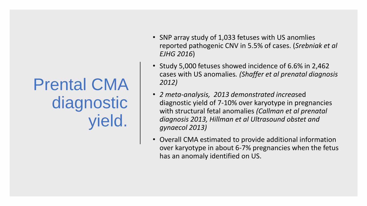
Prental CMA diagnostic
yield.
• SNP array study of 1,033 fetuses with US anomlies reported pathogenic CNV in 5.5% of cases. (Srebniak et al EJHG 2016)
• Study 5,000 fetuses showed incidence of 6.6% in 2,462 cases with US anomalies. (Shaffer et al prenatal diagnosis 2012)
• 2 meta-analysis, 2013 demonstrated increased diagnostic yield of 7-10% over karyotype in pregnancies with structural fetal anomalies (Callman et al prenatal diagnosis 2013, Hillman et al Ultrasound obstet and gynaecol 2013)
• Overall CMA estimated to provide additional information over karyotype in about 6-7% pregnancies when the fetus has an anomaly identified on US.
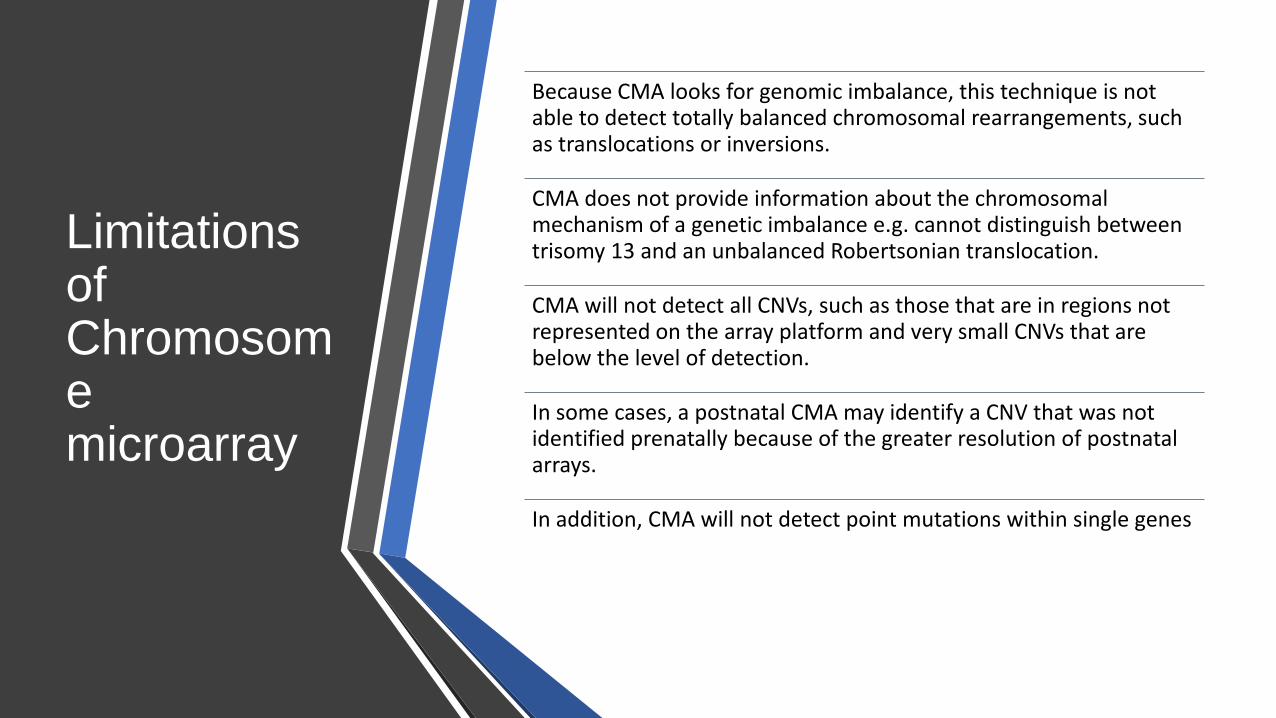
Limitations of Chromosome microarray
Because CMA looks for genomic imbalance, this technique is not able to detect totally balanced chromosomal rearrangements, such as translocations or inversions.
CMA does not provide information about the chromosomal mechanism of a genetic imbalance e.g. cannot distinguish between trisomy 13 and an unbalanced Robertsonian translocation.
CMA will not detect all CNVs, such as those that are in regions not represented on the array platform and very small CNVs that are below the level of detection.
In some cases, a postnatal CMA may identify a CNV that was not identified prenatally because of the greater resolution of postnatal arrays.
In addition, CMA will not detect point mutations within single genes
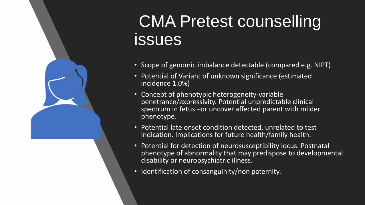
CMA Pretest counselling issues
• Scope of genomic imbalance detectable (compared e.g. NIPT)
• Potential of Variant of unknown significance (estimated incidence 1.0%)
• Concept of phenotypic heterogeneity-variable penetrance/expressivity. Potential unpredictable clinical spectrum in fetus –or uncover affected parent with milder phenotype.
• Potential late onset condition detected, unrelated to test indication. Implications for future health/family health.
• Potential for detection of neurosusceptibility locus. Postnatal phenotype of abnormality that may predispose to developmental disability or neuropsychiatric illness.
• Identification of consanguinity/non paternity.
Joint Committee on Genomics in Medicine. June 2015.
Recommendations for the use of chromosome microarray in pregnancy
Prenatal microarray
indications.
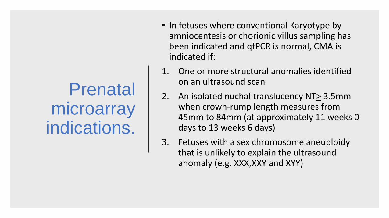
• In fetuses where conventional Karyotype by amniocentesis or chorionic villus sampling has been indicated and qfPCR is normal, CMA is indicated if:
1. One or more structural anomalies identified on an ultrasound scan
2. An isolated nuchal translucency NT> 3.5mm when crown-rump length measures from 45mm to 84mm (at approximately 11 weeks 0 days to 13 weeks 6 days)
3. Fetuses with a sex chromosome aneuploidy that is unlikely to explain the ultrasound anomaly (e.g. XXX,XXY and XYY)
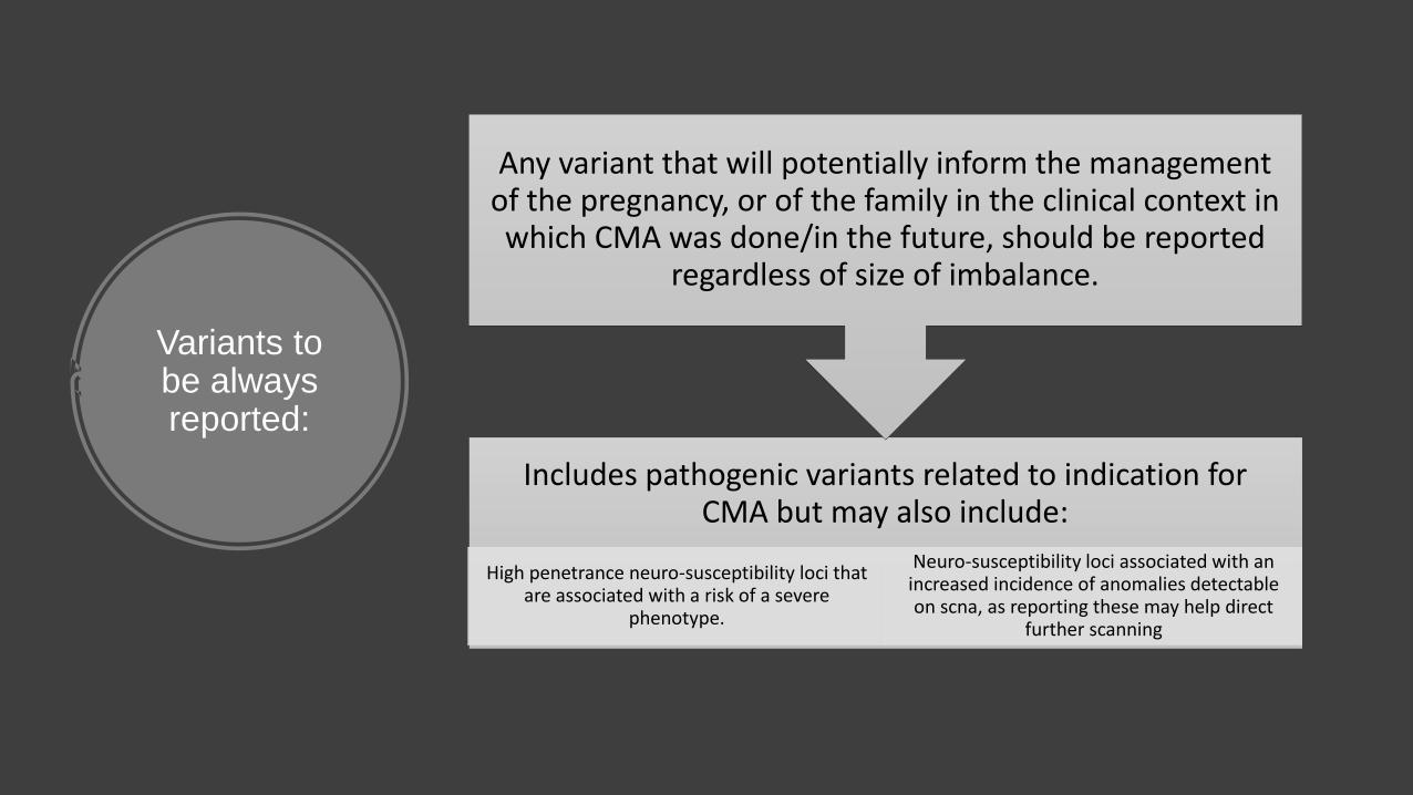
Variants to be always reported:
Includes pathogenic variants related to indication for
CMA but may also include:
High penetrance neuro-susceptibility loci that are associated with a risk of a severe
phenotype.
Neuro-susceptibility loci associated with an increased incidence of anomalies detectable on scna, as reporting these may help direct
further scanning
Any variant that will potentially inform the management of the pregnancy, or of the family in the clinical context in which CMA was done/in the future, should be reported
regardless of size of imbalance.
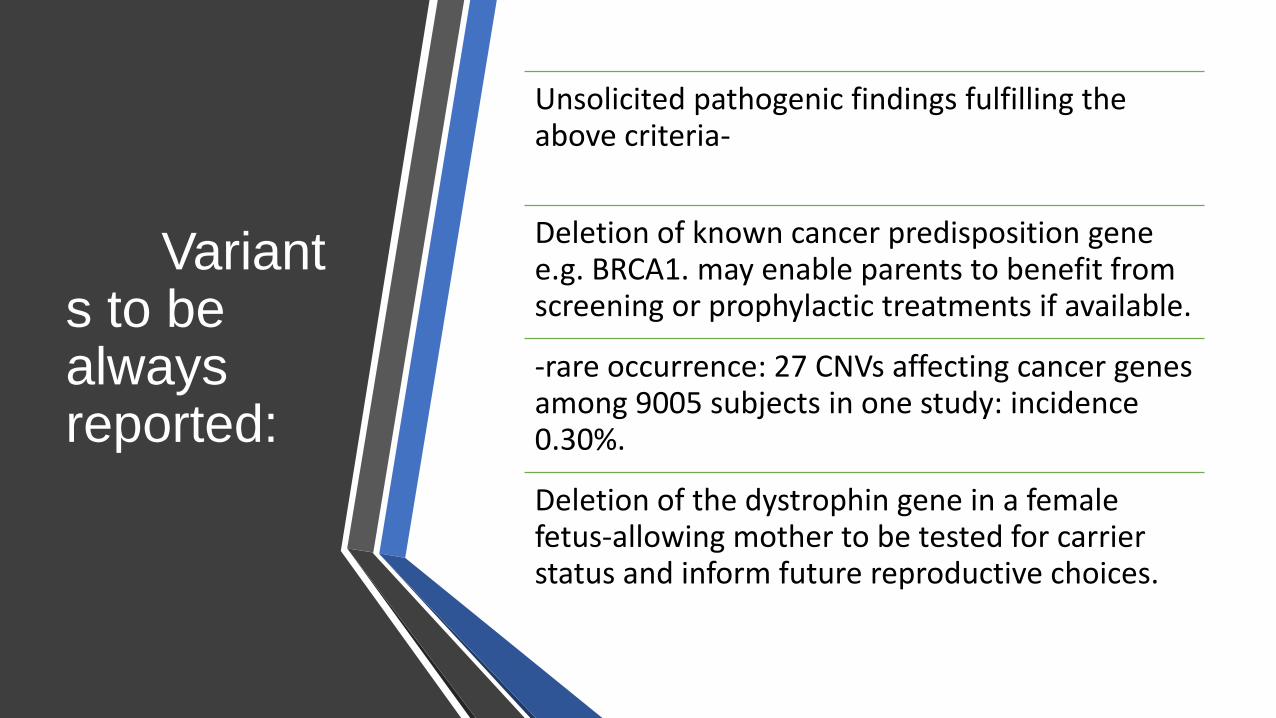
Variants to be always reported:
Unsolicited pathogenic findings fulfilling the above criteria-
Deletion of known cancer predisposition gene e.g. BRCA1. may enable parents to benefit from screening or prophylactic treatments if available.
-rare occurrence: 27 CNVs affecting cancer genes among 9005 subjects in one study: incidence 0.30%.
Deletion of the dystrophin gene in a female fetus-allowing mother to be tested for carrier status and inform future reproductive choices.
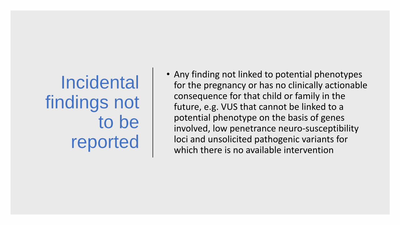
Incidental findings not
to be reported
• Any finding not linked to potential phenotypes for the pregnancy or has no clinically actionable consequence for that child or family in the future, e.g. VUS that cannot be linked to a potential phenotype on the basis of genes involved, low penetrance neuro-susceptibility loci and unsolicited pathogenic variants for which there is no available intervention
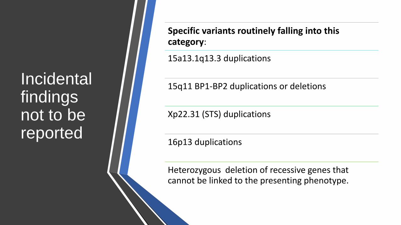
Incidental findings not to be reported
Specific variants routinely falling into this category:
15a13.1q13.3 duplications
15q11 BP1-BP2 duplications or deletions
Xp22.31 (STS) duplications
16p13 duplications
Heterozygous deletion of recessive genes that cannot be linked to the presenting phenotype.
UK Clinical Genetics services. 2018
Survey on Current Reporting Practices in Genetic Services for Prenatal Microarray
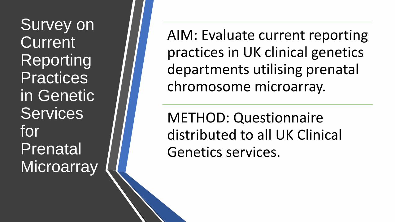
Survey on Current Reporting Practices in Genetic Services for Prenatal Microarray
AIM: Evaluate current reporting practices in UK clinical genetics departments utilising prenatal chromosome microarray.
METHOD: Questionnaire distributed to all UK Clinical Genetics services.
Survey 1.
1. Which Clinical Genetics Service are you reporting from?
ARE YOU OFFERING PRENATAL MICROARRAY TESTING-IF SO ON WHICH PATIENTS?
All CVS and amnios including those referred for a single gene test or raised SS only?
Only those with an anomaly on scan or a raised NT?
Another arrangement? Please describe.
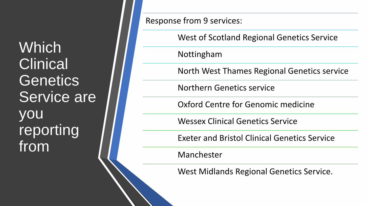
Which Clinical Genetics Service are you reporting from
Response from 9 services:
West of Scotland Regional Genetics Service
Nottingham
North West Thames Regional Genetics service
Northern Genetics service
Oxford Centre for Genomic medicine
Wessex Clinical Genetics Service
Exeter and Bristol Clinical Genetics Service
Manchester
West Midlands Regional Genetics Service.
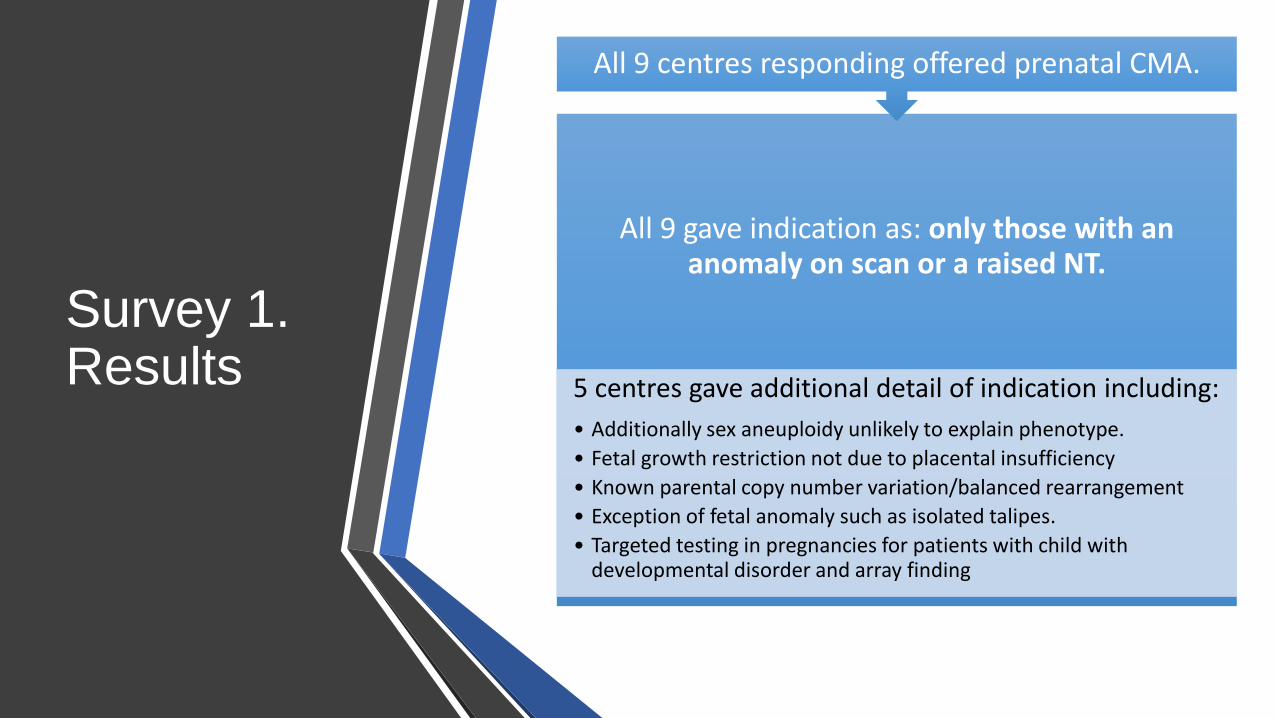
Survey 1. Results
All 9 gave indication as: only those with an anomaly on scan or a raised NT.
5 centres gave additional detail of indication including:
• Additionally sex aneuploidy unlikely to explain phenotype.
• Fetal growth restriction not due to placental insufficiency
• Known parental copy number variation/balanced rearrangement
• Exception of fetal anomaly such as isolated talipes.
• Targeted testing in pregnancies for patients with child with developmental disorder and array finding
All 9 centres responding offered prenatal CMA.
Survey 2.
2. If you offer testing, when did you start doing this testing?
How many tests have you done in total?
How many samples do you analyse on average over one year- 01/04 to 31/03?
How many pathogenic CNVs (pCNV) have you reported in total?
How many pCNVs have you reported on average in one year 01/04-31/03?
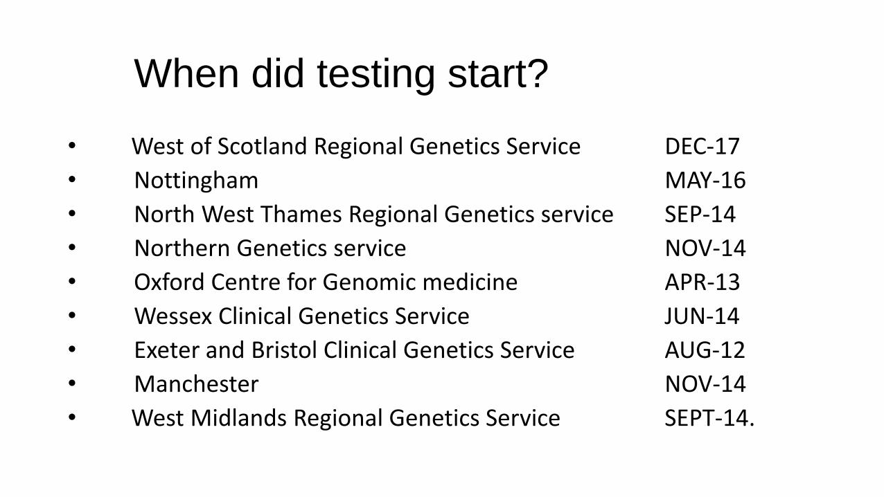
When did testing start?
• West of Scotland Regional Genetics Service DEC-17
• Nottingham MAY-16
• North West Thames Regional Genetics service SEP-14
• Northern Genetics service NOV-14
• Oxford Centre for Genomic medicine APR-13
• Wessex Clinical Genetics Service JUN-14
• Exeter and Bristol Clinical Genetics Service AUG-12
• Manchester NOV-14
• West Midlands Regional Genetics Service SEPT-14.
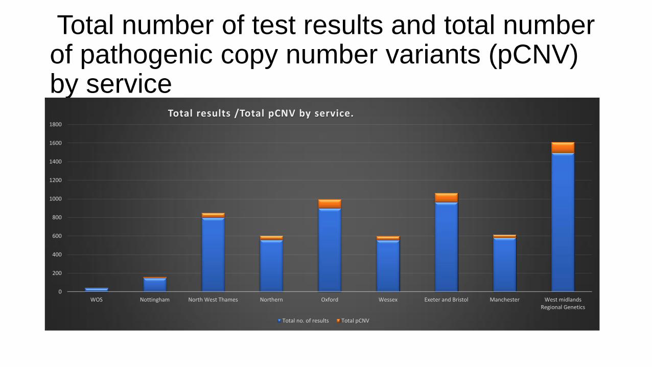
Total number of test results and total number of pathogenic copy number variants (pCNV) by service
0
200
400
600
800
1000
1200
1400
1600
1800
WOS Nottingham North West Thames Northern Oxford Wessex Exeter and Bristol Manchester West midlandsRegional Genetics
Total results /Total pCNV by service.
Total no. of results Total pCNV
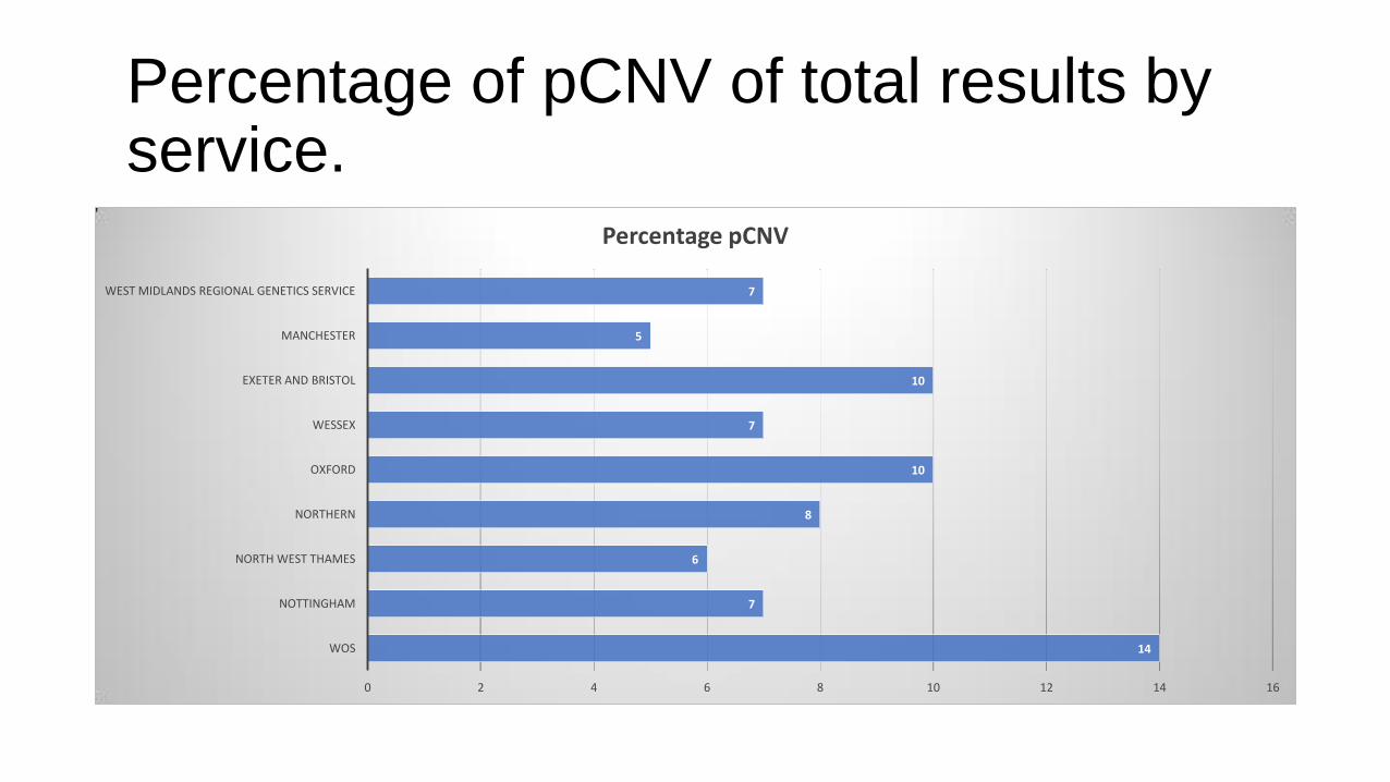
Percentage of pCNV of total results by service.
14
7
6
8
10
7
10
5
7
0 2 4 6 8 10 12 14 16
WOS
NOTTINGHAM
NORTH WEST THAMES
NORTHERN
OXFORD
WESSEX
EXETER AND BRISTOL
MANCHESTER
WEST MIDLANDS REGIONAL GENETICS SERVICE
Percentage pCNV
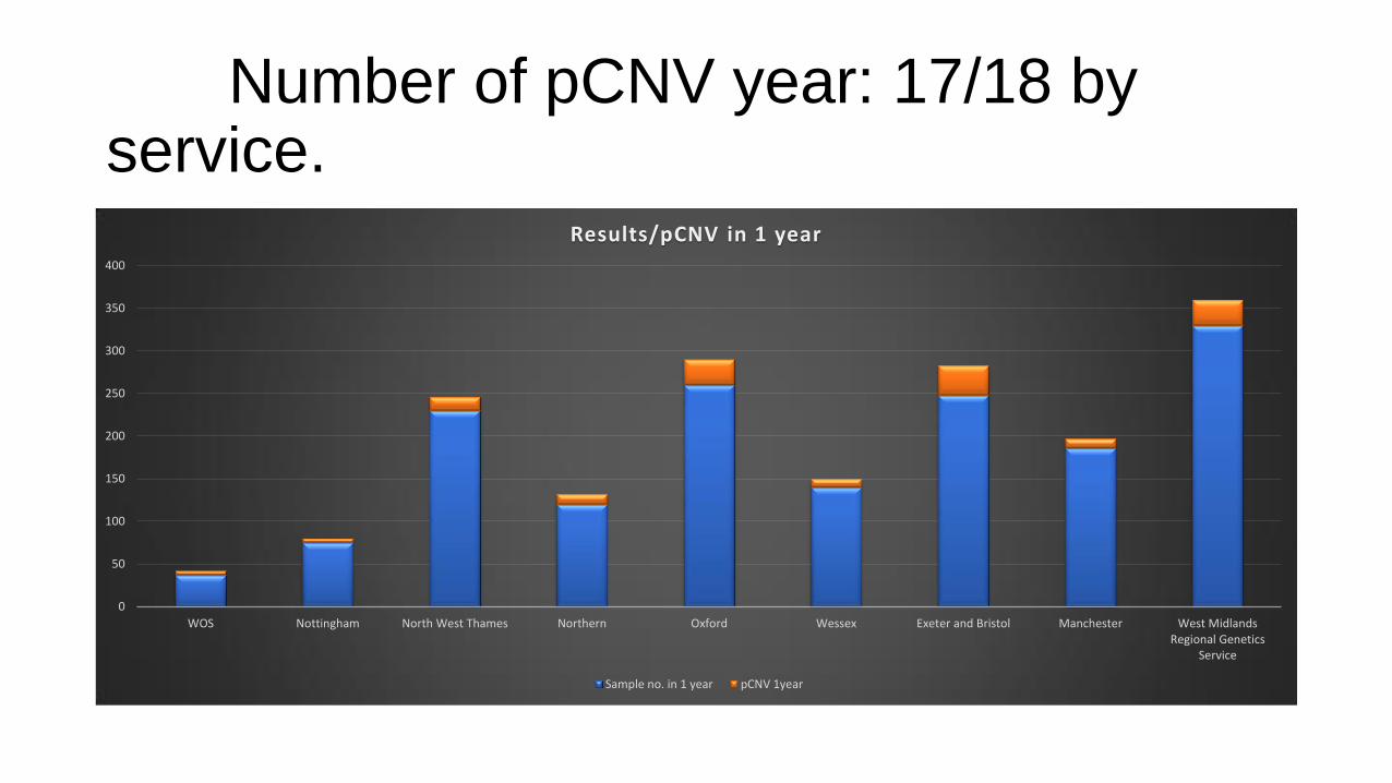
Number of pCNV year: 17/18 by service.
0
50
100
150
200
250
300
350
400
WOS Nottingham North West Thames Northern Oxford Wessex Exeter and Bristol Manchester West MidlandsRegional Genetics
Service
Results/pCNV in 1 year
Sample no. in 1 year pCNV 1year
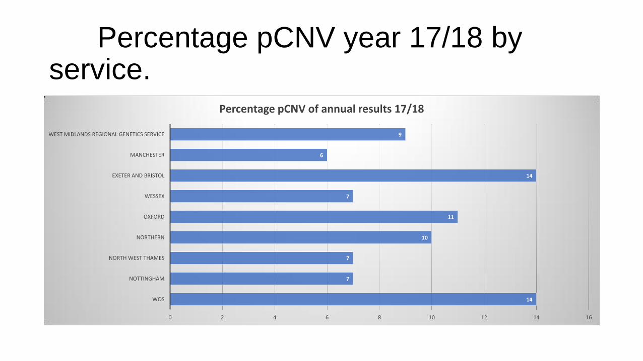
Percentage pCNV year 17/18 by service.
14
7
7
10
11
7
14
6
9
0 2 4 6 8 10 12 14 16
WOS
NOTTINGHAM
NORTH WEST THAMES
NORTHERN
OXFORD
WESSEX
EXETER AND BRISTOL
MANCHESTER
WEST MIDLANDS REGIONAL GENETICS SERVICE
Percentage pCNV of annual results 17/18
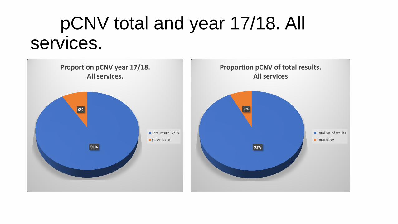
pCNV total and year 17/18. All services.
91%
9%
Proportion pCNV year 17/18. All services.
Total result 17/18
pCNV 17/18
93%
7%
Proportion pCNV of total results. All services
Total No. of results
Total pCNV
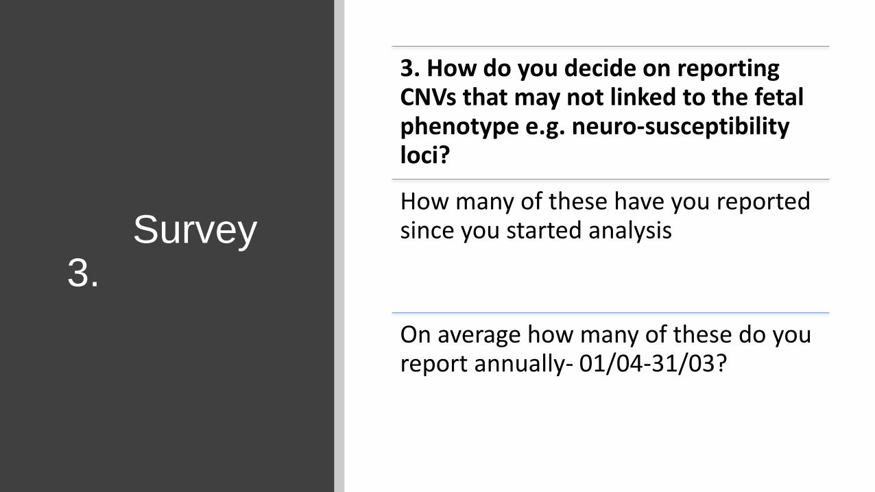
Survey 3.
3. How do you decide on reporting CNVs that may not linked to the fetal phenotype e.g. neuro-susceptibility loci?
How many of these have you reported since you started analysis
On average how many of these do you report annually- 01/04-31/03?
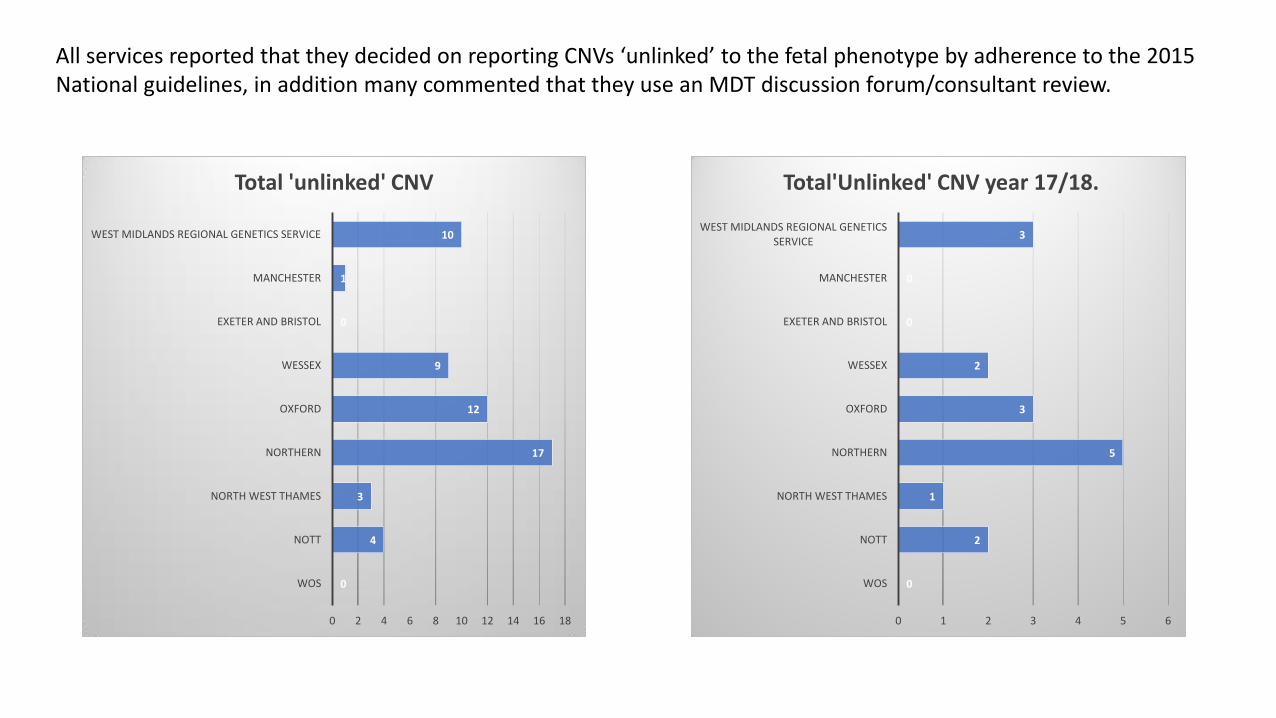
All services reported that they decided on reporting CNVs ‘unlinked’ to the fetal phenotype by adherence to the 2015 National guidelines, in addition many commented that they use an MDT discussion forum/consultant review.
0
4
3
17
12
9
0
1
10
0 2 4 6 8 10 12 14 16 18
WOS
NOTT
NORTH WEST THAMES
NORTHERN
OXFORD
WESSEX
EXETER AND BRISTOL
MANCHESTER
WEST MIDLANDS REGIONAL GENETICS SERVICE
Total 'unlinked' CNV
0
2
1
5
3
2
0
0
3
0 1 2 3 4 5 6
WOS
NOTT
NORTH WEST THAMES
NORTHERN
OXFORD
WESSEX
EXETER AND BRISTOL
MANCHESTER
WEST MIDLANDS REGIONAL GENETICS SERVICE
Total'Unlinked' CNV year 17/18.
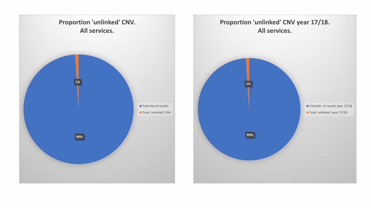
99%
1%
Proportion 'unlinked' CNV. All services.
Total No of results
Total 'unlinked' CNV
99%
1%
Proportion 'unlinked' CNV year 17/18. All services.
Total No. of results year 17/18
Total 'unlinked' year 17/18
Survey 4.
4. How many actionable incidental findings have you reported since you started analysis? e.g. BRCA1 deletions
How many have you reported on average annually 01/04-31/03
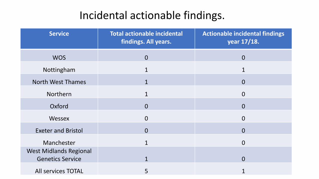
Service Total actionable incidental findings. All years.
Actionable incidental findings year 17/18.
WOS 0 0
Nottingham 1 1
North West Thames 1 0
Northern 1 0
Oxford 0 0
Wessex 0 0
Exeter and Bristol 0 0
Manchester 1 0 West Midlands Regional
Genetics Service 1 0
All services TOTAL 5 1
Incidental actionable findings.

Survey 5.
5.What do you do about reporting results where the significance is uncertain?
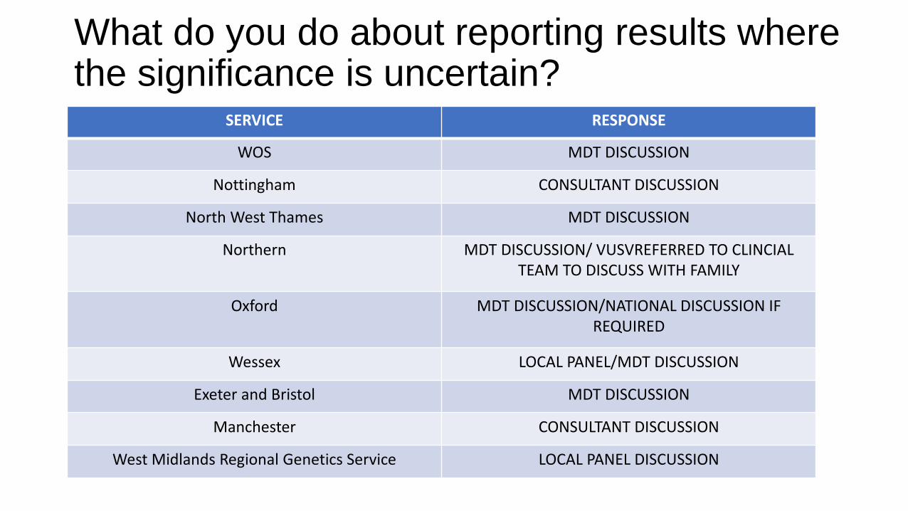
What do you do about reporting results where the significance is uncertain? SERVICE RESPONSE
WOS MDT DISCUSSION
Nottingham CONSULTANT DISCUSSION
North West Thames MDT DISCUSSION
Northern MDT DISCUSSION/ VUSVREFERRED TO CLINCIAL TEAM TO DISCUSS WITH FAMILY
Oxford MDT DISCUSSION/NATIONAL DISCUSSION IF REQUIRED
Wessex LOCAL PANEL/MDT DISCUSSION
Exeter and Bristol MDT DISCUSSION
Manchester CONSULTANT DISCUSSION
West Midlands Regional Genetics Service LOCAL PANEL DISCUSSION
Survey 6.
6. Do you have a local group that reviews these results?
If so, what health professionals are involved with this group?
Do you record discussions around these results?
If you record them, where do your record them
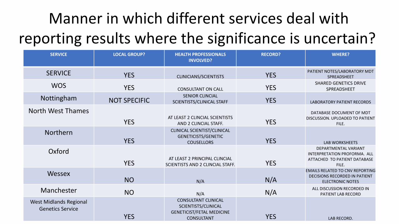
SERVICE LOCAL GROUP? HEALTH PROFESSIONALS INVOLVED?
RECORD? WHERE?
SERVICE YES CLINICIANS/SCIENTISTS YES PATIENT NOTES/LABORATORY MDT
SPREADSHEET
WOS YES CONSULTANT ON CALL YES SHARED GENETICS DRIVE
SPREADSHEET
Nottingham NOT SPECIFIC SENIOR CLINCIAL
SCIENTISTS/CLINICAL STAFF YES LABORATORY PATIENT RECORDS
North West Thames
YES AT LEAST 2 CLINCIAL SCIENTISTS
AND 2 CLINCIAL STAFF. YES
DATABASE DOCUMENT OF MDT DISCUSSION. UPLOADED TO PATIENT
FILE.
Northern YES
CLINICAL SCIENTIST/CLINICAL GENETICISTS/GENETIC
COUSELLORS YES LAB WORKSHEETS
Oxford
YES AT LEAST 2 PRINCIPAL CLINCIAL
SCIENTISTS AND 2 CLINCIAL STAFF. YES
DEPARTMENTAL VARIANT INTERPRETATION PROFORMA. ALL ATTACHED TO PATIENT DATABASE
FILE.
Wessex NO N/A N/A
EMAILS RELATED TO CNV REPORTING DECISIONS RECORDED IN PATIENT
ELECTRONIC NOTES
Manchester NO N/A N/A ALL DISCUSSION RECORDED IN
PATIENT LAB RECORD
West Midlands Regional Genetics Service
YES
CONSULTANT CLINICAL SCIENTISTS/CLINICAL
GENETICIST/FETAL MEDICINE CONSULTANT YES LAB RECORD.
Manner in which different services deal with reporting results where the significance is uncertain?
Conclusion
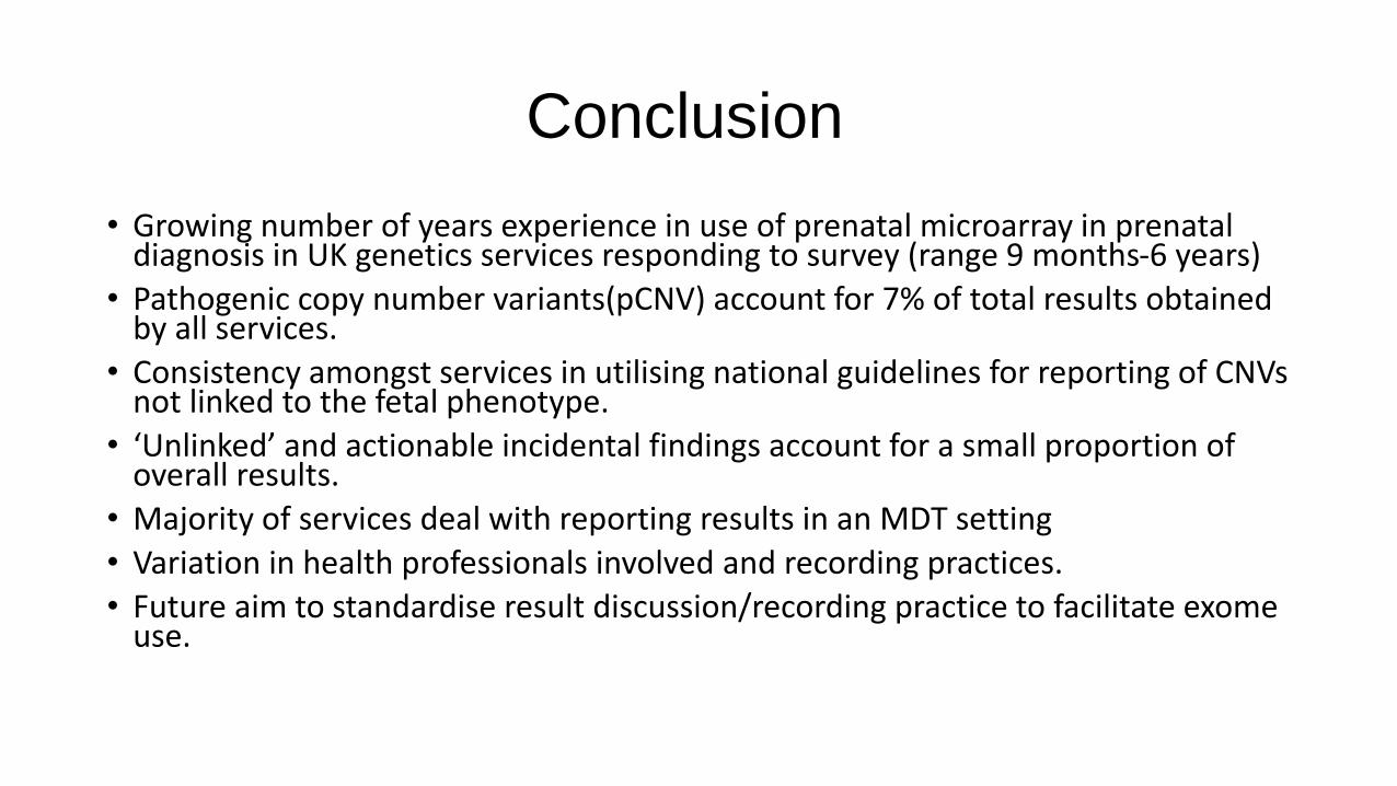
• Growing number of years experience in use of prenatal microarray in prenatal diagnosis in UK genetics services responding to survey (range 9 months-6 years)
• Pathogenic copy number variants(pCNV) account for 7% of total results obtained by all services.
• Consistency amongst services in utilising national guidelines for reporting of CNVs not linked to the fetal phenotype.
• ‘Unlinked’ and actionable incidental findings account for a small proportion of overall results.
• Majority of services deal with reporting results in an MDT setting
• Variation in health professionals involved and recording practices.
• Future aim to standardise result discussion/recording practice to facilitate exome use.