prenatal diagnosis
DESCRIPTION
shareTRANSCRIPT
Prenatal DiagnosisPrenatal Diagnosis
ObjectivesObjectives
Managing the remaining weeks of the pregnancy Managing the remaining weeks of the pregnancy Determining the outcome of the pregnancy Determining the outcome of the pregnancy Planning for possible complications with the birth Planning for possible complications with the birth
process process Planning for problems that may occur in the Planning for problems that may occur in the
newborn infant newborn infant Deciding whether to continue the pregnancy Deciding whether to continue the pregnancy Finding conditions that may affect future Finding conditions that may affect future
pregnancy in order to provide reassurance and pregnancy in order to provide reassurance and reduce anxiety esp among high risk groupsreduce anxiety esp among high risk groups
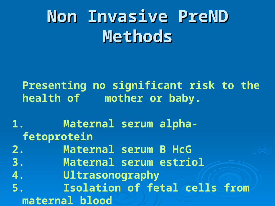
Non Invasive PreND MethodsNon Invasive PreND Methods
Presenting no significant risk to the health of mother or baby.
1. Maternal serum alpha-fetoprotein2. Maternal serum B HcG3. Maternal serum estriol4. Ultrasonography5. Isolation of fetal cells from maternal blood
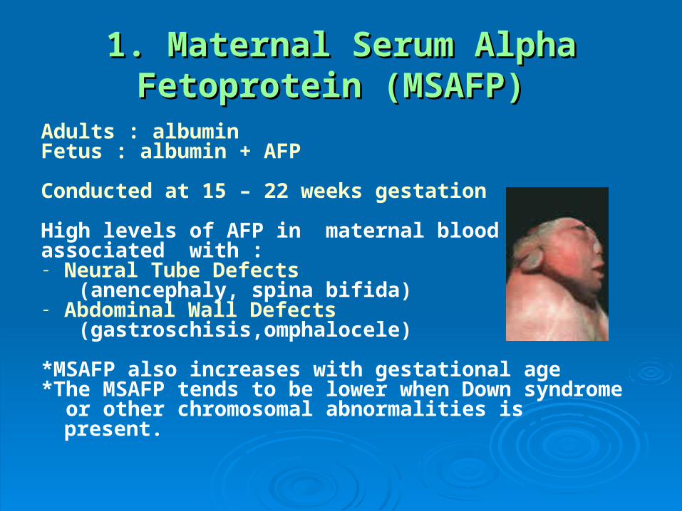
1. Maternal Serum Alpha Fetoprotein 1. Maternal Serum Alpha Fetoprotein (MSAFP) (MSAFP)
Adults : albuminFetus : albumin + AFP
Conducted at 15 – 22 weeks gestation
High levels of AFP in maternal blood are associated with :- Neural Tube Defects (anencephaly, spina bifida)- Abdominal Wall Defects (gastroschisis,omphalocele)
*MSAFP also increases with gestational age *The MSAFP tends to be lower when Down syndrome or other chromosomal abnormalities is present.
2. Maternal Serum B HcG2. Maternal Serum B HcG
11stst trimester when threatened abortion or ectopic trimester when threatened abortion or ectopic pregnancy is suspected, the amount of beta-HCG will be pregnancy is suspected, the amount of beta-HCG will be lower than expected lower than expected
22ndnd trimester, the beta-HCG can be used in conjunction trimester, the beta-HCG can be used in conjunction with the MSAFP to screen for chromosomal with the MSAFP to screen for chromosomal abnormalities, and Down syndrome in particular. abnormalities, and Down syndrome in particular.
An elevated beta-HCG coupled with a decreased An elevated beta-HCG coupled with a decreased MSAFP suggests Down syndrome MSAFP suggests Down syndrome
Very high levels of HCG suggest trophoblastic disease Very high levels of HCG suggest trophoblastic disease (molar pregnancy). (molar pregnancy).
The absence of a fetus on USG along with an elevated The absence of a fetus on USG along with an elevated HCG suggests a hydatidiform mole. HCG suggests a hydatidiform mole.
3. Maternal Serum Estriol3. Maternal Serum Estriol
If the estriol level drops, the fetus is If the estriol level drops, the fetus is threatened and delivery may be necessary threatened and delivery may be necessary emergently emergently
Estriol tends to be lower when Down Estriol tends to be lower when Down syndrome is present and when there is syndrome is present and when there is adrenal hypoplasia with anencephaly. adrenal hypoplasia with anencephaly.
Made by fetal adrenal glands , excreted by maternal kidney/liver
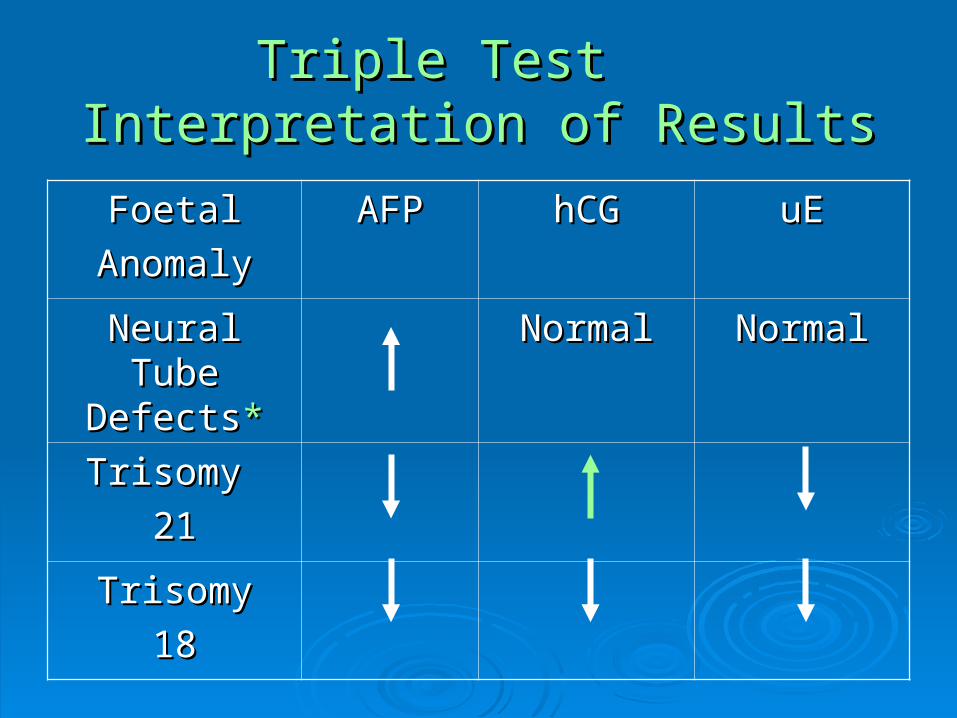
Triple Test Triple Test Interpretation of ResultsInterpretation of Results
FoetalFoetal
AnomalyAnomaly
AFPAFP hCGhCG uEuE
Neural Tube Neural Tube DefectsDefects**
NormalNormal NormalNormal
Trisomy Trisomy
2121
TrisomyTrisomy
1818
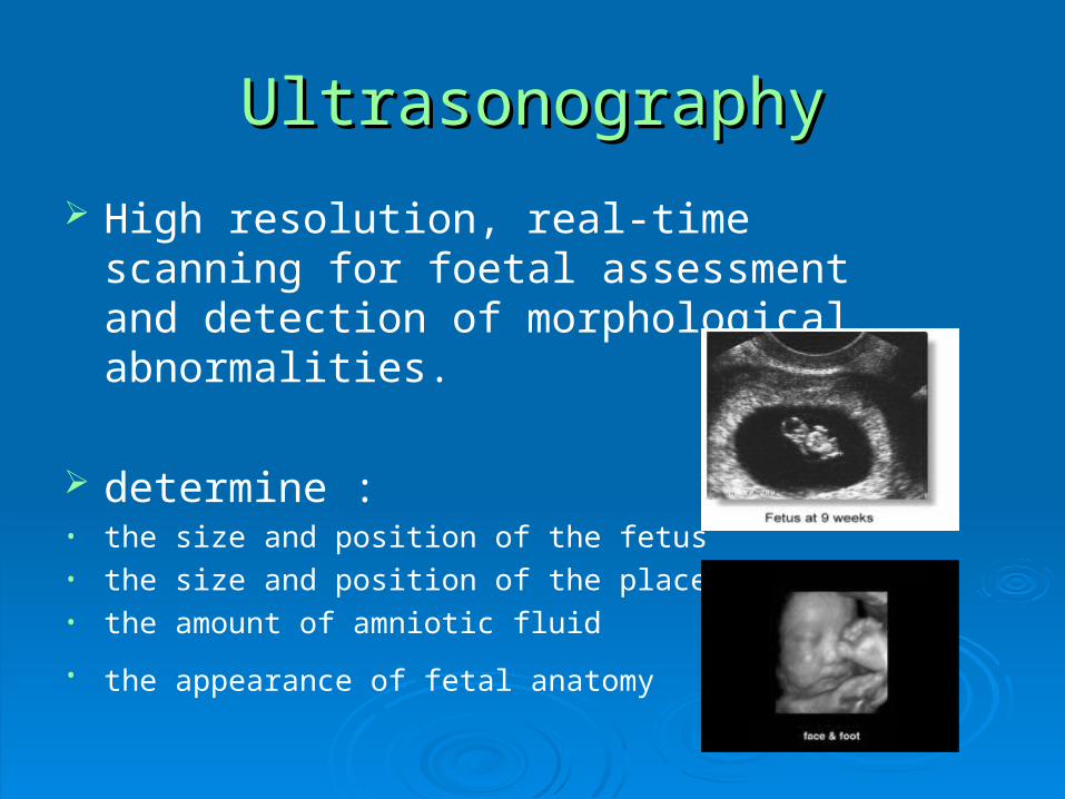
UltrasonographyUltrasonography
High resolution, real-time scanning for foetal assessment and detection of morphological abnormalities.
determine :• the size and position of the fetus• the size and position of the placenta• the amount of amniotic fluid
• the appearance of fetal anatomy
5. Isolation of foetal cells from maternal blood
Foetal cells are found in the maternal circulation as well as cell-free DNA.
Difficult to get many fetal blood cells Difficult to get many fetal blood cells Not a frequently used detection method
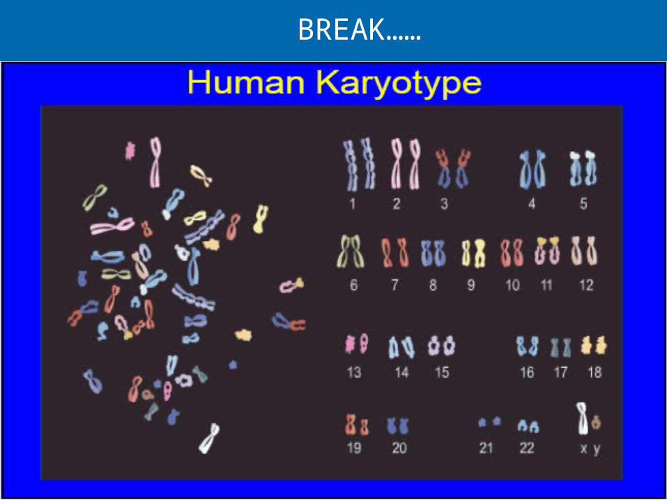
BREAK……
Invasive PreNDInvasive PreND
Routine ultrasound suggests abnormality.Routine ultrasound suggests abnormality.Advanced maternal age, risk of trisomy 21 & Advanced maternal age, risk of trisomy 21 & trisomy 18.trisomy 18.Previous affected child with chromosomal Previous affected child with chromosomal abnormality.abnormality.Structural chromosome abnormality in either Structural chromosome abnormality in either parent.parent.Family history of genetic disorder where DNA or Family history of genetic disorder where DNA or biochemical test is available.biochemical test is available.Family history of an X linked disorder (with no Family history of an X linked disorder (with no specific prenatal test).specific prenatal test).
Associated with RISK to fetus
Invasive PreNDInvasive PreND
1.1. Amniocentesis (0.5-1.0% incr. risk of Amniocentesis (0.5-1.0% incr. risk of pregnancy loss)pregnancy loss)
2.2. Chorionic villus samplingChorionic villus sampling
3.3. Cordocentesis - direct sampling of foetal Cordocentesis - direct sampling of foetal blood from cordblood from cord
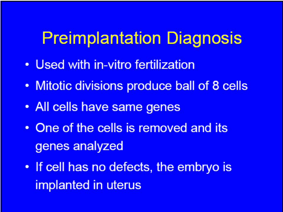
4.4. Preimplantation genetic diagnosis - IVFPreimplantation genetic diagnosis - IVF
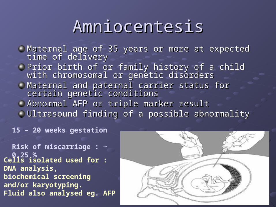
AmniocentesisAmniocentesisMaternal age of 35 years or more at expected time of deliveryMaternal age of 35 years or more at expected time of deliveryPrior birth of or family history of a child with chromosomal or Prior birth of or family history of a child with chromosomal or genetic disorders genetic disorders Maternal and paternal carrier status for certain genetic Maternal and paternal carrier status for certain genetic conditions conditions Abnormal AFP or triple marker result Abnormal AFP or triple marker result Ultrasound finding of a possible abnormality Ultrasound finding of a possible abnormality
15 – 20 weeks gestation
Risk of miscarriage : ~ 0.25 %
Cells isolated used for : DNA analysis, biochemical screening and/or karyotyping. Fluid also analysed eg. AFP
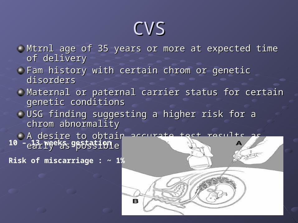
CVSCVSMtrnl age of 35 years or more at expected time of delivery Mtrnl age of 35 years or more at expected time of delivery Fam history with certain chrom or genetic disorders Fam history with certain chrom or genetic disorders Maternal or paternal carrier status for certain genetic conditions Maternal or paternal carrier status for certain genetic conditions USG finding suggesting a higher risk for a chrom abnormality USG finding suggesting a higher risk for a chrom abnormality A desire to obtain accurate test results as early as possible in A desire to obtain accurate test results as early as possible in pregnancy pregnancy
10 – 13 weeks gestation
Risk of miscarriage : ~ 1%
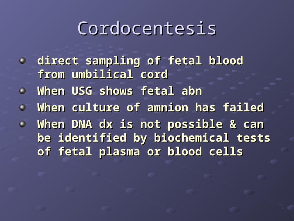
CordocentesisCordocentesis
direct sampling of fetal blood from direct sampling of fetal blood from umbilical cordumbilical cord
When USG shows fetal abnWhen USG shows fetal abn
When culture of amnion has failedWhen culture of amnion has failed
When DNA dx is not possible & can be When DNA dx is not possible & can be identified by biochemical tests of fetal identified by biochemical tests of fetal plasma or blood cellsplasma or blood cells


Thank You !!Thank You !!