prenatal diagnosis of - achondroplasia - cpa chennai · prenatal diagnosis of - achondroplasia dr....
TRANSCRIPT
Prenatal Diagnosis of -Achondroplasia
Dr. Subapriya Kandasamy, MS (O&G)
Fellow in fetal medicine
Ms. Gayathri J
Fellow in genetics
Dr. Sujatha Jagadeesh
Dysmorphologist and consultant geneticist
Achondroplasia
• Primary defect is abnormal endochondral ossification
• Most common nonlethal skeletal dysplasia
• Incidence - 1:10,000- 1:50,000
Case
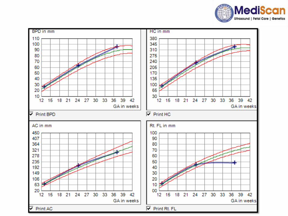
• Mrs X, primi gravida at 37 weeks of gestation was referred to MediScan for 2nd opinion in view of short long bones.
• Non consanguineous marriage. No high risk factors
• No significant findings in family pedigree (no short statureness)
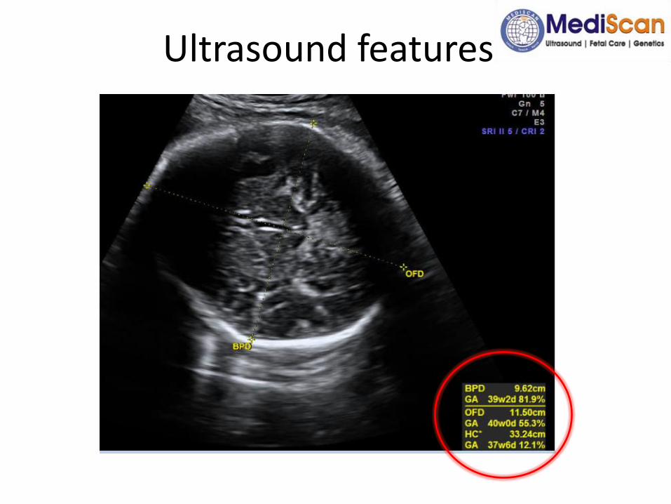
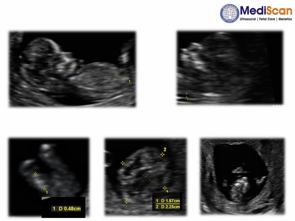
Ultrasound features
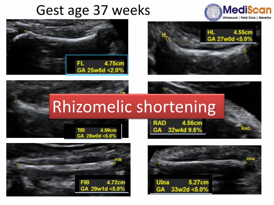
Gest age 37 weeks
Rhizomelic shortening
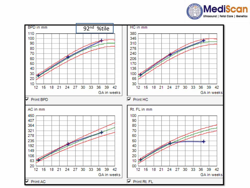
292nd %tile
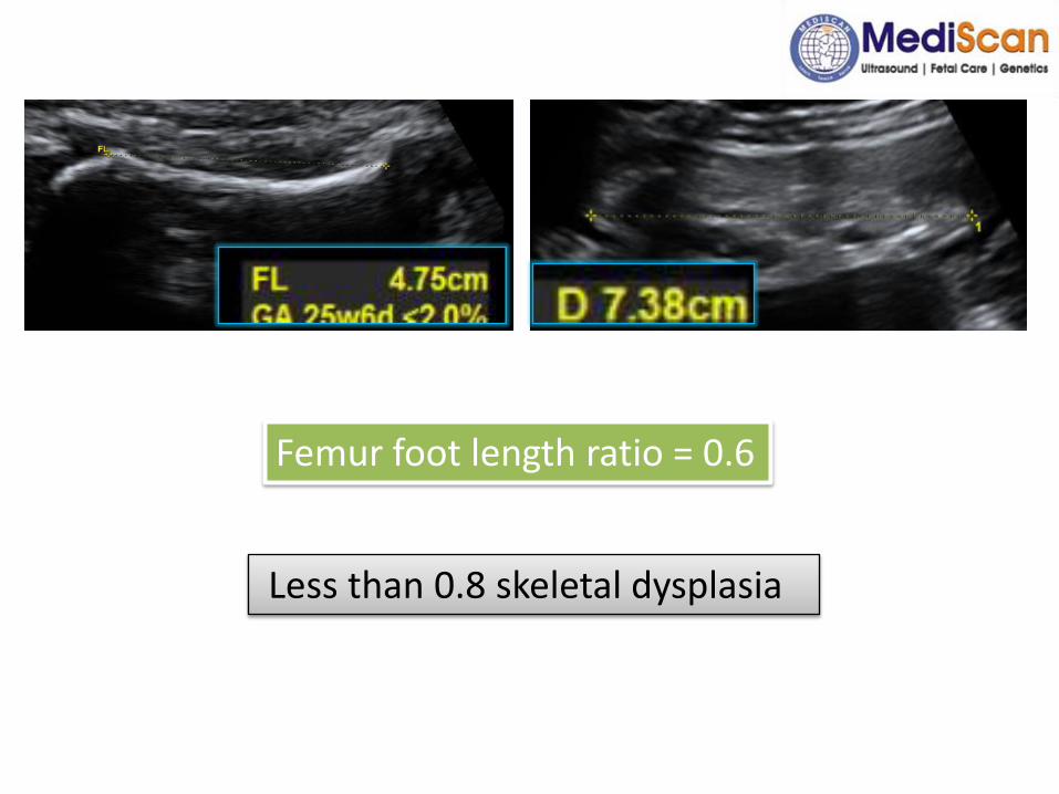
Femur foot length ratio = 0.6
Less than 0.8 skeletal dysplasia
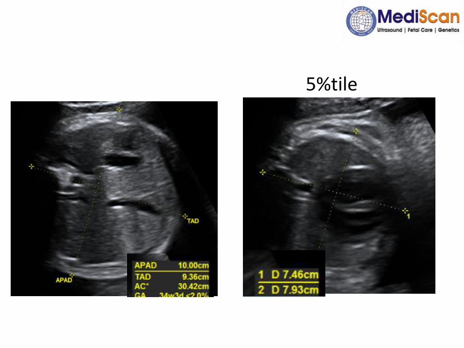
5%tile
Ultrasound features
Her ultrasound corresponds to 37 weeks with following features:
– Hydramnios
– Femur length falls less than -4SD for 37 wks.
– All long bones less than 5th %tile for 37 wks.
• Suggestive of skeletal dysplasia
Suggested: postnatal evaluation at genetic clinic
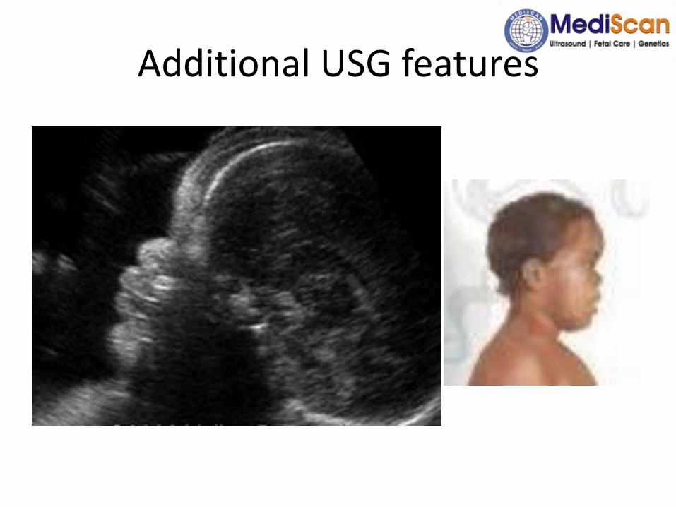
Additional USG features
Postnatal evaluation
1.History : Family pedigree, obstetric history, h/o consanguinity
2.Clinical examination:
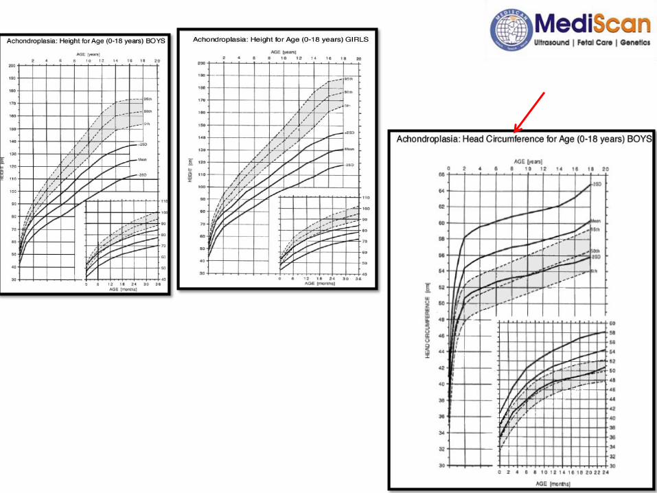
– To recognize short stature – by growth charts
– Determine site of disproportion –
• (a) Measuring body proportions i.e. upper to lower segment ratio (1.7 at birth, 1.3 at 3 yrs, 1.1 at 6yrs, 1 at 10 yrs).
• (b) Arm span – less than ht by 2.5cm at birth, is equal to ht at 11yrs, more than ht later on
• (c) Upper limbs extending upto midthigh - normal
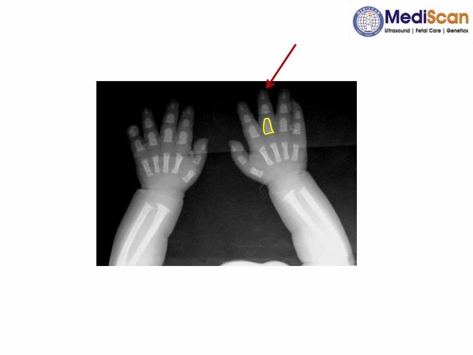
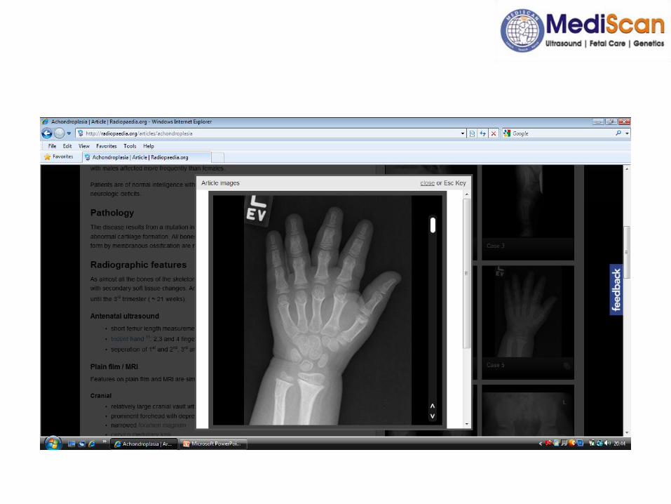
3. Identify dysmorphism: macrocephaly, prominent forehead, flat nasal bridge, trident hand
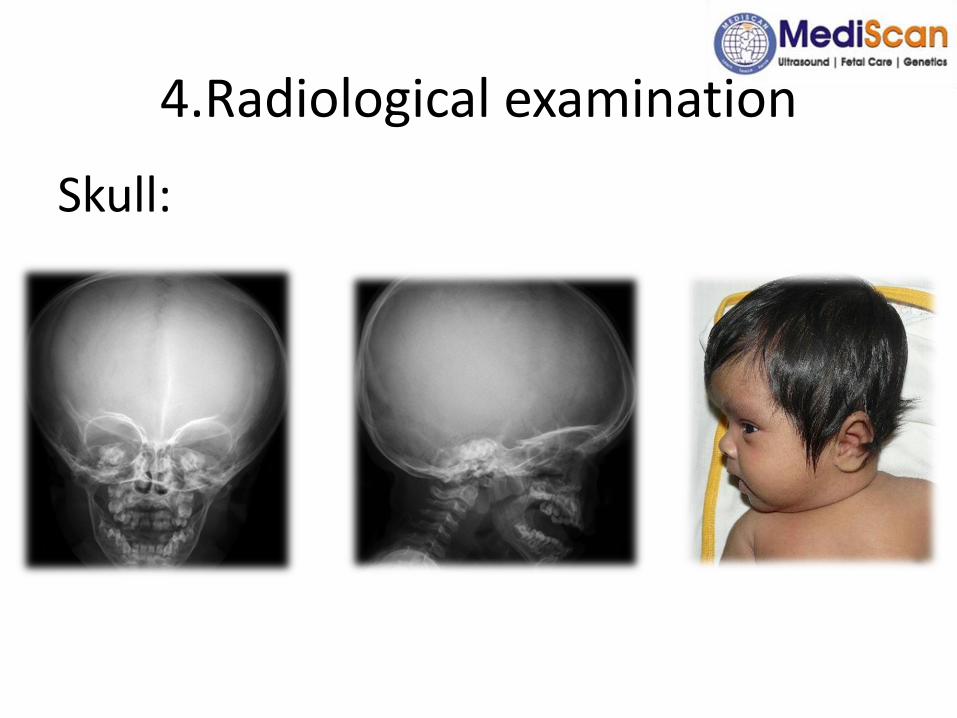
4.Radiological examination
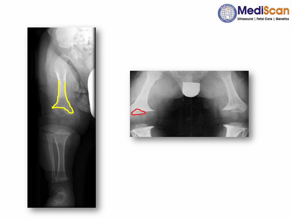
Skull:
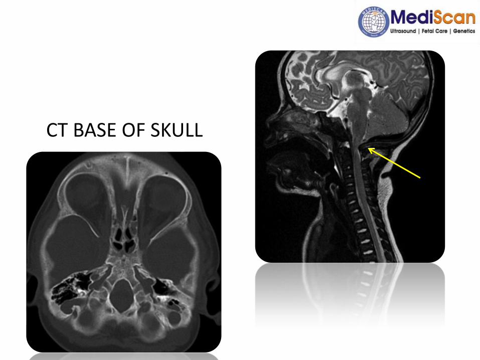
CT BASE OF SKULL
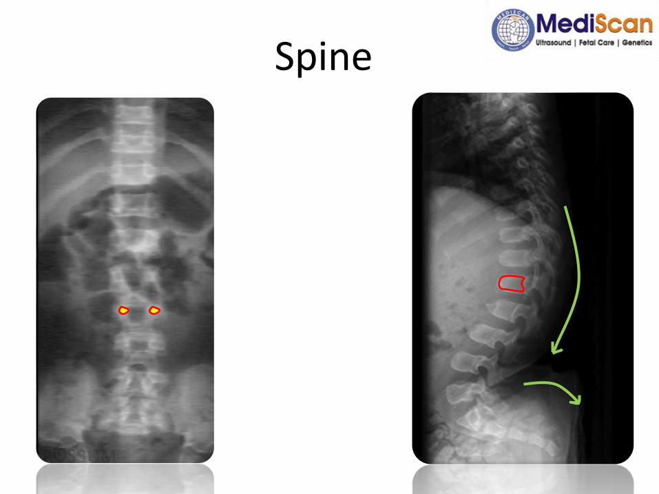
Spine
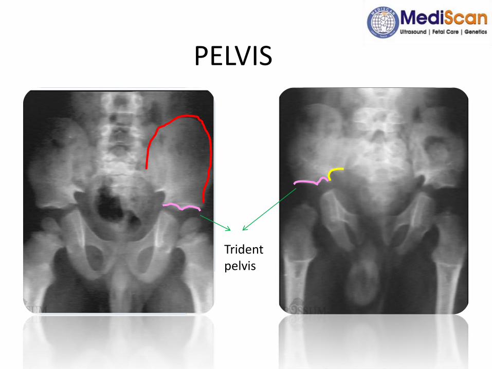
PELVIS
Trident pelvis
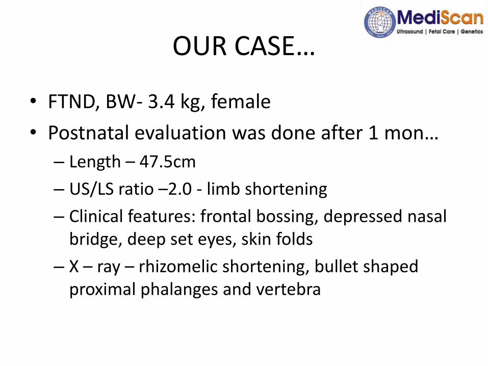
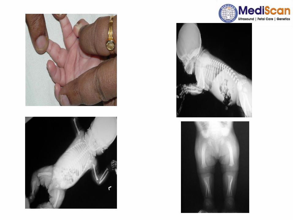
OUR CASE…
• FTND, BW- 3.4 kg, female
• Postnatal evaluation was done after 1 mon…
– Length – 47.5cm
– US/LS ratio –2.0 - limb shortening
– Clinical features: frontal bossing, depressed nasal bridge, deep set eyes, skin folds
– X – ray – rhizomelic shortening, bullet shaped proximal phalanges and vertebra
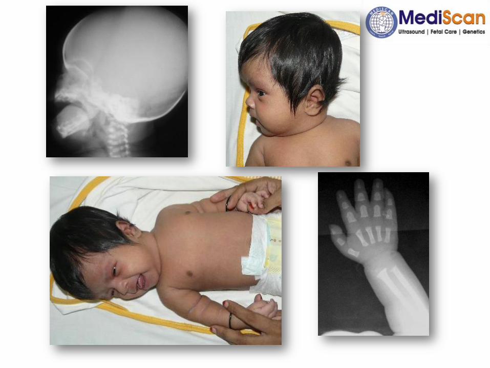
• The clinical features were suggestive of achondroplasia
• Major difficulty in antenatal diagnosis of achondroplasia - not recognized in most cases until 3rd trimester.
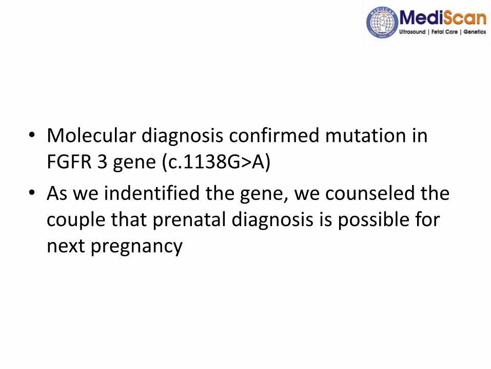
• Molecular diagnosis confirmed mutation in FGFR 3 gene (c.1138G>A)
• As we indentified the gene, we counseled the couple that prenatal diagnosis is possible for next pregnancy
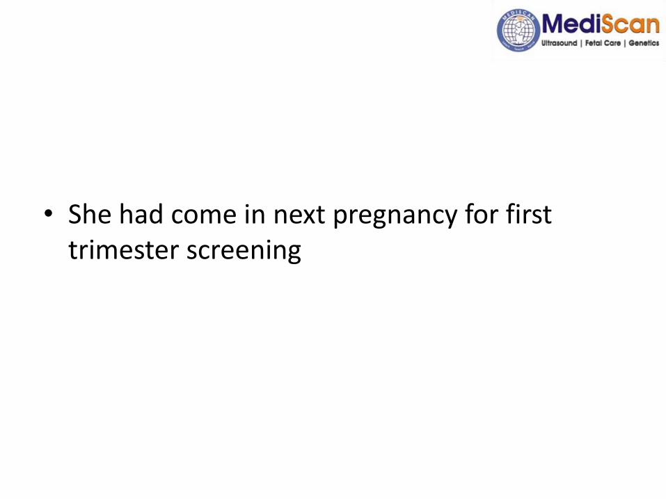
• She had come in next pregnancy for first trimester screening
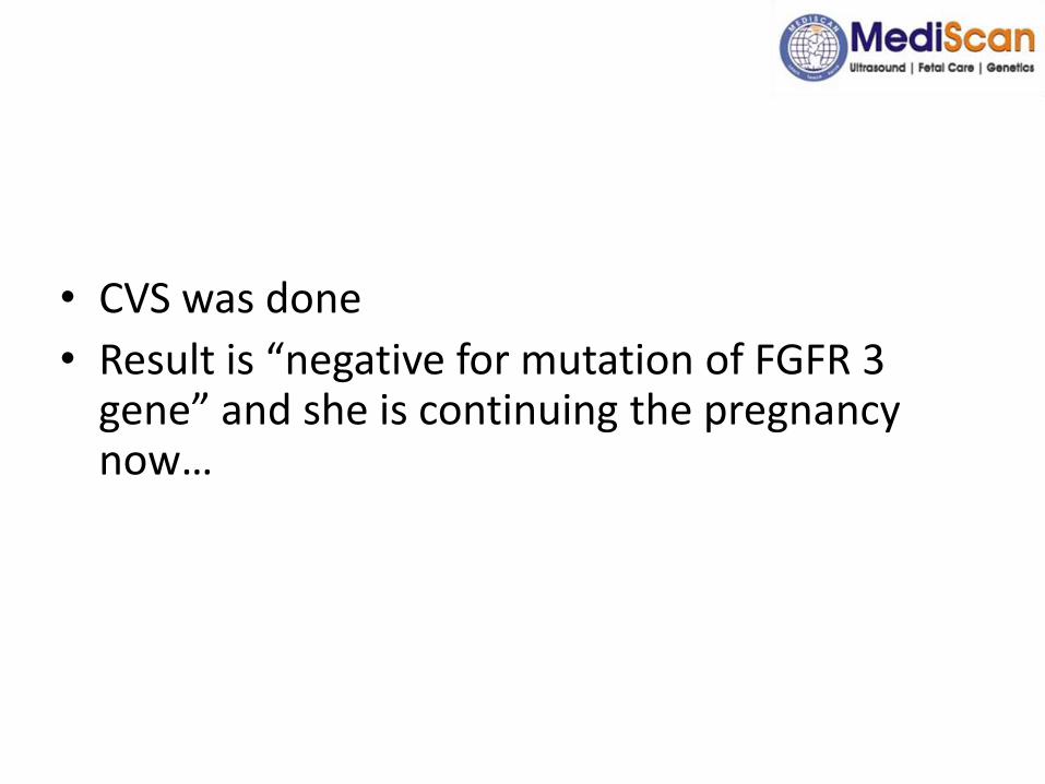
• CVS was done
• Result is “negative for mutation of FGFR 3 gene” and she is continuing the pregnancy now…
Highlights…
• Presence of Skeletal dysplasia
• Lethal – non lethal
• But exact diagnosis can be confirmed only after birth
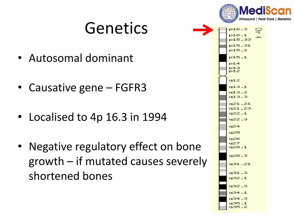
Genetics
• Autosomal dominant
• Causative gene – FGFR3
• Localised to 4p 16.3 in 1994
• Negative regulatory effect on bone growth – if mutated causes severelyshortened bones
• 85% of mutations occur de novo
• Related to advanced paternal age
• Mutation occurs during spermatogenesis. There may be some regulatory mechanism that occurs during oogenesis
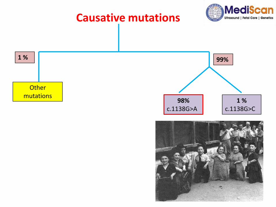
Causative mutations
Other mutations
99%
98%c.1138G>A
1 %c.1138G>C
1 %
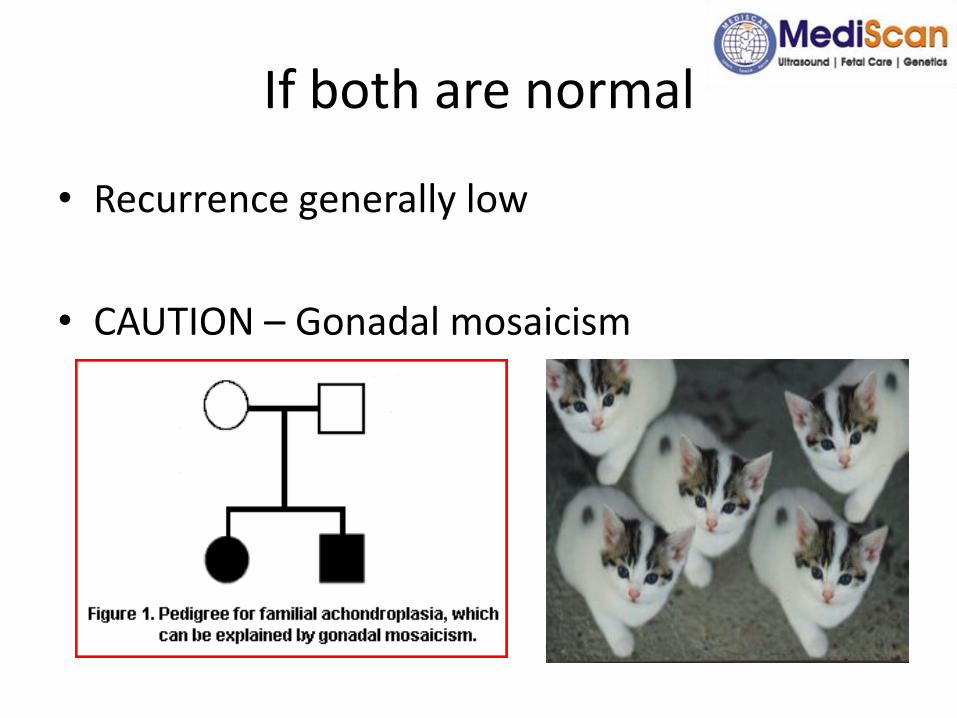
If both are normal
• Recurrence generally low
• CAUTION – Gonadal mosaicism
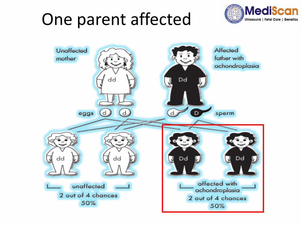
One parent affected
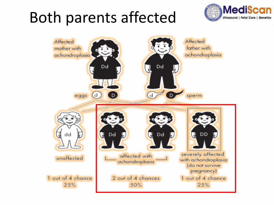
Both parents affected
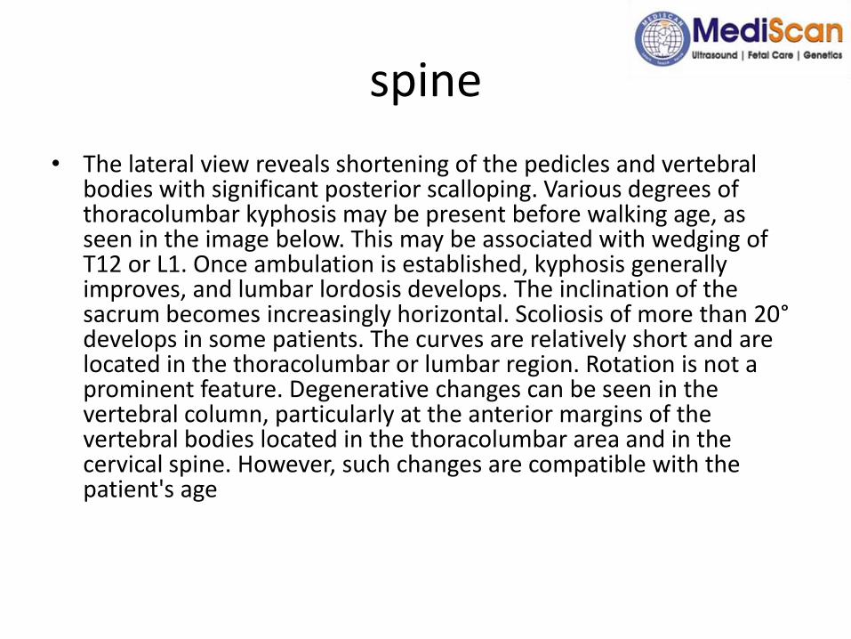
spine
• The lateral view reveals shortening of the pedicles and vertebral bodies with significant posterior scalloping. Various degrees of thoracolumbar kyphosis may be present before walking age, as seen in the image below. This may be associated with wedging of T12 or L1. Once ambulation is established, kyphosis generally improves, and lumbar lordosis develops. The inclination of the sacrum becomes increasingly horizontal. Scoliosis of more than 20°develops in some patients. The curves are relatively short and are located in the thoracolumbar or lumbar region. Rotation is not a prominent feature. Degenerative changes can be seen in the vertebral column, particularly at the anterior margins of the vertebral bodies located in the thoracolumbar area and in the cervical spine. However, such changes are compatible with the patient's age
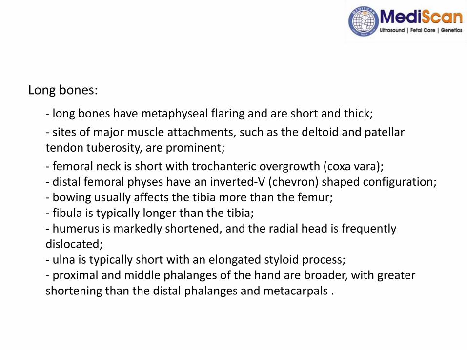
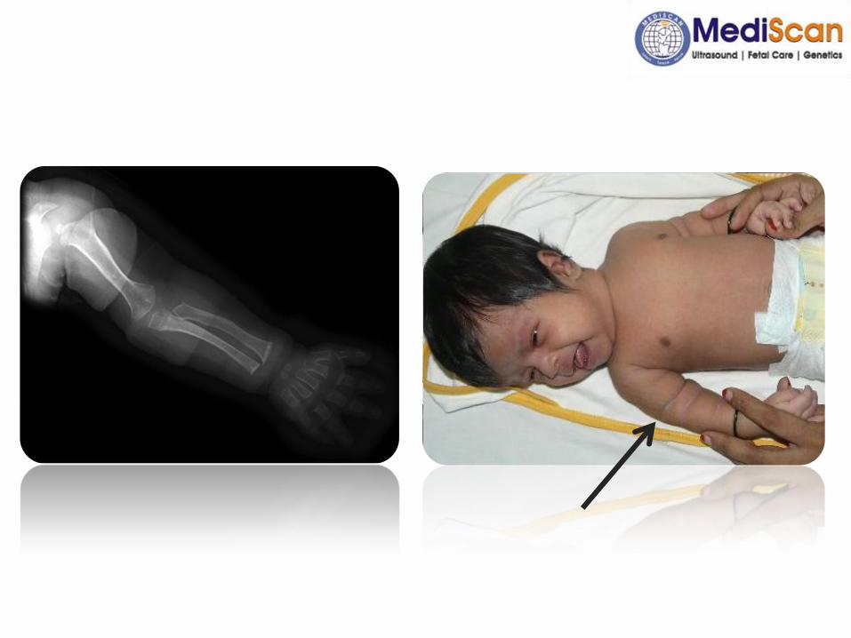
Long bones:
- long bones have metaphyseal flaring and are short and thick;
- sites of major muscle attachments, such as the deltoid and patellar tendon tuberosity, are prominent;
- femoral neck is short with trochanteric overgrowth (coxa vara); - distal femoral physes have an inverted-V (chevron) shaped configuration;- bowing usually affects the tibia more than the femur;- fibula is typically longer than the tibia;- humerus is markedly shortened, and the radial head is frequently dislocated;- ulna is typically short with an elongated styloid process;- proximal and middle phalanges of the hand are broader, with greater shortening than the distal phalanges and metacarpals .

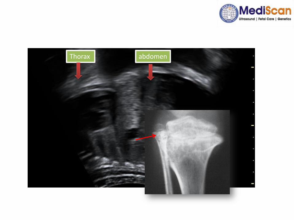
Thorax abdomen