prenatal diagnostic sonography abend weinger marta md neonatology department bnai zion hospital
TRANSCRIPT
Prenatal Diagnostic SonographyPrenatal Diagnostic Sonography
Abend Weinger Marta MDAbend Weinger Marta MD
Neonatology department Neonatology department
Bnai Zion Hospital Bnai Zion Hospital
To date the prevalent practice is to report the US findings and to state the risk of malformed baby
The professional liability oblige to report The report may cause a great deal of anxiety to
mother Both sides of argument are well intentioned What is the good way?
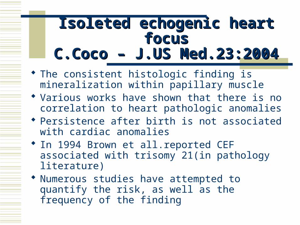
Isoleted echogenic heart focusIsoleted echogenic heart focusC.Coco – J.US Med.23:2004C.Coco – J.US Med.23:2004
The consistent histologic finding is mineralization within papillary muscle
Various works have shown that there is no correlation to heart pathologic anomalies
Persistence after birth is not associated with cardiac anomalies
In 1994 Brown et all.reported CEF associated with trisomy 21(in pathology literature)
Numerous studies have attempted to quantify the risk, as well as the frequency of the finding
Objective: to evaluate the risk of Down syndrome in fetuses with heart echogenic focus and likelihood ratios in an unselected population
Method: prospective evaluation of 12,672 second trimester sonographic features were examined. Population with echogenic focus was checked for chromosomal anomalies
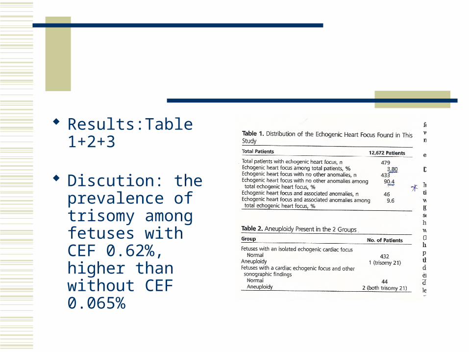
Results:Table 1+2+3
Discution: the prevalence of trisomy among fetuses with CEF 0.62%, higher than without CEF 0.065%
The decision to performed amniocentesis on all fetuses with CEF- fetal loss 1/200
If restricted to CEF+associated anomalies only 46 kariotype need- calculated fetal loss 0.23%
Conclusion: CEF+ other major or minor anomalies justifies amniocentesis
Amniocentesis need not to be offer to patients at low risk with CEF
The significans of isolated choroid plexus cysts
In 1984 was published the initial report on prenatal US detection of choroid plexus cyst
It was thought to be a finding without pathologic significance
Two years later the association with trisomy 18 was first suggested
In nearly two decades a large volume of literature has been published
The clinical dilemma is between invasive genetic testing against potential missing a major aneuploidy
Two recent review articles summarized the literature: nine of the articles in 1 of these reviews recommended genetic testing, whereas 11 others concluded that was not indicated
AIUM published standards for second trimester antenatal US examination
Some people recommend extended anatomic survey when ICPC found (EX : fetal hands)
Choroid plexus cyst and Echogenic intracardiac focus. J/US Med 23:2004
These two markers have been the source of the greatest controversy as signs for aneuploidy
The EIF is considered a soft marker for trisomies 21 and 13, whereas the CPC is considered marker for trisomy 18
Large no. of practitioners feel that the reporting of these 2 findings cause more harm than good
The AIUM and SRU offer a compromise as guide line:
The recommendation is: When CPC or EIF is identified without other minor
or major anomalies - In the absence of other risk factors this is considered a normal variant,and no further evaluation is recommended
Risk status is ascertained preferably by the patients biochemical screening test results (the so-called triple or quadruple markers) and maternal age
The AIUM guidelines should be strictly followed
Outcome of fetuses with Club Feet J.US Med.23:2004
Clubfoot (talipes equinovarus) – malformation of the bones of ankle and foot-inverted and rotated medially
One of the most common birth defect 1\1000 of live births
US DG: the bones of the foot lie in the same plane as the lower leg bones
CF is commonly associated with other fetal anomalies and aneuploidy. Could be syndrome or genetic defect
Other causes of CF: uteral anomalies, restricted environment in utero, olygohydramnios
The purpose of the study: to evaluate the outcome of fetuses with prenatally diagnosed clubfoot with attention to the difference between uni- and bilateral CF
Method:identification of all fetuses scanned during 3 years in whom CF was suspected .or dg.prenatally
Collection of maternal and NN medical records: unilateral or bilateral CF, gestational age at dg., other sonographic findings, outcome and neonatal findings at birth
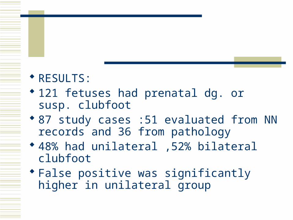
RESULTS: 121 fetuses had prenatal dg. or susp. clubfoot
87 study cases :51 evaluated from NN
records and 36 from pathology 48% had unilateral ,52% bilateral clubfoot False positive was significantly higher in
unilateral group
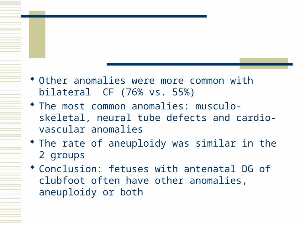
Other anomalies were more common with bilateral CF (76% vs. 55%)
The most common anomalies: musculo-skeletal, neural tube defects and cardio-vascular anomalies
The rate of aneuploidy was similar in the 2 groups Conclusion: fetuses with antenatal DG of clubfoot
often have other anomalies, aneuploidy or both
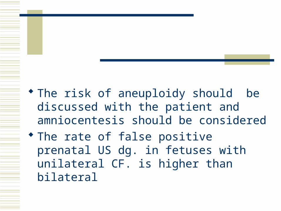
The risk of aneuploidy should be discussed with the patient and amniocentesis should be considered
The rate of false positive prenatal US dg. in fetuses with unilateral CF. is higher than bilateral
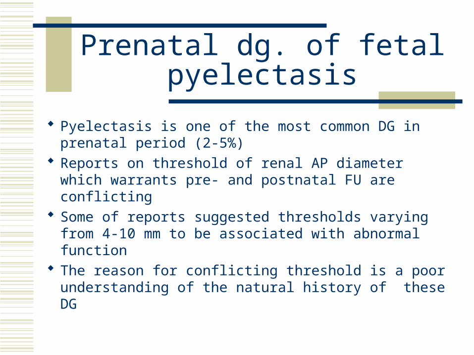
Prenatal dg. of fetal pyelectasis
Pyelectasis is one of the most common DG in prenatal period (2-5%)
Reports on threshold of renal AP diameter which warrants pre- and postnatal FU are conflicting
Some of reports suggested thresholds varying from 4-10 mm to be associated with abnormal function
The reason for conflicting threshold is a poor understanding of the natural history of these DG
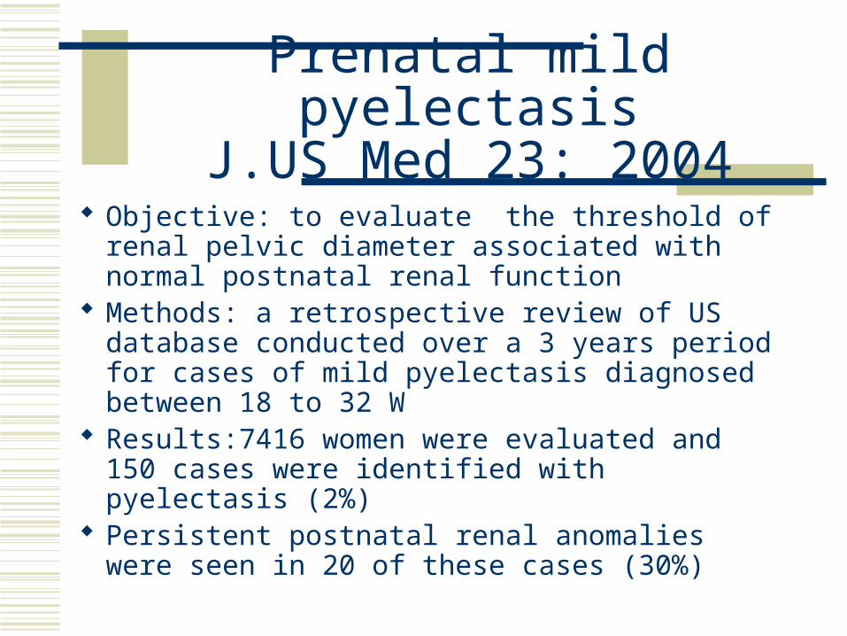
Prenatal mild pyelectasisJ.US Med 23: 2004
Objective: to evaluate the threshold of renal pelvic diameter associated with normal postnatal renal function
Methods: a retrospective review of US database conducted over a 3 years period for cases of mild pyelectasis diagnosed between 18 to 32 W
Results:7416 women were evaluated and 150 cases were identified with pyelectasis (2%)
Persistent postnatal renal anomalies were seen in 20 of these cases (30%)

For purpose of the study, cases with pyelectasis of greater than 10 mm, multiple anomalies and aneuploidy were excluded
The renal pelvic AP thresholds evaluated included diameters of 6,7,8,9 mm
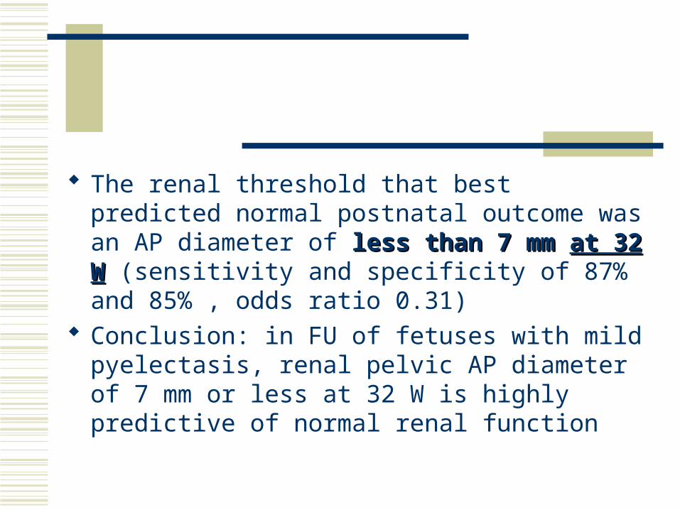
The renal threshold that best predicted normal postnatal outcome was an AP diameter of less than less than 7 mm 7 mm at 32 Wat 32 W (sensitivity and specificity of 87% and 85% , odds ratio 0.31)
Conclusion: in FU of fetuses with mild pyelectasis, renal pelvic AP diameter of 7 mm or less at 32 W is highly predictive of normal renal function
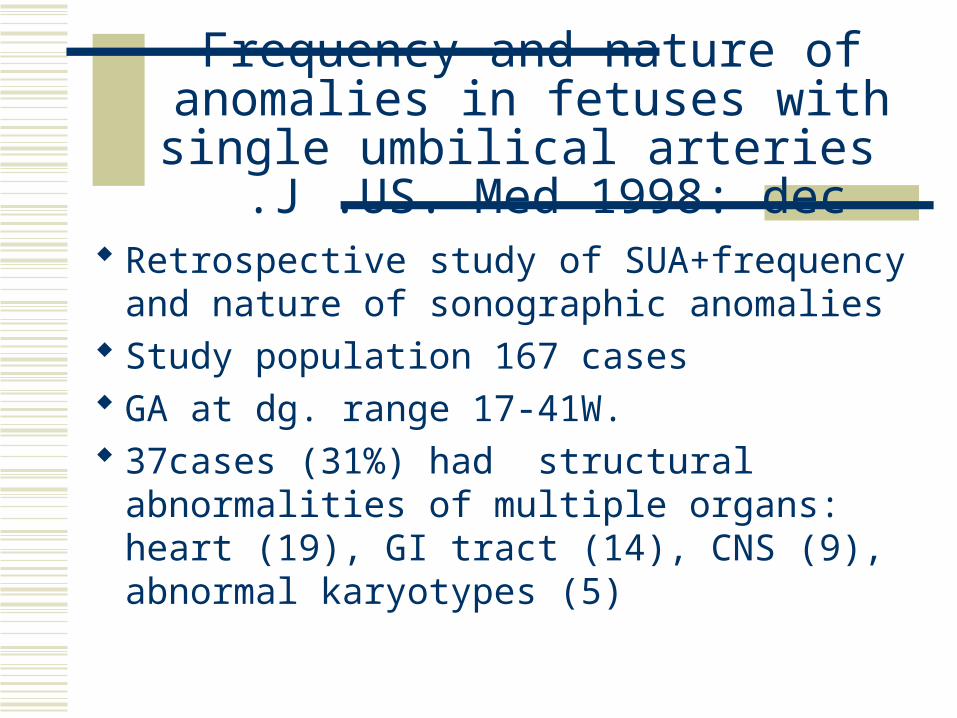
Frequency and nature of anomalies in fetuses with single umbilical arteries
J .US. Med 1998: dec .
Retrospective study of SUA+frequency and nature of sonographic anomalies
Study population 167 cases GA at dg. range 17-41W. 37cases (31%) had structural abnormalities
of multiple organs: heart (19), GI tract (14), CNS (9), abnormal karyotypes (5)
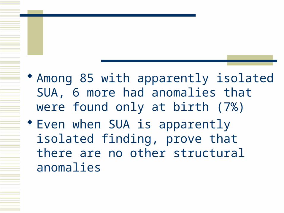
Among 85 with apparently isolated SUA, 6 more had anomalies that were found only at birth (7%)
Even when SUA is apparently isolated finding, prove that there are no other structural anomalies
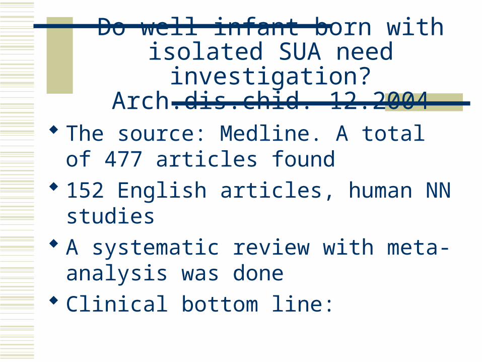
Do well infant born with isolated SUA need investigation?
Arch.dis.chid. 12.2004
The source: Medline. A total of 477 articles found
152 English articles, human NN studies A systematic review with meta-analysis was
done Clinical bottom line:
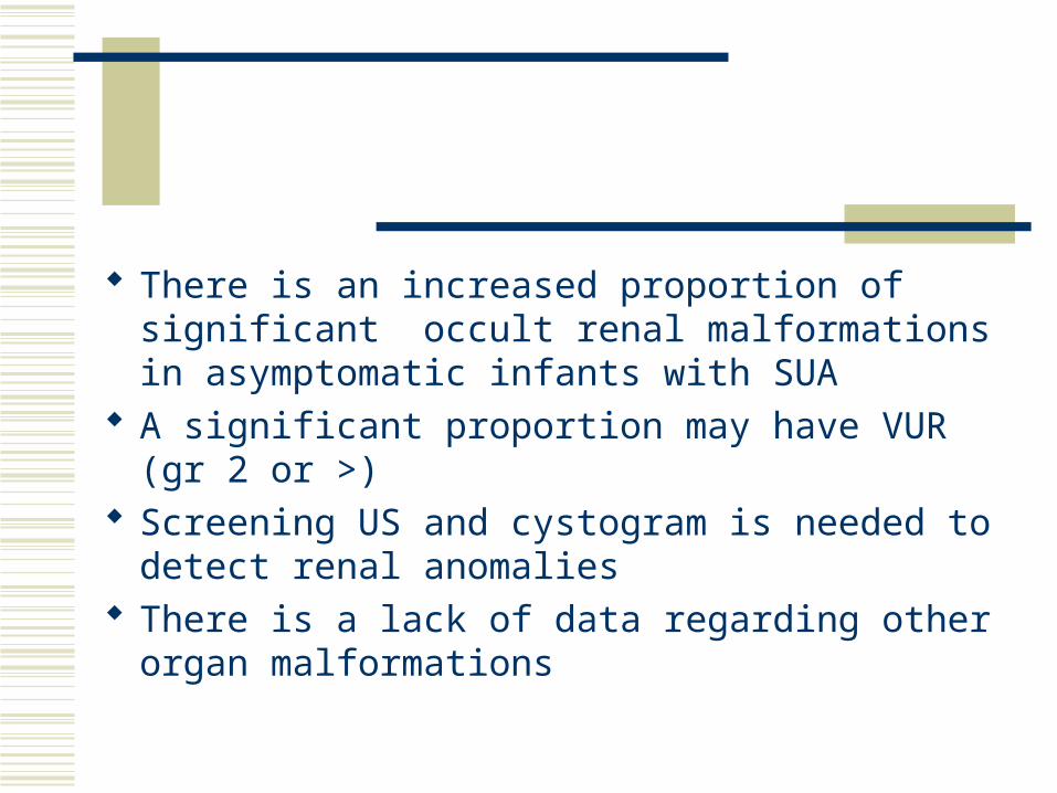
There is an increased proportion of significant occult renal malformations in asymptomatic infants with SUA
A significant proportion may have VUR (gr 2 or >) Screening US and cystogram is needed to detect
renal anomalies There is a lack of data regarding other organ
malformations