prenatal screening with cell-free dna
TRANSCRIPT
Prenatal screening with cell-free DNA
Slides adapted in part from Nancy Rose, MD
Disclosures & Objectives
I have no conflicts of interest to disclose
Describe the characteristics of cell-free DNA screening in comparison to other types of prenatal screening
Understand the need for confirmatory testing and pregnancy implications
Describe unexpected, particularly maternal, cell-free DNA test results and their clinical implications
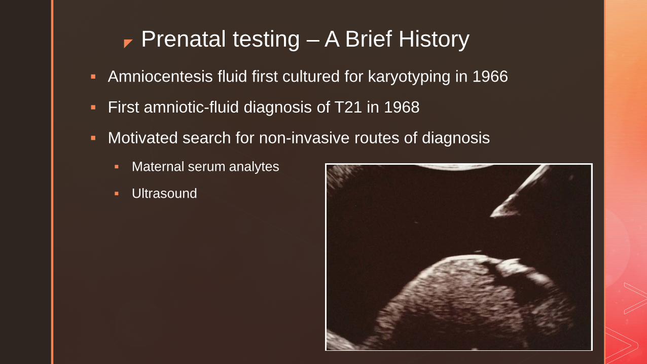
Prenatal testing – A Brief History Amniocentesis fluid first cultured for karyotyping in 1966
First amniotic-fluid diagnosis of T21 in 1968
Motivated search for non-invasive routes of diagnosis
Maternal serum analytes
Ultrasound
Types of non-DNA based genetic screening Maternal age
Ultrasound
First trimester screen Ultrasound nuchal translucency + maternal serum analytes – PAPP-A & hCG
10-14 weeks gestation
Quad screen Maternal serum analytes – AFP, hCG, Inhibin A, uE3
15-20/22 weeks gestation
Integrated screening and stepwise sequential or contingent screening First trimester US NT + PAPP-A
Second trimester quad screen analytes
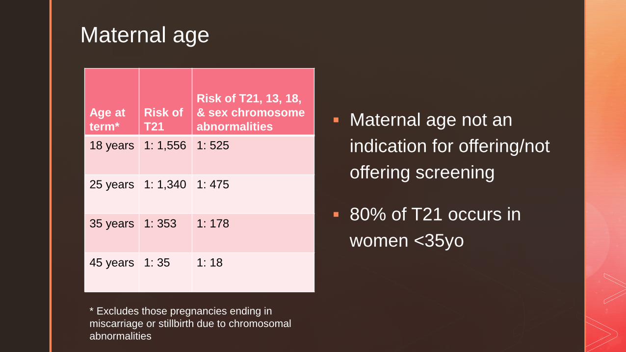
Maternal age
Age at term*
Risk of T21
Risk of T21, 13, 18, & sex chromosome abnormalities
18 years 1: 1,556 1: 525
25 years 1: 1,340 1: 475
35 years 1: 353 1: 178
45 years 1: 35 1: 18
* Excludes those pregnancies ending in miscarriage or stillbirth due to chromosomal abnormalities
Maternal age not an indication for offering/not offering screening
80% of T21 occurs in women <35yo
Cell-free DNA – A Brief History 1948: First identified Mandel and Metais 1966: Association of SLE and increased cfDNA
levels. 1977: Higher levels in oncology patients 1997: Lo and colleagues: fetal CFDNA in maternal
plasma. Applied concept of tumor fragments to fetal development
2008: Association of cfDNA and aneuploidy 2011: Rapid commercial development with little
transparency
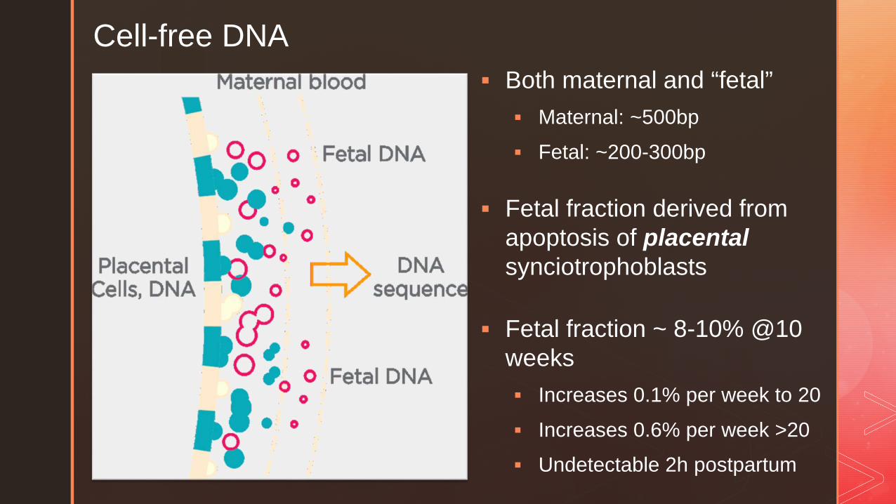
Cell-free DNA
Both maternal and “fetal” Maternal: ~500bp Fetal: ~200-300bp
Fetal fraction derived from apoptosis of placentalsynciotrophoblasts
Fetal fraction ~ 8-10% @10 weeks Increases 0.1% per week to 20 Increases 0.6% per week >20 Undetectable 2h postpartum
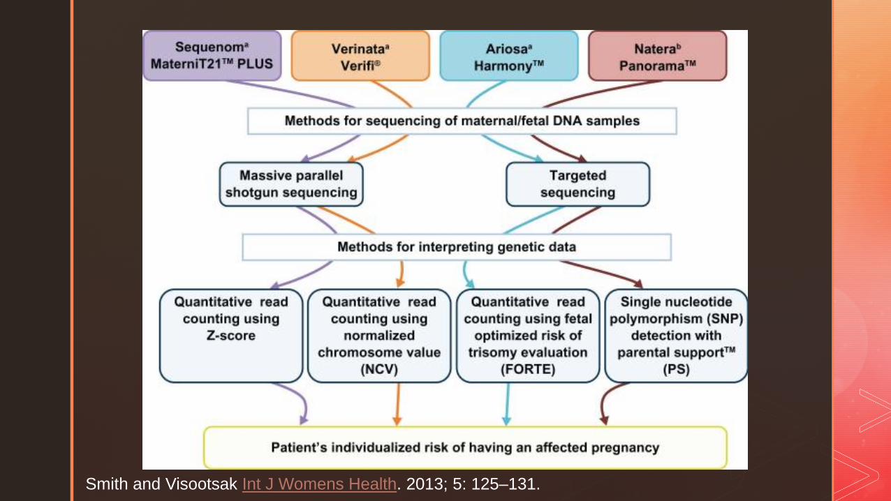
Smith and Visootsak Int J Womens Health. 2013; 5: 125–131.

Side comments about fetal fraction It’s not really fetal! It’s placental!
1-2% confined placental mosaicism (CPM)
Most commonly autosomal triploidy
Examples: triploidy rescue or monosomy rescue
It changes with maternal habitus
Decreases 0.5% for every 10lb increase between 80 and 200 lbs
Dilutional effect?
Maternal adipocyte apoptosis?
8-17% no call rate in BMI>35 vs 0.5-1% no call <35
It changes with aneuploidy
Increased or decreased (↑T21, ↓T13/18/45X)
Overall a low fetal fraction raises risk for aneuploidy
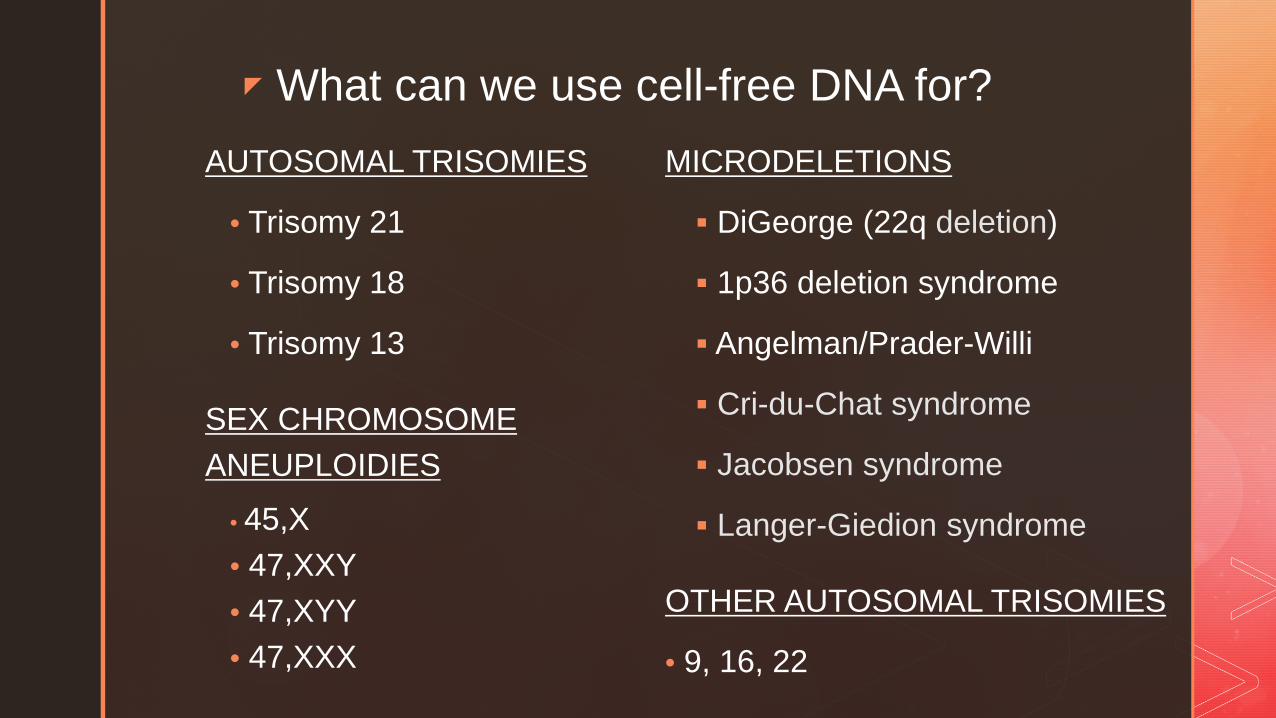
What can we use cell-free DNA for?AUTOSOMAL TRISOMIES
• Trisomy 21
• Trisomy 18
• Trisomy 13
SEX CHROMOSOME ANEUPLOIDIES
• 45,X• 47,XXY• 47,XYY• 47,XXX
MICRODELETIONS
DiGeorge (22q deletion)
1p36 deletion syndrome
Angelman/Prader-Willi
Cri-du-Chat syndrome
Jacobsen syndrome
Langer-Giedion syndrome
OTHER AUTOSOMAL TRISOMIES
• 9, 16, 22
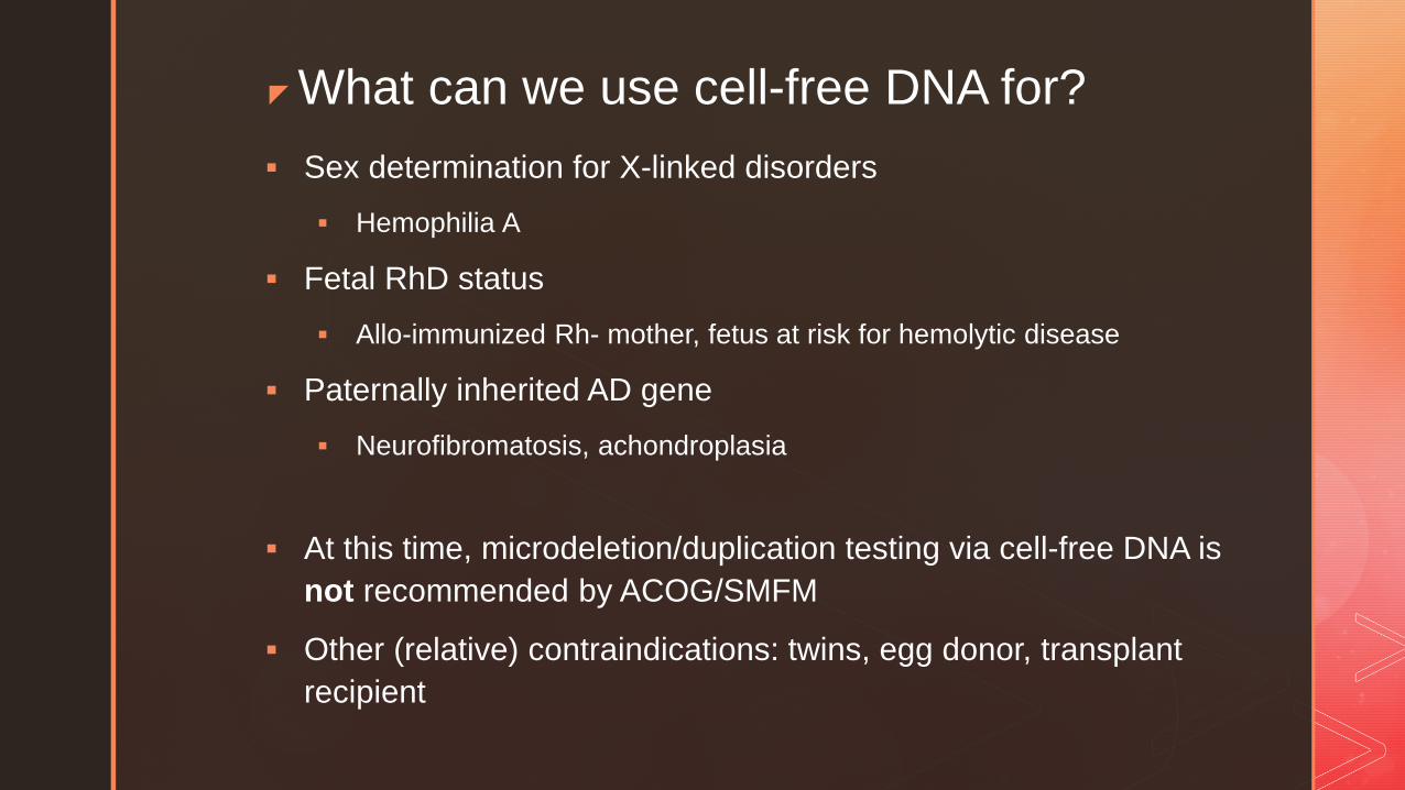
What can we use cell-free DNA for? Sex determination for X-linked disorders
Hemophilia A
Fetal RhD status Allo-immunized Rh- mother, fetus at risk for hemolytic disease
Paternally inherited AD gene Neurofibromatosis, achondroplasia
At this time, microdeletion/duplication testing via cell-free DNA is not recommended by ACOG/SMFM
Other (relative) contraindications: twins, egg donor, transplant recipient
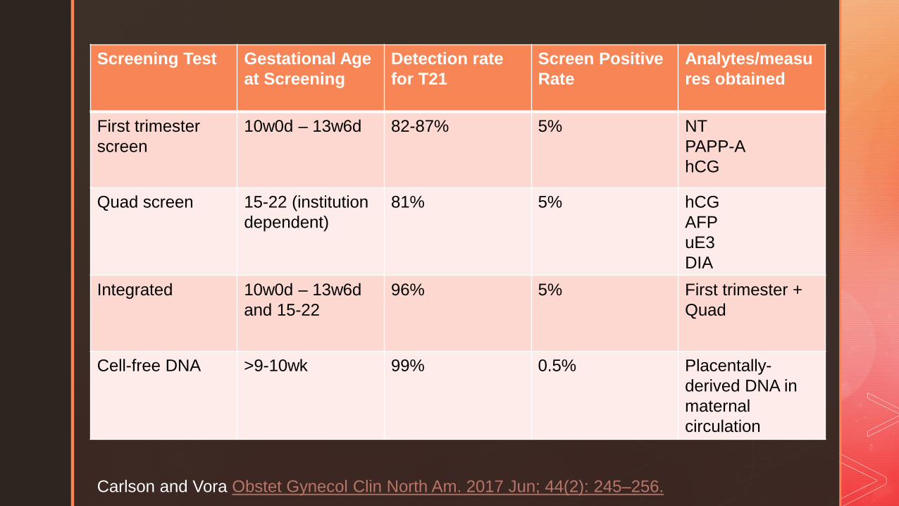
Screening Test Gestational Age at Screening
Detection rate for T21
Screen Positive Rate
Analytes/measures obtained
First trimester screen
10w0d – 13w6d 82-87% 5% NTPAPP-AhCG
Quad screen 15-22 (institution dependent)
81% 5% hCGAFPuE3DIA
Integrated 10w0d – 13w6d and 15-22
96% 5% First trimester + Quad
Cell-free DNA >9-10wk 99% 0.5% Placentally-derived DNA in maternal circulation
Carlson and Vora Obstet Gynecol Clin North Am. 2017 Jun; 44(2): 245–256.
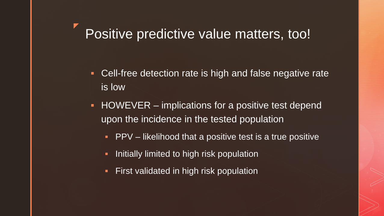
Positive predictive value matters, too!
Cell-free detection rate is high and false negative rate is low
HOWEVER – implications for a positive test depend upon the incidence in the tested population
PPV – likelihood that a positive test is a true positive
Initially limited to high risk population
First validated in high risk population
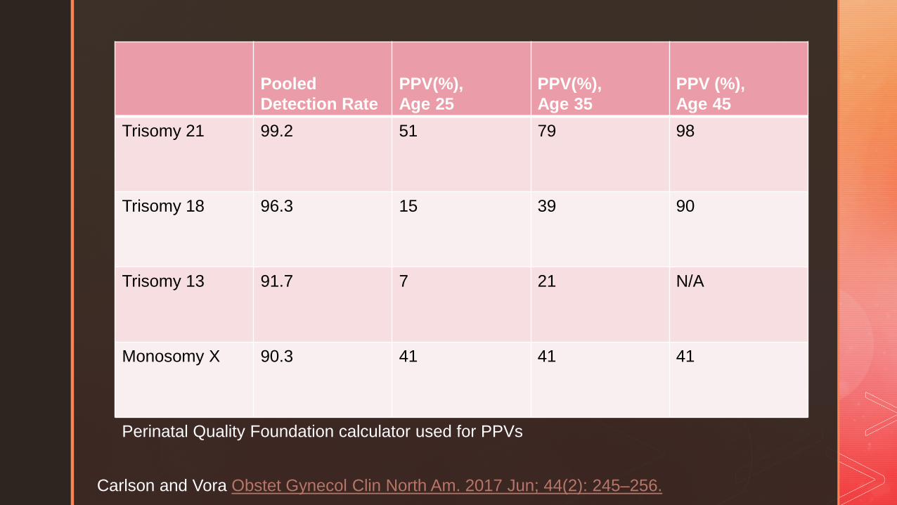
Pooled Detection Rate
PPV(%),Age 25
PPV(%), Age 35
PPV (%), Age 45
Trisomy 21 99.2 51 79 98
Trisomy 18 96.3 15 39 90
Trisomy 13 91.7 7 21 N/A
Monosomy X 90.3 41 41 41
Carlson and Vora Obstet Gynecol Clin North Am. 2017 Jun; 44(2): 245–256.
Perinatal Quality Foundation calculator used for PPVs
Clinical pretest counseling – by the book Screening should be offered to every mother at 1st
prenatal visit Cell free DNA may be offered to any mother Discuss pros/cons of each test
All tests are *pretty good*, better than age + US
Costs/insurance are important
Additional information from serum screen
Include how to interpret (broadly) positive/negative results Cover possible unexpected results Given volume of information, videos, pamphlets, and
infographics are being developed
What do I do with positive or no-call results? Positive test - requires confirmation before action No call test due to low FF in obese patient
Fetal fraction does not increase as much through pregnancy
>40% still have a no-call on subsequent test with BMI>35
Need additional testing (US, +/- amnio/CVS)
No call or indeterminate for other reasons High risk of aneuploidy (20-30%)
Offer genetic counseling and diagnostic testing
Repeat cell free DNA could be considered if very early in gestation (e.g., 11 weeks) but delays possible diagnosis
Clinical Cases
37yo G2P0 at 11 weeks estimated gestation, first prenatal visit
Normal BMI, no medical history, no history of aneuploidy or congenital defects
Viability ultrasound:
Size consistent with dates
No obvious abnormalities (e.g. acrania)
Would like genetic screening after discussion
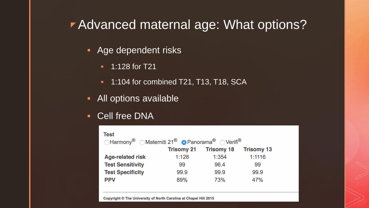
Advanced maternal age: What options?
Age dependent risks
1:128 for T21
1:104 for combined T21, T13, T18, SCA
All options available
Cell free DNA
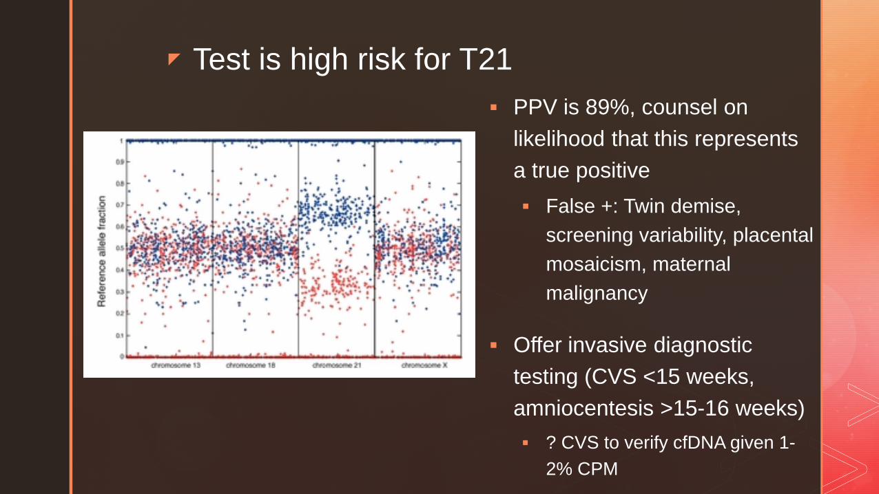
Test is high risk for T21 PPV is 89%, counsel on
likelihood that this represents a true positive False +: Twin demise,
screening variability, placental mosaicism, maternal malignancy
Offer invasive diagnostic testing (CVS <15 weeks, amniocentesis >15-16 weeks) ? CVS to verify cfDNA given 1-
2% CPM
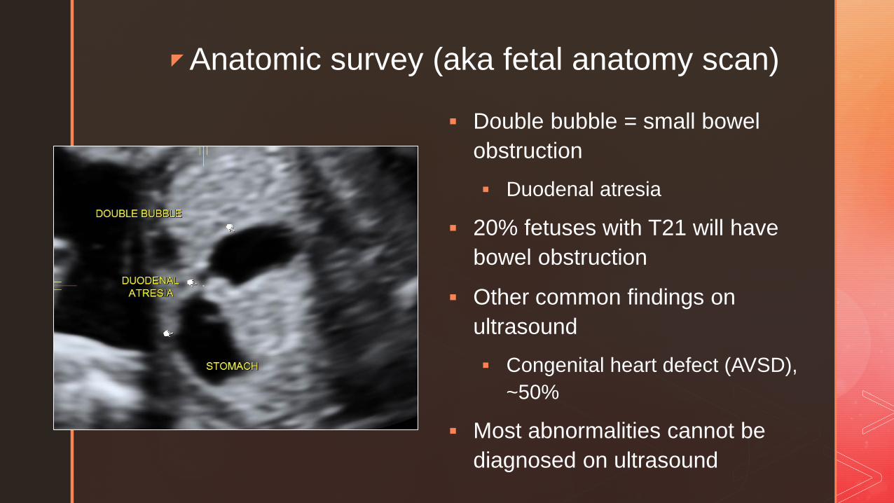
Anatomic survey (aka fetal anatomy scan)
Double bubble = small bowel obstruction Duodenal atresia
20% fetuses with T21 will have bowel obstruction
Other common findings on ultrasound Congenital heart defect (AVSD),
~50%
Most abnormalities cannot be diagnosed on ultrasound
Clinical case #2
25 yo G3P2 at 18 weeks gestation by last menstrual period presenting for anatomy scan
Declined genetic testing and ultrasound earlier in pregnancy
Normal BMI, no history of recurrent miscarriage, aneuploidy, or congenital defects
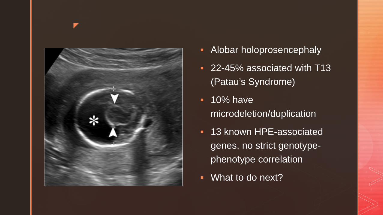
Alobar holoprosencephaly
22-45% associated with T13 (Patau’s Syndrome)
10% have microdeletion/duplication
13 known HPE-associated genes, no strict genotype-phenotype correlation
What to do next?
Ultrasound findings and cell-free DNA
Standard text-book answer: do not use cell free to evaluate anomalies
HOWEVER, when taking into account maternal preferences, some moms/families opt for cfDNA
Ultrasound findings very consistent with T13/18/21
Patient desires pregnancy to continue regardless of result
Desires not to perform invasive procedure for risk of pregnancy loss
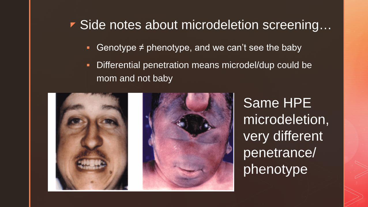
Side notes about microdeletion screening… Genotype ≠ phenotype, and we can’t see the baby
Differential penetration means microdel/dup could be mom and not baby
Same HPE microdeletion, very different penetrance/phenotype
Unintended maternal consequences
The test cannot distinguish a maternal from a fetal result!
45, X results can reflect older mothers, not fetuses
The microdeletion syndrome you identify may be maternal
Beware the patient with an organ transplant At least 26 cases of maternal cancer diagnosis
A tale of two monosomy Xs…
40 year old, first pregnancy
Nuchal translucency normal
CfDNA 45, X result
Sonogram: apparently normal female
Amnio: 46,XX
Maternal karyotype 45,X[5]/46,XX [45]
Interpretation: normal maternal aging
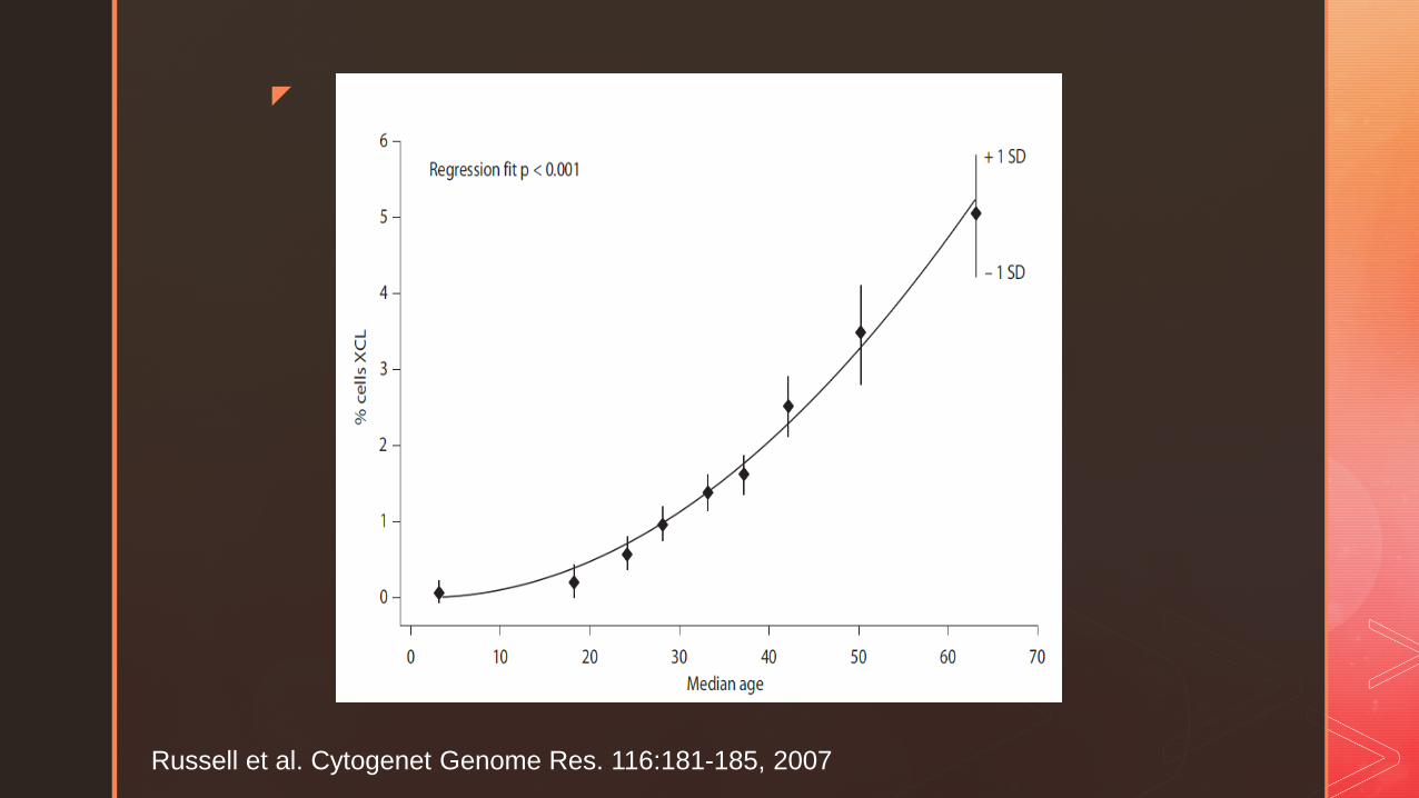
Russell et al. Cytogenet Genome Res. 116:181-185, 2007
35 Year old G1P0 cfDNA: 45, X Fetal karyotype: 46, XX Maternal karyotype:45 X (17), 46XX (35)
Phenotype: Mild hearing loss, bone density issues, bicuspid aortic
valve, short stature, normal intelligence
Clinical implications: ECHO Possible premature ovarian failure, with attendant
increase in cardiovascular disease Endocrine disorders
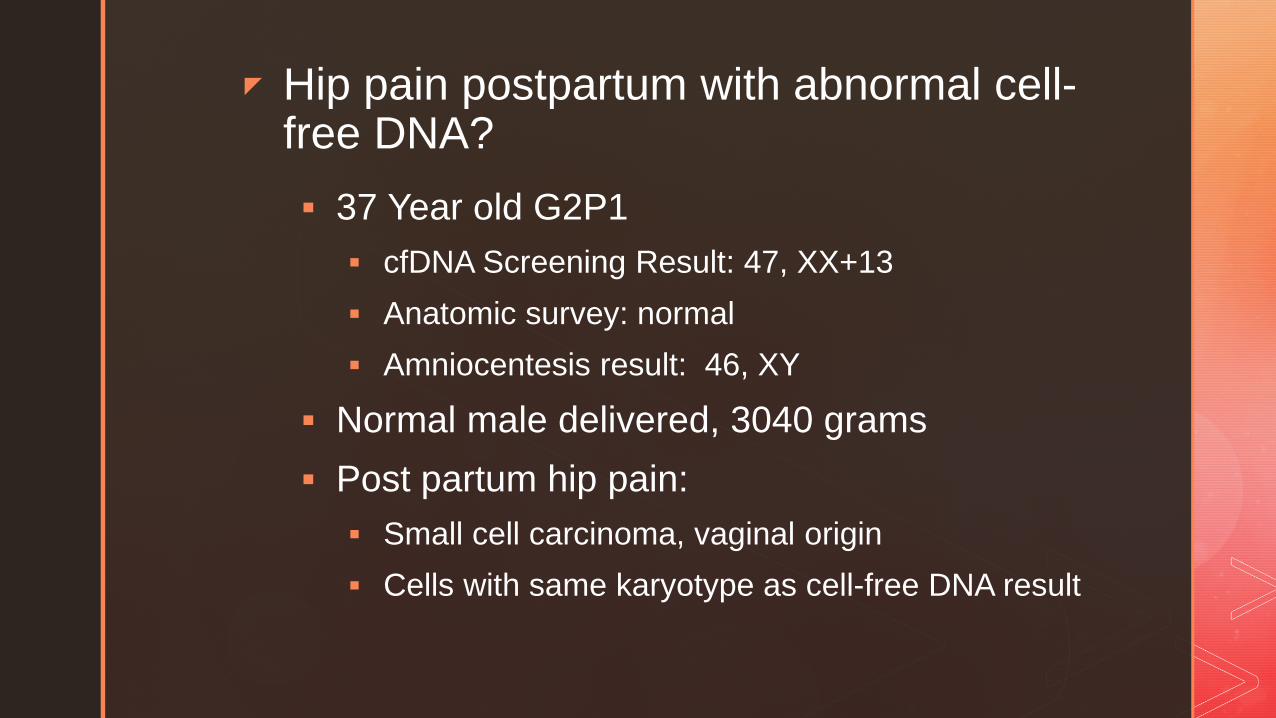
Hip pain postpartum with abnormal cell-free DNA? 37 Year old G2P1 cfDNA Screening Result: 47, XX+13 Anatomic survey: normal Amniocentesis result: 46, XY
Normal male delivered, 3040 grams Post partum hip pain: Small cell carcinoma, vaginal origin Cells with same karyotype as cell-free DNA result

Summary
Cell-free DNA screening for fetal aneuploidy is highly sensitive
Must be partnered with ultrasound or AFP for anatomic/structural evaluation
Positive predictive value falls with falling incidence
Positive tests need confirmation!
Range of applications is rapidly increasing
Clinical implications are equally wide ranging and require counseling *pre-test*
Thank you!
Thanks to Dr Toydemir, the fellows, medical directors, and lab staff who made my visit to ARUP educational and pleasant
Thanks to Nancy Rose, who gave feedback on this presentation