preoperative prediction of failure following two-stage revision for knee prosthetic joint infections
TRANSCRIPT

The Journal of Arthroplasty 29 (2014) 115–121
Contents lists available at ScienceDirect
The Journal of Arthroplasty
j ourna l homepage: www.arth rop lasty journa l .o rg
Preoperative Prediction of Failure Following Two-Stage Revision for Knee ProstheticJoint Infections
Fady Youssef Sabry, MD, MRCS, Leonard Buller, BA, Sarim Ahmed, MD,Alison K. Klika, MS, Wael K. Barsoum, MDDepartment of Orthopaedic Surgery, Orthopaedic and Rheumatologic Institute, Cleveland Clinic, Cleveland, Ohio
a b s t r a c ta r t i c l e i n f o
The Conflict of Interest statement associated with thidx.doi.org/10.1016/j.arth.2013.04.016.
Reprint requests: Alison K. Klika, MS, DepartmCleveland Clinic, 9500 Euclid Ave., Cleveland, OH 44195
0883-5403/2901-0024$36.00/0 – see front matter © 20http://dx.doi.org/10.1016/j.arth.2013.04.016
Article history:Received 19 March 2012Accepted 12 April 2013
Keywords:total knee arthroplastypredictive modelingnomogramrevisioninfection
While two-stage revision is the gold standard for treatment of knee prosthetic joint infection (PJI), it is notwithout risk. The purpose of this study was to develop a tool to preoperatively predict the probability that atwo-stage revision would fail to eradicate knee PJI. 3,809 surgical cases were retrospectively reviewed anddata were collected from 314 charts. Overall, 105 (33.4%) cases failed to eradicate PJI using this procedure.Univariate analysis identified multiple variables independently associated with reinfection. Logisticregression was used to generate a model (bootstrap-corrected concordance index of 0.773) predictingfailure of infection eradication. Preoperative knowledge of a high probability of failure may improve riskassessment, lead to more aggressive management, and allow for time to consider alternative therapies.
s article can be found at http://
ent of Orthopaedic Surgery,.
14 Elsevier Inc. All rights reserved.
© 2014 Elsevier Inc. All rights reserved.
PJI are the leading cause of implant failure requiring revision totalknee arthroplasty (TKA) [1]. These devastating complications areassociated with considerable morbidity [2,3] and economic burden[4]. The incidence of infection following primary TKA ranges from 0.4%to 4.0% [5,6] and increases nearly ten-fold for revision procedures [4].The overall number of PJI is projected to increase as the demand forprimary and revision TKA continues to grow [7].
The ultimate goal of PJI management is the restoration of a pain-free, functional joint with eradication of the infection. Variousprotocols are available for the management of PJI, ranging fromconservative treatment with antibiotic suppression [8], to surgicaloptions including irrigation and debridement with or withoutpolyethylene exchange [9,10], single-stage revision arthroplasty[10], two-stage revision arthroplasty with antibiotic-laden spacerplacement [11], resection arthroplasty [12], arthrodesis [13], andamputation [14]. Introduced by Insall et al. [11] in 1983, the two-stagerevision with antibiotic-laden spacer placement has become the gold-standard for management of PJI [15–17]. The two-stage revisioninvolves two separate procedures, one to remove the infectedprosthesis and implant an antibiotic-laden cement spacer, and asubsequent procedure to reimplant a new prosthesis followinginfection eradication.
While the successful eradication of infection reported for these two-stage revisions is relatively high [18–21], the procedure is not withoutlimitations. First, it requires a minimum of two surgical procedures,which carries a substantial financial burden [22]. Additionally, betweenthe two stages patients may experience substantial joint pain andreducedmobility [23]. Moreover, when removingwell-fixed prosthesesto implant the antibiotic spacer the risk of additionalmorbidity includesdegrading the bone stock, soft tissue deformation and fracture [24–26].The current guidelines for selecting the most effective and appropriatesurgical approach for a particular patient are based upon the duration ofsymptoms, condition of the implant and soft tissue evaluated duringsurgery and, if known, the infecting microorganism [27]. It is of criticalimportance to make good decisions which give patients the greatestprobability of successful infection eradication on the first attempt, asGardner et al.[10] have reported lowered success of infection eradica-tion with a two-stage revision following a failed debridement andpolyethylene exchange.
A tool to predict the probability of failure of a two-stage revisionmay trigger closer monitoring and more aggressive management ofthose patients predicted to fail, assisting surgeons in this decision-making process. This predictive ability may lead to greater success ineradication of infection on the first attempt, or consideration ofalternative treatments which may potentially reduce surgicalcomplications and costs. The specific aims of the study were to 1)identify independent predictors of failure to eradicate a PJI followinga two-staged revision, 2) generate Kaplan–Meier survival analyses fortwo-stage revisions stratified by the type of infecting organism, and3) develop a nomogram to preoperatively predict a patient’sprobability of failure to eradicate a PJI when managed with a two-stage revision procedure.
116 F.Y. Sabry et al. / The Journal of Arthroplasty 29 (2014) 115–121
Materials and Methods
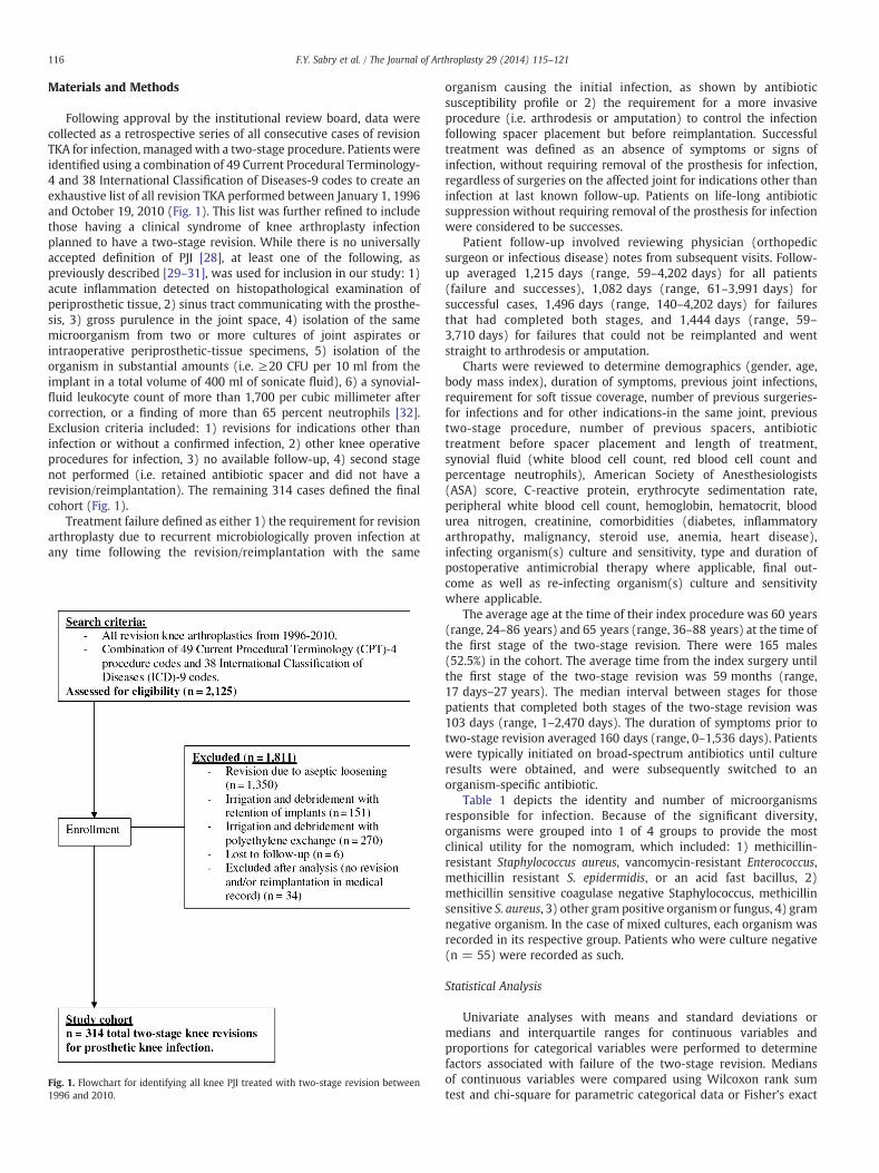
Following approval by the institutional review board, data werecollected as a retrospective series of all consecutive cases of revisionTKA for infection, managedwith a two-stage procedure. Patients wereidentified using a combination of 49 Current Procedural Terminology-4 and 38 International Classification of Diseases-9 codes to create anexhaustive list of all revision TKA performed between January 1, 1996and October 19, 2010 (Fig. 1). This list was further refined to includethose having a clinical syndrome of knee arthroplasty infectionplanned to have a two-stage revision. While there is no universallyaccepted definition of PJI [28], at least one of the following, aspreviously described [29–31], was used for inclusion in our study: 1)acute inflammation detected on histopathological examination ofperiprosthetic tissue, 2) sinus tract communicating with the prosthe-sis, 3) gross purulence in the joint space, 4) isolation of the samemicroorganism from two or more cultures of joint aspirates orintraoperative periprosthetic-tissue specimens, 5) isolation of theorganism in substantial amounts (i.e. ≥20 CFU per 10 ml from theimplant in a total volume of 400 ml of sonicate fluid), 6) a synovial-fluid leukocyte count of more than 1,700 per cubic millimeter aftercorrection, or a finding of more than 65 percent neutrophils [32].Exclusion criteria included: 1) revisions for indications other thaninfection or without a confirmed infection, 2) other knee operativeprocedures for infection, 3) no available follow-up, 4) second stagenot performed (i.e. retained antibiotic spacer and did not have arevision/reimplantation). The remaining 314 cases defined the finalcohort (Fig. 1).
Treatment failure defined as either 1) the requirement for revisionarthroplasty due to recurrent microbiologically proven infection atany time following the revision/reimplantation with the same
Fig. 1. Flowchart for identifying all knee PJI treated with two-stage revision between1996 and 2010.
organism causing the initial infection, as shown by antibioticsusceptibility profile or 2) the requirement for a more invasiveprocedure (i.e. arthrodesis or amputation) to control the infectionfollowing spacer placement but before reimplantation. Successfultreatment was defined as an absence of symptoms or signs ofinfection, without requiring removal of the prosthesis for infection,regardless of surgeries on the affected joint for indications other thaninfection at last known follow-up. Patients on life-long antibioticsuppression without requiring removal of the prosthesis for infectionwere considered to be successes.
Patient follow-up involved reviewing physician (orthopedicsurgeon or infectious disease) notes from subsequent visits. Follow-up averaged 1,215 days (range, 59–4,202 days) for all patients(failure and successes), 1,082 days (range, 61–3,991 days) forsuccessful cases, 1,496 days (range, 140–4,202 days) for failuresthat had completed both stages, and 1,444 days (range, 59–3,710 days) for failures that could not be reimplanted and wentstraight to arthrodesis or amputation.
Charts were reviewed to determine demographics (gender, age,body mass index), duration of symptoms, previous joint infections,requirement for soft tissue coverage, number of previous surgeries-for infections and for other indications-in the same joint, previoustwo-stage procedure, number of previous spacers, antibiotictreatment before spacer placement and length of treatment,synovial fluid (white blood cell count, red blood cell count andpercentage neutrophils), American Society of Anesthesiologists(ASA) score, C-reactive protein, erythrocyte sedimentation rate,peripheral white blood cell count, hemoglobin, hematocrit, bloodurea nitrogen, creatinine, comorbidities (diabetes, inflammatoryarthropathy, malignancy, steroid use, anemia, heart disease),infecting organism(s) culture and sensitivity, type and duration ofpostoperative antimicrobial therapy where applicable, final out-come as well as re-infecting organism(s) culture and sensitivitywhere applicable.
The average age at the time of their index procedure was 60 years(range, 24–86 years) and 65 years (range, 36–88 years) at the time ofthe first stage of the two-stage revision. There were 165 males(52.5%) in the cohort. The average time from the index surgery untilthe first stage of the two-stage revision was 59 months (range,17 days–27 years). The median interval between stages for thosepatients that completed both stages of the two-stage revision was103 days (range, 1–2,470 days). The duration of symptoms prior totwo-stage revision averaged 160 days (range, 0–1,536 days). Patientswere typically initiated on broad-spectrum antibiotics until cultureresults were obtained, and were subsequently switched to anorganism-specific antibiotic.
Table 1 depicts the identity and number of microorganismsresponsible for infection. Because of the significant diversity,organisms were grouped into 1 of 4 groups to provide the mostclinical utility for the nomogram, which included: 1) methicillin-resistant Staphylococcus aureus, vancomycin-resistant Enterococcus,methicillin resistant S. epidermidis, or an acid fast bacillus, 2)methicillin sensitive coagulase negative Staphylococcus, methicillinsensitive S. aureus, 3) other gram positive organism or fungus, 4) gramnegative organism. In the case of mixed cultures, each organism wasrecorded in its respective group. Patients who were culture negative(n = 55) were recorded as such.
Statistical Analysis
Univariate analyses with means and standard deviations ormedians and interquartile ranges for continuous variables andproportions for categorical variables were performed to determinefactors associated with failure of the two-stage revision. Mediansof continuous variables were compared using Wilcoxon rank sumtest and chi-square for parametric categorical data or Fisher’s exact

Table 1Number and Percentage of Total of Infecting Organisms in 314 Patients (55 PatientsWere Culture Negative and Patients With More Than 1 Organism Were Tallied in EachGroup).
Organism (n = 264)Number
(Percentage)
Group 1 45 (17.0)Methicillin-resistant S. aureus (MRSA) 35Methicillin-resistant S. epidermidis (MRSE) 2Vancomycin-resistant Enterococcus species 5Acid fast bacilli 3
Group 2 144 (54.5)Methicillin-sensitive coagulase negative Staphylococcus 92Methicillin-sensitive S. aureus (MSSA) 50Methicillin-sensitive S. epidermidis (MSSE) 2
Group 3 49 (18.6)Other gram positive 46Fungus 3
Group 4 26 (9.8)Gram negative 26
TOTAL 264 (100)
117F.Y. Sabry et al. / The Journal of Arthroplasty 29 (2014) 115–121
test for nonparametric categorical data. A P value ≤0.05 wasconsidered statistically significant. An unadjusted analysis wasperformed to assess the differences in clinical, demographic,microbiological, perioperative and medical comorbidity variablesbetween patients who failed treatment and those who had asuccessful outcome (SAS 9.2; SAS Institute, Cary, NC). Kaplan–Meier survival analysis (SAS 9.2, Cary, NC) was used to analyzethe overall success of two-stage procedure and was also stratifiedby infection group as well as duration of symptoms. Groups werecompared using the Wilcoxon significance test to report chi-squareand P values.
Cox regression was used to construct a predictive model for theprobability of failure of two-stage revision. Multiple imputations wereused to impute the missing values before fitting a model. Using thepredictive model, a nomogram was generated to calculate theprobability of failure (R software, Vienna, Austria). The model wasinternally validated by bootstrapping, a method for resampling, todetermine if changes needed to be made to the model. Bootstrappingis advantageous because the entire dataset is used for modeldevelopment and it has been shown to provide relatively unbiasedestimates of predictive accuracy with low variance, while also
Table 2Results of the Univariate Analyses, Comparing Continuous Variables for Patients With and W
Variable
Recurrent Infection
N Median (25th, 75th)
Age 105 65.4 (56.5, 72.2)BMI 101 32.1 (27.7, 42.3)Duration of symptoms 104 135.0 (41.5, 360.0)Time from index surgery 103 1391.0 (564.0, 3165.0)Number of previous surgeries 105 3.0 (2.0, 5.0)Synovial fluid WBC 55 39450.0 (23030.0, 68250.0Synovial fluid RBC 54 18712.5 (4961.0, 113400.0Synovial fluid % neutrophils 54 91.5 (87.0, 95.0)CRP 85 4.3 (2.5, 12.2)ESR 87 76.0 (42.0, 102.0)Peripheral WBC 105 8.7 (6.8, 13.0)Hemoglobin 105 9.7 (8.9, 10.8)Hematocrit 105 30.2 (27.1, 33.5)Platelets 91 272.0 (196.0, 332.0)BUN 105 15.0 (11.0, 18.0)Creatinine 105 0.9 (0.7, 1.2)Time to reimplantation 105 124.0 (84.0, 184.0)
BMI (body mass index), peripheral WBC (peripheral white blood cell count), RBC (red bloo(methicillin resistant S. aureus), VRE (vancomycin resistant Enterococcus), MRSE (methicilli⁎ P ≤ 0.05.
requiring fewer model fits than other internal validation techniques[33]. One thousand bootstrap samples were generated and a bias-corrected C statistics (AUC, area under the curve) was used to evaluatethe model fitting. No variable selection was performed based on thefindings reported by Harrell et al. [33]. To accurately assign a higherrisk to a patient that failed treatment for each discordant pair ofpatients we quantified discrimination using a bias-corrected concor-dance index for the internal validation.
Results
A total of 314 cases of two-stage to control knee PJI wereconfirmed eligible and included in this study. In 23 of these 314 cases(7%), the infection was unable to be eradicated and never proceededto reimplantation (i.e., patient went on to arthrodesis or amputation).Eighty-two of the 314 cases (26%) failed to eradicate PJI followingreimplantation. The total failure rate to eradicate PJI was 105 (33%)out of the total 314 cases included in the study. For the 291 cases thatcompleted the two stages, the average time to failure was 429 days(range, 9–3,886 days) following the second stage (reimplantation).The median interval between stages for those patients thatcompleted both stages of the two-stage revision was 103 days(range, 1–2,470 days) and cases with a recurrent infection had asignificantly longer median time to reimplantation (P = 0.036) thancases without a recurrent infection. Following the two-stage revision,the median [Interquartile range] duration of continuous postopera-tive intravenous antibiotics for all patients (successes and failures)was 43 days (Interquartile range, 41–56 days), for successes was42 days (Interquartile range, 41–55 days) and was 44 days (Inter-quartile range, 42–66 days) for the failures. The difference betweenaverage length of postoperative antibiotics was not statisticallysignificant (P = 0.14).
Results of the univariate analyses indicated that several variableswere significantly different between patients who did and did nothave a successful eradication of infection following a two-stagerevision knee procedure (Tables 2, 3, 4 and 5). Patients with arecurrent infection had a longer duration of symptoms, an increasedtime from the index TKA, a greater number of previous surgeries onthe same joint, a higher C-reactive protein, erythrocyte sedimentationrate or peripheral white blood cell count, a lower hemoglobin andhematocrit, the requirement for soft tissue coverage, and a history ofprevious infection in the same joint (Tables 2 and 3). When
ithout Recurrent Infection.
No Recurrent Infection
P ValueN Median (25th, 75th)
209 64.8 (56.8, 73.5) 0.57207 31.9 (27.2, 38.3) 0.23204 60.0 (30.0, 140.0) b0.001⁎203 805.0 (303.0, 2047.0) 0.003⁎209 2.0 (1.0, 3.0) b0.001⁎
) 105 32550.0 (12000.0, 63394.0) 0.33) 103 26775.0 (5250.0, 175350.0) 0.45
104 92.0 (80.5, 95.0) 0.28177 3.3 (1.3, 6.9) 0.005⁎171 53.0 (33.0, 88.0) 0.006⁎209 8.0 (6.5, 10.1) 0.015⁎209 10.5 (9.3, 11.6) 0.001⁎209 32.4 (28.9, 35.8) b0.001⁎190 274.5 (203.0, 333.0) 0.74207 15.0 (11.0, 20.0) 0.98208 0.8 (0.7, 1.1) 0.11209 96.0 (70.0, 161.0) 0.015⁎
d cell count), ESR (erythrocyte sedimentation rate), BUN (blood urea nitrogen), MRSAn resistant S. epidermidis), MSSA (methicillin sensitive S. aureus).

Table 3Results of the Univariate Analyses, Comparing Categorical Variables for Patients Withand Without Recurrent Infection.
Categorical Variable
RecurrentInfection(n = 105)
No RecurrentInfection(n = 209)
P ValueN (%) N (%)
Male gender 60 57.1 105 50.2 0.25Type of joint 0.45Left knee 55 52.4 100 47.8Right knee 50 47.6 109 52.2Soft tissue coverage required 13 12.3 5 2.4 b0.001⁎Previous infection anywhere 87 82.8 107 51.1 b0.001⁎Previous infection in same joint 75 71.4 90 43.1 b0.001⁎Previous two-stage revision 25 23.8 32 15.3 0.065Number of previous spacers 0.17
80 76.2 177 84.721 2.1 25 11.94 3.8 6 2.90 0 1 0.4
Preoperative antibiotics 37 35.2 91 43.5 0.16
⁎ P ≤ 0.05.
118 F.Y. Sabry et al. / The Journal of Arthroplasty 29 (2014) 115–121
considering medical comorbidities, patients with a recurrent infectionwere more likely to have a higher ASA score, diabetes, anemia, andheart disease (Table 4). In terms of infecting organisms, patients witha recurrent infection were more likely to have an infection with agroup 4 organism (Table 5).
Table 6 shows the cumulative Kaplan-Meier survival rates for allcases of two-stage revision. Fig. 2 depicts the cumulative survival ratesfollowing two-stage revision for infection with a group 1, group 2,group 3 or group 4 organism or a culture negative infection. Thedifference in infection free survival following a group 1 organisminfection is significantly lower than the survival rate following aculture negative infection (P = 0.0041) but not following infectionwith a group 2 organism (P = 0.09), a group 3 organism (P = 0.53),or a group 4 organism (P = 0.62). Additionally, the difference ininfection free survival following infection with a group 2 organism issignificantly lower than following a culture negative infection (P =0.0464), but not following a group 3 infection (P = 0.40). Theinfection free survival following a group 4 infection is significantlylower than following a group 2 infection (P = 0.04) and a culturenegative infection (P = 0.001).
Fig. 3 depicts the nomogram generated from the Cox regressionanalysis, predicting the probability of failure of the two-stageprocedure to eradicate the infection. The bias-corrected C statistic(AUC, area under the curve) of our predictive model for predicting thefailure of this procedure was 0.773.
Table 4Results of the Univariate Analyses, Comparing Medical Comorbidities for Patients Withand Without Recurrent Infection.
Categorical Variable
RecurrentInfection(n = 105)
No RecurrentInfection(n = 209)
P ValueN (%) N (%)
ASA score 0.029⁎2 15 14.2 41 19.63 60 57.1 129 61.74 14 13.3 11 5.2Diabetes 52 49.5 58 27.7 b0.001⁎Inflammatory arthropathy 11 10.4 19 9.1 0.69Malignancy 5 4.7 18 8.6 0.22Steroids 11 10.4 17 8.1 0.49Anemia 86 81.9 144 68.8 0.014⁎Heart disease 98 93.3 171 81.8 0.006⁎
⁎ P ≤ 0.05.
Discussion
This study identified variables independently associated withfailure of a two-stage revision to eradicate a knee PJI. A Kaplan-Meieranalysis, defining survivorship as infection free following reimplanta-tion, showed an overall survival rate of nearly 65% at 5 yearspostoperatively for all patients treated with this procedure. Theregression analysis was used to generate nomogram to predict apatient’s probability of failure following two-stage revision, which isstatistically supported by a high C statistic.
Results of the univariate analysis showed that failure of the two-stage procedure to eradicate infection was associated with multiplevariables. The present study found that patients with a recurrentinfection had a longer time from their index TKA than those patientswithout a recurrent infection, a relationship which has not beenpreviously described. The wide range in lengths of time from indexsurgery to the two-stage procedure is similar to the large rangespreviously observed by Gardner et al. [10] (7–2,543 days) and Choi etal. [23] (2 weeks–20 years). We found that an increased number ofprevious surgeries in the same knee was associated with failure,which is consistent with prior studies [6,34] including Bejon et al. [35],who found the success rate of first revisions was 89% versus 73% forre-revisions. Duration of symptoms has traditionally been consideredan important predictor of PJI control, with shorter durationsassociated with higher success rates [9,36,37], which is consistentwith our results. This study found soft tissue deficiencies around thejoint requiring plastic coverage procedures were associated withhigher failure rates, similar to the results of Segawa et al. [38], whoconcluded necrosis of the soft tissues around the joint complicated theprocess of treating those patients.
The results of this study indicate that compromised immunestatus and poor general health, specifically the presence of heartdisease, diabetes, anemia, systemic steroids or an inflammatoryarthropathy was associated with a higher risk of reinfectionfollowing two-stage revision. These findings are consistent withthose of Cierny et al. [39] and Peersman et al. [40] who showed ahigher incidence of reinfection among cases of poor host conditionsincluding malnutrition, immune deficiencies, chronic hypoxia anddiabetes mellitus. Higuera et al. [41] also demonstrated that chronicheart failure was significantly associated with higher incidence ofpostoperative complications including joint infections. Cases whichfailed treatment had a significantly longer time to reimplantation, afinding which has not previously been reported. We think that tworeasons may account for this result. The first is the fact that theprocess of reimplantation itself entails debridement, and it is widelyaccepted that debriding infections early facilitates infection control.Accordingly, doing the reimplantation (with the debridementinvolved in it) at an earlier stage might have helped in infectioneradication. The second reason is related to the antibiotic treatmentduring the interim period. Patients in our study were typicallymanaged with intravenous antibiotics during the interim period for6 weeks or until the inflammatory markers normalized, not thewhole interim period. This means that patients who had longerinterim periods spent more time without any antibiotic coverage. Webelieve that this finding requires more investigation to identify, ifpossible, optimum time to reimplantation.
No statistically significant differences were found betweensuccesses and failures of infection eradication in regards to bodymass index, age, inflammatory arthropathy, malignancy, steroid use,evidence of preoperative antibiotics or of a previous two-stagerevision. Notably, malignancy has previously been associated withthe risk of developing a PJI [42]. It is possible that the small number ofpatients suffering frommalignancy included in this study contributedto type II error. Furthermore, the fact that preoperative antibioticsadministration was not statistically significant in terms of failures orsuccesses is a particularly relevant finding that should be evaluated to

Table 5Results of the Univariate Analyses, Comparing Identity of the Original Infecting Organism for Patients With and Without Subsequent Recurrent Infection.
Categorical Variable
Recurrent Infection(n = 105)
No Recurrent Infection(n = 209)
P ValueN (%) N (%)
Group 1MRSA or VRE or MRSE or acid fast bacillus
20 19.1 25 11.9 0.091
Group 2Methicillin-sensitive coagulase-negative Staphylococcus or MSSA
45 42.8 99 47.3 0.45
Group 3Other gram positive or fungus
22 20.9 27 12.9 0.064
Group 4Gram negative
15 14.2 11 5.3 0.006⁎
⁎ P ≤ 0.05.
119F.Y. Sabry et al. / The Journal of Arthroplasty 29 (2014) 115–121
determine whether any specific preoperative antibiotic regimen canreduce the rate of recurrence.
The overall success rate of the two-stage revision for eradicatinginfection (66.6%) in this study is somewhat lower thanmost previouslypublished reports. For example, Bejon et al. [35] reported an overallsuccess rate of 83% in 152 PJI at an average follow-up of 5.75 years.Similarly, Goldman et al. [43] predicted a 10-year survivorship of77.4%. Moreover, Haleem et al. [44] found a 5-year survivorship freeof implant removal for reinfection of 93.5%. Smaller studies like thoseof Pietsch et al. [45], Insall et al. [11] and Durbhakula et al. [19] havealso reported success rates between 92% and 100% for the two-stagerevision procedure. We believe that one reason that may account forthe lower success rate in our study may be due to the fact that thepatient cohort in our study was considerably sick, as 85.6% had heartdisease, 52.5% had previous infection in the same joint and 18.2% hadprevious two-stage revision. Recent publications are showing trendstowards higher incidences of failures due to the increase in theantibiotic resistance that is being observed recently [46,47].
The results of this study support the idea that PJI with certainmicroorganisms have a poorer prognosis. For example, studiesindicate higher reinfection rates following two-stage revision forantibiotic resistant organisms [15,48]. This study found the overallfailure rate of patients treated with two-stage revision was 52.2% and44.4% for group 4 (gram negative) and group 1 (MRSA, MRSE, or VRE)organisms respectively, versus 31.5% and 40.0% for group 2 and group3 organisms, respectively.
Regarding the management of a culture negative infection, theliterature is conflicting. While a truly culture negative joint representsan absence of a causative organism, it may also represent failure toisolate anunusual pathogen [49]. Thiswas likely the case in the study byKilgus et al. [48] who described higher failure rates in culture-negativeinfections. In contrast and similar to our findings, Berbari et al. [50]reported high success rates in patients with culture negative results. Asmany surgeons agree that delaying treatment until culture resultsreturn is unreasonable, the use of our nomogram, which takes intoconsiderationmultiple variables in addition to culture result, should aidin the decision making until the infecting organism is identified.
The bias-corrected C statistic (AUC, are under the curve) of ourpredictive model for predicting failure of the two-stage revision wasvery good (0.773). The model generated in this study states that a
Table 6Cumulative Survival Rates for All Cases of Two-Stage Revision.
Time Percentage Survival and 95% CI
6 months 83.4 (79.1, 87.7)1 year 80.6 (76.0, 85.3)2 year 75.6 (70.4, 80.9)3 year 69.9 (63.7, 76.1)4 year 67.2 (60.5, 73.9)5 year 64.7 (57.5, 72.0)
patient’s best predicted probability of infection free survival is 95%.Instead of looking at a single variable, this tool considers the patient’sentire preoperative condition, aiding both the surgeon and the patientin making the appropriate decision.
Due to the retrospective nature of this study, a potential selectionbias exists as clarification of the rationale for deciding on a particularsurgical technique was impossible. This is due to the fact that nostandard treatment algorithm for the infected joint was used andtreatment decisions, both surgical and regarding the antibioticregimen, were dependent upon the surgical and infectious diseaseteam managing the patient. Also, lack of a standard treatmentprotocol in terms of the selection of antibiotic cement spacer resultedin diversity of the type of spacer used in the first stage of the two-stageprocedure, which may have had an impact on the infectioneradication rates. Given the low frequency of PJI, the benefits of aprospective approach-an alternative approach- would not outweighthe potential drawbacks of such a long-term study. The Cox regressionanalysis used to generate our nomogram included some patients witha relatively short follow-up, who may present with recurrentinfections in the future. However, previous studies have includedpatients with a similar range of follow-up time [9]. The low absolutenumber of gram negative infections (n = 26) in this cohort makesfurther evaluation of the influence of a gram negative infection on theoutcome of a two-stage revision necessary. Finally, this study did notaddress postoperative functionality, as the focus was on infectioncontrol. Therefore, no conclusions can be drawn from the effect ofthese procedures on the ultimate functionality of the joint.
In conclusion, this study identified variables independentlyassociated with failure to eradicate infection, multiple Kaplan-Meier
Fig. 2. Kaplan Meier survival analysis for two-stage revision for all infection types.

Fig. 3. Two-stage revision nomogram. Identify the patient’s value of a given parameter and draw a perpendicular line up to assign score. Identify total points’ score point on the totalpoints line then draw a vertical line down to obtain the probability of failure.
120 F.Y. Sabry et al. / The Journal of Arthroplasty 29 (2014) 115–121

121F.Y. Sabry et al. / The Journal of Arthroplasty 29 (2014) 115–121
survival analyses and an easy-to-use tool for predicting a patient’sprobability of failure to eradicate a knee PJI with a two-stage revision.In the future, the nomogram generated from this study should beprospectively compared to current treatment guidelines on candi-dates for two-stage revision to determine if use of the nomogramimproves PJI management.
References
1. Bozic KJ, Kurtz SM, Lau E, et al. The epidemiology of revision TKA in the UnitedStates. Clin Orthop Relat Res 2010;468:45.
2. Darouiche RO. Treatment of infections associated with surgical implants. N Engl JMed 2004;350:1422.
3. Bozic KJ, Ries MD. The impact of infection after total hip arthroplasty on hospitaland surgeon resource utilization. J Bone Joint Surg Am 2005;87:1746.
4. Kurtz SM, Lau E, Schmier J, et al. Infection burden for hip and knee arthroplasty inthe United States. J Arthroplasty 2008;23:984.
5. Blom AW, Brown J, Taylor AH, et al. Infection after TKA. J Bone Joint Surg Br2004;86:688.
6. Jamsen E, Huhtala H, Puolakka T, et al. Risk factors for infection after kneearthroplasty. A register-based analysis of 43,149 cases. J Bone Joint Surg Am2009;91:38.
7. Kurtz S, Ong K, Lau E, et al. Projections of primary and revision hip and kneearthroplasty in the United States from 2005 to 2030. J Bone Joint Surg Am 2007;89:780.
8. Moran E, Byren I, Atkins BL. The diagnosis and management of PJIs. J AntimicrobChemother 2010;65(Suppl 3):iii45.
9. Marculescu CE, Berbari EF, Hanssen AD, et al. Outcome of PJIs treated withdebridement and retention of components. Clin Infect Dis 2006;42:471.
10. Gardner J, Gioe TJ, Tatman P. Can this prosthesis be saved? Implant salvageattempts in infected primary TKA. Clin Orthop Relat Res 2011;469:970.
11. Insall JN, Thompson FM, Brause BD. Two-stage reimplantation for the salvage ofinfected TKA. J Bone Joint Surg Am 1983;65:1087.
12. Falahee MH, Matthews LS, Kaufer H. Resection arthroplasty as a salvage procedurefor a knee with infection after a total arthroplasty. J Bone Joint Surg Am 1987;69:1013.
13. Ellingsen DE, Rand JA. Intramedullary arthrodesis of the knee after failed TKA.J Bone Joint Surg Am 1994;76:870.
14. Sierra RJ, Trousdale RT, Pagnano MW. Above-the-knee amputation after a totalknee replacement: prevalence, etiology, and functional outcome. J Bone Joint SurgAm 2003;85-A:1000.
15. Mortazavi SM, Vegari D, Ho A, et al. Two-stage exchange arthroplasty for infectedTKA: predictors of failure. Clin Orthop Relat Res 2011;469:3049.
16. Siebel T, Kelm J, Porsch M, et al. Two-stage exchange of infected knee arthroplastywith an prosthesis-like interim cement spacer. Acta Orthop Belg 2002;68:150.
17. Gooding CR, Masri BA, Duncan CP, et al. Durable infection control and function withthe PROSTALAC spacer in two-stage revision for infected knee arthroplasty. ClinOrthop Relat Res 2011;469:985.
18. Laffer RR, Graber P, Ochsner PE, et al. Outcome of prosthetic knee-associatedinfection: evaluation of 40 consecutive episodes at a single centre. Clin MicrobiolInfect 2006;12:433.
19. Durbhakula SM, Czajka J, Fuchs MD, et al. Antibiotic-loaded articulating cementspacer in the 2-stage exchange of infected TKA. J Arthroplasty 2004;19:768.
20. Leone S, Borre S, Monforte A, et al. Consensus document on controversial issues inthe diagnosis and treatment of PJIs. Int J Infect Dis 2010;14(Suppl 4):S67.
21. Pitto RP, Castelli CC, Ferrari R, et al. Pre-formed articulating knee spacer in two-stage revision for the infected TKA. Int Orthop 2005;29:305.
22. Ure KJ, Amstutz HC, Nasser S, et al. Direct-exchange arthroplasty for the treatmentof infection after total hip replacement. An average ten-year follow-up. J Bone JointSurg Am 1998;80:961.
23. Choi HR, von Knoch F, Zurakowski D, et al. Can implant retention be recommendedfor treatment of infected TKA? Clin Orthop Relat Res 2011;469:961.
24. Pulido L, Ghanem E, Joshi A, et al. PeriPJI: the incidence, timing, and predisposingfactors. Clin Orthop Relat Res 2008;466:1710.
25. Garvin KL, Cordero GX. Infected TKA: diagnosis and treatment. Instr Course Lect2008;57:305.
26. Fisman DN, Reilly DT, Karchmer AW, et al. Clinical effectiveness and cost-effectiveness of 2 management strategies for infected total hip arthroplasty inthe elderly. Clin Infect Dis 2001;32:419.
27. Zimmerli W, Trampuz A, Ochsner PE. Prosthetic-joint infections. N Engl J Med2004;351:1645.
28. Barrack RL, Engh G, Rorabeck C, et al. Patient satisfaction and outcome after septicversus aseptic revision TKA. J Arthroplasty 2000;15:990.
29. Betsch BY, Eggli S, Siebenrock KA, et al. Treatment of joint prosthesis infection inaccordance with current recommendations improves outcome. Clin Infect Dis2008;46:1221.
30. Trampuz A, Hanssen AD, Osmon DR, et al. Synovial fluid leukocyte count anddifferential for the diagnosis of prosthetic knee infection. Am J Med 2004;117:556.
31. Parvizi J, Ghanem E, Sharkey P, et al. Diagnosis of infected total knee: findings of amulticenter database. Clin Orthop Relat Res 2008;466:2628.
32. Schinsky MF, Della Valle CJ, Sporer SM, et al. Perioperative testing for joint infectionin patients undergoing revision total hip arthroplasty. J Bone Joint Surg Am2008;90:1869.
33. Harrell Jr FE, Lee KL, Mark DB. Multivariable prognostic models: issues indeveloping models, evaluating assumptions and adequacy, and measuring andreducing errors. Stat Med 1996;15:361.
34. Maheshwari AV, Gioe TJ, Kalore NV, et al. Reinfection after prior stagedreimplantation for septic TKA: is salvage still possible? J Arthroplasty 2010;25:92.
35. Bejon P, Berendt A, Atkins BL, et al. Two-stage revision for PJI: predictors ofoutcome and the role of reimplantation microbiology. J Antimicrob Chemother2010;65:569.
36. Schoifet SD, Morrey BF. Treatment of infection after TKA by debridement withretention of the components. J Bone Joint Surg Am 1990;72:1383.
37. Hartman MB, Fehring TK, Jordan L, et al. Periprosthetic knee sepsis. The role ofirrigation and debridement. Clin Orthop Relat Res 1991;273:113.
38. Segawa H, Tsukayama DT, Kyle RF, et al. Infection after TKA. A retrospective study ofthe treatment of eighty-one infections. J Bone Joint Surg Am 1999;81:1434.
39. Cierny III G, DiPasquale D. Periprosthetic total joint infections: staging, treatment,and outcomes. Clin Orthop Relat Res 2002;403:23.
40. Peersman G, Laskin R, Davis J, et al. Infection in total knee replacement: aretrospective review of 6489 total knee replacements. Clin Orthop Relat Res2001;392:15.
41. Higuera CA, Elsharkawy K, Klika AK, et al. 2010 Mid-America OrthopaedicAssociation Physician in Training Award: predictors of early adverse outcomesafter knee and hip arthroplasty in geriatric patients. Clin Orthop Relat Res2011;469:1391.
42. Berbari EF, Hanssen AD, Duffy MC, et al. Risk factors for PJI: case–control study. ClinInfect Dis 1998;27:1247.
43. Goldman RT, Scuderi GR, Insall JN. 2-Stage reimplantation for infected total kneereplacement. Clin Orthop Relat Res 1996;331:118.
44. Haleem AA, Berry DJ, Hanssen AD. Mid-term to long-term followup of two-stagereimplantation for infected TKA. Clin Orthop Relat Res 2004;428:35.
45. Pietsch M, Wenisch C, Traussnig S, et al. Temporary articulating spacer withantibiotic-impregnated cement for an infected knee endoprosthesis. Orthopade2003;32:490.
46. Kurd MF, Ghanem E, Steinbrecher J, et al. Two-stage exchange knee arthroplasty:does resistance of the infecting organism influence the outcome? Clin Orthop RelatRes 2010;468:2060.
47. Parvizi J, Azzam K, Ghanem E, et al. Periprosthetic infection due to resistantstaphylococci: serious problems on the horizon. Clin Orthop Relat Res 2009;467:1732.
48. Kilgus DJ, Howe DJ, Strang A. Results of periprosthetic hip and knee infectionscaused by resistant bacteria. Clin Orthop Relat Res 2002;404:116.
49. Azzam K, Parvizi J, Jungkind D, et al. Microbiological, clinical, and surgical featuresof fungal PJIs: a multi-institutional experience. J Bone Joint Surg Am 2009;91(Suppl 6):142.
50. Berbari EF, Marculescu C, Sia I, et al. Culture-negative PJI. Clin Infect Dis 2007;45:1113.