preparation and characterization of superparamagnetic molecularly imprinted...
TRANSCRIPT

Preparation and Characterization of Superparamagnetic MolecularlyImprinted Polymers for Selective Adsorption and Separation ofVanillin in Food SamplesFangjian Ning,† Hailong Peng,*,†,‡ Liling Dong,† Zhong Zhang,§ Jinhua Li,§ Lingxin Chen,§
and Hua Xiong*,†
†State Key Laboratory of Food Science and Technology, Nanchang University, Nanchang, Jiangxi 330047, People’s Republic of China‡Department of Chemical Engineering, Nanchang University, Nanchang, Jiangxi 330031, People’s Republic of China§Key Laboratory of Coastal Environmental Processes and Ecological Remediation, Yantai Institute of Coastal Zone Research, ChineseAcademy of Sciences, Yantai, Shandong 264003, People’s Republic of China
ABSTRACT: Novel water-compatible superparamagnetic molecularly imprinted polymers (M-MIPs) were prepared by coatingsuperparamagnetic Fe3O4 nanoparticles with MIPs in a methanol−water reaction system. The M-MIPs were used for theselective adsorption and separation of vanillin from aqueous solution. The M-MIPs were characterized by X-ray powderdiffraction (XRD), Fourier transform infrared (FTIR) spectroscopy, thermogravimetric analysis (TGA), vibrating samplemagnetometry (VSM), and scanning electron microscopy (SEM). Results indicated that a core−shell structure of M-MIPs wasobtained by coating a layer of silica and MIPs on the surface of the Fe3O4 nanoparticles. The obtained M-MIPs possess a looseand porous structure and can be rapidly separated from the solution using a magnet. The adsorption experiments showed thatthe binding capacity of the M-MIPs was significantly higher than that of the superparamagnetic non-molecularly imprintedpolymers (M-NIPs). Meanwhile, the adsorption of M-MIPs reached equilibrium within 100 min, and the apparent maximumadsorption quantity (Qmax) and dissociation constant (Kd) were 64.12 μmol g
−1 and 58.82 μmol L−1, respectively. The Scatchardanalysis showed that homogeneous binding sites were formed on the M-MIP surface. The recoveries of 83.39−95.58% wereachieved when M-MIPs were used for the pre-concentration and selective separation of vanillin in spiked food samples. Theseresults provided the possibility for the separation and enrichment of vanillin from complicated food matrices by M-MIPs.
KEYWORDS: superparamagnetism, water-compatible, magnetic molecularly imprinted polymers, vanillin, selective adsorption
■ INTRODUCTION
Vanillin (3-methoxy-4-hydroxybenzaldehyde) is an importantflavor enhancer that is used to improve the fragrance ofcommercial foods, cosmetics, and pharmaceutical products.1−3
Synthetic vanillin, instead of natural vanillin, covers most of themarket requirement because of its simple synthesis process withlow-cost starting materials. Vanillin is used as an additive,particularly in infant formula foods, to conceal the unpleasantoriginal flavor to increase the appetite of babies. However,excessive intake of vanillin may lead to headache, ill feeling,vomiting, expiratory dyspnea, or even liver and kidney damage.4
Therefore, determining the appropriate dosage and controllingthe intake of vanillin are important.Several methods for detecting vanillin have been reported,
namely, ultraviolet−visible (UV−vis) spectrometry (UV),5,6
high-performance liquid chromatography (HPLC),7,8 liquidchromatography−mass spectrometry (LC−MS),9,10 and gaschromatography−mass spectrometry (GC−MS).11 UV-basedmethods do not involve complex pretreatment processes, easilydetect vanillin, but are insensitive to crude samples. Althoughchromatographic methods exhibit high sensitivity and specific-ity, they consume a significant amount of time in thepretreatment of samples and demand highly sophisticatedinstrumentation.6,12 Therefore, the development of a sensitiveand specific method for convenient pretreatment, separation,
and enrichment of vanillin from commercial samples isnecessary.Molecular imprinting technology is an attractive method for
molecular recognition with high sensibility and selectivity. Thistechnology has been extensively applied in various areas,including biomimetic sensors,13 solid-phase extraction,14
chemical and biochemical separation,15 and mimic enzymecatalysis.16 Three-dimensional cavities formed during thesynthesis process of molecularly imprinted polymers (MIPs)are complementary to the template in terms of shape, size, andfunctional groups, thereby enabling the re-identification oftarget molecules. In our previous study,17 MIPs for vanillinwere prepared by precipitation polymerization and applied tosolid-phase extraction. However, polymers have some dis-advantages, such as weak binding capacity, slow bindingkinetics, and complex separation processes. Moreover, Wanget al.18 used bulk polymerization to synthesize vanillin-imprinted polymers and investigated the influence of differentporogen agents. However, in this method, a series ofcomplicated post-treatments, including crushing, grinding,sieving, and centrifugation, are necessary. Given the heteroge-
Received: August 29, 2014Revised: October 28, 2014Accepted: October 28, 2014Published: October 28, 2014
Article
pubs.acs.org/JAFC
© 2014 American Chemical Society 11138 dx.doi.org/10.1021/jf504144g | J. Agric. Food Chem. 2014, 62, 11138−11145

neous internal distribution of binding sites, the mass transfer oftarget molecules from solution to cavities is obstructed by theadsorption of the template on the surface of MIPs, whichcauses low binding affinity.To overcome these problems, a surface-imprinting technique
combined with magnetic nanoparticles [superparamagneticMIPs (M-MIPs)] has been applied recently,19−21 in whichmost of the binding sites on the surface of polymers areexposed to facilitate the recognition and removal of thetemplate. In addition, magnetic support can be easily isolatedfrom the real samples using a magnet. However, traditionalpolymerization processes of MIPs are conducted in organicphase, thereby restricting its application in aqueous systems. Inrecent years, water-compatible MIPs have been developed. Forexample, tramadol-imprinted polymers embedding SiO2−Fe3O4 nanoparticles were prepared in double-distilled waterand then used to separate tramadol from human urine samples.The obtained MMIPNPs exhibit high sensitivity and lowdetection limits.22 Tao Jing et al.23 prepared lysozyme-imprinted magnetic nanoparticles immersed in phosphatebuffer (0.2 mol L−1, pH 6.2) and reused them 5 times. Thehigh adsorption capability and high saturation magnetization ofthe lysozyme-imprinted magnetic nanoparticles enable theirefficient separation from the crude sample. However, to thebest of our knowledge, only a few studies on M-MIPs of vanillinhave been conducted.In this paper, we report the use of a simple method to
prepare core−shell M-MIPs in a methanol−water reactionsystem. The obtained M-MIPs were used for the separation andpre-concentration of vanillin. The Fe3O4 nanoparticles weresynthesized by the alkaline co-precipitation method and thencoated by a SiO2 layer, in which the surface was modified by 3-methacryloxypropyltrimethoxysilane (MPS) to form end vinylbonds that provided reaction sites to induce the occurrence ofimprinted polymerization on the surface of the Fe3O4@SiO2@MPS. Finally, M-MIP particles were prepared for the separationand enrichment of vanillin from the aqueous solution beforefurther HPLC−UV analysis. These M-MIPs are water-compatible and highly selective, and they exhibit fast bindingkinetics and can be rapidly separated.
■ EXPERIMENTAL SECTIONReagents and Materials. Vanillin, methyl vanillin, ethyl vanillin,
ferrous sulfate heptahydrate (FeSO4·7H2O), anhydrous ferric chloride(FeCl3), ammonia (NH3·H2O), ethanol, methanol, and acetic acidwere all purchased from Sinopharm Chemical Reagent Co., Ltd.(Shanghai, China). Methacrylic acid (MAA), ethylene glycoldimethacrylamide (EGDMA), 2,2-azobis(isobutyronitrile) (AIBN),and MPS were obtained from Sigma (Shanghai, China). Tetraethox-ysilane (TEOS) was obtained from Aladdin (Shanghai, China). All ofthese chemicals were of analytical reagent grade.Preparation of Fe3O4@SiO2 Nanoparticles. The Fe3O4
magnetic nanoparticles were synthesized via the alkaline co-precipitation method. Briefly, 1.63 g of FeCl3 and 1.45 g of FeSO4·7H2O were dissolved in 100 mL of distilled water with vigorousstirring for 20 min under a nitrogen atmosphere. When thetemperature was increased to 60 °C, 5 mL of NH3·H2O (25−28%,w/w) was injected into the mixture rapidly, and then the solutionimmediately turned black. The reaction was allowed to proceed for 60min under a nitrogen atmosphere. The black product was separatedfrom the liquid using a permanent magnet and then washed withdistilled water several times until the pH of the washing water wasneutral.The obtained Fe3O4 nanoparticles were dispersed homogeneously
into 100 mL of distilled water by sonication for 20 min. The magnetic
dispersion (10 mL) was mixed with 80 mL of ethanol, followed by theaddition of 5 mL of ammonia (25−28%, w/w). Subsequently, 1 mL ofTEOS was added drop by drop, and the reaction was maintained for 6h at 40 °C. The obtained Fe3O4@SiO2 particles were collected using amagnet, rinsed alternately with ethanol and water 3 times, and thendried under vacuum for 24 h.
MPS, a silane coupling agent, was used to modify the silica surfaceto enhance the binding capacity between the magnetic particles andthe MIPs. In this procedure, 500 mg of Fe3O4@SiO2 particles wasdispersed in 50 mL of anhydrous toluene by sonication for 20 min,followed by the addition of 10 mL of MPS. The reaction wasconducted with reflux for 24 h at 50 °C under a nitrogen atmosphere.The resulting Fe3O4@SiO2−MPS was thoroughly washed with tolueneand dried under vacuum.
Preparation of M-MIPs. The M-MIPs were synthesized bydissolving 0.68 mmol of vanillin (as template) and 2.72 mmol of MAA(as a functional monomer) in 30 mL of methanol/water (9:1, v/v)solution. After vortex oscillation, the mixture was stored underrefrigeration at 4 °C overnight to form hydrogen bonds betweenvanillin and MAA. Subsequently, 100 mg of modified Fe3O4@SiO2, 6.8mmol of EGDMA (as a cross-linker), and 30 mg of AIBN (as aninitiator) were dispersed in 20 mL of methanol/water (9:1, v/v) bysonication for 25 min, followed by the addition of the vanillin−MAAmixture. After nitrogen gas injection, the solution was mixed at 50 °Cfor 8 h, then kept at 60 °C for 24 h, and further aged for 6 h at 80 °C.The polymer products were eluted with methanol/acetic acid (9:1, v/v) in a Soxhlet extractor for 48 h to remove the templates. Finally, theobtained M-MIPs were dried under vacuum for 24 h. Super-paramagnetic non-MIPs (M-NIPs, as a control) were prepared usingthe method described previously without the addition of vanillin.
Characterization. Fourier transform infrared (FTIR) spectra wererecorded using the FTIR spectrophotometer (Nicolet 5700, ThermoElectron Corporation, Waltham, MA). X-ray powder diffraction(XRD) patterns were recorded using an XRD analyzer (D8-FOCUS,Bruker, Karlsruhe, Germany). Transmission electron microscopy(TEM) images were obtained on a JEOL (JEM-2010HR, Japan)transmission electron microscope. The magnetic properties wereexamined by vibrating sample magnetometry (VSM, 7407, Lakeshore,Westerville, OH). The amounts of analytes were determined by aHPLC−UV (Agilent Technolologies 1260 Infinity) method. TheZORBAX SB-C18 column with a 250 × 4.6 mm inner diameter wasused as the analytical column. HPLC−UV conditions employed wereas follows: mobile phase, methanol/water/acetic acid (3:7:0.05, v/v/v); flow rate, 0.3 mL min−1; and detection wavelength, 279 nm, underroom temperature.
Binding Experiment. To evaluate the adsorption capacity of M-MIPs or M-NIPs, 10 mg of particles was dispersed in 5 mL of vanillinsolution with concentrations from 0.04 to 0.15 mmol L−1. The mixturewas incubated at room temperature for 24 h and then separated by amagnet to determine the concentration of vanillin in supernatantsolution.
The M-MIPs or M-NIPs (20 mg) were added to 30 mL of 0.08mmol L−1 vanillin solution to investigate their kinetic adsorptionproperties. The mixture was incubated in a shaker at roomtemperature, and samples were obtained at different times. Themicrospheres were then separated by a magnet to determine theresidual concentration of vanillin using a UV spectrophotometer at279 nm.
Selectivity experiments were conducted using vanillin, methylvanillin, and ethyl vanillin as structural analogues. M-MIPs (20 mg)or M-NIPs (20 mg) were dispersed into 2.0 mL methanol−watersolutions containing 0.08 mmol L−1 vanillin, methyl vanillin, and ethylvanillin. The amount of vanillin, methyl vanillin, and ethyl vanillin inthe supernatant were determined by UV after shaking for 12 h at roomtemperature. The recognition ability of M-MIPs was evaluated usingthe imprinting factor (α), which is defined in the following equation:
α = Q Q/MIP NIP
where QMIP and QNIP are the adsorption amounts of the templates oranalogues on M-MIPs and M-NIPs, respectively.
Journal of Agricultural and Food Chemistry Article
dx.doi.org/10.1021/jf504144g | J. Agric. Food Chem. 2014, 62, 11138−1114511139

Analysis of Vanillin in Real Samples. Several commercial foodsamples (bread, milk powder, biscuit, and chocolate) were purchasedfrom the local market in Nanchang. The food samples were ground to
a fine powder using a mortar and pestle. Food samples (2 g) unspikedor spiked with different concentrations of vanillin standard solutions(2 mL or 0.01, 0.04, or 0.08 mmol L−1) and using water (20 mL) were
Figure 1. Synthesis schematic of M-MIPs.
Figure 2. (A) FTIR spectra of Fe3O4@SiO2, MPS, and Fe3O4@SiO2−MPS, (B) XRD of Fe3O4, Fe3O4@SiO2, and M-MIPs, and (C)thermogravimetric curves of Fe3O4@SiO2 and M-MIPs.
Journal of Agricultural and Food Chemistry Article
dx.doi.org/10.1021/jf504144g | J. Agric. Food Chem. 2014, 62, 11138−1114511140

extracted for 30 min. The solutions were then centrifuged and filteredto obtain the extract solution. The M-MIP particles (20 mg) wereadded to the extract solution, which was then shaken at roomtemperature for 1 h. A magnet (N35 model) was used to separate theM-MIP particles from the solution. The supernatant was measured byHPLC−UV and determined the amount of vanillin. The food samplewithout spiking was used as a control group. Thus, the recovery offood samples with spiking has been subtracted by the level of vanillinin the control group. The vanillin adsorbed onto the M-MIP sorbentwas subsequently eluted with 2 mL of methanol/acetic acid (9:1, v/v).After desorption for 2 h, the template molecule vanillin and adsorbentwere easily and rapidly separated using a magnet. The solutioncontaining the template molecule vanillin was dissolved in 2 mL ofacetonitrile solvent. The recovery amounts of vanillin in food sampleswere then determined by HPLC−UV. All of the tests were conductedin triplicate, and the data were reported as the mean values. Inaddition, matrix interferences could be excluded using selective M-MIPs as the extracting agent. The absorbance values were measured bydeducting the absorbance of blank samples.
■ RESULTS AND DISCUSSION
Preparation of M-MIPs. The water-compatible M-MIPs ofvanillin were synthesized via a multi-step polymerizationmethod, as illustrated in Figure 1. Superparamagnetic Fe3O4nanoparticles were produced using the co-precipitationmethod24 and then coated with a thin film of silica to enhancethe dispersibility of magnetic particles and prevent oxidation.MPS was then allowed to react with the hydroxyl of Fe3O4@SiO2 to introduce vinyl groups, which induced the copoly-merization of MIPs to occur on the surface of the microspheresupon the addition of MAA. On the other hand, TEOS has a lotof −OH groups, which can make MPS graft easily onto theFe3O4−SiO2 surface. Finally, the M-MIPs were prepared afterthe templates were eluted with methanol/acetic acid solution.The obtained M-MIPs enabled the easy recognition, separation,and enrichment of the template from complex food samplesamples.FTIR Spectroscopy. Figure 2A shows the FTIR spectra of
Fe3O4, Fe3O4@SiO2, and Fe3O4@SiO2−MPS. The character-istic peak of Fe3O4 was observed at 580.57 cm−1. The peaks at
1097.03, 950.00, and 804.40 cm−1 were attributed to thestretching vibrations of Si−O−Si, Si−O−H, and Si−O,respectively, indicating the formation of the silica shell. Theabsorbance peaks from 2800 to 3000 cm−1 were attributed tothe stretching vibrations of the C−H bond from the methyl andmethylene groups. The peak at 1719.73 cm−1 was attributed tothe stretching vibration of the carbonyl group, which indicatedthe successful modification of MPS.
Crystalline Structure Analysis. Figure 2B shows the XRDpatterns of Fe3O4, Fe3O4@SiO2, and M-MIPs. A series ofcharacteristic peaks for Fe3O4 in the 2θ range of 10−80° wereobserved at 2θ = 30.32°, 35.42°, 43.39°, 53.60°, 57.22°, and62.79°. These peaks were indexed to the (220), (311), (400),(422), (511), and (440) crystalline planes, respectively, whichmatched well with the data for magnetite [Joint Committee onPowder Diffraction Standards (JCPDS) card 19-629]. How-ever, a few γ-Fe2O3 particles were obtained because of the
Figure 3. Hysteresis loops of the (A) Fe3O4 nanoparticle and (B) M-MIPs and (C) magnetic separation picture of M-MIPs.
Figure 4. SEM images of (A and B) M-MIPs and (C and D) M-NIPs.
Journal of Agricultural and Food Chemistry Article
dx.doi.org/10.1021/jf504144g | J. Agric. Food Chem. 2014, 62, 11138−1114511141
oxidation of Fe3O4 during the co-precipitation and silanizationprocesses.25−27 This finding was not considered in this study,because these particles exhibited similar magnetic propertiesand either phase allows for the growth of the silica shell on itssurface.28 Furthermore, by comparing the XRD patterns ofFe3O4 and Fe3O4@SiO2, some other characteristic peaks forSiO2 were observed in the 2θ range of 10−35°. Meanwhile, afew peaks could be observed in the M-MIP curve for thickMIPs, which significantly decreased the content of Fe3O4@SiO2 in all of the samples. This inference can be validated bythe results of thermogravimetric analysis (TGA) and VSM.TGA. The thermogravimetric curves of Fe3O4@SiO2 and M-
MIPs are shown in Figure 2C, which quantified theencapsulation efficiency of the Fe3O4@SiO2 particles thatwere covered with the M-MIPs. When the temperatureincreased to 200 °C, the weight of both samples decreasedby approximately 4.21%, because of dehydration of thephysically adsorbed water or solvent residues. A flat curve ofFe3O4@SiO2 was then observed from 200 to 800 °C. However,the MIPs decomposed rapidly at temperatures of >800 °C. The
decrease in mass was 89.83%, especially from 300 to 500 °C.The residual weight (5.96%) was attributed to the thermostableFe3O4@SiO2.
Magnetic Properties of Fe3O4 and M-MIPs. Themagnetic properties of Fe3O4 and M-MIPs were determinedusing VSM at room temperature. The magnetic hysteresis loopof the dried samples is shown in Figure 3. The saturationmagnetization (Ms) value of the M-MIPs was significantlydecreased to 0.90 emu g−1, in comparison to pure Fe3O4 (66.80emu g−1). The decrease in the magnetization value was due tothe formation of thick polymers that covered the M-MIPparticles. Moreover, hysteresis did not occur, and remanenceand coercivity were zero; thus, both samples were super-paramagnetic. As such, the M-MIPs with low Ms values couldbe easily attracted to an external magnetic field at the wall ofthe vial (Figure 3C). When the magnet was removed, the M-MIPs were re-dispersed into the solution.
Morphology Analysis. Figure 4 shows the SEM images ofthe M-MIPs and M-NIPs. In comparison to the irregular andcompact structure of the M-NIPs, the structure of the M-MIPs
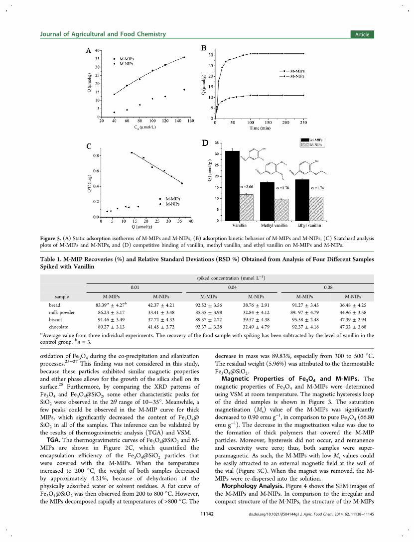
Figure 5. (A) Static adsorption isotherms of M-MIPs and M-NIPs, (B) adsorption kinetic behavior of M-MIPs and M-NIPs, (C) Scatchard analysisplots of M-MIPs and M-NIPs, and (D) competitive binding of vanillin, methyl vanillin, and ethyl vanillin on M-MIPs and M-NIPs.
Table 1. M-MIP Recoveries (%) and Relative Standard Deviations (RSD %) Obtained from Analysis of Four Different SamplesSpiked with Vanillin
spiked concentration (mmol L−1)
0.01 0.04 0.08
sample M-MIPs M-NIPs M-MIPs M-NIPs M-MIPs M-NIPs
bread 83.39a ± 4.27b 42.37 ± 4.21 92.52 ± 3.56 38.76 ± 2.91 91.27 ± 3.45 36.48 ± 4.25milk powder 86.23 ± 5.17 33.41 ± 3.48 85.35 ± 3.98 32.84 ± 4.12 89. 97 ± 4.79 44.96 ± 3.58biscuit 91.46 ± 3.49 37.72 ± 4.33 89.37 ± 2.72 39.57 ± 4.38 95.58 ± 2.48 47.39 ± 2.94chocolate 89.27 ± 3.13 41.45 ± 3.72 92.37 ± 3.28 32.49 ± 4.79 92.37 ± 4.18 47.32 ± 3.68
aAverage value from three individual experiments. The recovery of the food sample with spiking has been subtracted by the level of vanillin in thecontrol group. bn = 3.
Journal of Agricultural and Food Chemistry Article
dx.doi.org/10.1021/jf504144g | J. Agric. Food Chem. 2014, 62, 11138−1114511142

was loose and porous with many cavities, which facilitated themass transport and binding kinetics of the template molecules.Binding Properties of the M-MIPs. To estimate the
adsorption capability of the M-MIPs and M-NIPs, staticadsorption experiments were conducted using different initialconcentrations of vanillin solution. Figure 5A shows that thebinding amount of the M-MIPs and M-NIPs for vanillin wasincreased with the increase in the vanillin concentration.Moreover, the amount of vanillin bound to the M-MIPs wasmuch higher than that bound to the M-NIPs. The Scatchardequation was adopted to evaluate the binding ability of the M-MIPs and M-NIPs.29 The Scatchard equation is expressed asfollows:
= −QC
Q
KQK
max
d d
where Q is the amount of vanillin bound to M-MIPs atequilibrium, Qmax is the apparent maximum binding amount, C0is the initial concentration of vanillin, C is the concentration ofthe supernatant at equilibrium, and Kd is the dissociationconstant.As shown in Figure 5C, the Scatchard plot for the M-MIPs
was a single straight line with a good linear relationship (R2 =0.981), which indicated that the binding sites in the M-MIPswere almost homogeneous. As a control, the M-NIPs withnonlinear correlation possessed fewer homogeneous bindingsites. The linear regression equation for the M-MIPs was Q/C= 1.090 − 0.017Q. The Qmax and Kd were calculated to be 64.12μmol g−1 and 58.82 μmol L−1, respectively, on the basis of the
slope and intercept of the Scatchard plot. These valuesconfirmed the high affinity of the M-MIPs to vanillin.Kinetic adsorption experiments for M-MIPs and M-NIPs
were carried out to investigate the binding process. As shown inFigure 5B, the adsorption capacity of M-MIPs was much higherthan that of M-NIPs. The adsorption amount of M-MIPsincreased rapidly in the first 30 min, and the curve becamerelatively flat until reaching equilibrium after approximately 100min. A similar behavior was observed for the M-NIPs in Figure5B. Cavities with recognition sites on the surface of the M-MIPs were occupied rapidly by template molecules, whichincreased the difficulty of transferring vanillin into the innersites. Given that no specific recognition sites matched vanillinin terms of shape, size, and functional groups in the M-NIPs,the binding amount of the M-NIPs was obviously lower thanthat of the M-MIPs.To investigate the binding specificity of the M-MIPs, a
selectivity test was conducted using structural analogues ofvanillin, methyl vanillin, and ethyl vanillin as controlcompounds. As shown in Figure 5D, the adsorption capacityof the M-NIPs is similar and non-selective for the threecompounds because selective recognition sites are absent in theM-NIPs. However, the M-MIPs showed a significantly higherbinding capacity for vanillin than methyl vanillin and ethylvanillin. The binding capacities of the M-MIPs for vanillin,methyl vanillin, and ethyl vanillin were 31.4, 17.4, and 18.6μmol g−1, respectively. Accordingly, the imprinting factor values(α) of the M-MIPs for vanillin, methyl vanillin, and ethylvanillin were 2.66, 1.78, and 1.74, respectively. These results
Figure 6. HPLC−UV chromatograms of bread, milk powder, biscuit, and chocolate samples. The samples were (a) not spiked, (b) spiked with 0.01mmol L−1 vanillin and without extraction, (c) spiked with 0.01 mmol L−1 vanillin and with M-MIP extraction, and (d) spiked with 0.01 mmol L−1
vanillin and the remaining solution after extraction.
Journal of Agricultural and Food Chemistry Article
dx.doi.org/10.1021/jf504144g | J. Agric. Food Chem. 2014, 62, 11138−1114511143

indicated that the M-MIPs exhibited highly specific recognitionability for vanillin. The high selectivity for vanillin may be basedon two factors: (1) the first factor is the difference in chemicalstructures between vanillin, methyl vanillin, and ethyl vanillin,and (2) only vanillin can specifically match the binding sites ofthe template in terms of size and shape.Method Validation and Application to the Analysis of
Vanillin in Food Samples. To assess the practicalapplicability of the M-MIPs, four food samples (bread, milkpowder, biscuit, and chocolate) were used as real samples in theanalysis. At the three spiked vanillin concentrations of 0.01,0.04, and 0.08 mmol L−1, the extraction recoveries of the M-MIPs were 83.39−95.58% with relative standard deviations of2.48−5.17% (Table 1.). The determined limits of detection andquantification were 2.67 and 8.0 μmol L−1 at signal-to-noiseratios of 3 and 10, respectively. The regression equation wasattained, A(279 nm) = 8.33021x + 2.77378 (R2 = 0.9996) within0.1−5.0 mmol L−1. Moreover, the separation process wasfinished within 10 s when a magnet was used. The results ofHPLC−UV chromatograms (Figure 6) also indicated that M-MIPs had high selectivity and enrichment ability.In summary, novel superparamagnetic particles coated with
vanillin-imprinted polymer were synthesized using MAA as thefunctional monomer and EGDMA as the cross-linker. Theresultant M-MIPs showed some attractive characteristics, suchas higher binding capacity, faster binding kinetics, and quickerseparation. The analytical method based on M-MIPs wassuccessfully applied to vanillin analysis in spiked food sampleswith higher selectivity and concentration. Thus, the approachoffers a simple and straightforward technique for selectiveseparation and fast enrichment of vanillin from complicatedfood matrices.
■ AUTHOR INFORMATION
Corresponding Authors*Telephone/Fax: +86-791-86634810. E-mail: [email protected].*Telephone/Fax: +86-791-86634810. E-mail: [email protected].
FundingThis work was funded by the National Natural ScienceFoundation of China (21201098, 51102131, 31160317,21275158, and 21105117), the Jiangxi Department ofEducation Fund (GJJ13039), and the Specialized ResearchFund for the Doctoral Program of Higher Education(20113601110004).
NotesThe authors declare no competing financial interest.
■ REFERENCES(1) Zabkova, M.; Otero, M.; Minceva, M.; Zabka, M.; Rodrigues, A.E. Separation of synthetic vanillin at different pH onto polymericadsorbent Sephabeads SP206. Chem. Eng. Process. 2006, 45, 598−607.(2) Walton, N. J.; Narbad, A.; Faulds, C.; Williamson, G. Novelapproaches to the biosynthesis of vanillin. Curr. Opin. Biotechnol. 2000,11, 490−496.(3) Nadiah, M. Y.; Eri, T.; Takaomi, K. Molecularly imprintedpolymer particles having coordinated hydrogen bonding in covalent-imprinting for efficient recognition towards vanillin. Sep. Purif. Technol.2014, 122, 341−349.(4) Peng, H. L.; Wang, S. Q.; Zhang, Z.; Xiong, X.; Li, J. H.; Chen, L.X.; Li, Y. B. Molecularly imprinted photonic hydrogels as colorimetric
sensors for rapid and label-free detection of vanillin. J. Agric. FoodChem. 2012, 60, 1921−1928.(5) Zhang, G. W.; Ni, Y. N. Simultaneous spectrophotometricdetermination of vanillin and ethyl maltol in food by multivariatecalibration approach. J. Anal. Sci. 2005, 01, 20−23.(6) Su, L. Q.; Guo, X. L.; Han, S. A. Preparation and evaluation ofvanilli molecularly imprinted polymer microspheres by reversibleaddition−fragmentation chain transfer precipitation polymerization.Anal. Methods 2014, 6, 2512−2517.(7) Dong, Z. Z.; Gu, F. L.; Xu, F.; Wang, Q. H. Comparison of fourkinds of extraction techniques and kinetics of microwave-assistedextraction of vanillin from Vanilla planifolia Andrews. Food Chem.2014, 143, 54−61.(8) Waliszewski, K. N.; Pardio, V. T.; Ovando, S. L. A simple andrapid HPLC technique for vanillin determination in alcohol extract.Food Chem. 2007, 101, 1059−1062.(9) DeJager, L. S.; Perfetti, G. A.; Diachenko, G. W. Comparison ofheadspace−SPME−GC−MS and LC−MS for the detection andquantification of coumarin, vanillin, and ethyl vanillin in vanilla extractproducts. Food Chem. 2008, 107, 1701−1709.(10) Trenholm, R. A.; Vanderford, B. J.; Drewes, J. E.; Snyder, S. A.Determination of household chemicals using gas chromatography andliquid chromatography with tandem mass spectrometry. J. Chromatogr.A 2008, 1190, 253−262.(11) Sostaric, T.; Boyce, M. C.; Spickett, E. E. Analysis of the volatilecomponents in vanilla extracts and flavorings by solid-phasemicroextraction and gas chromatography. J. Agric. Food Chem. 2000,48, 5802−5807.(12) Shen, Y.; Han, C.; Liu, B.; Lin, Z. F.; Zhou, X. J.; Wang, C. J.;Zhu, Z. N. Determination of vanillin, ethyl vanillin, and coumarin ininfant formula by liquid chromatography−quadrupole linear ion trapmass spectrometry. J. Dairy Sci. 2014, 97, 679−686.(13) Song, X. L.; Xu, S. F.; Chen, L. X.; Wei, Y. Q.; Xiong, H. Recentadvances in molecularly imprinted polymers in food analysis. J. Appl.Polym. Sci. 2014, 131, 40766.(14) Jiang, T.; Zhao, L.; Chu, B.; Feng, Q.; Yan, W.; Lin, J. M.Molecularly imprinted solid-phase extraction for the selectivedetermination of 17β-estradiol in fishery samples with high perform-ance liquid chromatography. Talanta 2009, 78, 442−447.(15) Byun, H. S.; Youn, Y. N.; Yun, Y. H.; Yoon, S. D. Selectiveseparation of aspirin using molecularly imprinted polymers. Sep. Purif.Technol. 2010, 74, 144−153.(16) Guo, Y.; Wang, R. Y.; Chi, W. H.; Liu, S. A.; Shi, H. G.; Guo, T.Y. One-step synthesis of reactant-product-dual-template imprintedcapsules as phosphotriesterase mimetic enzymes for pesticideelimination. RSC Adv. 2014, 4, 7881−7884.(17) Wang, S.; Peng, H.; Xiong, H.; Ruan, X.; Huang, S.; Dong, L.Preparation and recognition mechanism of vanillin molecularlyimprinted polymer microspheres. Food Sci. 2012, 33, 1−7.(18) Wang, G. S.; Cao, Q. E.; Xiong, J.; Zhu, X. F.; Hou, N. B.; Ding,Z. T. Preparation and recognition properties of vanillin-imprintedpolymers. Helv. Chim. Acta 2006, 89, 3032−3040.(19) Kong, X.; Gao, R.; He, X.; Chen, L.; Zhang, Y. Synthesis andcharacterization of the core−shell magnetic molecularly imprintedpolymers (Fe3O4@MIPs) adsorbents for effective extraction anddetermination of sulfonamides in the poultry feed. J. Chromatogr. A2012, 1245, 8−16.(20) Lin, Z.; Cheng, W.; Li, Y.; Liu, Z.; Chen, X.; Huang, C. A novelsuperparamagnetic surface molecularly imprinted nanoparticle adopt-ing dummy template: An efficient solid-phase extraction adsorbent forbisphenol A. Anal. Chim. Acta 2012, 720, 71−76.(21) Luo, X.; Zhan, Y.; Huang, Y.; Yang, L.; Tu, X.; Luo, S. Removalof water-soluble acid dyes from water environment using a novelmagnetic molecularly imprinted polymer. J. Hazard. Mater. 2011, 187,274−282.(22) Madrakian, T.; Afkhami, A.; Mahmood-Kashani, H.; Ahmadi, M.Superparamagnetic surface molecularly imprinted nanoparticles forsensitive solid-phase extraction of tramadol from urine samples.Talanta 2013, 105, 255−261.
Journal of Agricultural and Food Chemistry Article
dx.doi.org/10.1021/jf504144g | J. Agric. Food Chem. 2014, 62, 11138−1114511144

(23) Jing, T.; Du, H.; Dai, Q.; Xia, H.; Niu, J.; Hao, Q.; Mei, S.;Zhou, Y. Magnetic molecularly imprinted nanoparticles for recognitionof lysozyme. Biosens. Bioelectron. 2010, 26, 301−306.(24) Dong, L. L.; Peng, H. L.; Wang, S. Q.; Zhang, Z.; Li, J. H.; Ai, F.R.; Zhao, Q.; Luo, M.; Xiong, H.; Chen, L. X. Thermally andmagnetically dual-responsive mesoporous silica nanospheres: Prepara-tion, characterization, and properties for the controlled release ofsophoridine. J. Appl. Polym. Sci. 2014, 131, 40477.(25) Ning, F. J.; Peng, H. L.; Li, J. H.; Chen, L. X.; Xiong, H.Molecularly imprinted polymer on magnetic graphene oxide for fastand selective extraction of 17β-estradiol. J. Agric. Food Chem. 2014, 62,7436−7443.(26) Liu, B.; Han, M.; Guan, G.; Wang, S.; Liu, R.; Zhang, Z. Highlycontrollable molecular imprinting at superparamagnetic iron oxidenanoparticles for ultrafast enrichment and separation. J. Phys. Chem. C2011, 115, 17320−1732.(27) Li, X. Y.; Huang, X. L.; Liu, D. P.; Wang, X.; Song, S. Y.; Zhou,L.; Zhang, H. J. Synthesis of 3D hierarchical Fe3O4/graphenecomposites with high lithium storage capacity and for controlleddrug delivery. J. Phys. Chem. C 2011, 115, 21567−21573.(28) Wang, X.; Wang, L.; He, X.; Zhang, Y.; Chen, L. A molecularlyimprinted polymer-coated nanocomposite of magnetic nanoparticlesfor estrone recognition. Talanta 2009, 78, 327−332.(29) Fan, J.; Wei, Y.; Wang, J.; Wu, C.; Shi, H. Study of molecularlyimprinted solid-phase extraction of diphenylguanidine and itsstructural analogs. Anal. Chim. Acta 2009, 639, 42−50.
Journal of Agricultural and Food Chemistry Article
dx.doi.org/10.1021/jf504144g | J. Agric. Food Chem. 2014, 62, 11138−1114511145