prepared by mrs. rana al-dahlawi rad 453 radiation protection course
Post on 21-Dec-2015
216 views
TRANSCRIPT

Prepared by
Mrs. Rana AL-Dahlawi
RAD 453
Radiation Protection Course

Safety in Various Imaging ModalitiesSafety in Various Imaging Modalities
N.M
MRI
US

Nuclear Medicine ImagingNuclear Medicine Imaging


Gamma Rays are used to form images in nuclearGamma Rays are used to form images in nuclear
medicine. They are emitted by radioactive atoms calledmedicine. They are emitted by radioactive atoms called
RadioisotopesRadioisotopes. . There are two principal hazards in nuclear medicineThere are two principal hazards in nuclear medicine
imaging: imaging:
1.Contamination.1.Contamination.
2. Radiation Exposure.2. Radiation Exposure.
ContaminationContamination is the uncontrolled spread of radioactive is the uncontrolled spread of radioactive
material. It is hazardous because radioactive material canmaterial. It is hazardous because radioactive material can
be inhaled or ingested. be inhaled or ingested. Some radioactive sources used in nuclear medicine poseSome radioactive sources used in nuclear medicine pose
an external radiation hazard. an external radiation hazard.

Hot LabHot Lab


Contamination control requires protective shielding suchContamination control requires protective shielding such
as disposable gloves, gowns, & shoe covers. as disposable gloves, gowns, & shoe covers. Contamination control requires periodic surveys with Contamination control requires periodic surveys with
a Geiger Counter or a portable scintillation counter. a Geiger Counter or a portable scintillation counter.
A chemical fume hood without a charcoal filter is requiredA chemical fume hood without a charcoal filter is required
for storage & use of radioactive gases such as for storage & use of radioactive gases such as 131131--׀׀ & &
Xe-133. Xe-133. Ingestion is minimized by frequent hand washing withIngestion is minimized by frequent hand washing with
soap & warm water. soap & warm water. No eating, drinking or smoking should be allowed inNo eating, drinking or smoking should be allowed in
radioactive materials areas. radioactive materials areas.


The syringe used for administrateRadiopharmaceutical
to the patientmust be shielded .

Patient misadministration can be avoided byPatient misadministration can be avoided byimplementing strict procedures:implementing strict procedures: 1. Physician orders must be verified.1. Physician orders must be verified.2. Mode of administration must be verified. 2. Mode of administration must be verified. 3.Quantity of radioactive material must be verified. 3.Quantity of radioactive material must be verified.
Quantity of radioactive material should be assayedQuantity of radioactive material should be assayedin a dose calibrator. Type of radioactive material mustin a dose calibrator. Type of radioactive material mustbe verified.be verified.
Patient misadministration includes using:Patient misadministration includes using:1.1. Wrong radioisotopes. Wrong radioisotopes. 2.2. Wrong quantity. Wrong quantity. 3.3. Wrong mode of administration. Wrong mode of administration.

Nursing mothers should cease breast feeding for at leastNursing mothers should cease breast feeding for at least
24 hours following Tc-99m diagnostic study. 24 hours following Tc-99m diagnostic study.
Radionuclide therapyRadionuclide therapy with I-131presents special radiation with I-131presents special radiation
protection problems. protection problems. I-131 in capsule form is preferred to that in liquid form.I-131 in capsule form is preferred to that in liquid form.
Iodine- 131 is secreted in urine, saliva & perspiration. Iodine- 131 is secreted in urine, saliva & perspiration.

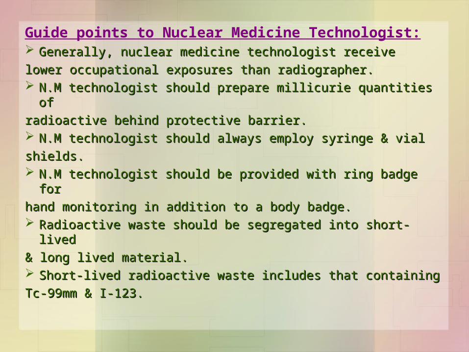
Guide points to Nuclear Medicine Technologist: Generally, nuclear medicine technologist receiveGenerally, nuclear medicine technologist receive
lower occupational exposures than radiographer. lower occupational exposures than radiographer. N.M technologist should prepare millicurie quantities ofN.M technologist should prepare millicurie quantities of
radioactiveradioactive behind protective barrier. behind protective barrier. N.M technologist should always employ syringe & vialN.M technologist should always employ syringe & vial
shields. shields. N.M technologist should be provided with ring badge forN.M technologist should be provided with ring badge for
hand monitoring in addition to a body badge. hand monitoring in addition to a body badge. Radioactive waste should be segregated into short-livedRadioactive waste should be segregated into short-lived
& long lived material. & long lived material. Short-lived radioactive waste includes that containing Short-lived radioactive waste includes that containing
Tc-99mm & I-123. Tc-99mm & I-123.

long-lived radioactive waste includes that containing long-lived radioactive waste includes that containing
Ga-67, I-131,& I-111. Ga-67, I-131,& I-111. Radioactive waste should be stored for decay beforeRadioactive waste should be stored for decay before
disposable in the normal solid waste system. disposable in the normal solid waste system.
Before depositing radioactive waste in a storage decayBefore depositing radioactive waste in a storage decay
receptacle, remove or obliterate all radiation symbols. receptacle, remove or obliterate all radiation symbols.
Radioactive waste must be stored for decay to the levelRadioactive waste must be stored for decay to the level
of natural background before disposable in a sanitaryof natural background before disposable in a sanitary
system. system.

Ring T.L.D is required toRing T.L.D is required tomonitoringmonitoring the accumulated dosethe accumulated dose
resulting from dealingresulting from dealingwith radioactivewith radioactive
materialsmaterials


Dose Calibrator used to assay amount of radioactivityDose Calibrator used to assay amount of radioactivity in a syringe. System is based on a calibrated ionizationin a syringe. System is based on a calibrated ionization
chamberchamber.

Survey instrument used to detect locationSurvey instrument used to detect location & & relative amount of radioactivity. System is based onrelative amount of radioactivity. System is based on a Geiger- Muller countera Geiger- Muller counter . .
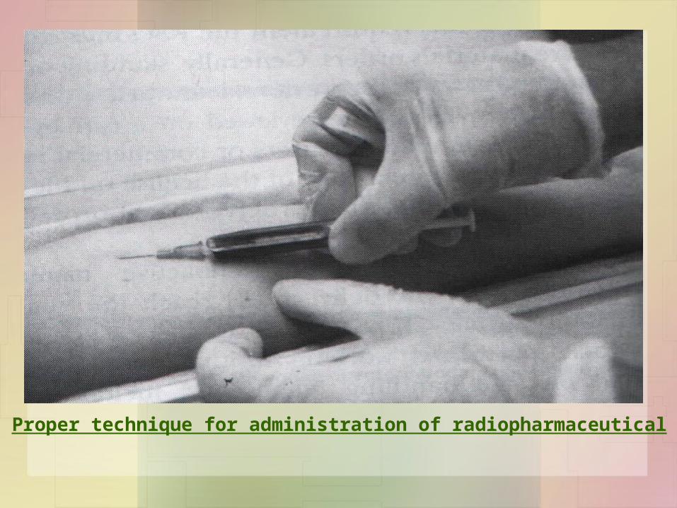
Proper technique for administration of radiopharmaceutical

Magnetic Resonance ImagingMagnetic Resonance Imaging

M.R.I is an imaging modality that doesn’t used ionizingM.R.I is an imaging modality that doesn’t used ionizingRadiation. It used low Radiofrequency therefore there is noRadiation. It used low Radiofrequency therefore there is norisk associated with exposing the patients to this frequency.risk associated with exposing the patients to this frequency.
M.R.I is based on excited protons in the body, thisM.R.I is based on excited protons in the body, thismodality has great contrast over the other modality. modality has great contrast over the other modality.
The primary hazard associated with the static magneticThe primary hazard associated with the static magneticfield is that of ferromagnetic attraction. field is that of ferromagnetic attraction.
When a ferromagnetic object, e.g. one containing iron orWhen a ferromagnetic object, e.g. one containing iron orsteel is close to the magnet if sufficiently close, this cansteel is close to the magnet if sufficiently close, this canturn this object into a dangerous projectile. turn this object into a dangerous projectile.

Practical Guidelines: Practical Guidelines: Everyone entering the magnet room should be carefullyEveryone entering the magnet room should be carefully
screened, using checklist &/or details questioning, toscreened, using checklist &/or details questioning, to
ensure that they don’t have any contraindications to M.R.Iensure that they don’t have any contraindications to M.R.I
either internally or about their person. either internally or about their person.
This includes the patient’s relatives, friend or nurse. This includes the patient’s relatives, friend or nurse.
The key to M.R.I safety in this respect is to be acutelyThe key to M.R.I safety in this respect is to be acutely
vigilant at all time. Metallic object taken into the bore of vigilant at all time. Metallic object taken into the bore of
a magnet may at worse cause serious injury or death & ata magnet may at worse cause serious injury or death & at
best may produce unwanted artifacts on the image. best may produce unwanted artifacts on the image.

M.R.I Contraindications:M.R.I Contraindications: Patients with implanted surgical clips or other Patients with implanted surgical clips or other
ferromagnetic material. ferromagnetic material.

Patients who have engaged in occupations or activitiesPatients who have engaged in occupations or activitiesthat may have caused the accidental lodging ofthat may have caused the accidental lodging offerromagnetic materials. e.g. metal-workers or who mayferromagnetic materials. e.g. metal-workers or who may
have embedded metal fragments from military dutieshave embedded metal fragments from military duties . .

Neonates & infants, for whom data establishing safety are Neonates & infants, for whom data establishing safety are lackinglacking . .
Patients with tattoos, including permanent eye-linerPatients with tattoos, including permanent eye-liner..

Patients with compromised thermoregulatory Patients with compromised thermoregulatory systems, e.g. neonate, low-birth-weight systems, e.g. neonate, low-birth-weight infants, certain cancer patients. infants, certain cancer patients.
Patients with metallic implants which may Patients with metallic implants which may cause artifacts in the images due to distortioncause artifacts in the images due to distortion
of the static magnetic field. of the static magnetic field.

Patients with prosthetic heart valves. Patients with prosthetic heart valves.
Pregnant patients (the safety of M.R.I has not been completely Pregnant patients (the safety of M.R.I has not been completely established for embryo & fetus)established for embryo & fetus)..

Diagnostic Ultrasound ImagingDiagnostic Ultrasound Imaging

There are tow types of responses to ultrasoundThere are tow types of responses to ultrasound
exposure- thermal responses and non thermal responses.exposure- thermal responses and non thermal responses.
Both thermal and non thermal responses have beenBoth thermal and non thermal responses have been
observed only at intensities much higher than thatobserved only at intensities much higher than that
employed in diagnostic ultrasound. employed in diagnostic ultrasound.
Thermal responses are heating of soft tissue and bone.Thermal responses are heating of soft tissue and bone.
Non thermal responses are mechanical in natureNon thermal responses are mechanical in nature
cavitation , tiny bubble formation, & membrane shearing.cavitation , tiny bubble formation, & membrane shearing.


There has never been a report of patient injury fromThere has never been a report of patient injury from
diagnostic ultrasound examination.diagnostic ultrasound examination.
There are no concerns of operator safety in applicationsThere are no concerns of operator safety in applications
of diagnostic ultrasound. of diagnostic ultrasound.
There are concerns of patient safety in diagnosticThere are concerns of patient safety in diagnostic
ultrasound because of expanding application &ultrasound because of expanding application &
increasing beam intensity.increasing beam intensity.
For more ultrasound beam, highest tissue heatingFor more ultrasound beam, highest tissue heating
occurs near the skin.occurs near the skin.

High intensity ultrasound waves can permanently alterHigh intensity ultrasound waves can permanently alter
the structure of the tissue through which they pass. the structure of the tissue through which they pass.
These effects range from slight warming of the tissues toThese effects range from slight warming of the tissues to
total necrosis & destruction. total necrosis & destruction.
The power level that produce these changes are manyThe power level that produce these changes are many
orders of magnitude greater than the power level that areorders of magnitude greater than the power level that are
used in diagnostic scanning. used in diagnostic scanning.
However, as far as our present knowledge goes noHowever, as far as our present knowledge goes no
immediate manifest injury or late effect has over occurred inimmediate manifest injury or late effect has over occurred in
human exposed to diagnostic level of medical ultrasound. human exposed to diagnostic level of medical ultrasound.

