presentation for phi sigma fall 2015
TRANSCRIPT

TARGETING Breast Cancer: What specific polymorphisms in the immune pathways of
breast cancer increase risk of breast carcinogenesis?
Caelie KernLyon, France
UNH Mentor: Dr. Charles WalkerForeign Mentor: Dr. David Cox
Centre Léon Bérard: Centre de Recherche en CancérologieInstitut National de la Santé et de la Recherche Médicale

CK3 – mRNA levels in OIS cellules
L106HMEC hTERT Rasv12 ± Doxycycline
L106: J2 J3 J4 J5Éléments du RLR pathway
qPCR
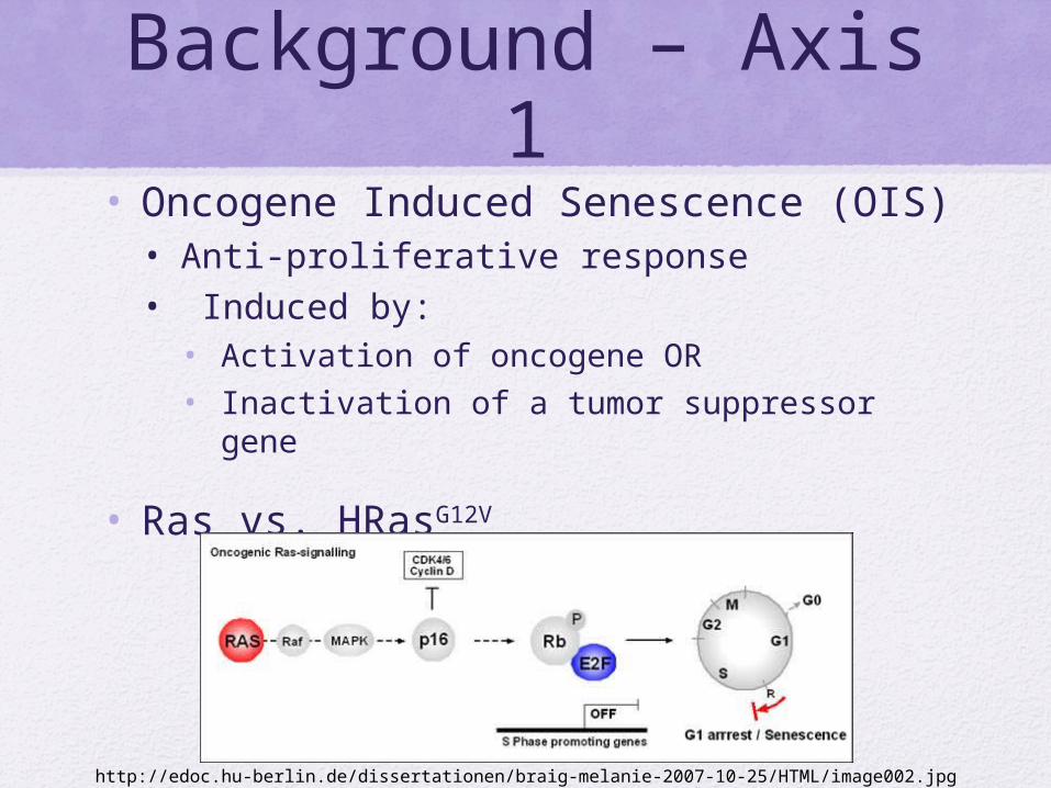
Background – Axis 1• Oncogene Induced Senescence (OIS)
• Anti-proliferative response• Induced by:
• Activation of oncogene OR• Inactivation of a tumor suppressor gene
• Ras vs. HRasG12V
http://edoc.hu-berlin.de/dissertationen/braig-melanie-2007-10-25/HTML/image002.jpg

Background – Axis 1
• RLR – Rig-like receptors• Respond to dsRNA • Trigger antiviral, antitumor,
and anti-proliferative response
• Activated pathways plays protective role against cancer
www.invivogen.com

Background – Axis 1• HTMM hTERT RasG12V
• Immortalized breast cancer cell line• Telomerase reverse transcriptase
• Mutated Ras via Lentivirus vector• Continuously proliferating
• Treatment with Doxycycline• Induces cellular senescence
https://www.systembio.com/support/resources/faqs/lenti

Cinétique d’inductionProtocoles
Echantillons à tester (L106)Cellules: HTMM Ras p34,5Conditions: J2, J3, J4, J5 ± DOX3 culture-dishes de 10Ø/conditionsPlatées à 0,5%
J-1 J0 J1 J2 J3 J4 J5
+Medium
Medium ± dox (Iug/ml)
0,5%: 2,5.104 cel/cult dish 10Ø
Contrôles positifs et négatifs (L106)Cellules: HTMM Ras p38Conditions: ± IFNα pendant 8 heures3 culture-dishes de 10Ø/conditionsPlatées à 50%
J-1(soir) J0J0+8heures
+Medium
Medium ± IFNα (I000 U/ml)
HTMMRas à 50%: 2,5.106 cel/cult dish 10Ø

Experimental procedureqPCR
RNA extrait: Macherey Nagel NucleoSpin RNA
Pour RT mRNA cDNA: BioRad iScript cDNA Synthesis Kit, 1ug mRNA utilisé
Primers: FW/RV à 10mM - 10uL de Primer FW + 10uL de Primer RV + 80uL d’H2O = 100uL
Échantillons: cDNA 1 ug/mL dilué 1/20
Master Mix: pour les échantillons et l’H2O en duplicata (soit 4 échantillons + 2 H2O)*2)Master Mix pour 12
each/gèneVol. Total: 15uL* Vol. Total :
SYBR Green 7,5 uL * 25 187,5 uL
Primers F/R (10uM) 0,4 uL * 25 10 uL
H2O 7,1 uL * 25 177,5 uL
Stage 1: 95 °C, 5minStage 2: 95 °C, 15s; 60 °C, 15s; 72 °C, 30sStage 3: 95 °C, 15s; 60° C, 20s; 95 °C, 15sSample volume: 20 uLData collection: Stage 2, Step 3Check t°: 0,7
Appareils:
• Eppendorf Thermocycler
• BioSystems StepOnePlus Real Time PCR System

RIG-I
L106 ± DoxFW: 5’ – AGC TCA GCT TGA TGA GGG ACA – 3’Rv: 5’ – GTC TGG CAT CTG GAA CAC CA – 3’
Primers from Ablasser JI 2009

RIG-I
J2 J3 J4 J50
0.5
1
1.5
2
2.5
3
3.5
4
mRNA Expression of RIG-I on L106 ± Dox
no doxavec dox
Fold
incr
ease
of
RIG
I mRN
A ex
pres
sion
(U
A)
no IFNa IFNa0
10
20
30
40
50
60
mRNA Expres-sion of RIG-I on
HTMM Ras ± IFN-α
Fold
incr
ease
of
RIG
-I m
RNA
expr
essi
on (
UA)

Results Axis 1• Proteins up-regulated due to
doxycycline treatment:• RIG-I, MDA-5, MAVS, IKKε, ISG20, CYLD,
NLRP3, ULBP2
• Conclusion:• Cells treated with doxycycline exhibit:
• Change in morphology• Changes in expression levels of RLR
Pathway Senescence!

CK6 – In Vitro Generation of XCR1+ DC from Cord Blood CD34+ Cells
Phéno de Différenciation et Activation par Ligands STING

Background – Axis 2• Dendritic cells
• Antigen-presenting cells• Interface between innate and adaptive immune
systems
• BDCA3+ CD141+• DC subset• Superior capacity to present Ag• Activated BDCA3+ = stronger response
• Excellent target for vaccine against cancer
http://www.blog.bryanmjones.com/2013/12/dendritic-cell-illustration.html
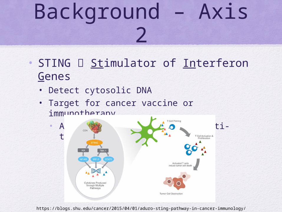
Background – Axis 2• STING Stimulator of Interferon Genes
• Detect cytosolic DNA• Target for cancer vaccine or immunotherapy
• Activation triggers natural anti-tumor response
https://blogs.shu.edu/cancer/2015/04/01/aduro-sting-pathway-in-cancer-immunology/

Echantillons à tester (CK2)Cellules: 1. CBO26 – 500,000 cel/cult2. CBO+55 – 400,000 cel/cult 4. C97.58 - ?#5. C94_79 – 300 000 cellules6. C96-180 – 350 000 cellules7. C96-163 – 350 000 cellules
Purification HSC
J0 J7 J13 J17
Décongélation HSCMise en
différenciationAjoute cytokines de différenciation
Prolifération
StemSpan +SCF: 100ng/mLFlt3-L: 100ng/mLIL-3: 20ng/mLIL-6: 20ng/mL
RMPIc: 20 uL/puitGM-CSF: 20ng/mLSCF: 20ng/mLFlt3-L: 100ng/mLIL-4: 100ng/mL
RMPIc +GM-CSF: 20ng/mLSCF: 20ng/mLFlt3-L: 100ng/mLIL-4: 100ng/mL
Phéno de différenciation
Conditions: J0- J7 2x 25cm flasks J7-J17 96 puits plaque (U-bottom)
Prolifération et Différenciation Protocole

Donneur 1
Donneur 2
FSC/SSC Singulet FSC
Gate sur les cellules vivantes =Cellules DAPI- CD11b/BDCA3 CD11b/Clec9A BDCA3/Clec9A
FSC/SSC Singulet FSC
Gate sur les cellules vivantes =Cellules DAPI- CD11b/BDCA3 CD11b/Clec9A BDCA3/Clec9A
Petit Phenotype – FACSFluorescence-Activated Cell Sorting

Methods – Axis 2• BDCA3+ Activation
• Increases expression of surface protein CLEC9A
• Controls:• Lipofectamine• Poly:IC
• Experimental Treatments: Cyclic Dinucleotides• 2,3’ - cGAMP• 2,3’ - cGAMP PS2

J18 J20
Activation par ligandsPhéno
d’activation
0,1 ug/ml 2-3' cGAMP1 ug/ml 2-3' cGAMP10 ug/ml 2-3' cGAMP 0,1 ug/ml 2-3' cGAMP PS2 1 ug/ml 2-3' cGAMP PS210 ug/ml 2-3' cGAMP PS2
Mileau
PBS1xPoly ICLipopolysaccharideLipofectamine
Donneur 1
0,1 ug/ml 2-3' cGAMP1 ug/ml 2-3' cGAMP10 ug/ml 2-3' cGAMP50 ug/ml 2-3’ cGAMP 0,1 ug/ml 2-3' cGAMP PS2 1 ug/ml 2-3' cGAMP PS210 ug/ml 2-3' cGAMP PS250 ug/ml 2-3’ cGAMP PS2Mileau
PBS 1xPoly IC LipopolysaccharideLipofectamine Donneur 2
Echantillons à tester (CK2)Cellules: 1. CBO26 – 500,000 cel/cult2. CBO+55 – 400,000 cel/cult 4. C97.58 - ?#5. C94_79 – 300 000 cellules6. C96-180 – 350 000 cellules7. C96-163 – 350 000 cellules
Conditions: J0- J7 2x 25cm flasks J7-J17 96 puits plaque (U-bottom)
Activation Protocole

No stimulation
cGAMP ps2 1ug/ml
cGAMP ps2 10ug/ml
cGAMP ps2 50ug/ml
CLEC9A CLEC9ACD11b/BDCA3

Results Axis 2• Success of cord blood differentiation:
• Donor 1: 3% BDCA3+• Donor 2: 12% BDCA3+
• cGAMP PS > cGAMP• Increased expression of CLEC9A
• Problem:• Fixation technique reduced viability of cells
Referenceshttp://www.jbc.org/content/282/21/15315
http://jem.rupress.org/content/207/6/1247
http://www.cs.tau.ac.il/~spike/maps/spike00028.html
http://www.ncbi.nlm.nih.gov/pubmed/19330805
http://www.ncbi.nlm.nih.gov/pmc/articles/PMC3354961/
http://www.ncbi.nlm.nih.gov/pubmed/20032638
http://www.abcam.com/protocols/flow-cytometry-immunophenotyping
http://www.nature.com/nrc/journal/v6/n6/fig_tab/nrc1884_F1.html
http://edoc.hu-berlin.de/dissertationen/braig-melanie-2007-10-25/HTML/chapter1.html
https://blogs.shu.edu/cancer/2015/04/01/aduro-sting-pathway-in-cancer-immunology/
http://www.invivogen.com/review-rlr
https://books.google.com/books?id=Vwu7BAAAQBAJ&pg=PA115&lpg=PA115&dq=RLR+and+cancer&source=bl&ots=I75ndUvA_H&sig=Nq8bVnXDs9voVHtKeOpz-QVtb1s&hl=en&sa=X&ved=0CEgQ6AEwCGoVChMIlpaP8-yjyAIVgTI-Ch15jAku#v=onepage&q=RLR%20and%20cancer&f=false