prevalence and characterization of cardiac … · prevalence and characterization of cardiac...
TRANSCRIPT

PREVALENCE AND CHARACTERIZATION OF CARDIAC PATHOLOGY INDUCED BY THE PARASITIC NEMATODE PHILOMETRA SALTATRIX IN JUVENILE BLUEFISH OF THE HUDSON RIVER ESTUARY, NEW YORK
A Final Report of the Tibor T. Polgar Fellowship Program
Sarah E. Koske
Polgar Fellow
School of Veterinary Medicine
University of Wisconsin-Madison Madison, WI 53706
Project Advisor:
Francis Juanes Department of Environmental Conservation
University of Massachusetts-Amherst Amherst, MA 01003
Koske, S.E. and Juanes, F. 2012. Prevalence and Characterization of Cardiac Pathology Induced by the Parasitic Nematode Philometra saltatrix in Juvenile Bluefish of the Hudson River Estuary, New York. Section IV: 1-45 pp. in D.J. Yozzo, S.H. Fernald and H. Andreyko (eds.), Final Reports of the Tibor T. Polgar Fellowship Program, 2011. Hudson River Foundation.
IV-1
ABSTRACT
Philometra saltatrix is a nematode parasite of bluefish that infects the ovaries of
adult bluefish as well as the pericardial cavity of juveniles. Ovarian infection has been
linked to decreased fecundity, and pericardial infection has been described as fatal via
constrictive pericarditis. The life cycle of this parasite is unknown.
Eighty juvenile bluefish sampled from the Hudson River Estuary (HRE) between
July and October 2010 were examined systematically for the parasite. Histopathology
revealed pericardial infections were associated with chronic granulomatous
inflammation, pericardial thickening and fibrosis, and pericardial adhesions over a range
of severities. Mild pathology was associated with viable female worms, while the most
severe pathology was associated with dead gravid females with or without the release of
larvae in the pericardial cavity. Overall prevalence of infection in the pericardium was
67.5% (54/80). Overall prevalence of fatal severity infection was 25% (20/80). No
difference in overall prevalence, severity of infection, or prevalence of fatal infection was
observed between the spring and summer-spawned bluefish cohorts. No difference in
condition factor was observed between parasitized and non-parasitized juveniles.
Pericardial infection with Philometra saltatrix should be considered an important
source of mortality in juvenile bluefish of the HRE. In severely affected fish, constrictive
pericarditis has the potential to impact ability to hunt and forage, pursue normal
migratory behavior, and could result in increased susceptibility to predation. Future
studies using full calendar year sampling periods, as well as experimental infections, are
needed in order to fully evaluate the effects of this parasite on juvenile bluefish.
IV-2

TABLE OF CONTENTS
Abstract………………………………………………………………………… IV-2 Table of contents……………………………………………………………….. IV-3 Lists of figures and tables…………………………………………………........ IV-5 Introduction…………………………………………………………………….. IV-6 The host-parasite interaction…………………………………………… IV-8 Methods………………………………………………………………………… IV-10 Sampling locations……………………………………………………… IV-10 Sample processing and pathology scoring……………………………… IV-12 Histopathology………………………………………………………….. IV-16 Data analysis……………………………………………………………. IV-17 Results…………….……………………………………………………………. IV-18
Cohort assignment……….……………………………………………… IV-18
Prevalence……….……………………………………………………… IV-19
Pathology……….…………………………………………………….... IV-21
Condition factor……….………………………………………………... IV-23
Histopathology……….………………………………………………… IV-23 Discussion………………………………………………………………………. IV-27 Pathology and pathophysiology…………..…………………………….. IV-27 Histopathology………………………………………………………….. IV-28 Pathophysiology………………………………………………………… IV-29 The “grade three” infection……………………………………………... IV-32
IV-3
Parasite ecology……………………………………………………...… IV-33 Condition factor and infection…………………………………………. IV-37 Conclusions…………………………………………………………………….. IV-39 Recommendations……………………………………………………………… IV-40 Acknowledgements…………..………………………………………………… IV-42 Literature cited…………………………………………………………………. IV-43
IV-4
LIST OF FIGURES AND TABLES Figure 1 - Section of the lower Hudson River illustrating bluefish
sampling locations…………………………………………….… IV-11 Figure 2 - Example photographs of Gross Pathology Scores………..…...… IV-15 Figure 3 - Weekly pericardial infection prevalence by cohort over
sampling period ……………………………………………..….. IV-20 Figure 4 - Representative histopathology photomicrographs………….…… IV-26 Table 1 - Pathological characteristics evaluated on gross dissection…….… IV-14 Table 2 - Prevalence of Final Heart Pathology scores in juvenile
bluefish of the Hudson River Estuary………………………….... IV-21
IV-5
INTRODUCTION
The mechanisms of recruitment in marine fishes have been a topic of much
interest in the last 20 years (Rothschild 1986; Beyer 1989; Beamish and McFarlane
1989). Hjort (1914) proposed that the year-class strength of marine fish populations is
determined in the very early life stages when mortality rates are high. Small variations in
juvenile mortality rates can affect recruitment processes resulting in large fluctuations in
abundance and survival to the adult population (Houde 1987). Factors that influence
variable mortality rates in juveniles include egg size, larval and juvenile growth rates,
predation, parasitism, and food availability (Juanes and Conover 1995; Chambers and
Trippel 1997).
One species that is vulnerable to recruitment variability is the bluefish,
Pomatomus saltatrix. Bluefish is a globally important, and highly migratory pelagic
species found worldwide in subtropical and temperate waters (Juanes et al. 1996). Adults
can reach one meter in length, and are important to the recreational game fishing industry
and as a major marine predator. Along the United States coast, bluefish occur seasonally
in the western Atlantic Ocean from Maine to Florida (Kendall and Walford 1979)
migrating in loosely aggregated schools of similarly sized individuals (Olla and
Studholme 1971). Bluefish reproduce multiple times along the eastern coast of the United
States during annual spawning migrations. Although the exact temporal and
spatial patterns of bluefish spawning remain uncertain, at least two cohorts (spring and
summer) of juveniles are evident as a result of spawning over the continental shelf (Hare
and Cowen 1996). The spring-spawned cohort results from spawning between Cape
Hatteras, NC and Cape Canaveral, FL (March – May). The summer-spawned cohort
IV-6

originates from spawning between Cape Hatteras and Cape Cod, Massachusetts (June –
August).
The Hudson River estuary is an important juvenile bluefish nursery area, and is
used by the spring and summer cohorts of juvenile bluefish throughout the summer.
Juvenile bluefish typically migrate into the Hudson River estuary (HRE) in June and
emigrate by November. Otolith microstructure analysis has revealed that in early
summer, juvenile bluefish abundance is dominated by the spring cohort, while late
summer and early fall catches are comprised mostly of summer-spawned juveniles
(Stormer and Juanes 2008).
The relative contribution of the spring and summer cohort to the western Atlantic
population varies and is the current topic of some debate (Juanes et al. 1996; Hare and
Cowen 1996; Munch and Conover 2000; Conover et al. 2003). In the 1950s, the relative
abundance of spring and summer-spawned cohorts was nearly equal (Lassiter 1962).
From 1973-1995, spring-spawned bluefish dominated the cohort structure of juvenile
bluefish inhabiting the Mid-Atlantic Bight (Munch and Conover 2000). However, for
reasons unknown, an apparent shift in recruitment has favored the summer-spawned
cohort since the mid 1990s (Conover et al. 2003). Unfortunately, the summer-spawned
cohort does not appear to be contributing proportionally beyond the juvenile stage
(Conover et al. 2003). The apparent recruitment failure of a large portion of the summer
cohort coupled with disproportionate abundance of spring-spawned juveniles has been
implicated in the recent decline in adult stock size (Klein-MacPhee 2002).
IV-7

The host-parasite interaction
Parasitic infections by nematodes in marine fishes may make wild populations
vulnerable to other biotic and abiotic stressors such as predation and water
contamination. Conversely, environmental stressors such as coastal pollution, and climate
change may make fish more vulnerable to infections through immunosuppression (Sures
and Knopf 2004b). Infection may increase mortality, affect growth and reproduction, or
compromise the condition of a fish.
Philometra saltatrix is a nematode parasite of the superfamily Dracunculoidea,
family Philometridae, that appears to be specific to bluefish (Moravec et al. 2008).
Dracunculoids, in general, are mostly parasites of various tissues and body cavities, and
depending on species, can be found in skin, subcutaneous tissue, skeletal muscle, eyes,
orbits, gills, swim bladder, kidneys, gonads, and circulatory system (Moravec 2004).
Philometra saltatrix has been found in the adult female bluefish ovary (Ramachandran
1973; Clarke et al. 2006), as well as the pericardial cavity of juvenile bluefish (Cheung et
al. 1984).
Gravid female worms reach a maximum of 300mm in length (Koske and
Pinkerton 2010, unpublished research) and 300μm in width (Clarke et al. 2006).
Subgravid females range from 36-75mm in length and 231-462μm in width. Mature male
worms are 2-3mm long (Moravec et al. 2008) while juvenile male and female worms
average 2mm in length (Clarke et al. 2006). The presence of Philometra saltatrix in the
pericardial sac of juvenile bluefish and the ovary of adult females has led to the
hypothesis that gravid females migrate to the ovary in adult female bluefish near the time
of spawning, most likely under hormonal cues (Clarke et al. 2006). However, the life
IV-8
cycle of this parasite remains unclear. Infections by Philometrid nematodes have been
associated with ovarian damage in adults of several marine species (Ramachandran 1975;
Hine and Anderson 1981; Oliva et al. 1992). In bluefish, Clarke et al. (2006) and Burak
(2007) documented a variety of ovarian disorders associated with Philometra saltatrix
infection including hemorrhage, inflammation, edema, fibrosis, and follicular atresia.
In addition, Koske and Pinkerton (2010) reported that severe infections with
Philometra saltatrix in the pericardial cavity of juvenile bluefish cause severe chronic
granulomatous inflammation and pericardial fibrosis, especially after the death of the
gravid female parasite, with or without release of larvae into the pericardial cavity.
Severe chronic inflammation leads to diffuse pericardial and epicardial thickening and
fibrosis, a condition called constrictive pericarditis, where the fused, thickened
pericardium restricts proper filling of the ventricle (Koske and Pinkerton 2010). This
condition is capable of leading to eventual heart failure in affected individuals (Asher and
Klein 2002). In mammals, symptoms of this condition include exercise intolerance, heart
failure with liver congestion and ascites, and sudden collapse. By extension, we also
expect the same physiological changes to occur in fish.
Clarke et al. (2006) observed significantly greater Philometra saltatrix prevalence
in the ovaries of summer-spawning than in spring-spawning adult bluefish. It is possible
that infection of bluefish offspring is associated with proximity to infected adults,
possibly leading to increased vulnerability of summer-spawned juveniles to infection.
Moreover, if both ovarian and pericardial forms of infection cause severe pathologies, the
interaction between summer-spawned juvenile bluefish and Philometra saltatrix may be a
causal mechanism for the recent recruitment failure exhibited by the summer cohort.
IV-9
Characterization of the nature and severity of pathology induced by infection with
Philometra saltatrix in the pericardial cavity of juveniles, as well as estimation of the
prevalence of fatal infection, will help reveal the true impact of this parasite in bluefish.
METHODS
Sampling locations
Juvenile age-0 bluefish were sampled bi-weekly from late July 2010 to October
2010 from stations along a 65km stretch of the lower Hudson River (Figure 1) by the
New York State Department of Environmental Conservation.
IV-10

Figure 1. Section of the lower Hudson River illustrating bluefish sampling locations. Straight lines indicate sampling stations.
IV-11
Fish classified as juveniles were those spawned during the year 2010 of both
spring and summer-spawned cohorts, and ranged in length between 53-190 mm fork
length. Bluefish were collected with a 61m x 3m beach seine with 13 mm stretched mesh
wings and a 6 mm stretched mesh bag. Seine hauls were set from a boat and parallel to
shore. Catches were processed on shore, with juvenile bluefish preserved frozen for later
dissection and analysis.
Sample Processing and Pathology Scoring
Frozen bluefish were thawed in cold water in groups of three to six fish at a time
to minimize autolysis during room temperature conditions in the laboratory. The
sampling date, cruise identification number, station number along the river, total length
(mm), fork length (mm), standard length (mm), and wet weight (g) were recorded and the
fish assigned a fish identification number. The external surfaces of the fish, the oral
cavity, and the gills were examined for evidence of gross pathology such as traumatic
lesions, external parasites, or signs of systemic disease. Any findings were noted for
consideration in conjunction with later results if necessary.
The ventrum of the fish was incised with mayo scissors from the vent to the base
of the opercula, carefully avoiding internal organs, especially the heart and pericardium.
The surfaces of the internal organs, including intestine, stomach, pyloric caeca, liver,
kidney, swim bladder, and coelomic cavity were examined closely for gross pathology,
degree of autolysis, and evidence of nematode or other parasitic infections both with the
naked eye and under a dissecting microscope. Any observed pathology was noted, as was
IV-12

the location of any parasites found, including Philometra saltatrix. If Philometra saltatrix
adults or larvae were found in the coelomic cavity, including on the mesenteries of
organs or on the body wall itself, the reproductive stage of the parasite was determined
using visualization of the uterine contents under a dissecting and compound microscope.
The three stages included: immature (I), as described by a small, immature uterus
containing small, undeveloped eggs; subgravid (SG), described by a mature worm with
uterus containing well-developed eggs; and gravid (G), described by a uterus containing a
mix of well-developed eggs and hatched larvae, or solely hatched larvae (after Moravec
and de Buron 2009).
The intact pericardium was examined closely for presence of Philometra saltatrix
adults and larvae. Digital photographs were taken to document the gross in situ
appearance of the heart and nematode position, physical condition of the parasites,
relative numbers, and any associated pathology.
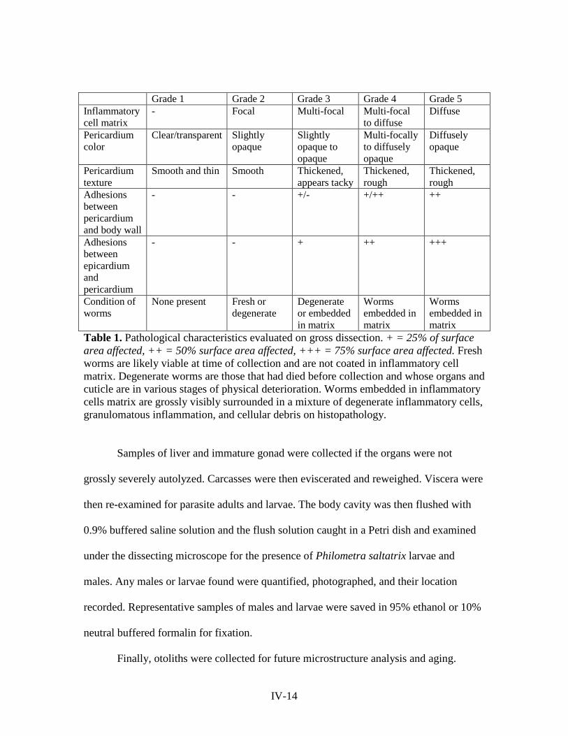
Gross pathological findings were recorded and the heart assigned a gross
pathology severity score of 0-5 (Figure 2) based on the severity of pathology in six
specific areas outlined in Table 1. A score of 0 indicated no infection/pathology and a
score of 5 indicated severe infection and pathology.
The heart and pericardium were carefully excised intact and as a whole and
transferred to a jar containing 10% neutral buffered formalin (Globe Scientific, NJ, USA)
for fixation. If the pericardium broke during transfer, the nematodes were individually
reproductively staged as described above.
IV-13
Grade 1 Grade 2 Grade 3 Grade 4 Grade 5 Inflammatory cell matrix
- Focal Multi-focal Multi-focal to diffuse
Diffuse
Pericardium color
Clear/transparent Slightly opaque
Slightly opaque to opaque
Multi-focally to diffusely opaque
Diffusely opaque
Pericardium texture
Smooth and thin Smooth Thickened, appears tacky
Thickened, rough
Thickened, rough
Adhesions between pericardium and body wall
- - +/- +/++ ++
Adhesions between epicardium and pericardium
- - + ++ +++
Condition of worms
None present Fresh or degenerate
Degenerate or embedded in matrix
Worms embedded in matrix
Worms embedded in matrix
Table 1. Pathological characteristics evaluated on gross dissection. + = 25% of surface area affected, ++ = 50% surface area affected, +++ = 75% surface area affected. Fresh worms are likely viable at time of collection and are not coated in inflammatory cell matrix. Degenerate worms are those that had died before collection and whose organs and cuticle are in various stages of physical deterioration. Worms embedded in inflammatory cells matrix are grossly visibly surrounded in a mixture of degenerate inflammatory cells, granulomatous inflammation, and cellular debris on histopathology.
Samples of liver and immature gonad were collected if the organs were not
grossly severely autolyzed. Carcasses were then eviscerated and reweighed. Viscera were
then re-examined for parasite adults and larvae. The body cavity was then flushed with
0.9% buffered saline solution and the flush solution caught in a Petri dish and examined
under the dissecting microscope for the presence of Philometra saltatrix larvae and
males. Any males or larvae found were quantified, photographed, and their location
recorded. Representative samples of males and larvae were saved in 95% ethanol or 10%
neutral buffered formalin for fixation.
Finally, otoliths were collected for future microstructure analysis and aging.
IV-14
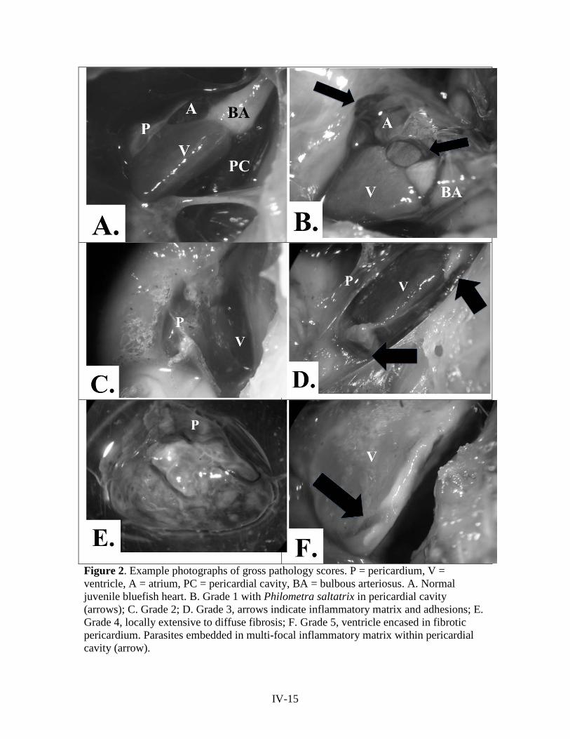
Figure 2. Example photographs of gross pathology scores. P = pericardium, V = ventricle, A = atrium, PC = pericardial cavity, BA = bulbous arteriosus. A. Normal juvenile bluefish heart. B. Grade 1 with Philometra saltatrix in pericardial cavity (arrows); C. Grade 2; D. Grade 3, arrows indicate inflammatory matrix and adhesions; E. Grade 4, locally extensive to diffuse fibrosis; F. Grade 5, ventricle encased in fibrotic pericardium. Parasites embedded in multi-focal inflammatory matrix within pericardial cavity (arrow).
IV-15
Histopathology
Formalin-fixed hearts were sectioned using a sectioning blade and included a
uniform section through the bulbous arteriosus, ventricle, and atrium. The sections were
placed in fine mesh tissue cassettes in 10% buffered formalin and submitted for
histological processing according to standard techniques and staining with hematoxylin
and eosin. Hearts were examined via high-power light field microscopy and pathological
findings and changes described for each sample. Slides with profound autolysis that
precluded accurate description of the cellular characteristics as well as those with
incomplete sectioning of all three heart divisions were omitted from histologic
assessment.
Slides were evaluated to assess severity and extent of inflammation, degree and
extent of pericardial fibrosis, physical condition of worms present in the pericardial
cavity, reproductive stages of worms present, and cellular character and extent of
inflammatory cell matrix present.
Histopathologic examination was used to confirm or amend the gross pathology
score assigned at initial dissection, based on the criteria above. The score that resulted
after any amendments was termed the Final Pathology Score (FPS). Final pathology
scores of grade four or five were determined to be of fatal severity based on the severity
and extent of pericardial fibrosis.
Slides of liver from both normal and pericardial cavity-infected fish were
submitted for processing as well as immature gonad samples.
IV-16
Data Analysis
Cohort assignment was accomplished by creating a length-frequency distribution
for each month sampled using R statistical software, and the distribution analyzed to
separate the fish into spring and summer-spawned cohorts by size.
Condition factor was calculated using the established formula:
K=weight in grams * 100
(fork length in cm)3
Infection in the pericardium was defined as the presence of any sex or stage
nematode in the pericardial cavity. Overall prevalence of pericardial infection was
defined as the number of fish from both cohorts, combined, with any sex or stage worm
present in the pericardial cavity divided by the total number of fish examined from both
cohorts combined. Prevalence of infection in the spring and summer-spawned cohorts
individually was defined by the number of fish infected in the pericardial cavity in the
cohort of interest divided by the total number of fish examined from the cohort of
interest.
The prevalence of pericardial cavity infection over time in a cohort was calculated
as the number of fish infected in the pericardium from the cohort of interest sampled
during a specific week period divided by the number of fish from the cohort of interest
sampled during that week period.
R statistical software was used for graphical representation of data and statistical
analysis. Fish fork length, condition factor, and mass were tested for normality using the
IV-17

Shapiro-Wilk normality test. Percent prevalence of infection, Final Pathology Scores
(FPS), and numbers of infected individuals between cohorts were compared using
Pearson's Chi-squared test with Yates' continuity correction. Means of non-normally
distributed data were analyzed non-parametrically using a Wilcoxon rank sum test with
continuity correction. A Welch Two Sample t-test was used to compare means among
normally distributed data. Spearman's rank correlation rho was used to assess correlation
between non-normally distributed numeric variables. A p value ≤ 0.05 was considered
significant in all tests.
Data was analyzed with both cohorts combined, and with cohorts individually
subsetted into spring and summer.
RESULTS
Cohort assignment
A total of 80 juvenile bluefish were processed. Fork lengths for all fish were not
normally distributed (Shapiro-Wilk normality test, W = 0.9672, p-value = 0.03742).
Length-frequency distribution analysis by month resulted in bimodal distributions with
two distinct length populations of juveniles representing the two cohorts. The mean fork
length (FL) for the spring cohort was 149.48mm (n=45) and 105.51mm (n=35) for the
summer cohort. Spring cohort fork length was normally distributed, while summer cohort
FL was not. A Wilcoxon rank sum test with continuity correction confirmed the
difference between the mean fork lengths of the cohorts was significant (W = 1427.5, p-
value = 5.531e-10).
IV-18
Prevalence
The overall prevalence of infection in the pericardium was 67.5% (54/80). While
the spring-spawned cohort prevalence of pericardial infection was 68.89% (31/45) and
the summer-spawned 65.71% (23/35), the difference in infection prevalence between
cohorts was not significant (Pearson's Chi-squared test with Yates' continuity correction,
X-squared = 0.0036, df = 1, p-value = 0.952). When looking at only infected fish,
35.48% (11/31) of the spring cohort and 39.13% (9/23) of the summer cohort had
potentially fatal infections. No significant difference in the number or percent prevalence
of fatal grade infections in infected fish existed between cohorts (Pearson's Chi-squared
test with Yates' continuity correction, p-value = 0.9916, p-value = 0.6984, respectively).
The percent prevalence of pericardial infection over time indicated that
prevalence increased in the spring cohort over mid-summer and declined to zero as the
spring cohort migrated out of the estuary (Figure 3). The spring cohort was no longer part
of samples collected after 9/27/2010. The summer cohort was first found in samples
beginning 8/2/2010. Infection prevalence was at 100% for the summer cohort when the
sampling period for the year ended on 10/25/2010.
IV-19

Figure 3. Weekly pericardial infection prevalence by cohort over sampling period.
A significant difference was observed in the mean fork length of fish infected in
the pericardium versus those not infected in the pericardium (Wilcoxon rank sum test
with continuity correction, p-value = 0.0004). Larger fish were more likely to be infected
in the pericardium, with a mean value of 140.65mm, versus those not infected
(mean=108.7mm FL). Only fish 97mm FL or larger were found infected in the
pericardium.
Fish infected in the pericardium also had a larger mean mass than those not
infected in the pericardium (Wilcoxon rank sum test with continuity correction, p-value =
0.0002).
0
20
40
60
80
100
120
7/25/06 8/25/06 9/25/06
Prev
alen
ce (%
)
Sampling date (week of)
Spring cohort, %Prevalence
Summer cohort, %Prevalence
IV-20
Pathology
Histopathologic examination supported the assigned Gross Pathology Score in
100% of cases and increased the pathology score in 12.12% (4/33) of cases. The revised
score resulting from the combination of Gross Pathology Score (GPS) with
histopathology, the Final Pathology Score (FPS) was used in further pathology analyses.
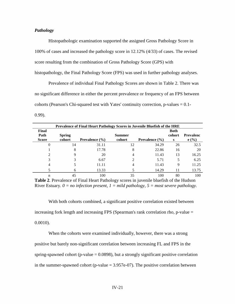
Prevalence of individual Final Pathology Scores are shown in Table 2. There was
no significant difference in either the percent prevalence or frequency of an FPS between
cohorts (Pearson's Chi-squared test with Yates' continuity correction, p-values = 0.1-
0.99).
Prevalence of Final Heart Pathology Scores in Juvenile Bluefish of the HRE Final Path Score
Spring cohort Prevalence (%)
Summer cohort Prevalence (%)
Both cohort
s Prevalenc
e (%) 0 14 31.11 12 34.29 26 32.5 1 8 17.78 8 22.86 16 20 2 9 20 4 11.43 13 16.25 3 3 6.67 2 5.71 5 6.25 4 5 11.11 4 11.43 9 11.25 5 6 13.33 5 14.29 11 13.75 n 45 100 35 100 80 100
Table 2. Prevalence of Final Heart Pathology scores in juvenile bluefish of the Hudson River Estuary. 0 = no infection present, 1 = mild pathology, 5 = most severe pathology.
With both cohorts combined, a significant positive correlation existed between
increasing fork length and increasing FPS (Spearman's rank correlation rho, p-value =
0.0010).
When the cohorts were examined individually, however, there was a strong
positive but barely non-significant correlation between increasing FL and FPS in the
spring-spawned cohort (p-value = 0.0898), but a strongly significant positive correlation
in the summer-spawned cohort (p-value = 3.957e-07). The positive correlation between
IV-21
increasing fork length and increasing Gross Pathology Score (GPS) was significant for
both cohorts combined and for the summer cohort, but barely non-significant for the
spring cohort alone (Spearman's rank correlation rho, both p-value = 0.0011; summer p-
value = 4.254e-07; spring p-value = 0.0565).
Overall prevalence of fatal infection (grades 4 and 5) was 25% (20/80) in juvenile
bluefish of the HRE during the 2010 sampling period. The average FPS for the spring and
summer cohorts were 1.89 and 1.80, respectively. No significant difference in the number
of fatal (grade 4 and 5) versus low-grade infections (grade 1 and 2) was observed
between cohorts (Pearson's Chi-squared test with Yates' continuity correction, X-squared
= 0.0018, df = 1, p-value = 0.9665). When looking at only infected fish, 35.48% (11/31)
of the spring cohort and 39.13% (9/23) of the summer cohort had potentially fatal
infections. The difference was not statistically significant (Pearson's Chi-squared test
with Yates' continuity correction, X-squared = 1e-04, df = 1, p-value = 0.9916).
A significant positive correlation existed between increasing FPS and collection
date later in the year for both cohorts combined and for the summer cohort alone
(Spearman's rank correlation rho, both p-value = 0.0080; summer p-value = 0.0012) but
not for the spring cohort (p-value = 0.1837). The FPS peaked for the spring cohort
between days 230-240 (mid to late August) and at day 270 (9/27/2010) for the summer
cohort. The peak refers to the time period with the greatest frequency of fatal severity
infections. Fatal severity infections were seen occasionally after the peak, but not with
equal frequency.
IV-22
Condition factor
Condition factor was normally distributed (Shapiro-Wilk normality test, W =
0.9945, p-value = 0.9834). Analysis of condition factor of fish infected in the heart versus
those not infected in heart revealed that infected fish have a higher condition factor than
those not infected in the heart. Or, alternatively, fish with a higher condition factor are
more often infected in the heart (Welch Two Sample t-test, p-value = 0.0015). When the
cohorts were examined individually, this was barely non-significant for the spring cohort
(normally distributed; p-value = 0.1403), and was very significant for the summer cohort
(not normally distributed; Spearman’s rank correlation rho, p-value = 3.779e-05). There
was no significant difference in the condition factor of fish with low-grade (grades 1 and
2) versus fatal grade (grades 4 and 5) infections (normally distributed; Welch Two
Sample t-test, p-value = 0.747). The mean condition factor of fish with fatal infections
was higher in the spring cohort than in the summer cohort (p-value = 0.0017). There was
no significant difference in the condition factor of those with low-grade infections
between cohorts (p-value = 0.2659).
Histopathology
Mild to moderate degrees of autolysis were present across all slides. Philometra
saltatrix females were often located at the base of the heart within the pericardial cavity,
wrapped around the bulbous arteriosus, which is a location associated with less cardiac
movement. This was also the most common site of inflammation and fibrosis. No
evidence was found of direct parasite damage to the cardiac musculature itself, nor any
evidence of myocardial invasion, attachment, pericardial rupture, or migration out of the
IV-23

pericardial cavity. Immature, subgravid, and gravid female worms were all found within
the pericardial cavity.
Grades 1-5 varied in the severity and extent of inflammation, pericardial fibrosis,
physical condition of the parasites, and extent of adhesions between the heart surface
(epicardium) and the pericardium, or the pericardium and the body wall. In both grade 4
and grade 5 infections, the epicardium and pericardium were diffusely affected, including
that of the ventricle, atrium, and bulbous arteriosus. The epicardium and pericardium
were, within the affected areas, fused and severely thickened (up to 3 mm) due to severe
mesothelial hyperplasia and hypertrophy, abundant granulomatous inflammation, and
immature fibrous connective tissue (Figure 4d, 4e). The epicardium had severe and
diffuse papilliferous projections of mesothelium, which were compressed and contained
few adult nematodes and occasionally, many larval nematodes.
Adult and larval nematodes were surrounded by many epithelioid to attenuated
macrophages, few admixed multinucleated giant cells, small to moderate amounts of
cellular debris, and moderate amounts of immature fibrous connective tissue. Gravid
females were most commonly represented, as well as some subgravid females. Adult
nematodes were often degenerate, with collapse and fragmentation of tissues, loss of
differential staining, and replacement of structures by cellular debris or rarely mineral.
Many larvae were located outside adult nematodes, embedded in granulomatous
inflammation (Figure 4f). These larvae were often degenerate, with collapse and
fragmentation of tissues, loss of internal structures with retention of a collapsed cuticle,
and loss of differential staining. Some larvae were found within the remains of adults,
IV-24
surrounded in granulomatous inflammation. The extent and thickness of fibrosis was
severe, often with involvement of tissues surrounding the bulbous arteriosus.
In contrast, low-grade infections (grade 1 or 2) were associated with non-
degenerate, recently viable parasites of all reproductive stages. These were not embedded
in inflammatory cell matrix and were associated with mild inflammation, which was
typically focal and restricted to the area immediately surrounding the parasite. Focal
areas of pericardial mesothelial hyperplasia were present. Grade two infections involved
a more reactive and thickened epicardium, with mild to moderate multi-focal
granulomatous inflammation.
The degree of pericardial thickening in grade three infections was considerably
increased compared to a grade two infection, with moderate to severe focal inflammation
and moderate focal to locally-extensive pericardial fibrosis. Subgravid and gravid worms
were seen embedded in a matrix of granulomatous inflammation. Parasites embedded in
matrix were often in various states of degeneration. Discrete areas of inflammatory
matrix were often associated with focal adhesions of the pericardium to the epicardium or
body wall.
Slides of liver sections were markedly autolyzed due to the freezing and thawing
process, and were not read. Slides of immature gonad were composed of moderately
autolyzed ovarian tissue containing primordial follicles.
IV-25

Figure 4. Representative histopathology photomicrographs. P = pericardium, PC = pericardial cavity, E = epicardium, M = myocardium. A. Normal heart, 20x magnification; B. Gross grade 2: immature female (I) and subgravid female (SG) Philometra saltatrix, no inflammation; C. Mild inflammation (short arrow) and fibrosis (long arrow); D. Gross grade 5: parasite embedded in diffuse and severe inflammation (arrow), severe fibrosis, 2x magnification; E. (Inset D) Gross grade 5: subgravid female with eggs in uterus (long arrow) embedded in severe inflammation (short arrow), severe fibrosis, 10x magnification; F. Gross grade 5: larvae granuloma, 40x magnification.
IV-26
DISCUSSION
Pathology and pathophysiology
While Philometra saltatrix is responsible for considerable fatalities in juvenile
bluefish, its effects are not due to direct tissue damage to the heart caused by the parasite.
The severe cardiac pathology is instead due to the host’s immune response to the dead
parasite and/or release of larvae into the pericardial cavity. This pathology is in the form
of varying degrees and extents of pericardial inflammation and fibrosis.
The heart of a fish is divided into three “chambers”: a single atrium, a single
ventricle, and the bulbous arteriosus (conus arteriosus). Blood from the hind end of the
fish is drained through the liver to the atrium, which then empties into the ventricle. The
ventricle, the muscular pump of the heart, pumps the blood forward to the gills for
oxygenation through a fibroelastic tube called the bulbous arteriosus. The pericardium
consists of two very thin layers. One layer, the visceral pericardium, is technically
adhered very closely to the surface of the heart and the second layer, the parietal
pericardium, makes up what is commonly known as the pericardium. The two layers
attach back together at the base of the heart around the bulbous arteriosus. The space
between these two layers is called the pericardial cavity. Normally, the parietal
pericardium is a single cell layer thick but tough (Figure 4a), and the pericardial cavity
contains a very small amount of clear, serous fluid, which acts as a lubricant (McGavin
and Zachary 2007).
IV-27
Histopathology
Histopathology served to further characterize the microscopic changes associated
with pericardial infection and understand the pathophysiology behind the changes
observed. Low-grade infections associated with live, non-degenerate worms induced
minimal pathology, and would not produce systemic signs of disease. Low-grade
infections were also associated with immature and subgravid female worms (Figure 4b).
The most severe pathology was associated with gravid worms that had died in the
pericardial cavity before or after releasing larvae into the pericardial cavity. Thus, the
majority of the inflammatory response happens after the worm dies or releases larvae.
The presence of macrophages, multi-nucleated giant cells, and cellular debris
indicates the inflammatory process seen is chronic in nature and directed at a large,
difficult to breakdown nidus of inflammation. Cellular debris indicates a longstanding
inflammatory response as well as bystander cell damage and cellular remodeling.
Granuloma formation, or the layering of fibrous connective tissue around an area of
inflammation, is a hallmark of chronic inflammation.
The presence of larval granulomas in some fish, or individual/clusters of larvae
surrounded by granulomatous inflammation, were observed both free in the pericardial
cavity indicating the release of larvae, and also within the degenerate cuticle of a dead
gravid female, indicating that the inflammatory response continued after the death of the
female (Figure 4f).
The matrix surrounding dead mature worms, formed by inflammatory cells and
cellular debris, anchored the pericardium to the epicardium frequently and facilitated the
organization of fibrosis and adhesions (Figure 4d).
IV-28

Pathophysiology
A fatal pericardial infection with Philometra saltatrix is defined by the severity
and extent of pericardial fibrosis present. Fibrosis is a natural response to chronic
infection and inflammation. A foreign body, such as a large nematode or nematode
larvae, which is incapable of being quickly broken down and cleared by the cells of the
immune system, elicits a demonstrated chain of responses from the body. These include
persistent active inflammation, tissue destruction, collagen deposition and fibrosis in an
attempt to repair tissue.
Early on, the response is mediated by certain types of white blood cells designed
for the chemical disintegration of foreign material. Over time, the predominant form of
white blood cell changes to those involved in the consumption and sequestration of
foreign material too large to be broken down. Chronic inflammation can be detrimental,
however. The long-term presence of inflammatory cells in an area, as well as their
chemical inflammatory mediators can cause damage to bystander tissues in the area. The
body tries to protect itself from damage by walling off the foreign object and laying down
connective tissue in the area to prevent further damage. It is important to note that
fibrosis, or the synthesis of collagen fibers leading to connective tissue formation is an
irreversible process. The degree and extent of the fibrosis, and how it affects nearby
tissues, is central to gauging if an infection will be fatal.
The transformation of the normal, thin, elastic, pliable pericardium to a thickened,
fibrous, inelastic pericardium due to chronic, severe inflammation can result in a
condition called constrictive pericarditis. As the fish grows or requires increased cardiac
IV-29
output, this inflexible pericardium restricts proper filling of the ventricle during diastole,
or the filling phase of the heart. While the prognosis for constrictive pericarditis in
mammals is generally poor (Asher and Klein 2002), the myocardium, or heart muscle
itself, is generally unaffected in these situations. Therefore, systolic function of the
ventricle is generally unaffected. Once the filling potential of the ventricle reaches its
filling constraint, however, filling ceases and congestion of blood behind the heart occurs
(Asher and Klein 2002).
Moderate to severe, or severe fibrosis of the pericardium, when diffuse, is
considered fatal based on inadequate ability of the heart to fill with blood during diastole.
Therefore, grade 4 or 5 infections, both grossly and histopathologically, are considered
fatal based on the degree and extent of pericardial fibrosis. Focal fibrosis is more
forgiving as the heart contracts and expands, and a fish with focal fibrosis of the
pericardium may be able to grow and function normally. Therefore, those fish with focal
fibrosis are not included in this calculation.
In mammals, constrictive pericarditis produces signs of exercise intolerance, poor
peripheral tissue perfusion, hepatic congestion, and eventual heart failure due to the
inability of the heart to meet the circulatory demands of the body. In fish, these changes
would be manifested as decreased ability to hunt and forage, decreased ability to pursue
normal migratory behavior, and increased susceptibility to predation. The effects would
become more pronounced as the fish outgrows the size and cardiac output of its confined
heart.
IV-30

Using histopathology as the definitive scoring tool, the prevalence of fatal
infection (grades 4 and 5) in all juvenile bluefish sampled from the HRE in 2010 was
25% (20/80). Of all infected fish, 37.04% (20/54) of infections were potentially fatal.
The 25% annual fatality rate is an approximation based on a relatively small
sample size from a portion of the year (late July to October). Ideally, fish should be
sampled over the entire calendar year sampling interval with a fixed number sampled
from each sampling location. Although the collection netting technique used was
designed to optimize random sampling of the fish population, one could argue this
fatality rate may be artificially increased due to the inability of clinically-affected grade 4
and 5 infected fish to escape sampling nets. However, the presence of uninfected/score-0
fish in the nets (see Table 2), as well as grades 1-3, makes this unlikely. Also, the peak
FPS occurred in mid to late August for the spring cohort and late September for the
summer, while sampling continued after those dates. A higher frequency of severe
infections would be expected at the end of the sampling period for both cohorts, along
with a relative decrease in the frequency of grade 0-3 of that cohort, if the prevalence of
clinically affected fish was increased. Instead, there is a decrease in the frequency of
severe pathology, indicating either resolving infections or the deaths of severely affected
individuals.
An important result of this study is the ability to employ a quick gross inspection
technique of pathology in the field with confidence in the agreement between GPS and
histopathology-verified FPS. There was always 100% agreement with the gross findings,
and histopathology is likely to support or increase the grade of severity. Thus, for
researchers assessing prevalence and severity of this infection in juvenile bluefish, it
IV-31
would not be necessary to submit individual histopathology samples to arrive at a
definitive severity grade. This means that the criteria included in the gross pathology
scoring system, which are designed to estimate the severity and extent of inflammation,
fibrosis, chronicity, and outcome, are also representative of the cellular changes.
The “grade 3” infection
Of the adjustments made to gross pathology scores, two grade one infections out
of 19 were increased to grade two, which is not surprising based on the small amount of
visible gross pathology. Also, two grade three infections out of seven were increased to
grade four. This is interesting because the degree of cellular change was more advanced
compared to the gross appearance. Yet, there were also grade three infections with a
similar gross appearance which were less severely affected on a microscopic level.
Across several of these grade three infections, a common finding involved degenerate
worms encased in inflammatory matrix. In the progression of infection from gross grade
one to grade five, the most severe inflammation and fibrosis is expected to be associated
with dead gravid females in grade five infections. After death, the worms begin to
degenerate and are removed slowly by the immune system.
However, in some grade three infections, the worms were degenerate and the
inflammatory response less severe and less extensive. This could indicate that some gross
grade three infections may be instead sequential to a grade five infection, reflecting a
state of resolving infection. This is supported by the presence of larval granulomas and
well-encapsulated degenerate worms, indicating a passage of time since the death of the
gravid female. Fibrosis of the pericardium was still present but focal to multi-focal in
IV-32
extent, which is expected because it is irreversible. Thus, both resolving infections of
non-fatal severity and progressing grade two infections could have the same gross grade
three pathology score, and would require histological examination. Progressing grade two
infections would involve live or very recently dead worms.
This sharing of grade 3 GPS by progressing and resolving infections is likely
responsible for the loss of significant positive correlation with increasing fork length and
increasing final pathology score in the spring cohort. The spring cohort is likely the only
cohort affected by this phenomenon because a longer window of the infection cycle is
seen during the sampling period, and the severe summer cohort infections have not yet
begun to resolve during the sampling period.
Parasite Ecology
Juvenile bluefish likely acquire the infection close to their spawning grounds
through the consumption of a copepod carrying an infective larva (Moravec 2004; Bryan
et al. 2008). Since a single adult arises from a single infective larva, multiple larvae are
acquired to result in multi-worm infections. The difference in the sexual maturity level
(immature, subgravid, or gravid) is a function of how recently the infective larva was
consumed (assuming a constant rate of parasite maturation). All females found in the
pericardial cavity were fertilized, indicating the presence of male nematodes in the fish’s
body as well. Recent research has shown males to be present in the body cavity and
gonad in juvenile bluefish (Koske et al., unpublished research).
The weekly percent prevalence of infection seen over the course of the sampling
period (Figure 3), which occurs in two major waves, reflects the staggered arrival of the
IV-33

cohorts in the estuary. After the fish acquires the parasite, at least two months pass before
infection is first visible in the pericardial cavity. For example, the summer cohort appears
in the HRE in early August but the first infection in the summer cohort is not found until
six weeks later, when prevalence suddenly approaches 100%. This is likely a function of
the close time proximity in which members of the summer cohort become infected and
the maturation time of the parasite. If the sampling period were extended earlier in the
year, a similar trend would likely be observed in the spring cohort.
Given the fact that the parasite’s reproductive strategy is likely linked to the off-
shore spawning of adult bluefish (Clarke et al. 2006; Moravec 2004) and the parasite is
likely acquired off-shore, not within the HRE, the parasite likely matures to its adult form
during the fish’s migration into, and early residence in, the estuary. This hypothesis is
supported by the fact that a significant trend exists linking increasing fork length with
increasing pathology score. If the parasite was acquired in the estuary, this trend would
be obscured by recurring waves of grade one infections and the progression of those
infections. Instead, there is a significant correlation in both cohorts with greater
pathology scores and larger fork lengths with collection dates later in the year. These
results point to a single infection event in each cohort. Also, evidence of re-infection,
defined by the co-existence of immature females and degenerate gravid females in the
pericardial cavity, was not observed during the entirety of this sampling period,
decreasing the likelihood the parasite is acquired in the HRE.
The overall prevalence of infection in the pericardium of all fish sampled was
67.5% (54/80). Studies evaluating the prevalence of parasitic infection in a natural wild
population are rare and thus comparable studies of other nematode parasites are few.
IV-34
With a prevalence of 67.5%, it is unusual to be lacking reports of occurrence from the
rest of the 1980s and 1990s. Cheung (1984) described an 80% prevalence in fish
examined at the New York Aquarium; however, his sample size was not reported in the
available abstract. Obviously, this parasite is widespread in the Hudson River estuary and
not localized to specific geographical areas of the Hudson River.
Importantly, during this sampling window, both the spring and summer cohorts
were equally infected in terms of prevalence and severity of infection. This indicates that
the summer cohort is not preferentially infected, and mortality associated with this
infection affects both cohorts equally. This is not in agreement with the proposition by
Clarke et al. (2006) who hypothesized that the summer cohort may be preferentially
infected due to a higher occurrence and intensity of Philometra saltatrix in the gonad of
summer-spawning adults.
Because the exit of the summer cohort from the HRE was not observed during the
sampling period, the summer cohort could possibly experience an increase in mortality
not seen in this study. However, it is also possible that the spring cohort experiences
mortality events as infection continues to progress when the fish have migrated back out
to sea and are effectively lost to the study. Thus, the ability to definitively evaluate and
compare mortality between cohorts in a natural setting may not exist. Experimental
infection and monitoring over time would give a better indication of mortality rates in
general, but the natural factors such as seasonal prey availability and water quality and
temperature that may affect infection rates between cohorts would be lost in this study.
During their residence in the HRE, juveniles grow in length, mass, and condition.
The smallest fish were not infected in the heart, and longer and heavier fish were more
IV-35

likely to be infected in the heart. This reflects the time interval between initial infection
and when the infection is grossly observable. Fish infected in the heart also had a higher
condition factor, which is not surprising given that condition factor is dependent on both
mass and length. Thus, the condition factor of the spring cohort is also expected to be
higher than for the summer because of their greater relative length and mass.
Gross pathology scores also increase with fork length, which reflects the fish
acquiring the parasite when small and having the infection develop and become more
severe as it grows over the course of the year. The fact that there is a trend for high
degree pathology in larger fish, in addition to the discovery that this correlates also with
time of year, is indicative that larger fish are not preferentially affected, but rather that the
score is the result of a natural long-standing infection. The final pathology score, which is
adjusted to reflect the histopathology findings, also increases with length and mass for the
same reason.
The histopathologic findings observed in 2010 juveniles of the HRE and larger
juveniles sampled from the New York Bight (NYB) in 2009 are similar (Koske and
Pinkerton 2010), although the prevalence of fatal infection in the NYB was only 9.09%.
This possibly reflects a further loss of severely infected individuals after migration out to
sea, or instead a reflection of the smaller sample size (n=33) in the NYB study.
In this sampling period, data on the spring cohort was collected for ten weeks, and
13 weeks for the summer cohort. However, of those 13 weeks, the summer cohort was
infected for only six weeks. The prevalence and average pathology score data were taken
from the entirety of the sampling period, not just the weeks where infection was present.
Thus, the comparison between the results of a 10-week and a 13-week study showed
IV-36

equal prevalence and pathology scores between cohorts. Without a sampling period that
includes both the arrivals and departures of both cohorts from the HRE, it cannot
definitively be said that one cohort is not preferentially affected over the other. However,
within the sampling period of this study, no significant difference was observed between
cohorts in the overall prevalence of pericardial infection, the prevalence of high versus
low-grade FPS, or the number or prevalence of fatal infections.
Condition factor and infection
Results showed that fish infected in the heart had higher condition factors than
those not infected in the heart. The formula used to calculate condition factor is
dependent on both fork length and mass. Since condition factor increases as fish FL
increases, the link between infection in the pericardium with both increased condition
factor and increased FL is not surprising. While one might expect a parasitized fish to
have a lower condition factor than a non-parasitized fish, the results show that fish are
grossly infected in the pericardium only after reaching 97mm FL and a condition factor
of 1.068. This could indicate two possibilities: 1) fish are infected early in life and
infection becomes apparent only around the age when condition factor approaches 1.0; 2)
the parasite might arrest in development within the fish until the fish reaches a size and
condition appropriate for optimal parasite development.
This also indicates that a severe pericardial infection does not induce significant
loss of condition while the fish resides in the estuary. Some loss of condition may be
present and not reflected here, however, because the increasing length of the fish would
mask any loss of mass due to the method of calculation. The fact that mass continues to
IV-37

increase with increasing FPS also indicates that infection does not significantly impact
fish mass. Generally, a parasite that routinely induced fatality or severe condition loss of
its host would not be evolutionarily successful due to host loss. Philometra saltatrix
infection may not even result in loss of condition. Knowing this infection can be fatal
through cardiac insufficiency and heart failure, any difference in condition may only
become apparent in severely infected fish just before the death of the fish, or not at all if
the fish succumbed to predation. These fatalities, of course, would occur out at sea and
are not measurable.
Suspected changes in the severely affected juvenile’s ability to successfully
forage, complete natural migrations, and escape predation are based on the
pathophysiology of heart failure. Moles (2003) described decreased prey capture rates in
Dolly Varden (Salvelinus malma) parasitized by the Philometrid nematode Philonema
agubernaculum in the body cavity. Parasitized fish captured only 32% of available prey
versus 64% by non-parasitized Dolly Varden. No difference was observed in the
condition of the parasitized versus non-parasitized Dolly Varden. Moles (2003)
hypothesized that while the effects of infection might be minimal when food supply is
abundant, in a limited food supply environment, the non-parasitized fish likely fare
better, and thus infection has the potential to alter predator-prey relationships.
Bluefish is a species heavily dependent on burst swimming and ambush during
normal feeding. Eventual decreased cardiac output and oxygenation of peripheral tissues
would lead to changes in cellular metabolism, thereby causing early muscle fatigue and
affecting foraging ability. Since constrictive pericarditis compromises the cardiac output
IV-38
of an affected fish, infection with Philometra saltatrix could also predispose affected fish
to fatalities associated with low dissolved oxygen content (McGladdery et al. 1988).
CONCLUSIONS
Pericardial cavity infection by Philometra saltatrix is an important source of
mortality in juveniles of the Hudson River Estuary, potentially fatal in 25% of all fish
sampled. The pathologic consequences of infection, which include varying degrees of
severity of constrictive pericarditis, have the potential over time to severely impact the
foraging and migration capabilities of severely affected growing fish. The most severe
pathology is induced after the death of the parasite and/or release of larvae into the
pericardial cavity. The severity of infection can be accurately assessed by quick gross
examination of changes seen in the character of the pericardium as described in this
study.
The results of this study show that both spring and summer-spawned cohorts are
equally affected in terms of prevalence of infection, severity of pathology, and prevalence
of fatal infections. Thus, this study suggests that Philometra saltatrix is not responsible
for the preferential decline in recruitment observed in the summer cohort.
Infection is likely acquired off-shore near spawning grounds and develops over
the course of the fish’s migration into the estuary. Over time, as the fish increases in size,
mass, and condition, the likelihood of infection in the pericardial cavity as well as the
severity of pathology increases. There was no evidence fish are re-infected in the estuary,
decreasing the likelihood of involvement of a paratenic host in transmission of the
infection in the estuary.
IV-39

RECOMMENDATIONS
Further study is needed to definitively quantify the negative effect on recruitment
caused by pericardial infection with Philometra saltatrix. Ideally, samples should be
collected from various locations in the HRE over the course of an entire calendar year in
order to observe the natural infection cycle in both cohorts.
Obtaining fresh samples of liver tissue at the time of processing could
histologically indicate whether passive congestion of blood is occurring in the liver of
grossly affected fish, which is a classic symptom of heart failure in mammals. This would
enable reliable extrapolation of other classic signs of heart failure to fish.
Optimally, in order to chronicle the story of infection from beginning to end and
note the timeline, as well as changes in health, growth rates, condition, and fecundity,
experimental infection should be pursued in a controlled environment over multiple
years. This would also enable the study of the health impacts on survivors of infection
and the whole of cohorts infected versus not infected.
Finally, it is important to note that the prevalence of infection likely varies from
year to year, depending on ocean current strengths, annual copepod crop, and water
temperature, as maturation rate of Philometrid species larvae in experimentally infected
copepods has been demonstrated to be heavily dependent on water temperature. Since the
presence of this parasite was not noted during the late 1980s and 1990s, it is possible that
infection prevalence has been steadily increasing since that time and Philometra saltatrix
infection in the pericardial cavity could be an already emergent and unmonitored source
of recruitment failure among juveniles of both cohorts. Fisheries biologists and
IV-40

conservation scientists should be involved in field monitoring of infection prevalence,
and additional effort should be directed toward elucidation of the life cycle of this
parasite to help direct future intervention.
IV-41

ACKNOWLEDGEMENTS
The authors would like to acknowledge the contributions of Dr. Timothy Yoshino
of the School of Veterinary Medicine, Director, Cellular and Molecular Parasitology
Training Program, University of Wisconsin-Madison for guidance on initial study design
and reproductive staging, and Dr. Marie Pinkerton, DVM, DACVP, Department of
Pathobiological Sciences, School of Veterinary Medicine, University of Wisconsin-
Madison for providing descriptive and diagnostic pathology services for the project along
with guidance on grading severity. We would also like to thank Amy Teffer, Department
of Environmental Conservation, UMass Amherst, for her assistance with statistical
analysis, and the New York State Department of Environmental Conservation for
sampling bluefish for the project. We would also like to thank Dr. František Moravec,
Institute of Parasitology, Biology Centre of the Academy of Sciences of the Czech
Republic, for his assistance confirming the identification of the parasite. Finally, David
Stormer, former Polgar fellow, Department of Environmental Conservation, UMass
Amherst, for his close guidance, instruction, and assistance with this project.
IV-42
LITERATURE CITED
Asher, C.R. and A.L. Klein. 2002. Diastolic heart failure: Restrictive cardiomyopathy,
constrictive pericarditis, and cardiac tamponade: Clinical and echocardiographic evaluation. Cardiology in Review. 10:4:218-299.
Beamish, R. and G. McFarlane (Eds.) 1989. Effects of ocean variability on recruitment
and an evaluation of parameters used in stock assessment models. Canadian Special Publication of Fisheries and Aquatic Sciences. 108.
Beyer, J.E. 1989. Recruitment stability and survival-simple size-specific theory with Examples from the early life dynamics of marine fish. Dana. 7:45-147.
Bryan, T.P., L.C. Tsoi, and I. de Buron. 2008. Development of the Philometrids
Philometra Overstreeti and Philometroides paralichthydis in the experimentally infected copepod Oithona colcarva. Folia Parasitologica. 55:313-315.
Burak, W.K. 2007. Infection of bluefish (Pomatomus saltatrix) ovaries by the
Drancunculoid nematode, Philometra saltatrix: prevalence, intensity, and effect on reproductive potential. Master’s thesis. Stony Brook University, Stony Brook, NY.
Cheung, P.J., R.F. Nigrelli, and G.D. Ruggieri. 1984. Philometra saltatrix infecting the heart of the 0 class bluefish, Pomatomus saltatrix (L.), from the New York coast. In S. F. Snieszko commemoration fish disease workshop, p. 27. Joint Workshop of Fish Health Section, AFS, and Midwest Disease Group, Little Rock, AR.
Clarke, L.M., A.D. M. Dove, and D.O. Conover. 2006. Prevalence, intensity, and effect
of a nematode (Philometra saltatrix) in the ovaries of bluefish (Pomatomus saltatrix). Fisheries Bulletin 104:118-124.
Conover, D.O., T. Gilmore, and S.B. Munch. 2003. Estimating the relative contribution
of spring and summer-spawned cohorts to the Atlantic coast bluefish stock. Transactions of the American Fisheries Society 132:1117–1124.
Chambers, R.C. and E.A. Trippel, (Eds.). 1997. Early Life History and Recruitment in
Fish Populations. Chapman & Hall, London. Hare, J.A., and R.K. Cowen. 1996. Transport mechanisms of bluefish (Pomatomus
Saltatrix larvae from South Atlantic Bight spawning grounds to Middle Atlantic Bight nursery habitats. Limnology and Oceanography. 41:1264-1280.
Hine, P.M., and C.D. Anderson. 1981. Diseases of the gonads and kidneys of New
IV-43
Zealand snapper, Chrysophrys auratus Forster (F. Sparidae). Pages 166-170 in M. E. Fowler, Editor. Wildlife diseases of the Pacific Basin and other countries. Academic Press, London.
Hjort, J. 1914. Fluctuations in the great fisheries of northern Europe viewed in the light of
biological research. Rapports et Proces-verbaux des Réunions. Conseil International pour l'Éxploration de la Mer . 20:1-228.
Houde, E.D. 1987. Fish early life dynamics and recruitment variability. American Fisheries Society Symposium. 2:17-29. Juanes, F., and D.O. Conover. 1995. Size-structured piscivory: advection and the linkage
between predator and prey recruitment in young-of-the-year bluefish. Marine Ecology Progress Services. 128: 287-304.
Juanes, F., J.A. Hare, and A.G. Miskiewicz. 1996. Comparing early life history strategies
of Pomatomus saltatrix: a global approach. Marine and Freshwater Research, 47:365-379.
Kendall, A.W. Jr., and L.A. Walford. 1979. Sources and distribution of bluefish,
Pomatomus saltatrix, larvae and juveniles off the east coast of the United States. Fisheries Bulletin. 77: 213-227.
Klein-MacPhee, G. 2002. Bluefish. Family Pomatomidae. Pages 400-406 in B.B.
Collette and G. Klein-MacPhee (Eds.). Bigelow and Schroeder’s Fishes of the Gulf of Maine Third Edition. Smithsonian Institution Press, Washington, D.C.
Koske, S.E. and M.E. Pinkerton. 2010. Pathology associated with pericardial
cavity infection by Philometra saltatrix (Nematoda, Philometridae) in juvenile bluefish (Pomatomus saltatrix). Poster session presented at: American College of Veterinary Pathologists Annual Meeting; 2010 October 30- November 3; Baltimore, Maryland.
Lassiter, R.R. 1962. Life history aspects of the bluefish, Pomatomus saltatrix (Linnaeus),
from the coast of North Carolina. M. Sc. thesis, North Carolina State College, Raleigh.
McGavin, M.D. and J.F. Zachary (Eds). 2007. Pathologic Basis of Veterinary Disease. Mosby/Elsevier, St. Louis. Fourth Edition. McGladdery, S.E., L. Murphy, B.D. Hicks, and S.K. Wagner. 1988. Pathology in Marine Science. pp. 305-315. London: Academic Press. Moles, A. 2003. Effect of Parasitism by Philonema agubernaculum (Nematoda: Philometridae) on the Ability of Dolly Varden to Capture
Prey in Fresh and Salt Water. Alaska Fishery Research Bulletin 10:2:119-123.
IV-44
Moravec, F. 2004. Some aspects of the taxonomy and biology of Dracunculoid nematodes parasitic in fishes: a review. Folia Parasitologica 51:1-13. Moravec, F. and I. De Buron. 2009. New data on three gonad-infecting species of
Philometra (Nematoda, Philometridae) from estuarine fishes in South Carolina, USA. Acta Parasitologica. 54:3:244-252.
Moravec, F., M. Magi, and F. Macchioni. 2008. Redescription of the
Gonad-infecting nematode Philometra saltatrix Ramachandran, 1973 (Philometridae) based on specimens from the type host Pomatomus saltatrix (L.) (Osteichthyes) from the Tuscan Sea, Italy. Folia Parasitologica 55:219-223.
Munch, S.B., and D.O. Conover. 2000. Recruitment dynamics of bluefish (Pomatomus
saltatrix) from Cape Hatteras to Cape Cod, 1973-1995. ICES Journal of Marine Science 57:393-402.
Oliva, M.E., A.S. Borquez, and A.N. Olivares. 1992. Sexual status of Paralabrax Humeralis (Serranidae ) and infection by Philometra sp. (Nematoda: Dracunculoidea). Journal of Fish Biology 40:979-980.
Olla, B.L., and A.L. Studholme. 1971. The effect of temperature on the activity of bluefish, Pomatomus saltatrix L. Biological Bulletin 141:337–349. Ramachandran, P. 1973. Philometra saltatrix sp n., infecting the gonads of the common
bluefish Pomatomus saltatrix (L.) off the New England coast of the United States. Zoologischer Anzeiger. 191:325-328.
Ramachandran, P. 1975. Philometra cephalus sp. N. infecting the gonads of the striped
mullet, Mugil cephalus L. from the Arabian Coast of Kerala, India, with a note on its pathology. Zoologischer Anzeiger. 191:140-144.
Rothschild, B.J. 1986. Dynamics of marine fish populations. Harvard University Press. Stormer, D.G., and F. Juanes. 2008. Cohort structure, growth, and energy dynamics of
Juvenile bluefish in the Hudson River estuary. Section VII: 26 pp. In S. H. Fernald, D. Yozzo and H. Andreyko (eds.), Final Reports of the Tibor T. Polgar Fellowship Program, 2008. Hudson River Foundation, New York, New York.
Sures, B. and K. Knopf. 2004b. Individual and combined effects of cadmium and
3,3’,4,4’,5 pentachlorobiphenyl (PBC 126) on the humoral immune response in European eel (Anguilla anguilla) experimentally infected with larvae of Anguillicola crassus (Nematoda). Parasitology 123: 179-184.
IV-45