prevalence of type 2 diabetes mellitus in adult …
TRANSCRIPT

PREVALENCE OF TYPE 2 DIABETES MELLITUS IN ADULT PATIENTS WITH HEPATITIS C VIRUS INFECTION AND ASSOCIATED LABORATORY MARKERS. EXPERIENCE AT RWANDA MILITARY HOSPITAL:
A cross sectional descriptive study
Submitted in partial fulfilment of requirements for the Degree of Master of
Medicine (M.MED) in Internal Medicine, University of Rwanda (UR).
Principal Investigator: Dr Anthony BAZATSINDA, UR Reg. No 213003790
March 2016

PREVALENCE OF TYPE 2 DIABETES MELLITUS IN ADULT PATIENTS WITH
HEPATITIS C VIRUS INFECTION AND ASSOCIATED LABORATORY MARKERS.
EXPERIENCE AT RWANDA MILITARY HOSPITAL:
A cross sectional descriptive study
Submitted in partial fulfilment of requirements for the Degree of Master of
Medicine (M.MED) in Internal Medicine, University of Rwanda (UR).
Principal Investigator: Dr Anthony BAZATSINDA, UR Reg. No 213003790
Supervisor: Dr Jules KABAHIZI
Co-supervisor: Dr Charlotte BAVUMA
March 2016

DECLARATION
I, Anthony Bazatsinda, hereby declare that this is my original work and has not been
presented for the award of a degree in any other university. I also declare that the intellectual
content of this thesis is the product of my own work, although I have received invaluable
assistance from my supervisors and others which I dully acknowledge.
Dr Anthony Bazatsinda MB ChB. (UR)
Department of Internal Medicine, University of Rwanda.
Signed.........................................................................Date...........................................
This research report has been presented with our full approval as supervisors:
Dr Jules Kabahizi (MB ChB), M.Med (Internal Medicine), Nephrologist , Chief consultant
King Faisal Hospital, Head of Internal Medicine department Rwanda Military Hospital, Senior
Lecturer, University of Rwanda.
Signed........................................................................Date.................................................
Dr Charlotte Bavuma (MB ChB, M.Med (Internal Medicine),Endocrinologist, PhD Candidate
in Diabetes Mellitus, Senior Lecturer , University of Rwanda.
Signed.......................................................................Date......................................................
i

DEDICATION
To God the Almighty, who is the provider of everything.
To my late Father, who couldn’t live long to witness this work.
To my great loving Mother, my Unique Wife Babrah UWIMBABAZI, beloved sisters and b
brothers.
To my M. Med friends for all their support and companionship.
ii

ACKNOWLEDGMENTS
I thank and acknowledge my supervisors, Dr Jules Kabahizi and Dr Charlotte Bavuma for
their assistance and guidance in planning and conducting this research.
Thanks are also due to the registrars, nurses in OPD in specialized hepatitis and diabetes
mellitus clinics, general medical wards, and Rwanda Military Hospital and Laboratory
department staff for their great laboratory work. In sincerely, I would like to thank the
consultants in Internal Medicine department for their comments in moving forward the
proposal approval and in the editing process.
I deeply acknowledge RMH to have financed this research and to have allowed me to
conduct the mentioned research in both OPD Specialized hepatitis and diabetes clinics and
general medical wards.
Many thanks to the Rwandan government for having enabled me to do M.Med program and
last but not least, this work would not have been feasible without the willingness and
cooperation of the patients.
iii

ACRONYMS
UR: University of Rwanda.
RMH: Rwanda Military Hospital.
T2DM: Type 2 Diabetes Mellitus.
HCV: Hepatitis C Virus infection.
ALT: Alanine aminotransferase (formerly SGPT).
AST: Aspartate aminotransferase (formerly SGOT).
GGT: Gamma glutamyl transferase.
ELISA: Enzyme-linked immunosorbent assay
BMI: Body Mass Index
SPSS: Statistical Package for Social Sciences.
WHO: World Health Organization.
OPD: Outpatient department.
M.MED: Master in Medicine.
IDF: International Diabetes Federation
IGT: Impaired Glucose Tolerance.
IFG: Impaired Fasting Glucose.
HDL: High-Density Lipoprotein.
LDL: Low-Density Lipoprotein.
CTGF: Connective Tissue Growth Factor.
NIDDM: Non-Insulin -Dependent Diabetes Mellitus.
KSA: Kingdom of Saudi Arabia
PCR: Polymerase chain reaction.
Th1: T helper lymphocytes.
iv

Contents DECLARATION .......................................................................................................................................... i
DEDICATION ............................................................................................................................................ ii
ACKNOWLEDGMENTS ............................................................................................................................ iii
ACRONYMS ............................................................................................................................................ iv
List of figures ......................................................................................................................................... vii
List of tables .......................................................................................................................................... vii
ABSTRACT ............................................................................................................................................. viii
Chapter I: INTRODUCTION ..................................................................................................................... 1
I.1 Theoretical framework ..................................................................................................................... 1
I.2 Background ................................................................................................................................... 2
I.3 Rationale ....................................................................................................................................... 5
I.4. Hypothesis and objectives ........................................................................................................... 5
I.4.1. Main objective ...................................................................................................................... 5
I.4.2. Specific objectives: ............................................................................................................... 6
Chapter II: Methodology ........................................................................................................................ 6
II.1 Study design ................................................................................................................................ 6
II.2 The study site .............................................................................................................................. 6
II.3 Study population ......................................................................................................................... 6
II.4 Inclusion criteria .......................................................................................................................... 6
II.5 Exclusion Criteria ........................................................................................................................ 7
II.6 Sample size .................................................................................................................................. 7
II.7. Recruitment procedure, data collection and sampling technique ............................................... 7
II.8. Data management ....................................................................................................................... 8
II.9. Data analysis .............................................................................................................................. 8
II.10. Dissemination of results ........................................................................................................... 8
II.11. Ethical consideration ................................................................................................................ 9
Chapter III: RESULTS ............................................................................................................................... 9
III.1. Baseline patient demographics (Ref Table 1) ........................................................................... 9
III.2. Prevalence of type 2 diabetes mellitus in HCV patients. ......................................................... 11
III.3. Laboratory markers and ultrasonography results (Ref Table 2) .............................................. 12
Chapter IV: DISCUSSION........................................................................................................................ 14
v

IV.1 Results discussion ..................................................................................................................... 14
IV.2 Study limitations ....................................................................................................................... 16
IV.3 Local relevance and generalizability ........................................................................................ 16
Chapter V. CONCLUSSION AND RECOMMENDATION ........................................................................... 17
REFERENCES .......................................................................................................................................... 18
APPENDICES .......................................................................................................................................... 22
Consent form ..................................................................................................................................... 22
Work sheet on T2DM/HCV. ............................................................................................................. 24
Time frame. ....................................................................................................................................... 28
Study budget. .................................................................................................................................... 29
Ethical clearance ............................................................................................................................... 31
vi

List of figures
Figure 1: Prevalence of diabetes in HCV patients. ..................................................................11
List of tables
Table 1: Baseline Patient Demographics .................................................................................10
Table 2: laboratory markers and ultrasonography findings .....................................................13
vii

ABSTRACT
Background: Type 2 diabetes Mellitus and hepatitis C co-morbidity remains a public
healthproblem globally and they are both associated with significant morbidity and mortality.
Anecdotally, in Rwanda referral hospitals, HCV infection has been found in different cases of
type 2 diabetes mellitus. Hence, the first study to be carried out in Rwanda Military Hospital
(RMH) to describe such cases. This study aims at determining the prevalence of diabetes
mellitus among adult patients with hepatitis C infection and assessment of the impact of
hepatitis C-type 2 diabetes co-morbidity on selected biochemistry and imaging parameters.
Methods: This was a 10 months cross sectional study including consecutive adult patients
(18years and above) with hepatitis C Virus infection who presented in Outpatients
specialized hepatitis C clinic and internal Medicine admission wards. Participants were
considered to have diabetes if they were on diabetic treatment or if they exhibited Fasting
blood glucose ≥ 126mg/dl and glycated haemoglobin ≥ 6.5%. All patients were tested for
lipid profile and liver function. They underwent also an abdomen ultrasounds. . SPSS version
16.0 was used for data analysis and we compared two groups (those with diabetes and those
without diabetes) regarding laboratory and imaging results.
Results: Among 298 participants with hepatitis C, 67 (22.48%) had type 2 diabetes.
Mostpatients with type 2 diabetes -hepatitis C co-morbidity showed increased levels of
Aspartate aminotransferase (AST) (53.7% of patients with co-morbidity versus 35.5% of
those with Hepatitis C alone; P<0.007), increased gamma glutamyltransferase (50.7% of
patients with co morbidity versus 30.4% of hepatitis C standalone group; P= 0.002) , reduced
levels of serum albumin (18.2% in co-morbid group versus 10% in isolate Hepatitis C group ;
P<0.005). High levels of total cholesterol and triglyceride were most prevalent in co-morbid
group; respectively 29.9% versus 3% (P<0.001) and 23.90% versus 2.60%, P<0.001). In
addition, a greatest number of patients with co affection had fatty liver as per ultrasound
(63.4% versus 36.60% HCV; P<0.001). Conclusion: Type 2 diabetes mellitus is prevalent in adult patients with hepatitis C atRwanda
Military Hospital and its co-existence with hepatitis C would have conjunctive negative
impact on liver function as well as on the lipid metabolism. However, there is a need of
further deep studies to understand the pathophysiological mechanism.
viii

Chapter I: INTRODUCTION
I.1 Theoretical framework.
Hepatitis C is an enveloped, single-stranded RNA flavivirus1. The spread is through blood
products, secretions, and sexual intercourse (even if there is little evidence for sexual
transmission). Some groups of people such as health workers, haemophiliacs, homosexuals,
intravenous drug abusers and patients on haemodialysis .have been reported to be at high risk
of hepatitis C infection1 : Up to 70-85% of the cases progress to chronic hepatitis
2, with
elevated risk of hepatocellular carcinoma, and abnormal liver histology even in carriers who
are asymptomatic1 . The worsening of the disease is in patients with concurrent HIV infection
and/or alcoholic cirrhosis1. Usually, screening of hepatitis infection is based on hepatitis viral
antibodies testing however these antibodies are detectable within 90 – 180 days and even if
detected they could not indicate active infection. To confirm that the patient has active
hepatitis C infection viral load is needed1.
Diabetes mellitus (DM) is a complex non communicable chronic disease including
carbohydrate, lipid and protein metabolism disorders7. It is characterized by raised blood
glucose due to defects in action of insulin, its secretion following progressive beta cells
destruction or both7. Diabetes Mellitus is currently classified as type 1 Diabetes Mellitus,
type2 Diabetes Mellitus, gestational Diabetes Mellitus and other secondary diabetes Mellitus
7. The World Health Organization (WHO) and National Diabetes Data Group have set criteria
for diabetic mellitus diagnosis 8-10
. According to WHO, 2 of the following criteria are
required to diagnose diabetes: diabetes mellitus symptoms plus random plasma glucose equal
or greater than 200md/dl (11.1 mmol/l), fasting plasma glucose equal or greater than 126
mg/dl (7.0 mmol/l), 2 hours plasma glucose equal or greater than 200 mg/dl (11.1 mmol/l)
during oral glucose tolerance test and glycated heamoglobin equal or greater than 6.5%..
Diabetes mellitus may be diagnosed following classic symptoms like frequent urination, thirst,
weight loss or symptoms of complication, however many individuals diabetes do not present with
symptoms, and their disease remains undiagnosed for many years, especially patients with type 2
diabetes11
. There is evidence that at the time of diagnosis, the patient with type 2 diabetes has
had the disease for at least 4 to 7 years11
. Furthermore, they might have complications of
1

diabetes: 25% would have diabetic retinopathy; 9%, diabetic neuropathy; and 8%, diabetic
nephropathy. Therefore, patients at high risk of type 2 diabetes should be identified and
screened regularly to avoid late diagnosis with complication. As hepatitis C infection might
be a predisposing factor to type 2 diabetes, screening of diabetes in patients with hepatitis C
and evaluation of the magnitude of this co-morbidity would be important for evidence based
decision making and clinical care,
I.2 Background
At present, there are approximately 170 million HCV chronic carriers around the world and
most of them are in the developing countries3-4
as reported in April 1998 by the World
Health Organization. Hepatitis C infection is not a new issue. Jaundice was recognized as a
sign of HCV many centuries before Christ4-5
. The first recognition of Hepatitis C as a
separate disease entity was in 1975 when the transfusion related hepatitis was very prevalent
and found not to be caused by only Hepatitis B virus and Hepatitis A viruses4-5
. At that time
it was called “non-A non-B Hepatitis4-5
.
The prevalence of viral hepatitis C is increasing tremendously over the past 3 decades However,
in developed countries cases of hepatitis C infection are reported to decrease3-4
. Transmission of
Hepatitis C Virus through blood transfusions in United States of America (USA) reduced from 3-
5% in 1992 to 0.001% per transfusion6. Even though the overall prevalence is decreasing, there is
increasing complications burden6. Hepatitis C virus infection
is a leading cause for cirrhosis, liver transplantation and Hepatocellular Carcinoma6.
The
estimated related deaths in USA per year is 12,000. Furthermore, in Japan, Southern Europe,
and North America. Hepatitis C Virus has been reported to play an important negative role in
causing chronic liver disease (CLD), and has become a major cause of liver cirrhosis and
primary liver cell carcinoma (PLCC) 6.
Hepatitis C virus has various genotypes Worldwide. Genotype 1 was the most frequent and
accounting for 46 percent of all infections, followed by genotypes 3 at 22 %, and genotypes 2 and
4 (13% each)7 . Subtype 1b accounts for 22% of all infections globally
7. There are variations
which are significant across the world with genotype 1 dominating in Europe, North America,
Latin America, Australia, Europe, Latin America and North America (53-71% of all cases) and
Genotype 3 accounting for 40 percent of all infections in Asia. Genotype 4 was most frequent (71
%) in Middle East and North Africa, but when Egypt was not included, genotype
2

4 accounted for 34 percent while genotype 1 accounted for 46 percent of infections across the
same region7. Egypt has a very high prevalence of genotype 4. To our knowledge, the
prevalence of Hepatitis C virus and its genotypes has not been evaluated in Rwanda
population, however, based on case findings in clinical practice, it might be high.
Type 2 Diabetes Mellitus accounts for more than 90% of all Diabetic Mellitus cases13
. More than
100 million people globally have diabetes and there are still many who are undiagnosed12
. The
overall approximate prevalence among adult diabetes in the USA is 5.8% -12.9 % (median 8.4 %)
14. This is in sharp contrast to other countries. For instance, there was a Nigerian survey on the
prevalence of type 2 diabetes mellitus and discovered it to be 2.7% among all Nigerians15
.
Globally, the diabetes mellitus prevalence has tremendously increased for the past two decades,
and in 1985 there was an approximation of about 30 million patients and in 2010 it was around
285 million16
. And based on this tremendous increase, according to the International Diabetes
Federation, by 2030 the diabetes mellitus is projected to be 438 million individuals worldwide43
.
It is true that the prevalence of type 1 and type 2 DM is globally rising. Yet, the type 2 Diabetes
Mellitus prevalence is vigorously increasing even faster than type 1 diabetes mellitus, due to
decreased activity as many countries are becoming more and more modernized and industrialized,
leading to increasing obesity and population aging16
.
Diabetes is particularly increasing in Low and Middle income countries (LMIC).
Approximated43
prevalence of diabetes mellitus in South Africa, Uganda, Kenya, and
Tanzania are 8.39%, 4.42%, 3.6%, and 7.95%, % respectively17
. In Rwanda, according to
recent Non communicable disease (NCD) risk factors step by step survey whose results are
not yet published but announced in NCD synergy conference in 2013, the prevalence of
diabetes was estimated to be 3%. In LMIC increasing prevalence of diabetes has been
associated to lifestyle modification such as physical inactivity, unhealthy diet and ageing 43
,
however the impact of some potential risk factors for diabetes such as hepatitis C virus has
not deeply assessed in these countries particularly in Rwanda. However there some evidence
on Diabetes and hepatitis C co-morbidity in non-African ethnic population. The chart below
show the hypothetic pathophysiology of hepatitis C and diabetes association.
3

Role of HCV in type 2 diabetes mellitus 18-19
Hepatitis C directly may cause liver fat deposition. These 3 events which include: Th1
lymphocytes immune-mediated response, TNF-Alpha system stimulation and liver steatosis
can be risk factors for liver fibrosis. The insulin resistance and type 2 diabetes mellitus
occurrence or development are associated with above 3 events. Alternatively, increased levels
of insulin in the blood affects the liver fibrosis course by stellate cells proliferation, hence
enabling the extracellular matrix production, and connective tissue growth factors expression.
Type 2 diabetes mellitus is the major etiology of nonalcoholic fatty liver disease18
and as a
result, it leads to liver fibrosis 19
. Therefore, having these two co-existent diseases can
exacerbate chronic hepatitis C and lead to advanced liver disease like cirrhosis and
hepatocellular carcinoma
4
20-21
.

Diabetes may have also an impact on hepatitis C infection outcome, Diabetes mellitus leads to
accelerated liver fibrosis progression and hepatocellular carcinoma development in chronic
hepatitis C. However, the effect of diabetes mellitus on the long-term outcome and the
synergistic interactions of many diabetic host factors on the liver fibrosis progression is
idiopathic20
. Diabetes mellitus has a significant effect on the long term outcome or prognosis
on hepatitis C by decreasing the time of hepatocellular carcinoma occurrence due to hepatitis
following transfusion (PTH) and death related to the liver. The existence of both obesity (BMI
≥25 kg/m (2) or hypertriglyceridemia (≥150 mg/dl) with diabetes mellitus has a synergistic
impact on the progression of liver fibrosis in patients with chronic hepatitis C. Hence, the
treatment of hypertriglyceridemia, obesity, and diabetes are keys to improve the chronic
hepatitis C prognosis20
.
I.3 Rationale
In our Rwandan population, there is no study showing the prevalence of type 2 diabetes
mellitus in HCV patients. However, the literature does support the association of type 2
Diabetes Mellitus and hepatitis C. It is of medical importance to know the existing correlation
of DM and HCV infection in Rwanda where there are no studies about the association.
Information on type 2 diabetes and hepatitis C co-morbidity would provide guidance to
clinicians and policy makers for implementation of evidence-based practices.
The study outcome will be evidence based towards our clinical practices. The prevalence of
Type 2 DM in HCV patients will create awareness of the existing correlation of DM and HCV.
Some of the laboratory markers for DM and HCV will be visited and used as a guide for the
overall treatment modalities of the disease. The study findings would be the basis for planning
the screening of Diabetes Mellitus in HCV Patients.
I.4. Hypothesis and objectives
There is a significant association of Type 2 diabetes Mellitus in our Hepatitis C population.
I.4.1. Main objective
To assess the burden of type 2 diabetes mellitus and Hepatitis C co morbidity in adult people
at Rwanda Military Hospital.
5

I.4.2. Specific objectives:
1. To determine the prevalence of type 2 diabetes mellitus among adults with hepatitis C
at RMH.
2. To compare the laboratory markers in T2DM /HCV versus with HCV alone .The
focus would be on comparison of Liver function tests (AST, ALT, GGT, INR, albumin and
bilirubin) and Lipid profiles (Total cholesterol, LDL, HDL, TRIGLYCERIDES).
3. To compare the abdominal ultrasound findings in Type 2 Diabetes Mellitus and HCV
group and in HCV alone.
Chapter II: METHODOLOGY
II.1 Study design
This is a cross sectional, descriptive study.
II.2 The study site
The study was conducted in Rwanda Military hospital in the specialized hepatitis clinic, as well
as patients with hepatitis C admitted to the general medical wards.
II.3 Study population
All Patients who are at least 18 years old or older, who were found positive for hepatitis C and
attended the hepatitis specialized clinic, and those who has been admitted to the general
medical wards in Rwanda Military Hospital were potential participants in the study. Patients
who declined to sign the consent form were excluded.
II.4 Inclusion criteria
All Patients aged 18 or greater with HCV infection who attended the hepatitis clinic, with
or without the presence of liver cirrhosis, and those who were found to have type 2 diabetes
mellitus among HCV.
6

II.5 Exclusion Criteria
All patients under age 18, with hepatitis B and/or HIV co-infection with HCV, or who
declined to consent were excluded.
II.6 Sample size
All HCV patients who attended specialized hepatitis clinic, as well as those admitted to the
general medical wards were consecutively screened for type 2 diabetes mellitus and HCV in
Rwanda Military Hospital during the study period of 10 months from April 2015 to January
2016. Sample size was calculated using the simple formula (Daniel, 1999) and the aim of this is to
determine enough sample size to estimate the population prevalence with good precision41
n= z²p (1-p)
d² n= minimal sample size required for the study.
z= 1.96 (normal deviate corresponding to 95% confidence interval)
d= 0.05 (degree of precision around the mean) P= 24% (represents prevalence of type 2 diabetes mellitus among hepatitis C patients in
Egypt22
) Thus n= 1.96² x 0.24 x
0.76 0.05² The minimum sample size was n=280.This was achieved in this study with 298 participants.
II.7. Recruitment procedure, data collection and sampling technique
Data were collected during the 10 months study period (from April 2015 to January 2016). The
investigator visited the specialized hepatitis clinics were all patients with hepatitis C (confirmed
by a positive, detectable viral load) were included in our study and seen in their respective days
of consultations and those who were admitted in general medical wards at RMH. Among these
hepatitis C patients, we screened for type 2 diabetes mellitus using fasting plasma glucose or
random glycemia and also glycosylated haemoglobin. During the study period, a focal person for
data collection helped in the recruitment of the study participants. Informed consent was obtained
from the patient or caretaker after detailed explanations of the
7

study content. After the study participant consent signature was obtained, the hospital
phlebotomists then collected the blood samples and sent them to the RMH Laboratory.
ELISA technique was used to test HCV antibodies using Cobas e 411machine. The following
laboratory parameters were also investigated: fasting plasma glucose and random glycemia
was done using Cobas c 311 /Cobas c 6000, glycosylated hemoglobin using Cobas c 311, and
lipid profile, transaminases, liver function tests except INR using Cobas c 311/6000, HCV
viral load using Cobas Ampliprep-0514 and INR using Diagnostica STAGO or Sysmex CA
1500. Genotype were not tested due to lack of reagents. The abdominal ultrasound was done
using MINDRAY Africhem machine to see the aspect of the liver for every participant who
was enrolled in the study.
II.8. Data management
The study data were collected using a worksheet (see appendix) and then entered into
personal computer for analysis using SPSS. The results of the research were kept in a
confidential, password protected electronic file accessible only by the investigator. Each
study participant was informed about his/her results individually.
II.9. Data analysis
Data was entered into the Epidata sheet for database creation and then exported to SPSS version
16.0 statistical package for analysis. Frequencies, means and proportions were calculated. To
compare the means, student t-test were used or its non-parametric equivalent if data was not
normally distributed. Statistical significance was taken at the level p< 0.05 and results were
presented in the form of frequency tables, bar graphs, linear graphs or charts as appropriate.
II.10. Dissemination of results
The results of the study will be strictly disseminated for educational purposes. Copies of the
study findings will be submitted to the University of Rwanda library and faculty of medicine.
The study will also be presented at scientific conferences.
8

II.11. Ethical consideration
An approval to carry out the study was obtained from the department of Internal Medicine,
University of Rwanda Ethical Review Committee. Rwanda Military Hospital Ethical
Committee has also approved to do the study.
Chapter III: RESULTS
III.1. Baseline patient demographics (Ref Table 1)
Two hundred and ninety-eight (298) patients met eligibility criteria and were enrolled in our
study from April 1, 2015 to January 31, 2016. No patients were lost to follow up. Therefore all
the patients were included in the study analyses. The majority of study participants were above 40
years of age (77.5%), compared to 22.5% aged 18-40 years (Table 1). Among participants in our
study, males were almost similar to females (51.01% and 48.99%) respectively. 54.03% of our
study participants were residing in the urban areas (Gasabo: 20.81%, Kicukiro: 20.47%,
Nyarugenge: 12.7%) compared to 45.97% of our study participants were residing in the rural
areas (with the most significant numbers in the districts of Bugesera 4.36% and Kirehe 3.36%). In
our study, the majority of participants were married 81.54%. In our study population, the
predominant Body Mass Index (BMI) was normal at 58.05% versus overweight at 40.60%. A
family history of type 2 diabetes mellitus among our study participants was 10.74%, which was
confounded by the fact that 32.21% did not know about the family history of type 2 diabetes
mellitus. The remaining 57.05% had no family history of type 2 diabetes mellitus.
9

Table 1: Baseline Patient Demographics
Variables N %
Age
18-40 years 67 22.5
>40 years 231 77.5
Sex
Male 152 51.01
Female 146 48.99
Locality
Urban 161 54.03
Rural 137 45.97
Marital status
Single 26 8.72
Married 243 81.54
Widow 28 9.4
Co-inhabitant 1 0.34
BMI
<18 4 1.35
18-24.9 173 58.05
25-29.9 121 40.6
Family history of diabetes
Yes 32 10.74
No 170 57.05
Unknown 96 32.21
Family history of HCV
Yes 19 6.38
No 230 77.18
Unknown 49 16.44
Jaundice
Yes 9 3.02
No 289 96.98
Abdominal distension
Yes 37 12.42
No 261 87.58
District distribution
Nyarugenge 38 12.75
Kicukiro 61 20.47
Gasabo 62 20.81
Rural districts 137 45.97
10

III.2. Prevalence of type 2 diabetes mellitus in HCV patients.
The total number of type 2 diabetes mellitus in our study was 67 out of 298 hepatitis C
patients, thus the prevalence of type 2 diabetes mellitus was 22.48%.
Figure 1: Prevalence of diabetes in HCV patients.
11
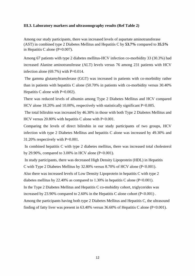
III.3. Laboratory markers and ultrasonography results (Ref Table 2)
Among our study participants, there was increased levels of aspartate aminotransferase
(AST) in combined type 2 Diabetes Mellitus and Hepatitis C by 53.7% compared to 35.5%
in Hepatitis C alone (P=0.007).
Among 67 patients with type 2 diabetes mellitus-HCV infection co-morbidity 33 (30.3%) had
increased Alanine aminotransferase (ALT) levels versus 76 among 231 patients with HCV
infection alone (69.7%) with P=0.014. The gamma glutamyltransferase (GGT) was increased in patients with co-morbidity rather
than in patients with hepatitis C alone (50.70% in patients with co-morbidity versus 30.40%
Hepatitis C alone with P=0.002). There was reduced levels of albumin among Type 2 Diabetes Mellitus and HCV compared
HCV alone 18.20% and 10.00%, respectively with statistically significant P=0.005. The total bilirubin was increased by 46.30% in those with both Type 2 Diabetes Mellitus and
HCV versus 20.80% with hepatitis C alone with P<0.001. Comparing the levels of direct bilirubin in our study participants of two groups, HCV
infection with type 2 Diabetes Mellitus and hepatitis C alone was increased by 49.30% and
31.20% respectively with P=0.001. In combined hepatitis C with type 2 diabetes mellitus, there was increased total cholesterol
by 29.90%, compared to 3.00% in HCV alone (P<0.001). In study participants, there was decreased High Density Lipoprotein (HDL) in Hepatitis
C with Type 2 Diabetes Mellitus by 32.80% versus 8.70% of HCV alone (P<0.001). Also there was increased levels of Low Density Lipoprotein in hepatitis C with type 2
diabetes mellitus by 22.40% as compared to 1.30% in hepatitis C alone (P<0.001).
In the Type 2 Diabetes Mellitus and Hepatitis C co-mobidity cohort, triglycerides was
increased by 23.90% compared to 2.60% in the Hepatitis C alone cohort (P<0.001) . Among the participants having both type 2 Diabetes Mellitus and Hepatitis C, the ultrasound
finding of fatty liver was present in 63.40% versus 36.60% of Hepatitis C alone (P<0.001).
12

Table 2: laboratory markers and ultrasonography findings
HCV/Diabetes n Hepatitis C alone n P value
(%) (%)
Laboratory
markers
Increased AST 36 (53.7) 82 (35.5) 0.007
( > 40 IU/L)
Increased ALT 33 (30.3) 76 (69.7) 0.014
( > 41 U/L)
Increased GGT 34 (50.7) 70 (30.4) 0.002
(>60 U/L)
Increased Total 31 (46.3) 48 (20.8) <0.001
bilirubin
(>17.1µmol/L)
Low Albumin 12 (18.2) 23 (10.0) 0.005
(< 3.97gms/dl)
Increased Direct 33 (49.3) 72 (31.2) 0.001
bilirubin
(>3.4 µmol/l)
Increased Total 20 (29.9) 7 (3.0) <0.001
cholesterol
(>5.2 mmol/l)
Low HDL 22 (32.8) 20 (8.7) <0.001
(<0.9 mmol/l)
Increased LDL 15 (22.4) 3 (1.3 <0.001
(>4.12mmol/l)
Increased 16 (23.9) 6 (2.6) <0.001
Triglyceride
(>2.26mmol/l)
Abdomen
Ultrasound result
Ascitis 34 (82.9) 7(17.1) <0.001
Fatty liver 26 (63.4) 15(36.6) <0.001
Cirrhosis 13 (81.2) 3(18.3) <0.001
13

Chapter IV: DISCUSSION
IV.1 Results discussion
In our study, the prevalence of type 2 diabetes mellitus was high; 22.48%. Most patients with
type 2 diabetes mellitus-hepatitis C co-morbidity showed liver enzymes and bilirubin
disturbance with low albumin, and impaired lipid profile than those with hepatitis C alone.
Furthermore, fatty liver and features of cirrhosis were most observed in co-morbid group.
The prevalence of type 2 diabetes mellitus in patients with hepatitis C was 22.5% in our study
population. It was relatively high. This could be due to the fact that most of our patients were
above 40 years, and age is a known risk factor for diabetes34
. Moreover, most of our patients
were from urban districts, which could have contributed to the patients’ high prevalence of
diabetes mellitus, being overweight (40.6 % of our population study). However this is higher
than the prevalence of diabetes in general Rwanda population (3% according to step by step
NCD risk factors survey’s preliminary results).Even though diabetes screening methods were
not similar, the prevalence of diabetes in hepatitis C patients is extremely higher (7 times )
than in general population.
This study outcome is similar to different other studies done in different places of the world. The
Mason et al: Multicenter study done in New Orleans showed that 21% of patients with hepatitis C
had type 2 diabetes23
. They also found that most of their participants who had co morbidity had
deranged liver functions tests23
. Raouf et al published a similar prevalence of type 2 diabetes
(24%) in the cohort Hepatitis C patients25
. In the Middle East and Europe, the prevalence of type
2 diabetes in hepatitis patients oscillated from 24% to 26%26-28
. It was noted, as per the study
done by Caronia et al that the prevalence of type 2 diabetes mellitus in Hepatitis C related
cirrhosis was 23.6%40
. The studies conducted by Akbar DH, Siddique AM, and Ahmed MM in
the Kingdom of Saudi Arabia reported similar prevalence of type 2 diabetes mellitus in HCV
patients (22% )30
. Zein CO, Mason AL, and Lecube A reported a range of 14-40% patients with
HCV related liver disease with concurrent type 2 diabetes mellitus31-33
.
Others reports showed higher prevalence of type 2 diabetes mellitus than ours probably due to
different demographic and lifestyle characteristics. For example Muhammad Sadik Memon et al
showed a prevalence of 31.5% of type 2 diabetes mellitus in hepatitis C seropositive patients34
and Samir Rouabhia et al done in Algeria showed also a higher prevalence of 39.1%35
14

compared to our study results. Obesity and physical inactivity which are common risk factors
for diabetes were more prevalent in their study population as compared to ours. This could
raise their type 2 diabetes prevalence.
Prevalence of type 2 diabetes mellitus in patients with hepatitis C infection is various due to
screening methodology and characteristics of population studies. However, it is found to be
sensibly high and suggests that hepatitis C infection may contribute to the rising burden of
diabetes on public health worldwide. Prevention of hepatitis C infection may contribute to
prevent type 2 diabetes mellitus.
There are also several studies examining the prevalence of liver function tests findings among
co affected diabetes/HCV cohort. In our study, we observed raised transaminases in the
patients with type 2 diabetes mellitus and hepatitis C. Samir Rouabhia et al found an
increased level of alanine aminotransferase in 51% of co-morbid patients as compared to our
findings (30.3%)35
. According to Bashir et al, the ALT levels were more elevated in patients
with Hepatitis C and type 2 diabetes co affection than Hepatitis C alone37
. A similar study
done by Tolman and associates came up with elevated liver enzymes ALT in 24% of patients
with type 2 diabetes mellitus and hepatitis C38
.
In our study participants, we observed lipids disorders in combined type 2 diabetes mellitus
and hepatitis C. In Bashir et al, HCV with type 2 diabetes mellitus showed a marked increase
in serum cholesterol and triglyceride level37
. However this may be due to the fact that
diabetes itself is a risk factor for dyslipidaemia but the combination of diabetes and
dyslipidaemia in patients with hepatitis C increase cardiovascular risk in this latter group.
Furthermore, fatty liver was most found in co-affected participants. In Wang et al, fatty liver
was observed in less number than we did however still high (35.7%) patients with both type 2
diabetes mellitus and hepatitis C39
. This is in accordance with the literature as diabetes
causes Nonalcoholic fatty liver disease (NAFLD) 42
.The co-existence of type 2 diabetes and
hepatitis C may increase liver deterioration and cardiovascular risk factors in patients with
hepatitis C. The addition of type 2 diabetes mellitus on hepatitis C can synergistically cause
liver fibrosis through the mechanism of insulin resistance18-19
.This should be taken into
consideration while managing hepatitis C patients.
15

IV.2 Study limitations
There are certain limitations of this study. The most important are small sample size, single-
Centered-hospital-based study and short period of 10 months which was unable to reflect the
actual incidence of type 2 diabetes mellitus in HCV Patients. In our study ,it was not possible to know in terms of hepatitis C infection and type 2 diabetes
mellitus which came first to help in the causality and risk factors from each other. Also in our study, we were not able to do liver biopsy for cirrhotic patients,
ultrasonographically diagnosed.
IV.3 Local relevance and generalizability
Based on our study results and despite its limitations, it is important therefore for every
patient with hepatitis C virus infection to also be screened for type 2 diabetes mellitus and
vice versa. Patients with hepatitis C infection found by early detection would help us to stop
the progression of the disease as nowadays we have proper treatment for infection for six
months (Ribavurin and sofosbivir) and Harvoni for three months. Thus preventing the co-
existence of type 2 diabetes mellitus induced by hepatitis C is essential. This can prevent the
activation of the immune system by Th1 lymphocytes to induce diabetes. Furthermore, our
study creates a significant local awareness of the association between type 2 diabetes mellitus
and hepatitis C infection. This is a great preventative approach for patients. There is also
needs to be simultaneous awareness among policy makers to put preventive measures on
hepatitis C and type 2 diabetes mellitus. Also these results will guard clinicians to educate patients about lifestyle modifications and
diet as it has a significant impact on these two diseases.
16

Chapter V. CONCLUSSION AND RECOMMENDATION
Type 2 diabetes mellitus is prevalent in adult patients with hepatitis C at Rwanda Military
Hospital and its co-existence with hepatitis C would have compounded negative impact on
liver function as well as on lipid metabolism. There is a need for further in-depth studies to
understand the pathophysiological mechanisms and causality.
As there was positive association between type 2 diabetes mellitus and HCV, it is necessary to
screen and control earlier in life for the presence of type 2 diabetes mellitus in HCV-infected
patients. It is also advisable to rule out HCV infection among the diabetic population.
To train all health care providers, particularly those working in the accident and
emergency department as well as in internal medicine, to better recognize hepatitis C and
diabetes mellitus as a co-morbidity.
To provide sufficient laboratory equipment to hospitals to be able to do HCV viral load for
early detection of the hepatitis C virus infection, which would be useful in the
management and prognostication of hepatitis patients.
To continue and support further studies on hepatitis C and type 2 diabetes mellitus
comorbidity in larger sample sizes, consider multicentre, for further generalizability
and guidance in the management of HCV and type 2 diabetes mellitus and look for
further correlations on the mechanism and causality.
17

REFERENCES
1. Eddleston M, Davidson R, Brent A, Wilkinson R. Oxford handbook of tropical medicine. 2012. p. 1–843.http://www.cdc.gov/hepatitis/hcv/cfaq.htm
2. Center for disease Control and prevention. Viral Hepatitis-hepatitis C information. Available
from: http://www.cdc.gov/hepatitis/hcv/cfaq.htm 3. Purcell RH. Hepatitis C virus; An Introduction. In: NIH Consensus Development Conference on Management of Hepatitis C. Available at: www.heplace.com/CCPurcell.html.
4. Sene D, Limal N, Cacoub P. Hepatitis C virus – associated extra hepatic manifestations. A
review. Metab Brain Dis2004; 19(3-4):357-81.liver disease. Jou Nutr. 2001; 131 (10):2805S- 2808S.
5. World Health organization. Fact Sheet No.164. Revised Oct 2000.
6. Armstrong GL, Wasley a, Simard EP, McQuillan GM, Kuhnert WL, Alter MJ. The prevalence
of hepatitis C virus infection in the United States, 1999 through 2002. Ann Intern Med [Internet].
2006;144(10):705–14. Available from: <Go to ISI>://000237636400001
7. Erin Gower, Chris Estes, Sarah Blach, Kathryn Razavi-Shearer, HomieRazavi. Global
epidemiology and genotype distribution of the hepatitis C virus infection. Journal of Hepatology
2014 vol. 61 j S45–S57 8. Olokoba, A.B., Bojuwoye, B.J., Katibi, I.A., Ajayi, A.O., Olokoba, L.B., Braimoh, K.T
etal (2007). Cholelithiasis and type 2 diabetes mellitus in Nigerians. South African
GastroenterologyReview. 5(3):14-17.
9. Susman JL, Helseth LD. Reducing the complications of type 2 diabetes mellitus: a patient-centered approach. American Family Physican.1997;56:471-80.
10. American Diabetes Association (ADA) Diabetes Guidelines. 2015; Summary Recommendations from NDEI. Am Diabetes Assoc. 2015;38(sup1):1. 11. World health organization. 2015. Fact sheet No 164. Updated in April 2004.
12. Harris MI, Klein R, Welborn TA, Knuiman MW. Onset of NIDDM occurs at least 4-7 yr before clinical diagnosis. Diabetes Care. Jul 1992;15(7):815-14.
13. Alberti, K.J.M.M., Mellander, A., Sevrano- Rios, M (1990). Guest editor’s introduction: An update on NIDDM IDF Bulletin. 3S:2.
14. Xu F, Mawokomatanda T, Flegel D, Pierannunzi C, Garvin W, Chowdhury P, et al.
Surveillance for Certain Health Behaviors Among States and Selected Local Areas — Behavioral
Risk Factor Surveillance System, United States, 2011. Morb Mortal Wkly report, Surveill Summ
(Washington, DC 2002). 2014;63(SS09):1–149.
15. Akinkugbe, O.O (1997). Final report of National expert committee on Non-communicable diseases. (Federal Ministry of Health and Social services, series 4): 64-90.
18

16. Harris MI, Klein R, Welborn TA, Knuiman MW. Onset of NIDDM occurs at least 4-7 yr
before clinical diagnosis. Diabetes Care. Jul 1992;15(7):815-9.
17. International Diabetes Federation 6th
edition. 2014 update. Available from: https://www.idf.org/sites/default/files/Atlas-poster-2014_EN.pdf
18. Angulo P. Nonalcoholic Fatty Liver Disease. 2002;346(16):1–11. Available from: papers3://publication/uuid/2C0516C5-6F77-4ECB-89D3-3B0F97C14810 19. Monto A, Alonzo J, Watson JJ, Grunfeld C, Wright TL. Steatosis in chronic hepatitis C:
relative contributions of obesity, diabetes mellitus, and alcohol. Hepatology [Internet].
2002;36(3):729–36. Available from: http://www.ncbi.nlm.nih.gov/pubmed/12198667
20. Kita Y, Mizukoshi E, Takamura T, Sakurai M, Takata Y. Impact of diabetes mellitus on prognosis of patients infected with hepatitis C virus. Vol. 56. 2007. p. 1682–8.
21. Lagiou P, Kuper H, Stuver S, Tzonou A, Trichopoulos D, Adami H. Role of diabetes mellitus in the etiology of hepatocellular carcinoma. J Natl Cancer Inst. 2000;92(13):1096–9. 22. Mona Abdel Raoufa, Zeinab A. Yousrya, Olfat M. Hindyb, Somayh S. Eissaa and Dalia
S. Soliman. Study of diabetes mellitus among patients with hepatitis C virus. The Egyptian
Journal of Internal Medicine 2012, 24:17–23.
23. Andrew L, Mason, Johnson Y, Nicole Hoang, Keping Qian, Graeme J. et al. Association
of Diabetes Mellitus and Chronic Hepatitis C Virus Infection.
24. Mason AL, Lau JY, Hoang N, Qian K, Alexander GJ, Xu L et al. Association of diabetes
mellitus and chronic hepatitis C virus infection. Hepatology 1999; 29: 328-333
25. Mona Abdel Raoufa , Zeinab A. Yousrya , Olfat M. Hindyb , Somayh S. Eissaa and
Dalia S. Solimanc. Study of diabetes mellitus among patients with hepatitis C virus. The Egyptian
Journalof Internal Medicine 2012, 24:17–23.
26. Grimbert S, Valensi P, Levy-Marchal C, Perret G, Richardet JP, Raffoux C, Trinchet JC, et
al. High prevalence of diabetes mellitus in patients with chronic hepatitis C. A case control study.
Gastroenterol Clin Biol 1996;20:544-548.
27. Ozyilkan E, Arslan M. Increased prevalence of diabetes mellitus in patients with chronic
hepatitis C virus infection. Am J Gastroenterol 1996;91:1480-1481.
28. Caronia S, Taylor K, Pagliaro L, Carr C, Palazzo U, O’Rahilly S, Alexander G. Strong
association between HCV and non-insulin dependent diabetes mellitus [Abstract]. J Hepatol
1996;25(Suppl 1):95.
29. Susman JL, Helseth LD. Reducing the complications of type 2 diabetes mellitus: a
patient centered approach. Am Fam Physican.1997;56:471-80.
30. Akbar DH, Siddique AM, Ahmed MM. Prevalence of type 2 diabetes in patients with
hepatitis C and B virus infection in Jeddah, Saudi Arabia. Med Princ Pract 2002;11:82-5.
31. Zein CO, Levy C, Basu A, Zein NN. Chronic hepatitis C and type II diabetes mellitus:
A prospective cross sectional study. American Journal Gastroentrol 2005;100:48-55.
19

32. Mason AL, Lau JY, Hoang N, Qian K, Alexander GJ, Xu L, et al. Association of diabetes
mellitus and chronic hepatitis C virus infection. Hepatology 1999;29:328-33.
33. Lecube a. Glucose Abnormalities in Patients with Hepatitis C Virus Infection:
Epidemiology and pathogenesis. Diabetes Care [Internet]. 2006;29(5):1140–9. Available from:
http://care.diabetesjournals.org/cgi/doi/10.2337/dc05-1995
34. Memon MS, Arain ZI, Naz F, Zaki M, Kumar S, Burney AA. Prevalence of type 2
diabetes mellitus in hepatitis C virus infected population: a Southeast Asian study. J Diabetes Res
[Internet]. 2013;2013:539361. Available from: http://www.ncbi.nlm.nih.gov/pubmed/23984431
35. Rouabhia S, Malek R, Bounecer H, Dekaken A, Bendali Amor F, Sadelaoud M, et al.
Prevalence of type 2 diabetes in Algerian patients with hepatitis C virus infection. World J Gastroenterol [Internet]. 2010;16(27):3427–31. Available from:
http://www.ncbi.nlm.nih.gov/pubmed/20632447
36. Knobler H, Schihmanter R, Zifroni A, Fenakel G, Schattner A. Increased risk of type 2
diabetes in noncirrhotic patients with chronic hepatitis C virus infection. Mayo Clin Proc
2000;75:355-359.
37. Bashir MF, Haider MS, Rashid N, Riaz S. Association of biochemical markers, hepatitis
C virus and diabetes mellitus in Pakistani Males. Trop J Pharm Res. 2013;12(5):845–50.
38. Tolman KG, Fonseca V, Dalpiaz A, Tan MH. Spectrum of liver disease in type 2 diabetesand
management of patients with diabetes and liver disease. Diabetes Care. 2007;30(3):734–43.
39. Wang C, Wang S, Yao W, Chang T. Community-based Study of Hepatitis C Virus Infection and Type 2 Diabetes : An Association Affected by Age and Hepatitis Severity Status. 2003;158(12):1154–60.
40. Simona Caronia,1 Kevin Taylor,2 Luigi Pagliaro,1 Colin Carr,2 Ugo Palazzo,1 Juraj Petrik,3 Stephen O’rahilly,2,4sarah Shore,5 Brian D. M. Tom,5 And Graeme J. M. Alexander4. Further Evidence for an Association between Non–Insulin-Dependent Diabetes Mellitus and
Chronic Hepatitis C Virus Infection.
41. Cochran WG (1977). Sampling Technique, 3rd
edition. New York: John Wiley & Sons.
Daniel WW (1999). Biostatistics: A foundation for analysis in the Health Sciences. 7th
edition.
New York: John Wiley & Sons.
Lwanga SK and Lemeshow S (1991). Sample Size Determination in Health Studies: A
Practical Manual. Geneva: World Health Organization.
Macfarlane SB (1997). Conducting a Descriptive Survey: 2.Choosing a Sampling
Strategy. Trop Doct, 27(1):14-21.
Naing L, Winn T and Rusli BN (2006).Sample Size Calculator for Prevalence Studies.
Available at: http://www.kck.usm.my/ppsg/stats_resources.htm
42. Harris MI, Flegal KM, Cowie CC, Eberhardt MS, Goldstein DE, Little RR, Wiedmeyer HM,
Byrd-Holt DD: Prevalence of diabetes, impaired fasting glucose, and impaired glucose
20

tolerance in U.S. adults: the Third National Health and Nutrition Examination Survey, 1988–
1994. Diabetes Care 21:518–524,1998.
43. Aguiree F, Brown A, Cho N, Dahlquist G, Aguiree, Brown, Cho, Dahlquist, Dodd, Dunning ...
& Whiting. IDF Diabetes Atlas. IDF Diabetes Atlas - sixth Ed [Internet]. 2013;155. Available
from: http://www.idf.org/sites/default/files/Atlas-poster-
2014_EN.pdf\nhttp://scholar.google.com/scholar?hl=en&btnG=Search&q=intitle:Diabetes+Atlas
#5\nhttp://dro.deakin.edu.au/view/DU:30060687\nhttp://hdl.handle.net/10536/DRO/DU:3006068
7
21

APPENDICES
Consent form
I, Dr Anthony BAZATSINDA postgraduate in college of medicine and health sciences,
school of medicine, internal medicine Department University of Rwanda, I hereby
conducting a study entitled prevalence of type 2 diabetes mellitus in adult patients with
hepatitis c virus infection and associated laboratory markers. Experience at Rwanda
Military Hospital.
I hereby asking patient/caretaker…………………………….. Permission of conducting
or participating him/herself in our study entitled as above for the interest of Rwandans
after completion of data collection and analysis of the results. There will be no
identification of patient’s names in this study so as he/she fears to be involved in the
study and I will keep confidentiality of every patient and I will put the data in office and
rock. No one else will be in contact with these data except me and investigator assistant.
Thereafter the results will be communicated to the patient later. In case of any emergency
to my patient during the study, always the patient is priority.
The study is about to see the relationship between HCV and diabetes mellitus, its
prevalence and associated laboratory markers. Results will help us in national protocol,
guidelines on treatment so as to improve management.
I ……………………..hereby confirm that i understand the content of this document and
the nature of this research project, i consent to participate in this research project. I
understand that I have the liberty to withdraw from the project at any time I desire.
We thank you to have accepted to participate in research project.
Name and Signature of the Participant
Investigator:
Dr Anthony BAZATSINDA
22
URUHUSHYA RWO GUKORERWAHO UBUSHAKASHATSI.
Njyewe, Muganga Anthony BAZATSINDA , umunyeshuri muri kaminuza y’u Rwanda
, mu kicikiro cya gatatu, mw’ishami ry’ubuvuzi , mu rwego rwo kurangiza icyo kiciro
,ndimo gukora ubushakashatsi .
None ndagusaba uburenganzira bwokugukoreraho ubushakashatsi kubijyanye n’indwara
y’isukari (Diabetes mellitus),twasanga uy’ifite tukaba twanagupima n’indwara
y’umwijima (Hepatitis C) ibi ni mu rwego rwo kumenya niba hari ihuriro izo ndwara
zombi zifitanye, kandi tugusanganye ubwo burwayi , tuzagufasha kubona ubuvuzi.
Ibizava mur’ubushakashatsi bizafasha urwego rw’igihugu rushizwe kwita kubarwayi
bizo ndwara zavuzwe hejuru
Ibi bikorwa mw’ibanga risesuye hagati yanjye na we gusa.
Tubashimiye uburyo mwakiriye icy’ikifuzo.
Njyewe. . . . . . . . . . . . . . . . . . .. . . . . . . . . . . . . .. . .. . nyuma y’ibisobanuro
mpawe k’ur’ubushakashatsi bugiye gukorwa, nemeye kuba nabukorerwaho
Amazina Umukono w’ukoreweho Umuganga
Ubushakashatsi . Anthony BAZATSINDA
Approved by:
Institutional review board
University of Rwanda
School of medicine and pharmacy
23

PREVALENCE OF TYPE 2 DIABETES MELLITUS IN ADULT PATIENTS WITH
HEPATITIS C VIRUS INFECTION AND ASSOCIATED LABORATORY
MARKERS. EXPERIENCE AT RWANDA MILITARY HOSPITAL.
Work sheet on T2DM/HCV.
Demographic data
1. Age in year a. 18-40
b. > 40
2. Gender a. male
b. female
3. Residence a. urban
b. rural
4. Marital status a. Single
b. Married
c. Separated
d. Divorced
e. Widow
5. Weight (Kg)
6. Height (m)
7. BMI (Kg/m2)
a.18
B.18-24.9
C.25-29.9
D.30-34.9
24

E.35-39.9
f. >40
8. Family history of diabetes
a. Yes
b. No
c. Don't know
9. He/she is T2DM
a. Yes
b. No
c. Don't know
10. He/she is HCV +
a. Yes
b. No
11. Family history of HCV
a. Yes
b. No
c. Don't know
12. Any colour change to jaundice
a. Yes
b. No
13. Abdominal distension
a. Yes
25

b. No
14. Ultrasonography findings
a. Normal
b. Ascites
c. Fatty liver
d. Fibrosis
e. Cirrhosis
f. Fatty liver and ascites
15. Risk factors
a. Previous blood transfusion
b. Previous surgical procedure
c. Tattooing
d. Multiple sexual partners
e. Illicit self-injection
f. Exposure to office or household contact of jaundice
g. Dental extraction
h. Scarification
i. Ear piecing
j. Circumcision
k. Uvulectomy by doctors native
26

. Laboratory markers
a. LFT’s - ALT a. Normal (1-41 U/L) b) Increased c) Decreased
- AST a) Normal (1-40 IU/L) b) Increased c) Decreased
- Albumin a) Normal (2.9-3.97 gms/dl) b) Increased c) Decreased
- INR a) Normal 0.8-1.2) b) Increased c) Decreased
- Total bilirubin a) Normal (0-17.1µmol/l) b) Increased c) Decreased
b. Lipid profile - Total cholesterol a) Normal (3.9-5.2mmol/l) b) Increased c) Decreased
- HDL a) Normal (0.9-1.68 mmol/l) b) Increased c) Decreased
- Triglyceride a) Normal (0.35-2.26 mmol/l) b) Increased c) Decreased
- LDL Normal (2.59-4.12) b) Increased c) Decreased
C. Hba1c a. <7
B.7.1-10
c. >10
d. HCV genotype
a. 1
b. 2
c. 3
d. 4
e. combination
e. HCV viral load according to National Genetics Institute assay
a. Negative (undetectable) <100 copies/ml
b. Low (>100-1,000,000)
c. Medium (1,000,000-5,000,000)
27

d. High (5,000,000-25,000,000)
e. Very high (>25,000,000)
Time frame.
The following was a proposed time-frame of the study process:
Number Activity Estimated Time
November 2014 to
1 Proposal Development and Presentation
5th- Jan 2015
2 Proposal Submission to the department for marking
January 2015
3 Submission of proposal for ethical approval February 2015
4 Pretesting March 2015
April 2015 to
5 Data Collection
January 2016
6 Data Analysis January 2016
7 Thesis writing January 2016
8 Thesis submission for correction February 2016
9 Thesis Final Submission March 2016
28

Total sample size is 298 participants. The Study was done a period in 10 months because
of the patient turnover in the
OPD specialized hepatitis and diabetes clinics and general medical ward.
Study budget.
Category Remarks Units Unit Cost Total
(Rwf) (Rwf)
Printing drafts
1000 100
100000
pages
Proposal
Proposal Copies
8copies
4500
36,000
Development
Literature review via 50,000
internet (40 hours)
Transport 100000 100,000
Viral load 298 75000 22,350,000
Lipid profile
– LDL
Laboratory - TRIGLICERIDE
Investigations. - Total cholesterol 298 15,000 4,470,000
- HDL
Genotype of HCV
298
NO NOT
REAGENTS
DONE
Stationery (pens, papers, 50000
etc).
Data Collection
Research assistant 1
1
100,000
each
for 10 months Assistant
month
1,000,000
29

Data Analysis Statistician 1 200,000 200,000
Printing drafts
1000 250,000 250,000
pages
Thesis Write Up
Printing Thesis 10 copies 25000 250,000
Contingencies 144280
5%
Grand total 29,000,280
30

Ethical clearance
31