prevention of huntington s disease-like behavioral
TRANSCRIPT

Research ArticlePrevention of Huntington’s Disease-Like Behavioral Deficits inR6/1 Mouse by Tolfenamic Acid Is Associated with Decreases inMutant Huntingtin and Oxidative Stress
Peng Liu,1 Yinjie Li,1 Wei Yang,1 Danyang Liu,1 Xuefei Ji,1 Tianyan Chi,1 Zhutao Guo,2
Lin Li,3 and Libo Zou 1
1Department of Pharmacology, Shenyang Pharmaceutical University, Shenyang 110016, China2Department of Clinical Pharmacy, Shenyang Pharmaceutical University, Shenyang 110016, China3Key Laboratory of Neurodegenerative Diseases (Capital Medical University), Ministry of Education, Beijing 100053, China
Correspondence should be addressed to Libo Zou; [email protected]
Received 19 October 2018; Revised 7 January 2019; Accepted 17 January 2019; Published 26 March 2019
Academic Editor: Ryuichi Morishita
Copyright © 2019 Peng Liu et al. This is an open access article distributed under the Creative Commons Attribution License, whichpermits unrestricted use, distribution, and reproduction in any medium, provided the original work is properly cited.
Tolfenamic acid is a nonsteroidal anti-inflammatory drug with neuroprotective properties, and it alleviates learning and memorydeficits in the APP transgenic mouse model of Alzheimer’s disease. However, whether tolfenamic acid can prevent motor andmemory dysfunction in transgenic animal models of Huntington’s disease (HD) remains unclear. To this end, tolfenamic acidwas orally administered to transgenic R6/1 mice from 10 to 20 weeks of age, followed by several behavioral tests to evaluatemotor and memory function. Tolfenamic acid improved motor coordination in R6/1 mice as tested by rotarod, grip strength,and locomotor behavior tests and attenuated memory dysfunction as analyzed using the novel object recognition test andpassive avoidance test. Tolfenamic acid decreased the expression of mutant huntingtin in the striatum of 20-week-old R6/1 miceby inhibiting specificity protein 1 expression and enhancing autophagic function. Furthermore, tolfenamic acid exhibitedantioxidant effects in both R6/1 mice and PC12 cell models. Collectively, these results suggest that tolfenamic acid has a goodtherapeutic effect on R6/1 mice, and may be a potentially useful agent in the treatment of HD.
1. Introduction
Huntington’s disease (HD) is an autosomal-dominantneurodegenerative disorder, the clinical hallmarks of whichinclude motor dysfunction, psychiatric disturbance, andcognitive deficits. HD is caused by abnormal expansion ofthe cytosine-adenine-guanine repeat in the IT15 genelocated on chromosome 4, resulting in the formation of apolyglutamine stretch in the N-terminus region of the Hun-tingtin protein (Htt) [1]. Mutant Htt (mHtt) causes selectiveneuronal loss in the brain. Mouse models of HD, most com-monly the R6 transgenic model that expresses a truncatedform of human Htt, have been primarily used to examineseveral therapeutic strategies [2]. Specificity protein 1 (Sp1)is a transcription factor, the target genes of which includeamyloid β precursor protein (APP), BACE1, Tau, and Htt,which all play vital roles in neurodegenerative diseases.
Because Sp1 promotes human Htt gene transcription [3–5],we hypothesized that the downregulation of Sp1-mediatedHtt transcription may alleviate the pathogenesis of HD.
Tolfenamic acid (TA) is a nonsteroidal anti-inflammatorydrug (NSAID) that decreases the expression and activity ofSP1 [6]. Previous studies have reported that tolfenamic acidalleviated cognitive deficits and downregulated the expressionof BACE1, APP, and phosphorylated tau in APP transgenicmice [7, 8]. Sankpal et al. reported that repeated administra-tion of tolfenamic acid in mice did not decrease body weightnor did it exhibit a toxic effect on several organs [9], whichsuggests that tolfenamic acid is safe for oral administration.However, whether tolfenamic acid can prevent HD-like symp-toms remains unclear.
In the present study, we investigated the effect of tolfe-namic acid on R6/1 transgenic mice. First, we assessedmotor function (rotarod test, grip strength test, and
HindawiOxidative Medicine and Cellular LongevityVolume 2019, Article ID 4032428, 13 pageshttps://doi.org/10.1155/2019/4032428

locomotor behavior test), memory function (novel objectrecognition test, Y maze test, and passive avoidance test),body weight, and brain weight. Subsequently, we testedthe effect of tolfenamic acid on huntingtin levels. To furtherunderstand the molecular mechanism of action of tolfe-namic acid, we tested the effect of tolfenamic acid onautophagy and oxidative stress in the brains of R6/1 trans-genic mice.
2. Materials and Methods
2.1. Materials. Tolfenamic acid (purity ≥98%) was purchasedfrom a commercial supplier (Abcam, Cambridge, MA, USA),and 3-nitropropionic acid (3-NP) was purchased fromSigma-Aldrich (MO, USA). The mouse monoclonal antibodyagainst huntingtin (EM48) was purchased from Millipore(CA, USA). The rabbit polyclonal antibodies against LC3,P62, and HO1; the mouse monoclonal antibody againstβ-actin; and horseradish peroxidase-conjugated secondaryantibodies were purchased from Proteintech (Wuhan,China). ML385, a specific Nrf2 inhibitor, was purchased fromMedChem Express (NJ, USA). Nrf2 small interfering RNA(siRNA) and rabbit polyclonal antibody against Sp1 werepurchased from Santa Cruz Biotechnology Inc. (CA, USA).The rabbit polyclonal antibody against NQO1 and D,L-buthionine-(S,R)-sulfoximine (BSO), a specific glutathionesynthase inhibitor, was purchased from Abcam (Cambridge,MA, USA).
2.2. Animals. B6.Cg-Tg(HDexon1)61Gpb/JNju mice (i.e.,R6/1) were procured fromNanjing Biomedical Research Insti-tution of Nanjing University (Nanjing, China), and wild type(WT) mice were used as control. The animals were housedin polyacrylic cages ( 30 0 cm length × 12 0 cmheight × 18 0cmwidth) under standard conditions with a 12h light/darkcycle and had ad libitum access to food and water. Bodyweight was recorded every 6-7days from 8 weeks of age untilthe animals were euthanized. All procedures involving ani-mals were performed in strict accordance with the P.R. Chinalegislation on the use and care of laboratory animals and theguidelines established by the Institute for Experimental Ani-mals at Shenyang Pharmaceutical University (permit number:SYPU-IACUC-C2016-2-25-183).
2.3. Drug and Treatment Schedule. The mice were dividedinto four groups of 8–10 animals each: control (WT mice);R6/1 mice (model group); R6/1 mice treated with tolfenamicacid (25mg/kg); and R6/1 mice treated with tolfenamic acid(50mg/kg). Ten-week-old mice were orally administeredtolfenamic acid or vehicle by gavage. After the behavioraltest, the mice were decapitated under ether anesthesia, andbrain tissue was extracted and dissected. The right half ofthe brain was used for immunohistochemical staining, whilestriatum from the left half was used for western blottinganalysis. The left half of the brain (except striatum) was usedto assess oxidative stress levels. The selection of two tolfe-namic acid doses was based on the study by Adwan et al.[8] and the authors’ previous study.
2.4. Locomotor Behavior Test. Locomotor activity wasassessed using a computer system and video camera whenthe mice were 20 weeks old. Mice were placed individually ina white PVC-enclosed chamber ( 25 cm long × 25 cmwide ×30 cmhigh) for 3min to acclimatize to the unfamiliar environ-ment, followed by recording of motor activity for 5min.Exploration distance, time, and number were recorded. Aftereach test, the floor was cleaned using ethanol (10%) to elimi-nate olfactory cues.
2.5. Grip Strength Test. The forelimb strength test was per-formed in mice 8 to 20 weeks old. Mice were grasped by theirback and drawn toward grip bars attached to a force sensor(Shandong Academy of Medical Sciences, China), and thenthey were allowed to grab the bars with both front paws.The mice were slowly pulled straight back with consistentforce until they released their grip. Grip strength was testedby the same investigator (Liu P.) three times to mitigate inter-rater differences in tensile strength, and the average value wasused for comparative analyses.
2.6. Rotarod Test. The rotarod test was performed asdescribed by van Dellen et al. [10] in mice from 8 to 20 weeksold. Two days before the test, mice were exposed to a trainingsession to acclimatize them to the rotarod procedure. On theday of the test, three separate trials began at an initial rate of3.5 rpm with an acceleration of 20 rpm/min to a maximum of30 rpm over a period of 180 sec in the rotarod apparatus(Shanghai Xinruan, China). The latency to fall values wererecorded, and the average time to fall was used in compara-tive analyses. After each test, the rods and separating wallswere cleaned using ethanol (10%) to eliminate olfactory cues.
2.7. Novel Object Recognition Test. The novel object recogni-tion test was performed at 20 weeks old, as described in theauthors’ previous report [11]. The apparatus consists of asquare box (length 50 cm × width 50 cm × height 15 cm). Onthe first two days, the mice were habituated to the equipmentfor 10min. On the test day, two identical objects, A1 and A2,were placed at the center of the box. The mouse was placed inthe box and permitted to explore for 5min. After a 1 h inter-trial interval, the familiar object A2, was replaced with anovel object B, and the mouse was permitted to explore theobjects for an additional 5min, which was the 1 h retentiontrial. After a 24 h retention interval, object B was replacedwith a novel object C, and the mouse was permitted toexplore the objects for 5min, which was a 24 h retention trial.The exploration time for each object was recorded. After eachtest, the floor was cleaned using ethanol (10%) to eliminateolfactory cues.
The preferential index (PI) was calculated as follows: timespent exploring the novel object/total exploration time.
2.8. Y Maze Test. The Y maze test was performed using20-week-old mice, as described in the authors’ previousreport [11]. The apparatus comprised three brown woodenarms (length 40 cm × height 12 cm × width 10 cm). The micewere placed (individually) at the end of an arm and permittedto explore for 5min. The total number of arm entries (n) andthe sequence of entries were recorded. Once the mouse
2 Oxidative Medicine and Cellular Longevity

entered three arms continuously, it was defined a “successfulalternation.” After each test, the floor was cleaned using eth-anol (10%) to eliminate olfactory cues. Alternation behaviorwas calculated as follows: number of successive alternations/n − 2 × 100.
2.9. Passive Avoidance Test. The passive avoidance test wasperformed using 20-week-old mice. The experimental deviceconsisted of a bright and dark room. A powerful light bulbwas hung at the top of the bright room to prompt the miceto enter into the dark room, the floor of which was equipped
15
20
25
30
35
8 9 10 11 12 13 14 15 16 17 18 19 20
Body
wei
ght (
g)
ControlModel
TA 25 mg/kgTA 50 mg/kg
Male ##
Weeks
(a)
10
15
20
25
30
8 9 10 11 12 13 14 15 16 17 18 19 20
Body
wei
ght (
g)
ControlModel
TA 25 mg/kgTA 50 mg/kg
Female##
Weeks
(b)
350
400
450
500
Control Model TA 25 mg/kg TA 50 mg/kg
Who
le b
rain
wei
ght (
mg)
##
⁎
⁎⁎
(c)
Figure 1: Effect of tolfenamic acid on body weight and brain weight in R6/1 mice. R6/1 mice exhibited a progressive decrease in body weight(a, b) and a decrease in brain weight (c) compared with control mice. Tolfenamic acid treatment attenuated losses in brain weight, but notbody weight. All results are expressed as mean ± SD. n = 7–8; ##p < 0 01 vs. control; ∗p < 0 05 and ∗∗p < 0 01 vs. model.
0
40
80
120
160
8 12 16 20
Grip
stre
ngth
(g)
WeeksControlModel
TA 25 mg/kgTA 50 mg/kg
####
(a)
0
50
100
150
200
8 10 12 14 16 18 20
Fall
of ti
me (
sec)
Weeks
ControlModel
TA 25 mg/kgTA 50 mg/kg
⁎ ⁎⁎
#
(b)
Figure 2: Effect of tolfenamic acid on motor deficits in R6/1 mice. R6/1 mice exhibited progressive weakening in muscle strength in the gripstrength test (a) and a decrease in fall latency time (b) in the rotarod test compared with control mice. Tolfenamic acid treatment improvedperformance in the rotarod test, but not in the grip strength test. All results are expressed as mean ± SD. n = 7 – 8; #p < 0 05 and ##p < 0 01versus control; ∗p < 0 05 and ∗∗p < 0 01 versus model.
3Oxidative Medicine and Cellular Longevity

with an electrified copper plate that could deliver a smallelectric shock (31V). The experiment was performed fortwo days. On the first day (training phase), the mice wereplaced individually into the bright room back to the holewithout electricity and free for 3min. The animals werethen driven into the darkroom through the alternating cur-rent. The normal reactions of the mice were to run back to
the bright room to avoid electric shock. Most of the ani-mals, again, or repeatedly ran into the darkroom but wereshocked and quickly ran back to the bright room. Thenumber of times the mice entered into the darkroom againafter a shock within 5min were recorded as error times.The method was the same as the training phase day ofthe test phase, performed after a 24h retention interval.
Control Model TA 25 mg/kg TA 50 mg/kg
Figure 3: Representative trace plot for the locomotor behavior test.
Table 1: Effect of tolfenamic acid on locomotor behavior in R6/1 mice.
Group Exploration distance (mm) Movement speed (mm/s) Exploration number Resting time (s) Exploration time (s)
Control 13,550 80 ± 956 50 45 17 ± 3 19 37 25 ± 2 86 32 43 ± 5 25 267 57 ± 5 25Model 2699 30 ± 674 67a 9 01 ± 2 25a 33 14 ± 4 19 187 72 ± 22 06a 111 87 ± 22 19a
TA 25mg/kg 6000 28 ± 757 01b 20 00 ± 2 52b 46 88 ± 4 36 130 22 ± 18 48b 169 79 ± 18 48b
TA 50mg/kg 7674 76 ± 1149 21c 25 58 ± 3 83c 39 57 ± 4 09 72 19 ± 17 46c 227 81 ± 17 46c
All of the results are expressed as the means ± SEM. n = 7 or 8. ap < 0 01 vs. the control group; bp < 0 05 and cp < 0 01 vs. the model group.
0
20
40
60
80
100
1 h 24 h
Pref
eren
tial i
ndex
(%)
ControlModel
TA 25 mg/kgTA 50 mg/kg
##⁎
(a)
0102030405060708090
Control Model TA25 mg/kg
TA50 mg/kg
Alte
rnat
ion
beha
viou
r (%
)
(b)
05
1015202530
Control Model TA25 mg/kg
TA50 mg/kg
Tota
l num
ber o
f arm
entr
ies
## ## ##
(c)
0123456
Control Model TA25 mg/kg
TA50 mg/kg
Erro
r tim
es
##
⁎
(d)
Figure 4: Effects of tolfenamic acid on cognitive dysfunction in R6/1 mice. R6/1 mice exhibited memory recall and visual recognition deficitsin the novel object recognition test (a), a decrease in spontaneous alternation behavior and arm entries in the Y maze test (b, c), and moreerror times in the passive avoidance test (d) compared to control mice. Tolfenamic acid treatment attenuated the cognitive deficits in thenovel object recognition test and the passive avoidance test, but not in the Y maze test. All results are expressed as the means ± SD. n = 7-8;##p < 0 01 versus control; ∗p < 0 05 versus model.
4 Oxidative Medicine and Cellular Longevity

00.20.40.60.8
11.2
Model TA 25 mg/kg TA 50 mg/kg m
Htt
level
(% o
f mod
el)
0
0.2
0.4
0.6
0.8
11.2
Control Model TA25 mg/kg
TA50 mg/kg
SP1
leve
l (%
of c
ontr
ol)
EM48
SP1
�훽-Actin -43 kDa
-250 kDa
-95 kDa
Control Model TA25 TA50
⁎⁎
⁎⁎
⁎⁎⁎
(a)
0
0.2
0.4
0.6
0.8
1
1.2
Control Model TA 25 mg/kg TA 50 mg/kg
EM48
cove
red
area
(%)
0
0.2
0.4
0.6
0.8
1
1.2
Control Model TA 25 mg/kg TA 50mg/kg
Mea
n In
tDen
of E
M48
(%)
TA 25 mg/kg
Control Model
TA 50 mg/kg
## ##
⁎⁎⁎⁎ ⁎⁎ ⁎⁎
(b)
Figure 5: Effect of tolfenamic acid treatment on the expression of mutant huntingtin (Htt) and Sp1 in the striatum. Both doses of tolfenamicacid (25 or 50mg/kg) significantly decreased the expression of mHtt and Sp1 (a) and decreased the intensity and positive area of theEM48-positive cell (b) in the striatum. All results are expressed as mean ± SEM. WB, n = 6; immunohistochemistry, n = 5, bar = 50μm.∗p < 0 05 and ∗∗p < 0 01 vs. model.
5Oxidative Medicine and Cellular Longevity
After each test, the floor was cleaned using ethanol (10%)to eliminate olfactory cues.
2.10. Estimation of Oxidative Stress. Malondialdehyde(MDA), nitrite, superoxide dismutase (SOD), catalase(CAT), total glutathione, and oxidized glutathione levels weremeasured from brain extracts prepared in cell lysis bufferusing commercially available assay kits (Nanjing JianchengBioengineering Institute, Nanjing, China) according to themanufacturer’s protocol. According to the kit instructions,the content of reduced gluthathione = total gluthathione − 2× oxidized gluthathione.
2.11. Isolation of Total RNA and Reverse Transcriptase-Polymerase Chain Reaction (RT-PCR) Analysis. RT-PCR wasperformed according to the method described in the authors’previous report [12]. Cerebral cortex total RNA was extractedusing TRIzol, and 2.0μg RNA was reverse transcribed by usinga complementary DNA (cDNA) synthesis system. cDNAproducts were amplified using PCR with specific primers ofglutathione peroxidase (GSH-Px) (forward (F): 5′-GGGACTACACCGAGATGAACG-3′; reverse (R): 5′-TCCGCAGGAAGGTAAAGAGC-3′); Nrf2 (F: 5′-CTTCCATTTACGGAGACCC-3′; R: 5′-GAGCACTGTGCCCTTGAGC-3′), andβ-actin (F: 5′-CTGTGCCCATCTACGAGGGCTAT-3′; R: 5′-TTTGATGTCACGCACGATTTCC-3′). The amplified PCR
products were separated on 1.5% agarose gels and then visual-ized with ethidium bromide under ultraviolet light. The bandintensity was quantified using Gel-Pro-Analyzer software.
2.12. Immunohistochemical Staining. Immunohistochemicalstaining was performed in accordance with the methoddescribed in the authors’ previous report [13]. Brain sectionswere incubated with EM48 (1 : 100) at 4°C overnight and thenwashed with phosphate-buffered saline (PBS) three times.The sections were incubated with biotin-labelled secondaryantibody at 37°C for 30min. The sections were treated withan avidin-biotin enzyme reagent and visualized using aDAB kit (Boster, Wuhan, China). The intensity and positivearea of each section were quantified using ImageJ software.
2.13. Western Blotting Analysis. Western blotting analysiswas performed in accordance with the method described inthe authors’ previous report [14]. Protein samples (30μg)were electrophoresed on an 8%–12% gradient sodium dode-cyl polyacrylamide gel, and then they were transferred toPVDF membranes. The membranes were first blocked with5% skim milk for 2 h at room temperature, incubated withprimary antibodies EM48 (1 : 100), Sp1 (1 : 1000), LC3(1 : 1000), P62 (1 : 1000), NQO1 (1 : 800), HO1 (1 : 800), andβ-actin (1 : 1000) at 4°C overnight, then incubated with sec-ondary antibody for 2 h at room temperature. Protein bandswere visualized using a commercially available electrochemi-luminescence kit. The band intensity was quantified usingImageJ software.
2.14. Cell Viability. PC12 cells were obtained from theNational Infrastructure of Cell Line Resource (Beijing,China). The cells were cultured in RPMI-1640 medium con-taining 10% fetal bovine serum in a humidified cell incubatorin an atmosphere of 5% CO2 at 37
°C. The viability of cells wastested using the MTT assay. PC12 cells were plated in 96-wellplates (1 × 10⁴ cells/well), and after 24 h of culture, the cellswere treated with tolfenamic acid (5 and 10μM)/ML385(5μM)/BSO (15μM) and cultured for an additional 24 h.Subsequently, the cells were incubated with 3-NP (15mM)for 8 h, followed by addition of 5mg/mL MTT to each well.After 4 h, the medium was removed and 150μL of dimethylsulfoxide was added to each well. The absorbance was mea-sured at a wavelength of 540nm using an ELISA plate reader(Thermo Fisher Scientific, Waltham, MA, USA).
Control siRNA/Nrf2 siRNA (60nM) were transfectedinto cells using Lipofectamine 2000 (Invitrogen, USA)according to the manufacturer’s protocol. After 24 h of incu-bation, the cells were treated with tolfenamic acid (5 and10μM) for an additional 24 h. Subsequently, the cells wereincubated with 3-NP (15mM) for 8 h, followed by cell viabil-ity analysis as described above.
The selection of tolfenamic acid/Nrf2 siRNA/ML385/B-SO/3-NP doses and action times were based on the studiesby Adwan et al., Huang et al., Zhang et al., Kulasekaran andGanapasam, Jiang et al., and Speen et al. [15–20] and our pre-liminary experiments.
2.15. Measurement of Reactive Oxygen Species Levels. PC12cells (1× 10⁴ cells per well) were incubated with tolfenamic
00.5
11.5
22.5
3
Control Model TA25 mg/kg
TA50 mg/kg
LC3
II/L
C3 I
leve
l(%
of c
ontr
ol)
00.20.40.60.8
11.21.41.6
Control Model TA25 mg/kg
TA50 mg/kg
P62
leve
l (%
of c
ontr
ol)
LC3
P62
β-Actin -43 kDa
-15 kDa
-62 kDa
Control Model TA25 TA50
⁎⁎⁎⁎
⁎⁎ ⁎⁎
##
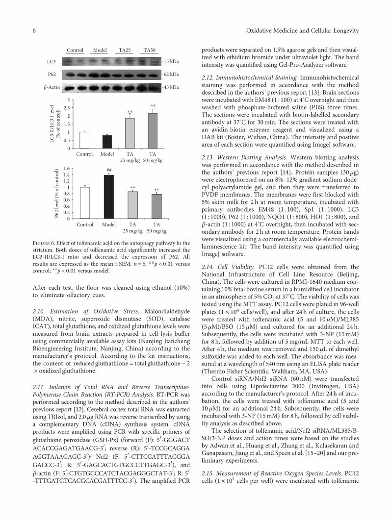
Figure 6: Effect of tolfenamic acid on the autophagy pathway in thestriatum. Both doses of tolfenamic acid significantly increased theLC3-II/LC3-I ratio and decreased the expression of P62. Allresults are expressed as the mean ± SEM. n = 6; ##p < 0 01 versuscontrol; ∗∗p < 0 01 versus model.
6 Oxidative Medicine and Cellular Longevity

acid (5 and 10μM) for 24 h, after which 3-NP (15mM) wasadded for an additional 8 h. After the drug treatments, thecells were incubated with 10μM DCF-DA, as the fluorescentprobe, at 37°C for 30 min. The level of reactive oxygen species(ROS) was measured by using a commercially available assaykit (Beyotime Biotechnology, China) according to the manu-facturer’s instructions. Fluorescence was measured using afluorometer (Thermo Fisher Scientific, USA) equipped witha 488 nm excitation filter and a 525nm emission filter.DCF-DA is poorly selective for O2
•-. Therefore, 50μM dihy-droethidium (DHE) was also used as an O2
•-fluorescent
probe. After 20min incubation at 37°C, cells were examinedunder a fluorescence microscope (Olympus, Japan).
2.16. Statistical Analysis. The data were analyzed using SPSSversion 21.0 (IBM Corporation, Armonk, NY, USA). Thestatistical significance of differences was determined usingone-way ANOVA followed by Fisher’s least significantdifference multiple comparison test with homogeneity ofvariance or Dunnett’s T3 test with heterogeneity of vari-ance. Experimental data are expressed as mean ± SD or SEM;p < 0 05 was considered to be statistically significant. Thepower calculation was performed using G∗Power 3.1.9.2(Heinrich-Heine-Universität Düsseldorf, Germany), and thevalue of 1 − β > 0 8 is acceptable.
3. Results
3.1. Effects of Tolfenamic Acid on Body and Brain Weight andMotor Deficits in R6/1 Mice. The body weight of the mice wastested as an index of general health, and brain weights weretested as an index of brain injury. Tolfenamic acid rescuedchanges in brain weight, but not body weight. The bodyweights of R6/1 mice progressively decreased from 15 to 20weeks of age (p < 0 01) (Figures 1(a) and 1(b)); however, tol-fenamic acid treatment did not result in any differences. Atthe beginning of the study, there were some differences in
body weight between the male and female R6/1 mice; there-fore, the data from male and female mice were analyzedindividually. Compared with the control group mice, R6/1group mice exhibited lighter brain weights (F 3, 21 = 20 789,p < 0 01; post hoc, p < 0 01) (Figure 1(c)). Tolfenamic acidtreatment appeared to prevent a decrease in brain weight(p < 0 05) (Figure 1(c)).
To assess muscle strength, a grip strength test was per-formed on the mice at 8, 12, 16, and 20 weeks of age. The gripstrength of mice in the model group gradually decreased: 8weeks, F 3, 33 = 0 181, p = 0 909; 12 weeks, F 3, 31 =1 798, p = 0 168; 16 weeks, F 3, 28 = 8 487, p < 0 01, posthoc, p < 0 01; and 20 weeks, F 3, 26 = 12 041, p < 0 01, posthoc, p < 0 01 (Figure 2(a)). Tolfenamic acid treatment didnot attenuate the weakening of muscle strength.
The rotarod test and locomotor behavior test were per-formed in R6/1 mice to evaluate motor coordination. Posthoc analyses revealed that mice in the model group exhib-ited a short latency from 14 weeks and gradually decreasedto 20 weeks (p < 0 05) (Figure 2(b)). Compared with themodel group, tolfenamic acid (50mg/kg) treatment signif-icantly increased the latency to fall values at 16 and 18weeks (p < 0 05) (Figure 2(b)). On the other hand, modelgroup mice exhibited higher immobility time comparedwith control group mice (Figure 3), which reflected loco-motor activity deficits in R6/1 mice (exploration distance:F 3, 21 = 25 899, p < 0 01, post hoc, p < 0 01; movementspeed: F 3, 21 = 25 894, p < 0 01, post hoc, p < 0 01;exploration number: F 3, 21 = 2 218, p = 0 110; restingtime: F 3, 21 = 16 495, p < 0 01, post hoc, p < 0 01; explo-ration time: F 3, 21 = 16 502, p < 0 01, post hoc, p < 0 01)(Table 1). Both doses of tolfenamic acid, however, rescuedthis change in locomotor activity (p < 0 05) (Table 1).
3.2. Effects of Tolfenamic Acid on Cognitive Dysfunction inR6/1 Mice. The novel object recognition test was performedin mice individually to evaluate recall memory. Compared
Table 2: Effect of tolfenamic acid on oxidative damage (lipid peroxidation, nitrite, superoxide dismutase, and catalase levels) in the brain ofR6/1 mice.
Group MDA (nmol/mg protein) NO (μmol/mg protein) SOD (U/mg protein) CAT (U/mg protein)
Control 9 10 ± 1 16 157 81 ± 17 67 27 33 ± 5 71 16 14 ± 2 57Model 12 23 ± 2 38 183 516 ± 34 48 23 52 ± 3 06 12 69 ± 5 26TA 25mg/kg 10 88 ± 1 57 181 20 ± 30 14 23 19 ± 3 69 12 97 ± 3 72TA 50mg/kg 9 80 ± 2 52 171 202 ± 26 43 27 82 ± 1 91 14 34 ± 2 45All of the results are expressed as the means ± SD. n = 5.
Table 3: Effect of tolfenamic acid on oxidative damage (glutathione levels) in the brain of R6/1 mice.
Group Total glutathione (μmol/L) Oxidized glutathione (μmol/L) Reduced glutathione (μmol/L)
Control 195 18 ± 26 99 51 05 ± 12 06 93 08 ± 13 99Model 214 23 ± 27 76 95 20 ± 12 21a 23 83 ± 15 14a
TA 25mg/kg 205 97 ± 9 01 70 50 ± 9 74b 64 97 ± 18 82b
TA 50mg/kg 201 94 ± 12 41 66 97 ± 8 62b 68 00 ± 18 67b
All of the results are expressed as the means ± SD. n = 5. ap < 0 01 vs. the control group; bp < 0 01 vs. the model group.
7Oxidative Medicine and Cellular Longevity

with the control group, the PI for novel object C wasdecreased in model group mice (F 3, 21 = 9 492, p < 0 01;post hoc, p < 0 01) (Figure 4(a)). More specifically, there were
memory recall and visual recognition impairments in20-week-old R6/1 mice. Treatment with 50mg/kg tolfenamicacid significantly prevented the decrease in PI (p < 0 01)(Figure 4(a)). In the Y maze test, treatment with 25 or50mg/kg tolfenamic acid neither alleviated nor worsenedspontaneous alternation behavior impairment in the mice(F 3, 21 = 2 510; p = 0 081) (Figure 4(b)). Furthermore,compared with the control group, model group and tolfe-namic acid group mice exhibited less locomotor behaviorduring exploration in the Y maze test (total number of armentries: F 3, 21 = 42 848, p < 0 01; post hoc, p < 0 01)(Figure 4(c)). In the passive avoidance test, compared withthe control group, the error times in model group mice weresignificantly increased (F 3, 21 = 3 632, p < 0 05, post hoc,p < 0 01) (Figure 4(d)). The error times in the tolfenamic acidgroup (50mg/kg) were less than that in R6/1 mice (P < 0 05,Figure 4(d)).
G∗Power software was used to perform the power calcu-lation for all of the behavioral experiments. The data are pro-vided as supporting information (SI.1-3). The value of1 − β > 0 8 was acceptable.
3.3. Effect of Tolfenamic Acid on the Expression of mHtt andAutophagy Pathway in the Striatum of R6/1 Mice. Westernblotting and immunohistochemistry methods were used toexamine the expression of mHtt in the striatum to investi-gate the neuroprotective effect of tolfenamic acid. Bothdoses of tolfenamic acid significantly decreased the expres-sion of mHtt and Sp1 (mHtt: F 3, 15 = 14 545, p < 0 01;post hoc, p < 0 01; Sp1: F 3, 20 = 7 222, p < 0 01, posthoc, p < 0 05) (Figure 5(a)). In immunohistochemistry,R6/1 mice exhibited high levels of EM48 labeling in thestriatum. EM48 labeling was reduced after TA treatment(positive area %: F 2, 12 = 76 893, p < 0 01, post hoc,p < 0 01; intensity %: F 2, 12 = 78 223, p < 0 01, post hoc,
00.20.40.60.8
11.2
Control Model TA25 mg/kg
TA50 mg/kg
GSH
-Px
mRN
A le
vel
(% o
f con
trol
)
00.20.40.60.8
11.2
Control Model TA25 mg/kg
TA50 mg/kg
Nrf2
mRN
A le
vel (
% o
f con
trol
)
GSH-Px
Nrf2
�훽-Actin
Control Model TA25 TA50
## ##
∗∗
Figure 7: Effect of tolfenamic acid on messenger RNA (mRNA) levels of glutathione peroxidase (GSH-Px) and Nrf2 in the cortex. R6/1 miceexhibited oxidative damage in the cortex. Tolfenamic acid increased the mRNA level of Nrf2, but had no effect on GSH-Px. All results areexpressed as mean ± SD. n = 3; ##p < 0 01 versus control; ∗∗p < 0 01 versus model.
00.20.40.60.8
11.2
Control Model TA25 mg/kg
TA50 mg/kg
NQ
O1
leve
l (%
of c
ontr
ol)
NQO1
HO1
�훽-Actin -43 kDa
-29 kDa
-28 kDa
Control Model TA25 TA50
00.20.40.60.8
11.21.4
Control Model TA25 mg/kg
TA50 mg/kg
HO
1 le
vel (
% o
f con
trol
)
##
##
⁎
⁎⁎
⁎⁎
Figure 8: Effect of tolfenamic acid on the expression of NQO1 andHO1 in the cerebral cortex. Tolfenamic acid significantly increasedthe expression of NQO1 and HO1. All results are expressedas mean ± SEM. n = 6. ##p < 0 01 versus control; ∗p < 0 05 and∗∗p < 0 01 versus model.
8 Oxidative Medicine and Cellular Longevity

p < 0 05) (Figure 5(b)). However, the mechanism of howtolfenamic acid cleared misfolded proteins remained unclearand, accordingly, the autophagy pathway was investigated.Compared with the model group, the LC3-II/LC3-I ratio inthe tolfenamic acid group was significantly increased(F 3, 20 = 10 665, p < 0 01, post hoc, p < 0 01) (Figure 6),suggesting that tolfenamic acid could increase autophagicfunction. P62 is an expendable substrate that decreases withautophagic upregulation. A decrease in P62 was also foundafter treatment with tolfenamic acid (F 3, 20 = 26 307,p < 0 01, post hoc, p < 0 01) (Figure 6).
3.4. Effect of Tolfenamic Acid on Oxidative Damage in R6/1Mice. Post hoc analyses revealed that there was no signifi-cant changes in the level of MDA, NO, CAT, or SODamong the groups (MDA: F 3, 16 = 2 357, p = 0 110; NO:F 3, 16 = 0 882, p = 0 471; CAT: F(3, 16)=0.922, p = 0 453;SOD: F 3, 16 = 2 020, p = 0 152) (Table 2), only the level ofGSH decreased in R6/1 mice (total GSH: F 3, 16 = 2 357,p = 0 548; oxidized GSH: F 3, 16 = 14 380, p < 0 01, posthoc, p < 0 01; reduced GSH: F 3, 16 = 14 578, p < 0 01, posthoc, p < 0 01) (Table 3). Tolfenamic acid treatment sig-nificantly increased GSH levels in R6/1 mice (p < 0 01)(Table 3). To reconfirm this result, the gene expression level
of GSH-Px in the cortex was tested using RT-PCR. TA nei-ther decreased nor enhanced messenger RNA (mRNA)levels of GSH-Px at 20 weeks (F 3, 8 = 117 345, p < 0 01,post hoc, p = 0 330) (Figure 7(a)). TA increased the mRNAlevel of the antioxidant gene Nrf2 (F 3, 8 = 269 915,p < 0 01, post hoc, p < 0 01) (Figure 7). NQO1 and HO1are two target genes of Nrf2. TA significantly increased theexpression of NQO1 and HO1 in the cerebral cortexof R6/1 mice (NQO1: F 3, 20 = 6 338, p < 0 05, posthoc, p < 0 01; HO1: F 3, 20 = 7 296, p < 0 01, post hoc,p < 0 05) (Figure 8).
3.5. Effect of Tolfenamic Acid on 3-NP-Induced Neurotoxicityin PC12 Cells. Oxidative stress is the major cause of cellularinjury in neurodegenerative disease. PC12 cells wereincubated with 3-NP as the in vitro model to test the effectof tolfenamic acid on 3-NP-induced neurotoxicity andoxidative stress. The selection of tolfenamic acid/Nrf2siRNA/ML385/BSO/3-NP doses and action times was basedon the studies by Adwan et al., Huang et al., Zhang et al.,Kulasekaran and Ganapasam, Jiang et al., and Speen et al.reported in [15–20] and our preliminary experiment.Pretreatment with tolfenamic acid for 24h significantly pre-vented PC12 cell death caused by 3-NP exposure (p < 0 01)
00.20.40.60.8
11.2
Control 3-NP TA 5 TA 10 ML385+ 3-NP
ML385+ TA 10
Cell
viab
ility
(% o
f con
trol
) ##⁎ ⁎
(a)
0
0.2
0.4
0.6
0.8
1
1.2
Control 3-NP TA 5 TA 10 Control 3-NP TA 5 TA 10
Cell
viab
ility
(% o
f con
trol)
⁎⁎
Control siRNA Nrf2 siRNA
(b)
0
0.2
0.4
0.6
0.8
1
1.2
Control 3-NP TA 10 BSO+ 3-NP
BSO+ TA 10
Cell
viab
ility
(% o
f con
trol
)
⁎⁎⁎ ⁎
(c)
Figure 9: Effect of tolfenamic acid on the viability of PC12 cells cultured with 3-nitropropionic acid (3-NP). PC12 cells were preincubatedwith control small interfering RNA (siRNA) or Nrf2 siRNA (60 nM). After 24 h of incubation, the cells were treated with tolfenamic acid(5 and 10μM) for 24 h and then with 3-NP (15mM) for 8 h (a). PC12 cells were treated with tolfenamic acid (5 and 10μM) with orwithout ML385 (5 μM) for 24 h and then with 3-NP (15mM) for 8 h (b). PC12 cells were treated with tolfenamic acid (10 μM) with orwithout BSO (15 μM) for 24 h and then with 3-NP (15mM) for 8 h (c). After treatment, cell survival was determined using the MTTassay. All of the results are expressed as the mean ± SD. n = 3; ∗∗p < 0 01 and ∗p < 0 05.
9Oxidative Medicine and Cellular Longevity
(Figures 9(a) and 9(b)). This protective effect was signifi-cantly blocked by Nrf2 siRNA and ML385 (a Nrf2 inhibi-tor) (Nrf2 siRNA: F(7, 16)=21.374, p < 0 01; post hoc,p < 0 01) (Figure 9(a)); (ML385: F(5, 12)=12.666, p < 0 01,post hoc, p < 0 05) (Figure 9(b)). BSO, a glutathione synthaseinhibitor, could partly block the protective effect of tolfenamicacid, but the difference was not statistically significant(F 4, 10 = 7 695, p < 0 01, post hoc, p = 0 263) (Figure 9(c)).These results suggest that tolfenamic acid exerted its neuro-protective effect through its antioxidant properties.
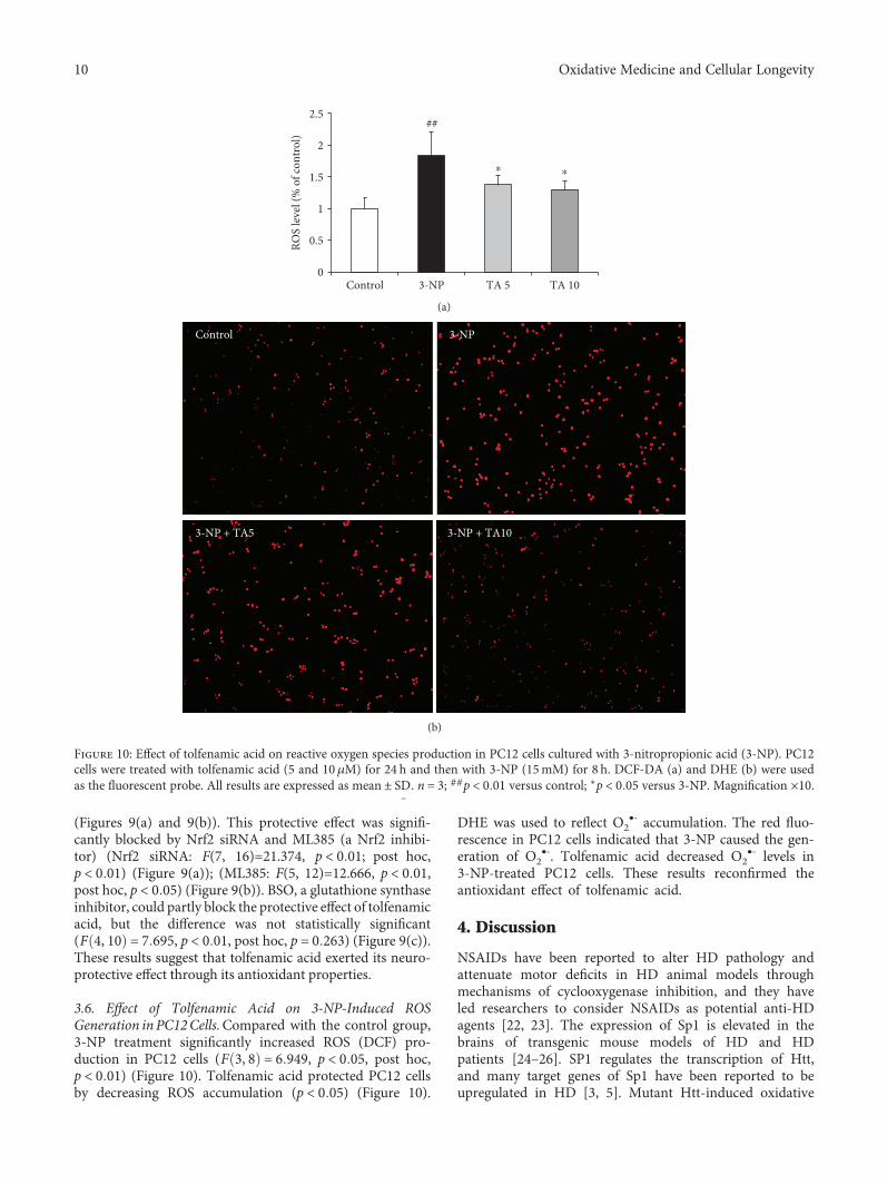
3.6. Effect of Tolfenamic Acid on 3-NP-Induced ROSGeneration in PC12Cells.Compared with the control group,3-NP treatment significantly increased ROS (DCF) pro-duction in PC12 cells (F 3, 8 = 6 949, p < 0 05, post hoc,p < 0 01) (Figure 10). Tolfenamic acid protected PC12 cellsby decreasing ROS accumulation (p < 0 05) (Figure 10).
DHE was used to reflect O2•- accumulation. The red fluo-
rescence in PC12 cells indicated that 3-NP caused the gen-eration of O2
•-. Tolfenamic acid decreased O2•- levels in
3-NP-treated PC12 cells. These results reconfirmed theantioxidant effect of tolfenamic acid.
4. Discussion
NSAIDs have been reported to alter HD pathology andattenuate motor deficits in HD animal models throughmechanisms of cyclooxygenase inhibition, and they haveled researchers to consider NSAIDs as potential anti-HDagents [22, 23]. The expression of Sp1 is elevated in thebrains of transgenic mouse models of HD and HDpatients [24–26]. SP1 regulates the transcription of Htt,and many target genes of Sp1 have been reported to beupregulated in HD [3, 5]. Mutant Htt-induced oxidative
0
0.5
1
1.5
2
2.5
Control 3-NP TA 5 TA 10RO
S le
vel (
% o
f con
trol
)
##
⁎ ⁎
(a)
Control 3-NP
3-NP + TA5 3-NP + TA10
(b)
Figure 10: Effect of tolfenamic acid on reactive oxygen species production in PC12 cells cultured with 3-nitropropionic acid (3-NP). PC12cells were treated with tolfenamic acid (5 and 10μM) for 24 h and then with 3-NP (15mM) for 8 h. DCF-DA (a) and DHE (b) were usedas the fluorescent probe. All results are expressed as mean ± SD̲. n = 3; ##p < 0 01 versus control; ∗p < 0 05 versus 3-NP. Magnification ×10.
10 Oxidative Medicine and Cellular Longevity

stress can activate Sp1 in neurons and glial cells [21]. Acti-vated Sp1 further exacerbates neuroinflammatory reactionand oxidative stress [27, 28]. Sp1 knockout HD transgenicmice live longer than their HD counterparts [25]. These datasuggest that the upregulation of Sp1 contributes to the pathol-ogy of HD, and that suppression of Sp1 may be beneficial.
Tolfenamic acid can induce the proteasome-dependentdegradation of SP transcription factors [6]. Previous studieshave demonstrated that Sp1 overexpression upregulatesAPP and BACE1 expression, which is involved in Alzhei-mer’s disease [7]. Tolfenamic acid can attenuate cognitivedeficits in APP transgenic mice after 2 weeks of administra-tion [7, 29]. These positive effects in cognitive behavior areregulated by inhibiting Sp1 and its target genes APP andBACE1. Thus, we hypothesized that tolfenamic acid couldinhibit Htt and, furthermore, attenuate motor and cognitivedeficits in HD mice.
R6/1 mice exhibit progressive locomotor coordinationdeficits, which begin at approximately 3 months of age [10,30]. In this study, motor impairment was assessed using therotarod test. We found that tolfenamic acid could inhibitthe progressive impairment of locomotor coordination.R6/1 mice also exhibited muscular weakness in the forelimbon the grip strength test and substantial locomotor activitydecrease in the locomotor behavior test. However, tolfenamicacid partly mitigated these impairments in R6/1 mice. Cogni-tive deficits appear before motor deficits in patients with HD[31]. HD patients experience a more serious decline of mem-ory recall function than memory storage, which is caused byneuronal and synaptic loss [32]. We used the novel objectrecognition test to evaluate the effect of tolfenamic acid onrecall memory [33]. We also used the Y maze and passiveavoidance tests to access the effect of TA on working andlong-term memory. R6/1 mice exhibited significant learningand memory deficits, and tolfenamic acid increased PI inthe novel object recognition test and decreased the errortimes in the passive avoidance test. Western blotting resultsrevealed that tolfenamic acid reduced Htt aggregation inthe striatum. Tolfenamic acid inhibited the expression ofSp1, which perhaps suggests that tolfenamic acid decreasedthe transcriptional level of mutant Htt in the brains of R6/1mice. Activating autophagic function also contributed tothe clearance of mutant Htt. LC3 transforms from form I toform II to serve as the recruiter of the autophagosome sub-strate P62 during the activation of autophagy. SP1 can blockautophagic flux via activating P62, and Sp1 inhibition willpromote autophagy [34, 35]. In this study, we found that tol-fenamic acid significantly increased the LC3-II/LC3-I ratioand decreased the level of P62. Therefore, tolfenamic acidinhibits the transcription factor Sp1 and activates the autoph-agy pathway, which may contribute to the clearance ofmutant Htt aggregates.
Another potentially important function of tolfenamicacid is the reduction of oxidative stress in the brain. mHttcauses inflammation, oxidative stress, lipid peroxidation,and mitochondrial dysfunction [36–38]. Oxidative stresscan cause cellular damage and neurodegeneration by induc-ing the production of ROS. Nrf2 regulates antioxidant geneexpression in response to oxidative stress [39]. We found that
tolfenamic acid treatment significantly increased mRNAlevels of Nrf2. NQO1 and HO1 are two vital target genes ofNrf2. We found that TA significantly increased the expres-sion of NQO1 and HO1 in the cerebral cortex of R6/1 mice.Previous studies have reported that oxidative stress caused byelevated levels of free radicals and depleted antioxidantenzymes cause neuronal damage in HD animal model brains[40, 41]. However, in this study, compared with WT mice,the content of MDA, NO, CAT, and SOD did not change sig-nificantly—only the level of GSH decreased in R6/1 mice.Tolfenamic acid treatment significantly attenuated the GSHlevel in R6/1 mice. Previous studies have reported that thelevel of GSH is decreased in HD patients [42]. GSH is pro-duced in the cytosol and transferred to the nuclei or mito-chondria. When GSH is oxidized, it becomes oxidized GSH.However, in vitro, we found that compared with the tolfe-namic acid treatment, the GSH synthase inhibitor BSO didnot significantly block the protective effect of tolfenamic acidin PC12 cells. Therefore, regulating the stabilization of GSHand oxidized GSH may be only one mechanism for tolfe-namic acid to cure HD, and will be investigated in futurestudies. Kulasekaran and Ganapasam reported that 3-NPcaused PC12 cell injury and induced significantly elevatedROS production [18]. Therefore, we used this in vitro cellmodel to reconfirm the antioxidant effect of tolfenamicacid. Tolfenamic acid significantly prevented 3-NP-induced neurotoxicity in PC12 cells, and this effect couldbe partly inhibited by Nrf2 siRNA or the specific Nrf2inhibitor—ML385. Tolfenamic acid also decreased ROSproduction in PC12 cells.
5. Conclusions
Collectively, the results of the present study suggest that tol-fenamic acid can attenuate motor and cognitive deficits inR6/1 transgenic mice. Tolfenamic acid could promote thedegradation of mHtt by inhibiting the transcription factorSp1 and enhancing autophagic function. Antioxidant pro-duction in the brains of R6/1 mice and in PC12 cells isanother important mechanism of tolfenamic acid. It has beenestablished that tolfenamic acid is safe for clinical use. There-fore, our data support tolfenamic acid as a potential candi-date for the treatment of HD.
Abbreviations
CAT: CatalaseGSH-Px: Glutathione peroxidaseHD: Huntington’s diseaseHO1: Heme oxygenase 1MDA: MalondialdehydemHtt: Mutant huntingtin proteinNO: NitriteNQO1: NAD(P)H quinine oxidoreductase 1Nrf2: Nuclear factor erythroid 2-related factor 2ROS: Reactive oxidative speciesSOD: Superoxide dismutaseSp1: Specificity protein 1TA: Tolfenamic acid.
11Oxidative Medicine and Cellular Longevity

Data Availability
The data used to support the findings of this study are avail-able from the corresponding author upon request.
Conflicts of Interest
The authors declare no competing financial interests.
Authors’ Contributions
P. Liu conceived the experiments, contributed to the researchdata, and drafted the manuscript. Y. Li, W. Yang, D. Liu, X. Ji,and Z. Guo contributed to the research data and data analy-sis. T. Chi and L. Li revised the manuscript. L. Zou revised themanuscript and supervised the analysis.
Acknowledgments
This work was supported by the Doctoral Scientific ResearchFoundation of Liaoning Province (No. 201601142), the Sci-entific Research Project of Liaoning Province (No. 51120424), the Open Foundation of Key Laboratory of Neurode-generative Diseases (Capital Medical University), ChinaMinistry of Education (No. 2016SJBX01), the University Stu-dents’ Innovation Undertaking Program of Liaoning Prov-ince (No. 201810163121), and the National Natural ScienceFoundation of China (No. 81703494).
Supplementary Materials
SI.1: the power (1 − β) value for the grip strength test androtarod test. SI.2: the power (1 − β) value for the locomotorbehavior test. SI.3: the power (1 − β) value for the passiveavoidance test, Y maze test, and novel object recognition test.(Supplementary Materials)
References
[1] S. Manoharan, G. J. Guillemin, R. S. Abiramasundari, M. M.Essa, M. Akbar, and M. D. Akbar, “The role of reactive oxygenspecies in the pathogenesis of Alzheimer’s disease, Parkinson’sdisease, and Huntington’s disease: a mini review,” OxidativeMedicine and Cellular Longevity, vol. 2016, Article ID8590578, 15 pages, 2016.
[2] T. Velusamy, A. S. Panneerselvam, M. Purushottam et al.,“Protective effect of antioxidants on neuronal dysfunctionand plasticity in Huntington’s disease,” Oxidative Medicineand Cellular Longevity, vol. 2017, Article ID 3279061, 15 pages,2017.
[3] A. S. Chen-Plotkin, G. Sadri-Vakili, G. J. Yohrling et al.,“Decreased association of the transcription factor Sp1 withgenes downregulated in Huntington’s disease,” Neurobiologyof Disease, vol. 22, no. 2, pp. 233–241, 2006.
[4] J. Bradford, J. Y. Shin, M. Roberts, C. E. Wang, X. J. Li, andS. Li, “Expression of mutant huntingtin in mouse brain astro-cytes causes age-dependent neurological symptoms,” Proceed-ings of the National Academy of Sciences, vol. 106, no. 52,pp. 22480–22485, 2009.
[5] R. Wang, Y. Luo, P. T. T. Ly et al., “Sp1 regulates human hun-tingtin gene expression,” Journal of Molecular Neuroscience,vol. 47, no. 2, pp. 311–321, 2012.
[6] M. Abdelrahim, C. H. Baker, J. L. Abbruzzese, and S. Safe,“Tolfenamic acid and pancreatic cancer growth, angiogenesis,and Sp protein degradation,” JNCI: Journal of the NationalCancer Institute, vol. 98, no. 12, pp. 855–868, 2006.
[7] G. M. Subaiea, L. I. Adwan, A. H. Ahmed, K. E. Stevens,and N. H. Zawia, “Short-term treatment with tolfenamicacid improves cognitive functions in Alzheimer’s diseasemice,” Neurobiology of Aging, vol. 34, no. 10, pp. 2421–2430, 2013.
[8] L. Adwan, G. M. Subaiea, R. Basha, and N. H. Zawia, “Tolfe-namic acid reduces tau and CDK5 levels: implications fordementia and tauopathies,” Journal of Neurochemistry,vol. 133, no. 2, pp. 266–272, 2015.
[9] U. T. Sankpal, C. M. Lee, S. F. Connelly et al., “Cellular andorganismal toxicity of the anti-cancer small molecule, tolfe-namic acid: a pre-clinical evaluation,” Cellular Physiologyand Biochemistry, vol. 32, no. 3, pp. 675–686, 2013.
[10] A. van Dellen, P. M. Cordery, T. L. Spires, C. Blakemore, andA. J. Hannan, “Wheel running from a juvenile age delays onsetof specific motor deficits but does not alter protein aggregatedensity in a mouse model of Huntington’s disease,” BMC Neu-roscience, vol. 9, no. 1, p. 34, 2008.
[11] G. Jin, L. H. Wang, X. F. Ji et al., “Xanthoceraside rescueslearning and memory deficits through attenuatingbeta-amyloid deposition and tau hyperphosphorylation inAPP mice,” Neuroscience Letters, vol. 573, pp. 58–63, 2014.
[12] Y. Qi, X. F. Ji, T. Y. Chi et al., “Xanthoceraside attenuates amy-loid β peptide 1-42-inducedmemory impairments by reducingneuroinflammatory responses in mice,” European Journal ofPharmacology, vol. 820, pp. 18–30, 2018.
[13] Q. Xu, X. F. Ji, T. Y. Chi et al., “Sigma 1 receptor activationregulates brain-derived neurotrophic factor through NR2A-CaMKIV-TORC1 pathway to rescue the impairment oflearning and memory induced by brain ischaemia/reperfu-sion,” Psychopharmacology, vol. 232, no. 10, pp. 1779–1791, 2015.
[14] P. Liu, R. Zhang, D. Liu et al., “Time-course investigation ofblood-brain barrier permeability and tight junction proteinchanges in a rat model of permanent focal ischemia,” Journalof Physiological Sciences, vol. 68, no. 2, pp. 121–127, 2018.
[15] L. Adwan, G. M. Subaiea, and N. H. Zawia, “Tolfenamic aciddownregulates BACE1 and protects against lead-inducedupregulation of Alzheimer’s disease related biomarkers,” Neu-ropharmacology, vol. 79, pp. 596–602, 2014.
[16] J. Y. Huang, Y. H. Yuan, J. Q. Yan et al., “20C, a bibenzyl com-pound isolated from Gastrodia elata, protects PC12 cellsagainst rotenone-induced apoptosis via activation of theNrf2/ARE/HO-1 signaling pathway,” Acta PharmacologicaSinica, vol. 37, no. 6, pp. 731–740, 2016.
[17] J. Zhang, W. Tong, H. Sun et al., “Nrf2-mediated neuroprotec-tion by MANF against 6-OHDA-induced cell damage viaPI3K/AKT/GSK3β pathway,” Experimental Gerontology,vol. 100, pp. 77–86, 2017.
[18] G. Kulasekaran and S. Ganapasam, “Neuroprotective efficacyof naringin on 3-nitropropionic acid-induced mitochondrialdysfunction through the modulation of Nrf2 signaling path-way in PC12 cells,” Molecular and Cellular Biochemistry,vol. 409, no. 1-2, pp. 199–211, 2015.
12 Oxidative Medicine and Cellular Longevity

[19] L. Jiang, H. Li, and N. Zhao, “Thymoquinone protects againstcobalt chloride-induced neurotoxicity via Nrf2/GCL-regulatedglutathione homeostasis,” Journal of Biological Regulators andHomeostatic Agents, vol. 31, no. 4, pp. 843–853, 2017.
[20] A. Speen, C. Jones, R. Patel et al., “Mechanisms of CDDO-imidazolide-mediated cytoprotection against acrolein-induced neurocytotoxicity in SH-SY5Y cells and primaryhuman astrocytes,” Toxicology Letters, vol. 238, no. 1, pp. 32–42, 2015.
[21] J. Cornett, L. Smith, M. Friedman, J. Y. Shin, X. J. Li, and S. H.Li, “Context-dependent dysregulation of transcription bymutant huntingtin,” Journal of Biological Chemistry, vol. 281,no. 47, pp. 36198–36204, 2006.
[22] P. Kumar, H. Kalonia, and A. Kumar, “Role of LOX/COX path-ways in 3-nitropropionic acid-induced Huntington’s disease-like symptoms in rats: protective effect of licofelone,” BritishJournal of Pharmacology, vol. 164, no. 2b, pp. 644–654, 2011.
[23] H. Kalonia, P. Kumar, and A. Kumar, “Licofelone attenuatesquinolinic acid induced Huntington like symptoms: possiblebehavioral, biochemical and cellular alterations,” Progress inNeuro-Psychopharmacology & Biological Psychiatry, vol. 35,no. 2, pp. 607–615, 2011.
[24] A. W. Dunah, H. Jeong, A. Griffin et al., “Sp1 and TAFII130transcriptional activity disrupted in early Huntington’s dis-ease,” Science, vol. 296, no. 5576, pp. 2238–2243, 2002.
[25] Z. Qiu, F. Norflus, B. Singh et al., “Sp1 is up-regulated in cellu-lar and transgenic models of Huntington disease, and itsreduction is neuroprotective,” Journal of Biological Chemistry,vol. 281, no. 24, pp. 16672–16680, 2006.
[26] M. Ravache, C. Weber, K. Mérienne, and Y. Trottier, “Tran-scriptional activation of REST by Sp1 in Huntington’s diseasemodels,” PLoS One, vol. 5, no. 12, article e14311, 2010.
[27] X. R. Mao, A. M. Moerman-Herzog, Y. Chen, and S. W. Bar-ger, “Unique aspects of transcriptional regulation in neu-rons—nuances in NFκB and Sp1-related factors,” Journal ofNeuroinflammation, vol. 6, no. 1, p. 16, 2009.
[28] H. Ryu, J. Lee, K. Zaman et al., “Sp1 and Sp3 are oxidativestress-inducible, antideath transcription factors in corticalneurons,” Journal of Neuroscience, vol. 23, no. 9, pp. 3597–3606, 2003.
[29] L. I. Adwan, R. Basha, M. Abdelrahim, G. M. Subaiea, andN. H. Zawia, “Tolfenamic acid interrupts the de novo synthesisof the β-amyloid precursor protein and lowers amyloid betavia a transcriptional pathway,” Current Alzheimer Research,vol. 8, no. 4, pp. 385–392, 2011.
[30] F. Kreilaus, A. S. Spiro, A. J. Hannan, B. Garner, and A. M. Jen-ner, “Therapeutic effects of anthocyanins and environmentalenrichment in R6/1 Huntington’s disease mice,” Journal ofHuntington’s Disease, vol. 5, no. 3, pp. 285–296, 2016.
[31] M. Anglada-Huguet, X. Xifró, A. Giralt, A. Zamora-Moratalla,E. D. Martín, and J. Alberch, “Prostaglandin E2 EP1 receptorantagonist improves motor deficits and rescues memorydecline in R6/1 mouse model of Huntington’s disease,”Molec-ular Neurobiology, vol. 49, no. 2, pp. 784–795, 2014.
[32] M. Kim, K.-H. Cho, M.-S. Shin et al., “Berberine preventsnigrostriatal dopaminergic neuronal loss and suppresses hip-pocampal apoptosis in mice with Parkinson’s disease,” Inter-national Journal of Molecular Medicine, vol. 33, no. 4,pp. 870–878, 2014.
[33] H. Kalonia, P. Kumar, A. Kumar, and B. Nehru, “Protectiveeffect of montelukast against quinolinic acid/malonic acid
induced neurotoxicity: possible behavioral, biochemical, mito-chondrial and tumor necrosis factor-α level alterations in rats,”Neuroscience, vol. 171, no. 1, pp. 284–299, 2010.
[34] X. W. Xu, C. W. Pan, X. M. Yang, L. Zhou, Z. Q. Zheng, andD. C. Li, “SP1 reduces autophagic flux through activatingp62 in gastric cancer cells,” Molecular Medicine Reports,vol. 17, no. 3, pp. 4633–4638, 2018.
[35] J. Kim, H. W. Lee, D. K. Rhee, J. C. Paton, and S. Pyo, “Pneu-molysin-induced autophagy contributes to inhibition of osteo-blast differentiation through downregulation of Sp1 in humanosteosarcoma cells,” Biochimica et Biophysica Acta (BBA) -General Subjects, vol. 1861, no. 11, pp. 2663–2673, 2017.
[36] A. Crotti, C. Benner, B. E. Kerman et al., “Mutant huntingtinpromotes autonomous microglia activation via myeloidlineage-determining factors,” Nature Neuroscience, vol. 17,no. 4, pp. 513–521, 2014.
[37] C. Vidoni, A. Castiglioni, C. Seca, E. Secomandi, M. A. B. Mel-one, and C. Isidoro, “Dopamine exacerbates mutant hunting-tin toxicity via oxidative-mediated inhibition of autophagy inSH-SY5Y neuroblastoma cells: beneficial effects of anti-oxidant therapeutics,” Neurochemistry International, vol. 101,pp. 132–143, 2016.
[38] M. Jodeiri Farshbaf and K. Ghaedi, “Huntington’s disease andmitochondria,” Neurotoxicity Research, vol. 32, no. 3, pp. 518–529, 2017.
[39] J. Zhao, L. Zhai, Z. Liu, S. Wu, and L. Xu, “Leptin level and oxi-dative stress contribute to obesity-induced low testosterone inmurine testicular tissue,” Oxidative Medicine and CellularLongevity, vol. 2014, Article ID 190945, 14 pages, 2014.
[40] A. M. Oliveira, S. M. Cardoso, M. Ribeiro, R. S. G. R. Seixas,A. M. S. Silva, and A. C. Rego, “Protective effects of 3-alkylluteolin derivatives are mediated by Nrf2 transcriptional activ-ity and decreased oxidative stress in Huntington’s diseasemouse striatal cells,” Neurochemistry International, vol. 91,pp. 1–12, 2015.
[41] A. Kumar, T. Chaudhary, and J. Mishra, “Minocycline modu-lates neuroprotective effect of hesperidin against quinolinicacid induced Huntington’s disease like symptoms in rats:behavioral, biochemical, cellular and histological evidences,”European Journal of Pharmacology, vol. 720, no. 1-3, pp. 16–28, 2013.
[42] D. J. Wright, T. Renoir, Z. M. Smith et al., “N-Acetylcysteineimproves mitochondrial function and ameliorates behavioraldeficits in the R6/1 mouse model of Huntington’s disease,”Translational Psychiatry, vol. 5, no. 1, p. e492, 2015.
13Oxidative Medicine and Cellular Longevity

Stem Cells International
Hindawiwww.hindawi.com Volume 2018
Hindawiwww.hindawi.com Volume 2018
MEDIATORSINFLAMMATION
of
EndocrinologyInternational Journal of
Hindawiwww.hindawi.com Volume 2018
Hindawiwww.hindawi.com Volume 2018
Disease Markers
Hindawiwww.hindawi.com Volume 2018
BioMed Research International
OncologyJournal of
Hindawiwww.hindawi.com Volume 2013
Hindawiwww.hindawi.com Volume 2018
Oxidative Medicine and Cellular Longevity
Hindawiwww.hindawi.com Volume 2018
PPAR Research
Hindawi Publishing Corporation http://www.hindawi.com Volume 2013Hindawiwww.hindawi.com
The Scientific World Journal
Volume 2018
Immunology ResearchHindawiwww.hindawi.com Volume 2018
Journal of
ObesityJournal of
Hindawiwww.hindawi.com Volume 2018
Hindawiwww.hindawi.com Volume 2018
Computational and Mathematical Methods in Medicine
Hindawiwww.hindawi.com Volume 2018
Behavioural Neurology
OphthalmologyJournal of
Hindawiwww.hindawi.com Volume 2018
Diabetes ResearchJournal of
Hindawiwww.hindawi.com Volume 2018
Hindawiwww.hindawi.com Volume 2018
Research and TreatmentAIDS
Hindawiwww.hindawi.com Volume 2018
Gastroenterology Research and Practice
Hindawiwww.hindawi.com Volume 2018
Parkinson’s Disease
Evidence-Based Complementary andAlternative Medicine
Volume 2018Hindawiwww.hindawi.com
Submit your manuscripts atwww.hindawi.com