primary dural lymphoblastic b-cell lymphoma: a rare subtype of aggressive dural lymphoma ·...
TRANSCRIPT

CASE REPORT
Primary dural lymphoblastic B-cell lymphoma: a rare subtypeof aggressive dural lymphoma
Christine Saraceni1 & Nicole Agostino1 & Shereen Gheith2
Received: 5 June 2015 /Accepted: 30 July 2015 /Published online: 14 August 2015# Springer-Verlag Berlin Heidelberg 2015
Abstract Lymphomas arising in the dura mater repre-sent a rare subset of primary central nervous systemlymphoma (PCNSL). The majority of primary durallymphoma (PDL) are low-grade marginal zone lympho-mas (MZL) characterized by an indolent course and fa-vorable long-term outcomes. Primary aggressive durallymphomas are exceedingly rare with a paucity of casesreported in the literature. Herein, we describe a case ofprimary lymphoblastic dural lymphoma. To our knowledge,this is the second report of an isolated dural B-lymphoblasticlymphoma (B-LBL) in an immunocompetent patient.
Keywords B-lymphoblastic lymphoma . Primary CNSlymphoma . Dural lymphoma . Acute lymphoblasticlymphoma . B-ALL
Introduction
Primary dural lymphoma is a rare form of PCNSL, mostlydescribed by case reports. The incidence of PDL is reportedas 0.6–3 % of all primary central nervous system lymphomas(incidence variable in studies) [1, 2]. Aggressive NHL
dural subtypes are unique as most aggressive lympho-mas of the meninges are associated with brain paren-chymal or systemic involvement. The majority of PDLare low-grade, B-cell marginal zone lymphoma (MZL)with rare cases of low-grade follicular subtypes reported[1]. Aggressive dural lymphomas including diffuse largeB-cell lymphoma (DLBCL) and lymphoblastic subtypesare limited to case reports and small case series [1, 3–6](Table 1). PDL have favorable outcomes and the indo-lent forms are biologically distinct from parenchymalPCNSL or systemic lymphoma metastatic to the CNS[1]. Whether B-lymphoblastic subtypes will follow thisfavorable trajectory remains unclear. The paucity ofdural lymphoblastic lymphoma cases in the literaturemake standardized treatment and prognosticationdifficult.
Case history
A 42-year-old immunocompetent male presented withseveral weeks of a left frontal headache, lack of coor-dination, and tendency to drift to the left while driving.Magnetic resonance imaging (MRI) revealed a large en-hancing extra-axial mass along the right posterior supe-rior parietal convexity invading the calvarium (Fig. 1a).A second 18-mm focus of enhancement was also notedwithin the left parietal calvarium distinct from the tumorbulk (Fig. 1b). The tumor was protruding from the outertable of the calvarium and in areas, invaded the menin-ges and infiltrated into the subdural space. Excision ofthe tumor was performed and submitted for pathology.A diagnosis of B-LBL was rendered. Complete bloodcount was only notable for a mild thrombocytosis:platelet count 454,000/μL which was present since
* Christine [email protected]
1 Department of Hematology Oncology, Lehigh Valley HealthNetwork, John and Dorothy Morgan Cancer Center,1240 S. Cedar Crest Blvd, Suite 401, Allentown, PA 18103, USA
2 Department of Pathology, Lehigh Valley Health Network,Allentown, PA 18103, USA
J Hematopathol (2016) 9:35–39DOI 10.1007/s12308-015-0257-0
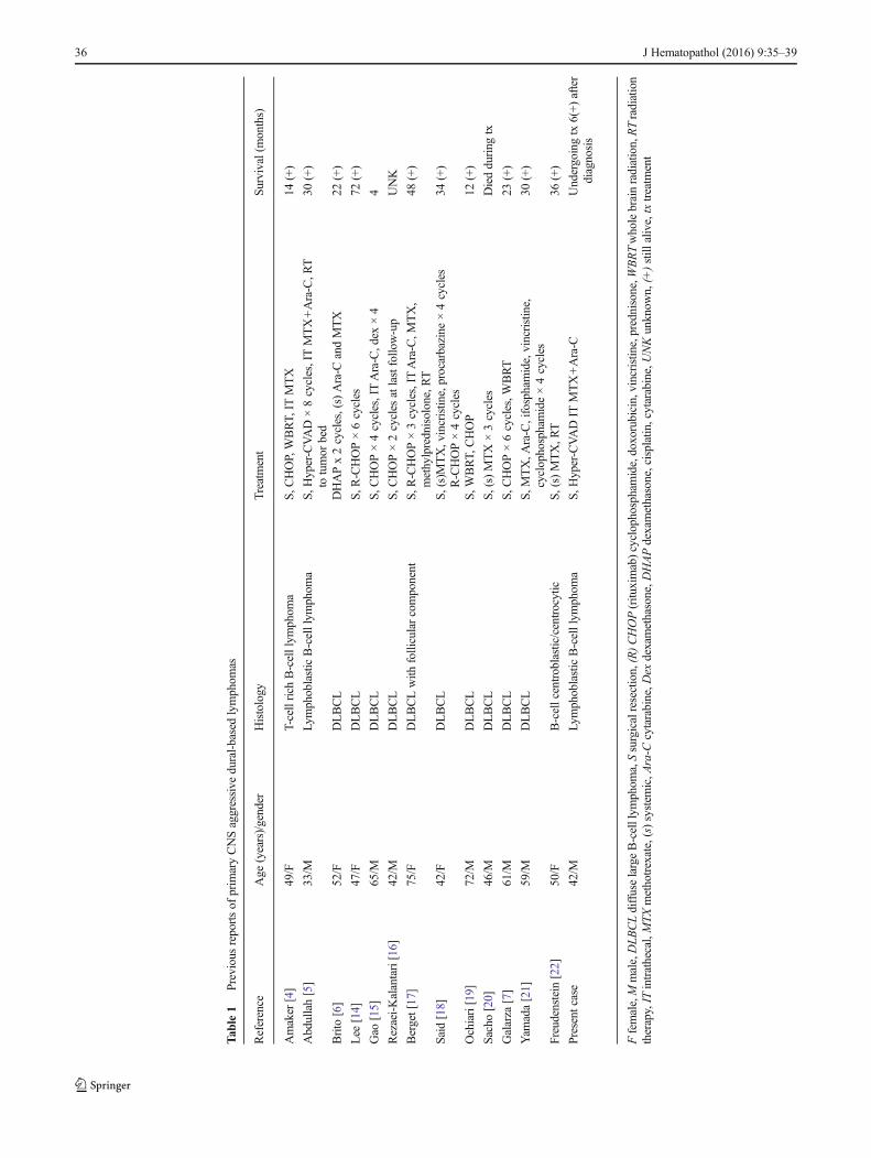
Tab
le1
Previous
reportsof
prim
aryCNSaggressive
dural-basedlymphom
as
Reference
Age
(years)/gender
Histology
Treatment
Survival(months)
Amaker
[4]
49/F
T-cellrich
B-celllym
phom
aS,C
HOP,WBRT,
ITMTX
14(+)
Abdullah[5]
33/M
Lym
phoblasticB-celllym
phom
aS,H
yper-CVAD×8cycles,ITMTX+Ara-C,R
Tto
tumor
bed
30(+)
Brito
[6]
52/F
DLBCL
DHAPx2cycles,(s)Ara-C
andMTX
22(+)
Lee
[14]
47/F
DLBCL
S,R
-CHOP×6cycles
72(+)
Gao
[15]
65/M
DLBCL
S,C
HOP×4cycles,ITAra-C,dex
×4
4
Rezaei-Kalantari[16]
42/M
DLBCL
S,C
HOP×2cycles
atlastfollo
w-up
UNK
Berget[17]
75/F
DLBCLwith
follicularcomponent
S,R-CHOP×3cycles,ITAra-C,M
TX,
methylprednisolone,RT
48(+)
Said[18]
42/F
DLBCL
S,(s)MTX,vincristin
e,procarbazine
×4cycles
R-CHOP×4cycles
34(+)
Ochiari[19]
72/M
DLBCL
S,WBRT,
CHOP
12(+)
Sacho
[20]
46/M
DLBCL
S,(s)MTX×3cycles
Diedduring
tx
Galarza
[7]
61/M
DLBCL
S,C
HOP×6cycles,W
BRT
23(+)
Yam
ada[21]
59/M
DLBCL
S,M
TX,A
ra-C,ifosphamide,vincristine,
cyclophosphamide×4cycles
30(+)
Freudenstein
[22]
50/F
B-cellcentroblastic/centrocytic
S,(s)MTX,R
T36
(+)
Presentcase
42/M
Lym
phoblasticB-celllym
phom
aS,H
yper-CVADIT
MTX+Ara-C
Undergoingtx
6(+)after
diagnosis
Ffemale,M
male,DLBCLdiffuselargeB-celllym
phom
a,Ssurgicalresection,(R)CHOP(ritu
ximab)cyclophosphamide,doxorubicin,vincristine,prednisone,W
BRTwholebrainradiation,RTradiation
therapy,IT
intrathecal,MTX
methotrexate,(s)system
ic,A
ra-C
cytarabine,D
exdexamethasone,D
HAPdexamethasone,cisplatin,cytarabine,UNKunknow
n,(+)still
alive,txtreatm
ent
36 J Hematopathol (2016) 9:35–39

2007. His thrombocytosis fluctuated in the range of450,000–650,000/μL. Computed tomography (CT) ofthe chest, abdomen, and pelvis and positron emissiontomography (PET) revealed no abnormalities. Cerebro-spinal fluid examination was unremarkable.
The patient was treated with hyper-CVAD (fractionatedcyclophosphamide, vincristine, doxorubicin, and dexametha-sone) alternating with high-dose cytarabine and methotrexatein conjunction with prophylactic intrathecal methotrexateand cytarabine. Six months since diagnosis, he has tolerated7 cycles of treatment without complication and will receiveconsolidative radiation therapy at systemic therapy comple-tion. MRI of the brain after 5 months showed no evidenceof recurrence.
Results
Tumor resection pathology Touch imprint from the braintumor biopsy revealed numerous small to medium-sized lym-phoid cells with scant cytoplasm, irregular nuclei with openvesicular chromatin, and occasional prominent nucleoli re-sembling blasts (Fig. 2a). Hematoxylin and eosin (H&E) stainshowed sheets of numerous small/medium lymphoid cellswith frequent apoptotic bodies and occasional mitosis(Fig. 2b). Immunohistochemistry (IHC) was performed ondeparaffinized, formalin-fixed brain tissue and showed thelymphoma cells to be diffusely positive for CD45, BCL-2,CD79a, PAX5, CD 10, and TDT and negative for CD20,CD3, CD5, BCL-6, BCL-1, and EBER (EBV by in situ hy-bridization) (Fig. 2c). Ki-67 showed high proliferation index.Fluorescence in situ hybridization (FISH) analysis onparaffin-embedded sections of brain tissue showed copy gainof BCL-2 (81 % cells) and MALT1 (77 % cells) with loss ofCEP9 (65 % cells) and ETV6 (68 % cells). No BCL-6,CCND1/IGH, or MYC/IGH rearrangements were found.
Bone marrow biopsy Bone marrow biopsy revealedtrilineage hematopoiesis and slight megakaryocytic hyperpla-sia without morphologic or immunophenotypic evidence of ahematolymphoid neoplasm. Bone marrow molecular studiesperformed by polymerase chain reaction (PCR) were negativefor JAK2 V617F, calreticulin and MPL codon 505 and 515gene mutations, cytogenetics showed 46, XY [7], myelopro-liferative disorder FISH panel showed normal signal patternsfor CHIC2, PDGFRB, FGFR1, and BCR/ABL.
Peripheral blood Molecular studies by PCR were negativefor JAK 2 exon 12 mutation.
Discussion
Primary dural-based lymphoblastic lymphoma are sparselyreported in the literature. The pathogenesis of PDL is poorlyunderstood as the dura is devoid of lymphoid tissue. Postulat-ed mechanisms, although this has not been definitively prov-en, include the action of an antigen-independent environmen-tal stimulus or an autoimmune mechanism that could initiatean inflammatory response with subsequent selection of a clon-al lymphoid population [1, 8].
The optimal induction therapy for adult B-LBL hasnot been fully delineated. The most commonly usedinduction regimes are derived from pediatric protocolsand include intensive chemotherapy with CNS prophy-laxis followed by prolonged maintenance therapy. ALL(acute lymphoblastic leukemia)-specific chemotherapy regi-mens have been favored over lymphoma-specific regimensfor high risk patients with LBL [9–11].
Fig. 1 MRI (T1 axial flare, post-contrast). a Large extra-axial enhancingmass along the right parietal convexity measuring 8.7×5.9×6.6 cm. bSeparate abnormal foci of enhancement within the left calvarium distinctfrom the larger mass
J Hematopathol (2016) 9:35–39 37

In our case, complete resection of the tumor was performedfollowed by treatment with an ALL protocol using hyper-CVAD chemotherapy and intrathecal CNS prophylaxis witheach course. He also underwent surveillance with bone mar-row biopsy after four courses of treatment which remainednegative. The role of consolidative radiation therapy in ourparticular case is less well defined. Prior reports of patientswith B-LBL of various sites reveal high rates of completeremission and overall survival [12]. The prognosis of duralDLBCL based on limited case reports, appears favorable [6].An interesting aspect of our case was the patient’s history ofthrombocytosis and findings of slight megakaryocytic hyper-plasia on bone marrow biopsy. This raised initial concern foran underlying myeloproliferative disorder (MPD). Concur-rence of B-lymphoblastic leukemia and myeloproliferativeneoplasm harboring a MPLW515S mutation has been previ-ously reported [13]. However, the MPL mutation was notdetected in our patient.
To our knowledge, this is the second reported case of B-LBL with isolated dural involvement in the absence of othersystemic sites of disease. The majority of B-LBL present
systemically with concomitant bone marrow involvementand/or leukemic component. As we gain further insight intothe natural history and pathogenesis of primary (aggressive)dural lymphoma, our understanding of its behavior will broad-en and improve our therapeutic approach.
Compliance with ethical standards
Ethical approval All procedures performed were in accordance withthe ethical standards of the institutional guidelines and with the 1964Helsinki declaration and its later amendments or comparable ethicalstandards.
Informed consent Informed consent was obtained from the patientincluded in this case report.
Conflict of interest Christine Saraceni declares no conflicts of interest.Nicole Agostino declares no conflicts of interest, and Shereen Gheithdeclares no conflicts of interest.
CD20
TdTPAX5
CD79a
a
c
bFig. 2 a Touch imprint showingmultiple lymphoblasts with small-to medium-sized cells, scantcytoplasm and open vesicularchromatin (original magnification×1000). b Hematoxylin and eosinstains showing sheets of smalllymphoid cells with frequentapoptotic bodies and occasionalmitosis (original magnification×500). c The tumor cells arediffusely reactive for CD79a,PAX5, and TDT and negative forCD20 (original magnification×200)
38 J Hematopathol (2016) 9:35–39
References
1. Iwamoto FM, Abrey LE (2006) Primary dural lymphomas: a re-view. Neurosurg Focus 21(5):E5
2. Zimmerman HM (1975) Malignant lymphomas of the nervous sys-tem. Acta Neuro Pathol Suppl 6:69–74
3. Clark AJ, Lee K, BroaddusWC,MartinMJ, Ghatak NR, GrossmanCE, Baker S Jr, Baykal A (2010) Primary brain T-cell lymphoma ofthe lymphoblastic type presenting as altered mental status. ActaNeurochir 152:163–168
4. Amaker BH, Chatak NR, Jebraili SA, Ferreira-Gonzalez A,Kornstein MJ (2000) Primary T-cell rich B-cell lymphomamasquerading as a meningioma. Arch Pathol Lab Med 124:1700–1703
5. Abdullah S, Morgensztern D, Rosado MF, Lossos IS (2005)Primary lymphoblastic B-cell lymphoma of the cranial dura mater,a case report and review of the literature. Leukemia & Lymphoma46(11):1651–1657
6. Brito AB, Reis F, de Souza CA, Vassallo J, Lima CS (2014)Intracranial primary dural diffuse large B-cell lymphoma success-fully treated with chemotherapy. Int J Clin Exp Med 7(2):456–460
7. GalarzaM, Gazzeri R, Elfeky HA, Johnson RR 2nd (2006) Primarydiffuse large B-cell lymphoma of the dura mater and cranial vault.Case report and literature review. Neurosurg Focus 21(5):E10,Review
8. McCann KJ, Ashton-Key M, Smith K, Stevenson FK, OttensmeierCH (2009) Primary central nervous system lymphoma: tumor-related clones exist in the blood and bone marrow with evidencefor separate development. Blood 113(19):4677–4680
9. Coleman CN, Picozzi VJ, Cox RS, McWhirter K, Weiss LM,Cohen JR, YuKP, Rosenberg SA. (1896) Treatment of lymphoblas-tic lymphoma in adults. JCO 4:1628–1637
10. Thomas DA, O’Brien S, Cortes J, Giles FJ, Faderi S, Verstovsek S,Ferrajoli A, Koller C, Beran M, Pierce S, Ha CS, Cabanillas F,Keating MJ, Kantarjian H (2004) Outcome with hyper-CVAD reg-imens in lymphoblastic lymphoma. Blood 104(6):1624–1630
11. Shi Y, Zhou S, He X, Han X, Wu S, Pan F, Liu P, Liu Y, Lei Y,Zhang H, Yang J, Qin Y, Zhang C, Yang S, Zhao L, Luo K, Wu G,Sun Y, Shi Y (2015) Autologous hematopoietic stem cell
transplantation in chemotherapy-sensitive lymphoblastic lympho-ma, treatment outcome and prognostic factor analysis. Chin JCancer Res 27(1):66–73
12. Maitra A, McKenna RW, Weinberg AG, Schneider NR, Kroft SH(2001) Precursor B-cell lymphoblastic lymphoma. Am J ClinPathol 115:868–875
13. Tao J, Zhang X, Lancet J, Bennett JM, Cai L, Papenhausen P,Moscinski L, Zhang L (2014) Concurrence of B-lymphoblasticleukemia and myeloproliferative neoplasm with copy neutral lossof heterozygosity at chromosome 1p harboring a MPLW515S mu-tation. Cancer Genet 207(10–12):489–494
14. Lee WH, Kim B, KimMS (2014) A case of primary dural lympho-ma: diffuse large B-cell type. Turk Neurosurg 24(5):799–803
15. Gao Y, Zhong C, Jiang J (2014) Primary diffuse large B-cell lym-phoma of the dura mimicking a meningioma with intervening skullbone invasion. J Neurooncol 120(1):215–217
16. Rezaei-Kalantari K, Samimi K, Jafari M, Karimi MA, Ansari K,Davoodi M, Nabi-Meybodi M, Gorkian M (2012) Primary diffuselarge B cell lymphoma of the cranial vault. Iran J Radiol 9(2):88–92
17. Berget E, Helgeland L, Lehmann AK, Smievoll AI, Vintermyr OK,Mork SJ (2013) Acta. Oncol 52(5):1047–1049
18. Said R, Rizk S, Dai Q. Clinical challenges of primary diffuse largeB-cell lymphoma of the dura: case report and literature review(2011) ISRN Hematol 2011; 945212. doi: 10.5402/2011/945212.Epub 2011 Apr 27
19. Ochiari H, Kawano H,Miyaoka R, Kawano N, Shimao Y, KawasakiK (2010) Primary diffuse large B-cell lymphomas of thetemporoparietal dura mater and scalp without intervening skull boneinvasion. Neurol Med Chir 50(7):595–598
20. Sacho RH, Kogels M, du Plessis D, Jowitt S, Josan VA (2010)Primary diffuse large B-cell central nervous system lymphoma pre-senting as an acute space-occupying subdural mass. J Neurosurg113(2):384–387
21. Yamada SM, Ikawa N, Toyonaga S, Nakabayashi H, Chang-ParkK, Shimizu K(2006) Primary malignant B-cell type dural lympho-ma: case report. Surg Neurol 66(5):539–543
22. Freudenstein D, Bornemann A, Ernemann U, Boldt R, Duffner F(2000) Intracranial malignant B-cell lymphoma of the dura. ClinNeuropathol 19(1):34–37
J Hematopathol (2016) 9:35–39 39