primary structure of p-type atpases involved in copper … · primary structure of two p-type...
TRANSCRIPT

THE JOURNAL OF BIOLOGICAL CHEMISTRY 0 1993 by The American Society for Biochemistry and Molecular Biology, Inc
Vol. 268, NO. 17, Issue of June 15, PP. 12775-12779, 1993 Printed in U.S.A.
Primary Structure of Two P-type ATPases Involved in Copper Homeostasis in Enterococcus hirue*
(Received for publication, February 18, 1993, and in revised form, March 29, 1993)
Alex Odermatt$, Heinrich Sutert., Reto Krapfs, and Marc SoliozH From the $Department of Clinical Pharmacology, University of Berne, 3010 Berne and the $Cantonal Hospital of St. Gallen, 9007 St. Gallen, Switzerland
We cloned an operon, copAB, from Enterococcus hirae encoding two P-type ATPases of 727 and 745 amino acids, respectively. Both enzymes display heavy metal ion binding motifs in their polar N-ter- minal region. With an antibody against CopB, we showed on Western blots that expression of the op- eron is induced by either low or high ambient copper concentrations. Disruption of the copA gene renders the cells dependent, whereas copper disruption of copB results in a copper-sensitive phenotype. CopA exhibits 35% sequence similarity to CopB and 43% similarity to the ATPase encoded by the recently cloned human Mcl gene, a gene responsible for the Menkes inborn error of copper metabolism. Our re- sults imply that CopA and CopB are heavy metal ion ATPases that regulate the cytoplasmic copper activ- ity, with CopA serving in the uptake and CopB in the extrusion of copper.
Regulation of intracellular copper activity is crucially impor- tant for cell viability. Copper ions are essential growth ele- ments through their function as cofactors in various redox en- zymes such as lysyl oxidase, cytochrome c oxidase, superoxide dismutase, or dopamine p-hydroxylase. However, copper is very toxic to both eukaryotic and prokaryotic cells. The mechanisms whereby cells effect copper homeostasis are poorly understood.
Genes involved in the control of cytoplasmic copper levels have been identified in different bacteria (1-5). In Escherichia coli, several chromosomal genes involved in copper homeostasis as well as plasmid-borne copper resistance genes have been identified (6). Regulation of cytoplasmic copper activity appears to involve influx and efflux pathways in addition to copper modification in the cytoplasm (7). In copper-resistant strains of Pseudomonas syringae pv. tomato, isolated from copper-treated tomato cultures, four plasmid-borne genes have been impli- cated in copper metabolism (8). Two of the gene products, CopA and CopC, were shown to be periplasmic copper binding pro- teins that accumulate in cells challenged with high copper lev- els, thus acting as copper scavengers (9). Similarly, in eukary- otes, ubiquitous small cysteine-rich proteins, the metal- lothionines, can sequester cytoplasmic copper in an inactive form (10).
We cloned two P-type ATPases, CopA and CopB, that are apparently involved in copper homeostasis in the Gram-posi-
* This work was supported by Grants 31-28577.90 (to M. S.) and 31-25370.88 (to R. K.) from the Swiss National Foundation. The costs of publication of this article were defrayed in part by the payment of page charges. This article must therefore be hereby marked “aduertisement” in accordance with 18 U.S.C. Section 1734 solely to indicate this fact.
The nucleotide sequence(s) reported in this paper has been submitted to the GenBankTMIEMBL Data Bank with accession number(s) L13292. 1 To whom correspondence should be addressed: Dept. of Clinical
Pharmacology, Murtenstrasse 35, 3010 Berne, Switzerland.
tive bacterium Enterococcus hirae. Expression of these en- zymes is regulated by the ambient copper concentration. CopA shows extensive sequence similarity to the recently cloned hu- man Menkes gene that encodes a P-type ATPase, believed to be a copper pump. Copper-translocating ATPases represent a novel mechanism by which cells regulate their cytoplasmic cop- per concentration.
MATERIALS AND METHODS
The copB gene has been cloned inadvertently from a genomic E. hirae gene bank in E. coli while trying to clone the gene of a potassium ATPase, using an antiserum with low specificity (11). The original clone, pZ3, did not contain the complete copAR sequence and was extended upstream by chromosome crawling. For this, the genomic PvuI frag- ment, whose 3’-end would overlap with our clone, was amplified and cloned as follows. Total genomic E. hirae DNA was digested with PuuI and circularized with T4 ligase. The unknown region of the desired circularized PuuI fragment was then amplified by the polymerase chain reaction, primed from within the known sequence stretch of this PuuI fragment (the two primers, directed away from each other, corre- sponded, respectively, to positions 363-344 and 656-675 of the sequence in Fig. 1). The 2.1-kilobase product of the polymerase chain reaction was made blunt ended with Klenow polymerase and cloned into the SmaI site of M13 mp18. M13 phages, produced in E. coli from two or more such independently generated clones, were then sequenced in both di- rections by the method of Sanger et al. (12), synthesizing the required primers to generate overlapping sequences.
To measure protein expression, cells were grown to 0.7 OD units (546 nm) in 1 ml of 1% Na2HP0,.2H,0, 1% trypticase peptone, 0.5% yeast extract, and 1% glucose, semi-anaerobically, i.e. the cultures were sealed but not deoxygenated. Following induction with the respective agents for 1 h under the same conditions, cell extracts were prepared by centrifuging the cultures and adding to the cell pellet 50 pl of 10 mg/ml lysozyme, 1 mM EDTA, 10 mM Tris-C1, pH 8. After incubation for 10 min at room temperature, 10 pl of 1 mg/ml DNase I in 100 mM MgCl, were added, and incubation continued for 5 min. Samples of these extracts containing the same amount of protein were separated on sodium dode- cy1 sulfate gels (13), which were subjected to Western blotting as de- scribed (14).
Growth curves were monitored in 1-ml cultures at 550 nm in the media given above. The cultures were inoculated from frozen stocks of logarithmically growing cells 1 h later, followed by addition of either AgNO, or CuSO, as detailed under “Results and Discussion.”
Antibodies against CopB were raised as follows. The DNA sequence coding for the 357 C-terminal amino acids of CopB was excised from the original clone pZ3 (11) with BstEII and SalI, made blunt ended with Klenow polymerase and cloned into the SmaI site of pEX3 to generate a P-galactosidase-CopB fusion protein. Details of the procedures to iso- late the fusion protein and to generate rabbit polyclonal antibodies have been described (15).
For gene disruptions, an erythromycin resistance gene was excised from pVA838 (16) with Hind111 and AuaI and cloned into the following restriction sites, indicated in Fig. 1: SpeI-BstEII for copA-copB disrup- tion; Asp700-1612 by partial digestion for copA disruption; NcoI for copB disruption. All cloning was blunt end by filling the restriction sites with Henow polymerase. These constructs were propagated in E. coli for plasmid isolation as described (17). The modified copAE sequences containing the erythromycin resistance marker were excised from the plasmids, purified by preparative DNA gel electrophoresis, and 1 pg of such linear DNA was introduced into wild-type E. hirae cells by elec-
12775
12776 Copper ATPases of E. hirae
-60 TCAAGCGACAGAGATTTGTCAAGCAATCAATGAATTAGGCTATC~GCA~GTG~TTTG
241 GCTGAGGAGAAACAAACCTATTTAAGRAAAATGAAGTTTGATCTTATCTTTAGTGCGATC 81 A~aG~UGluLySGlnThrTyrLeuArqLysMetLysPheAs~euIlePheserA~~~~e
301 TTGACTCTACCCTTGATGTTAGCAATGATTGCCATGATGCTTGGAAGTCATGGACCAATT 101 ~~UTh~LeUPrOLeUMetLeuAlaMetIleAlaMetMetMetLeuGl~erHis~lypr~
361 ~GTCGTTCTTCCATCTGTCTCTTGTGCAGTTGCTCTTTGCTTTGCCTGTGCAATTTTAT 121 ValSerPhePheHisLeuSerLeuValGlnLeuLeuPh~AlaLeuProva~inp~e~yr
421 GTAGGTTGGCGCTTTTATAGGAGCCTATCATGCATTI\ 141 ValGlyTrpArgPheTyrLysGlyAlaTyrHisAlaLeuLysThrLysAlaProAs~et
481 GATGTCTTAGTTGCGATTG;AACATCTGCAGCCTTCG~ATTAAG~ATTT~TAATGGTTTT 161 As~a~LeuValA~aIle~~yThrSerAlaAlaPheAlaLeuSerIleTyrAsnGlyPhe
541 TCCCTAGCCATTCCCATGATCTTTATTTTGAAAGTAGCAG~AT~AT~A~TA~~TTGATT 181 ~ r o S e r H i s S e r H i s A s ~ ~ u T y r P h ~ G l u s e r ~ e r S e r M e t I l e l l e ~ h ~ ~ e u ~ ~ e
601 TTACTTGGRAAGTATTTAGACATACAGC;~AAAAGT&CAGGCGATG~~ATC-CA~ 201 ~ ~ y s T y r L e U G l u H i s ~ r A l a L y s S e r L y s T h r G l y A s p A l a I l e L y s G l n
661 ATGATGTCCCTTCAGACICAGCACAAGTTTTAAGAGATGGCAAAGAAGAGACGATT 221 MetMetSerLeuGlnThrLysThrAlaGlnValLeuArgAspGlyLysGluGluThrIle
721 GCAATTGATGAGGTCATGATCGATGACATCTTAGTGATTCGTCCTGGTGAACAAGTACCT 241 A~alleAspGluVa~MetIleAspAspIleLeuValIleArqProGlyGluGlnvalP~o
781 ACAGATGGACGGATCATTGCTGGCACTAGTGCATTGGATGAAAGCATGTTGACAGGAGAA 261 ThrASpGlyArqIleIleAlaGlyThrSerAlaLeuA~pGluSerMetLeuThrGlyGlu
841 AGTGTACCTGTTGAGAAAAAAGWGATATGGTTTTTGGTGGAACGATCAATACCAAT 281 SerValProValGluLysLysGluLysAspMetValPheGlyGlyThrIleAsnThrAsn
901 GGATTGATCCAAATACAAGTTTCTCAGATAGGAAAAGATACGGTGTTGGCACAAATCATC 301 GlyLeuIleGlnlleGlnValSerGlnIleG~yLysAspThrValLeuAlaGlnIleIle
961 CAAATGGTGGAAGATGCTCAAGGAAGTAAAGCACCGATCCAACAAATTGCTGACAAGATT 32i GlnMetValGluAspAlaGlnGlySerLysAlaProIleGlnGlnIleAlaAspLysIle
. SpeI .
PVUI 1021 TCA GGATTTTCGTACCGATCGTTTTGTTTTTAGCATTGGTGACACTATTAGTTACAGGA
341 Se lyIlePheValProlleValLeuPheLeuAlaLeuAl~LeuValThrLeuL~uvalThrGly
1081 TGGCTCACGAAGGATTGGCAGTTAGCATTGCTTCATAGTGTGTCCGTTTTAGTCATTGCT 361 ~hrLysAspTrpGlnLeuAlaLeuLeuHisSerValSe~alLeuValIleAla
1141 TGCCCATGTGCGCTTGGTTTAGCAACACCAACTGCCATCATGGTAGGAACAGGGGTCGGT 381 CysProCysAlaLeuG1yLeuAlaThrProThrAlaIleMetValGl~hrGlyValGly
1201 GCTCATAATGGGATATTGATCAAAGGTGGCGAAGCGTTAGAAGGAGCCGCTCATCTAAAT 401 AlaHisAsnGlyIleLeuIleLysGlyGlyGluAlaLeuGluGlyAlaAlanisLeuAsn
1261 AGTATTATTTTGGATAAAACTGGAACGATTACACAAGGCCGACCAGAAGTAACAGATGTC 421 SerIleI~eLeuAspLysThrGlyThrIleThrGlnGlyArgProGluValThrAspVal
1321 ATCGGTCCC-GAGATCATTTCCCTTTTCTATTCCCTTGAACATGCTTCTGAACATCCT 441 IleGlyProLysGluIleIleSerLeuPheTyrSerLeuGluHisAlaSerGluHisPro
1381 TTAGGAAAAGCTATTGTTGCTTATGGTGCAAAAGTCGGTGCRAAAACTCAGCCAATCACT 461 LeuGlyLysAlaIleValAlaTyrGlyAl~LysValGlyAlaLysThrGlnProIleThr
1441 GATTTTGTTGCTCATCCTGGTGCAGGAATCAGTGGAACGATCAACGGTGTTCATTA~TT 481 AspPheValAlaHisProGlyAlaGlyIleSerGlyThrIleAsnGlyValHisTyrPhe
1501 GCTGGGACTAGAAAGAGACTAGCTGAAATGAACCTTTCATTTGATGAATTTCAAGAACAG 501 AlaGlyThrArqLysArgLeuAlaGluHetAsnLeuSerPheAspGluPheGlnGluGln
1561 GCGTTAGAATTAGAACAGGCAGGGAAAACAGTGATGTTTTTAGCCAACGAAGAACAGGTT . Asp700 .
521 AlaLeUGluLeuG1uGlnAlaGlyLysTh~ValMetPheLeuAlaAsnGluGluGlnVal
1621 CTTGGAATGATTGCCGTTGCTGATCAAATCAAAGAAGATGCAAAACAAGCAATCGAGCAA 541 LeuGlyMetIleAlaValA~aAspGlnIleLysGluAspAlaLysCl~AlaIleGluGln
1681 CTACAACRAAAAGGTGTCGATGTGTTTATGGTTACGGGAGATAATCAACGAGCCGCTCAA 561 LeuGlnGlnLysGlyValAspValPheM~tValThrGlyA~pAsnGlnArgAlaAlaGln
1741 GCAATCGGCAAI\CAAGTAGGAATTGATTCCGACCATATCTTTGCAGAAGTTTTACCTGAA 581 AlaIleGlyLysGlnValGlyIleAspSerAspHisIlePheAlaGluValLeuProGlu
1801 GAAAAAGCCAACTATGTAGAAAAACTACAGAAAGCTGGLAAGAAAGTTGGCATGGTCGGT 601 GluLysAlaAsnTyrValGluLysL.euGlnLysAlaGlyLysLysValGlyMetValG~y
1861 GATGGAATCAATGATGCCCCAGCGCTACGTTTAGCAGATGTTGGGATTGCAATGGGAAGT 621 AspGlylleAsnAspAlaProAlaLeuArgLeuAlaAspValGlyIleAlaMe~GlySer
1921 GGRACCGATATTGCGATGGRCAGCTGATGTGACATTAATGAATAGTCATTTAACTTCT 641 GlyThrAspIleAlaMetGluThrAlaAspValThrLeuMetAsnSerHisL~uThrSer
1981 A T C A A T C A A A T G A T T T C T T T A T C A G C T G C C A C A T T A A A A T T T G T T T 661 IleAsnGlnMetIleSerLeuSerAlaAlaThrLeuLysLysIleLysGlnAs~
Asp700 .
PVUI 2041 T G G G C A T T C A T T T A T A A T R S G I \ T C G G G A T T C C T T _ T T G C C C C A
681 TrpAlaphelleTyrAsnThrlleGlyIleProPheAlaAlaPheGlyPheLe~snPro ~
2101 ATCATTGCTGGTGGCGCAATGGCCTTTAGTTCAATCAGTGTATTATTGAATTCTTTAAGC 701 ~llelleAlaGlyGlyAlaMetAlaPheSerSerIleSerValLeuLeuAsnSerLeuSer
2161 TTAAATCGAAAAACGATCATAAATCGTTTCAGAGGLAAGSIGATGAATAA . copa .
721 WsnArgLysThrIleLys 727 MetAsnAs
2221 TGGAATAGATCCTGAGAATGAAACAAAT?,AAAAGGGCGCTATTGGAAAGAATCCTGAGGA 4 nGlyIleAspProGl~snGluThrAsnLysLysGlyAlaIleGlyLysAsnProGluGl
2281 AAAAATAACTGTAGMCAAACGAATICCAAGAATAATTTACAGGAACATGGAAAAATGGA
24 uLysIleThrValGluGlnThrAsnThrLysAsnRsnLeuGlnGluHisGlyLysM~tG1
2341 AAATATGGATCAACACCATACGCATGGACACATGGAACGGCACCAACAGATGGACCATGG . Ne01 .
44 uAsnMetAspGlnHisHisThrHisGlyHisMetGluArqHisGlnGlnMetAspHisGl
2401 A C A C A T G A G C G G A A T G G A T C A T A G C C A T A T G G A T C A T G A A T C A 64 yHisMetserGlyMetAspHisSerHisMetAspHisGluAspMetSerGlyMetAsnHi
2461 TAGCCACATGGGTCATGATATGAGTGGAATGGATCATTCTATGCACATGGGGAACTT 84 ~HisMetGlyHisGluAsnMetSerGlyMetAspHisSerMetHisMetGlyAsnPh
2521 TAAGC~TTTTGGCTTTCTCTGATTTTAGCGATTCC;ATTATTCTTTTTTCACCGA~ 104 eLySGlnLy~PheTr$euSerLauIleLeuAlaIleProllelleLeuPheSerP~oMe
2581 GATGGGGAT;~TCCTTTCCTTTTCAAGTGA~GTTTCCTGG~TCTAATTGG~TGGTGTTGG~ 124 tMetGlyMetSerPh~ProPheG1nValThrPheProGlySerAsnTr~a1ValLeuVa
2641 TCTGGCAACGATTTTATTTATTTATGGCGGACAACCATT~TAAGCGGAGCCAAAATGGA 144 1LeuAlaThrIleLeuPheIleTyrGlyGlyGlnProPheLeuSerGlyAl~ysMetG1
~
2701 ATTGAAACAAAAAAGTCCAGCAATGATGACACTGATTGCTATGGGGATTACCGTCGCATA 164 uLeuLysGlnLysSerProAl~etMetThrLeulleAlaMetGlyIleThrValAlaTy
2761 GTTTATAGTGTGTA TCTTTTA AGCCAACCTCATCAACCCCCATACACATGTCA GGA 184 ~-+llaAsnLeuI.leAsnProHisThrHi+&
2821 TTTTTTCTGGGAATTAGCAACGTTAATCGTAATCATGTTATTGGGACATTGGATCGAAAT 204 pPhePheTrpGluLeuAlaThrLeuIleVallleMetLeuLe~lyHisTrpIleGluMe
2881 G A A T G C A G T C T C T A A T G C C A G C G A T G C T T T A C A 4 A A A T T A T C 224 tAsnAlaVa1SerAsnAlaSerAspAlaLeuGlnLysLeuAlaGluLeuLeuProGluSe
2941 GGTAAAACGATTGAAAAAAGACGGAACTGAAGAAACCGTCTCTTTAAAAGAAGTCCATGA 244 rValLysArgLeuLysLysAspGlyThrGluGluThrValSerLeuLysGluValHisGl
3001 AGGTGATCGTCTAATTGTTCGTGCTGGAGACAAGATGCCAACGGATGGGACGATCGACAA 264 uGlyAspArgLeuIleValArqAlaGlyA~pLysMetProThrAspGlyThrll~AspLy
3061 A G G A C A T A C C A T T G T T G A T G A A T C T G C T G T T A C G G G T G A G C A 284 sGlyHisThrlleValAspGluSerAlaValThrGlyGluSerLysGlyValLysLysG1
3121 AGTGGGCGATTCGGTCATTGGTGGATCAATTAATGGCGACGGAACAATTG~TTACTGT 304 nValGlyAspSerValIleGlyGlySerIleAsnG~yA~pGlyThrIleGluIleThrVa
3181 AACAGGTACTGGCGAAAATGGTTACCTTGC.~GTAATGGAGATGGTACGAAAAGCCCA 324 1ThrGlyThrGlyGluAsnGlyTyrLeuAlaLysVal~etGluMetValArgLysAlaGl
3241 AGGAGITCTAAATTAGAGTTTCTATCAGATAAAGTAGCAAMTGGTTATTTTATGT 344 nGlyGluLy~serLysLeuGluPheLeuSerAspLysValAl~LysTr~uPheTyrVa
BStEII .
3301 3 64
3361 384
3421 404
3481 424
3541 444
3601 464
3661 484
3721 504
3781 524
3841 544
3901 564
3961 584
4021 604
4081 624
4141 644
4201 6 6 4
4261 684
4321 704
4381 724
4441 744
4501
4621 4561
4681 4141 4801 4861 4921
GGCTTTAGTAGTTGGGATCATCGCCTTTAiTGCTTGGCTCTTCCTAGCAAATTTACCAGA lAlaLeuValValGlyIleIleAlaPheIleAlaTrpLeuPheLeuAl~snLeuProAs
TGCACTAGAACGAATGGTCACCGTGTTCATCATTGCTTGTCCGCATGCACTGGGACTTGC pAlaLeuGluArgMe~alThrValPheIleIleAlaCysProHisAlaLeUGlyLeuAl
. BstEII
GATTGGGATMTGAATTATTTAAAAG~~-~~GATTACTCCTTATCAAGCACAGGAACA alleGlyIleMetAsnTyrLeuLysGluL~~LysIleThrProTyrGlnAlaGlnGluG1
AAAAAATTTAGCAGGTGTTGGTTTAGAAGCAACTGTGGAAGACAAAGATGTTUAATTAT nLysAsnLeuAlaGlyValGlyLeuGluAlaThrValGluAspLysAspValLysIleI1
TAATGAAAAAGAAGCAPAACGTTTAGGACTWTCGACCCTGAACGATTAAMAACTA eAsnGluLysGluAlaLysArgLeuGlyLeuLysIleAspProGluArgLeULysAsnTy
TGAAGCTCRRGGAAACACTGTCAGCTTTTTAGTAGTTTCAGATAAATTAGTGGCTGTGAT rGluAlaGlnGlyAsnThrValSerPheLeuValValSerAspLysLeuValAlavall1
TGCTCTAGGAGATGTCATTAAACCAGAAGCAAAAGAGTTTATCCAAGCGATCAAAGAAA~ eAlaLeUGlyAspValIleLysProGluAlaLysGluPheIleGlnAlaIleLysGluLy
A A A T A T T A T C C C A G T C A T G T T G A C T G G G G A T A A C C C C A G C A G T A G C C G A sAsnIleIlePrOValMetLeuThrGlyRSPAspAsnProLysAl~AlaGlnAlaValAlaGl
ATATCTTGGAATCAATGAATATTACGGCGGTCTACTGCCAGATGAC@+.GAGGCGATCGT uTyrLeuGlyIleAsnGluTyrTyrGlyGlyLeuLeuProAspAspLysGluAlalleVa
GCCAAGCTTAGCACGGGCAACGATAGGTATGGCAATCGGAGCAGGAACTGATATTGCGAT aProSerLeuAlaArqAlaThrIleGlyMetAlaIleGlyAlaGlyThrAspIl~AlaIl
TGATTCTGCAGATGTTGTCTTAACGAACAGTGACCCCAAGATATCTTGCATTTCTTAGA eAspSerAlaAspValValLeuTh~A~~SerAspProLysAspIleLeuHisPheLeuGl
ATTAGCAAAAGAAACAAGAAGWTGATCCAAAATCTTTGGTGGGGCGCTGGTTATAA uLeuAlaLysGluThrArqArgLysMetIleGlnAsnLeuTrpTrpGlyAlaGlyTyrAs
TATTATTGCTATTCCTTTAGCAGCAGGA4?CTTAGCACCAATCGGACTTATTTTAAGCCC ~leIleAlaIleProLeuAlaAlaGlyIleLeuAlaProIleGlyLeuIleLe~erPr
AGCAGTGGGAGCAGTCTTGATGTCACTAAGTACAGTGGTCGTTGCGCTCAACGCCTTAAC $laValGlyAlaValLeuMetSerLeuS~rThrValValValAlaLeuAsnAlaLeuTh
s y s 145 TTTAAAATRRGTTGTTAACGTTACTTGAT?.AACGAGTGAGTGTAAAAATAGWGAC
AATCTTTAAGGACAAACACAAGCAAATTTATTCAAATGAAAACTGGCAAGCAGCTATTTT
ATGAAAAGAAAAAATGGCAGGAGAAMGTTGAGAGAGGGATTCAAGAAATAATCAGTAAC TCAGCGAATCAATTTTAGCAGGTCGTT~-~GATTGTCTTTTTTATTTATGTAGACAAA
TAGCAGTAGTTMCTTTTTCGATCTGTTACAATTAGAATTTAATGAATTTCGTTTGAATTA CATAGTTGTTCCCATTGTACGAAATTTTGC.~CAGTTCAATGTGTGACGGTAAG~
AAGAAG~TGTTTTTTTCGTAATT~.~TATATAGGGAGATGTATTAATCTATAGTT GATCGTGCTCCTGAAACACACAGTAAAALXTAAGTGAGGGAACGCCAATGAAGTGGCAAA GATTATTGTCAMAUTGTACAAAATCTATYTATATT 4957
FIG. 1.
Copper ATPases of E. hirae
METAL BINDING
12777
* * * ION TRANSDUCTION
C P C O L G L A T C P C A L [ V I SIT C P m A L G L A I 1 * * * * * * *
P H O S P H O R Y L A T I O N S I T E
C o p A L D K T G T I T G R - 4 3 4 Menkes F D K T G T I T H G T -1053 CadA -1; - 4 2 4 C o p 0 L D K T G T L T Q G K - 4 4 9
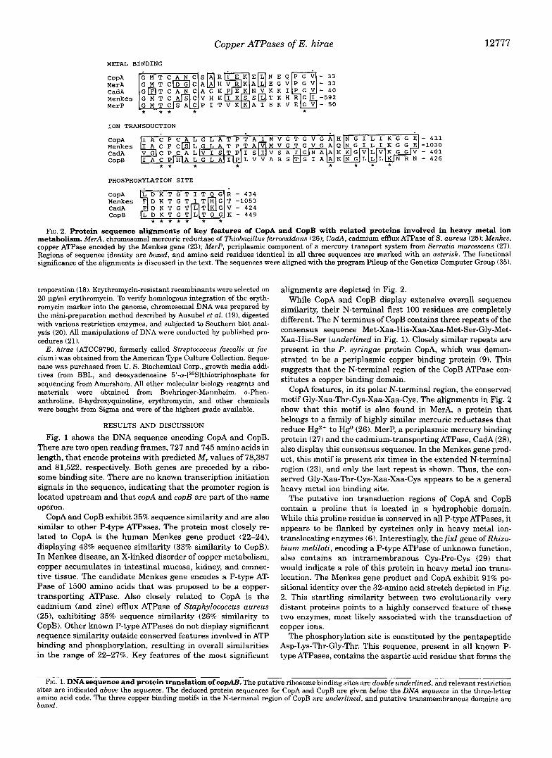
FIG. 2. Protein sequence alignments of key features of CopA and CopB with related proteins involved in heavy metal ion metabolism. MerA, chromosomal mercuric reductase of Thiobacillus ferrooxidans (26); CadA, cadmium efflux ATPase of S. aureus (25); Menkes, copper ATPase encoded by the Menkes gene (23); MerP, periplasmic component of a mercury transport system from Serratia marcescens (27). Regions of sequence identity are boxed, and amino acid residues identical in all three sequences are marked with an asterisk. The functional significance of the alignments is discussed in the text. The sequences were aligned with the program Pileup of the Genetics Computer Group (35).
troporation (18). Erythromycin-resistant recombinants were selected on 20 pg/ml erythromycin. To verify homologous integration of the eryth- romycin marker into the genome, chromosomal DNA was prepared by the mini-preparation method described by Ausubel et al. (19), digested with various restriction enzymes, and subjected to Southern blot anal- ysis (20). All manipulations of DNA were conducted by published pro- cedures (21).
E . hirae (ATCC9790, formerly called Streptococcus faecalis or fae- cium) was obtained from the American Type Culture Collection. Seque- nase was purchased from U. S. Biochemical Corp., growth media addi- tives from BBL, and deoxyadenosine 5’-a-[35Slthiotripho~phate for sequencing from Amersham. All other molecular biology reagents and materials were obtained from Boehringer-Mannheim. o-Phen- anthroline, 8-hydroxyquinoline, erythromycin, and other chemicals were bought from Sigma and were of the highest grade available.
RESULTS AND DISCUSSION
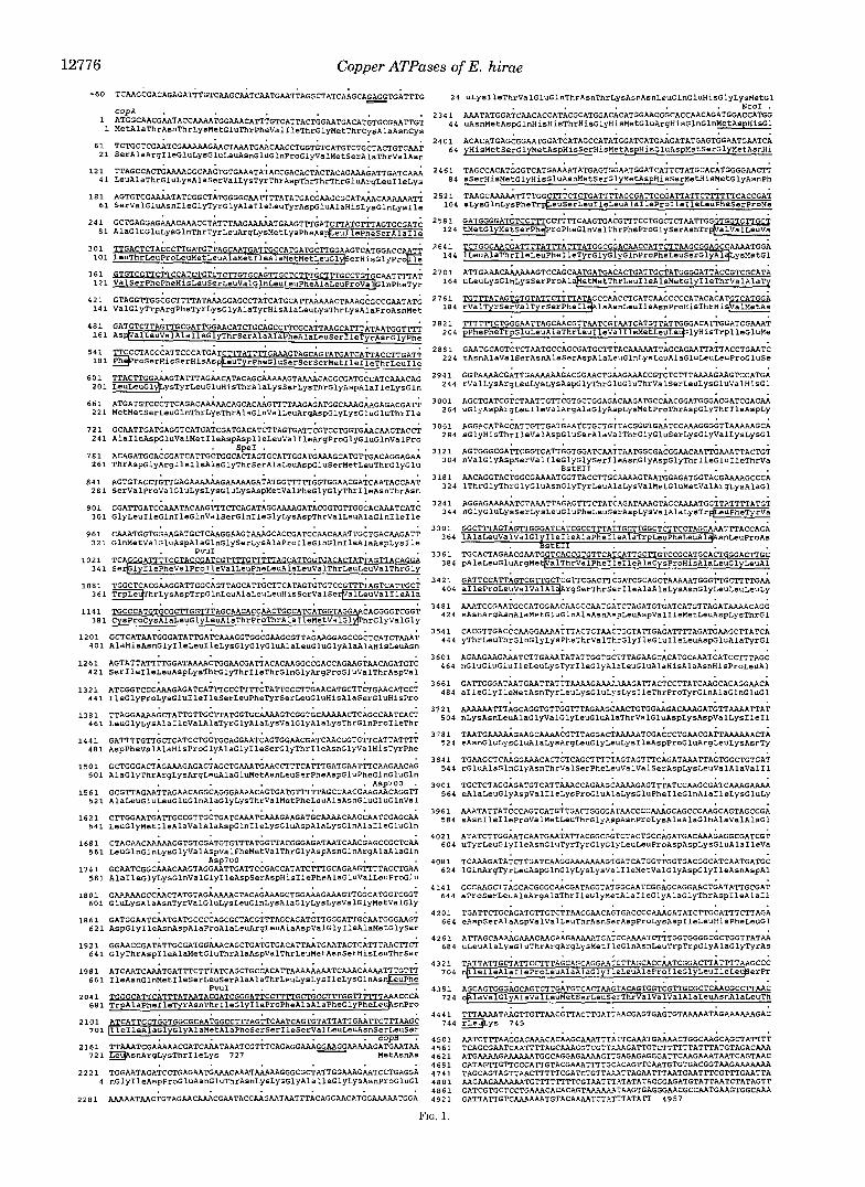
Fig. 1 shows the DNA sequence encoding CopA and CopB. There are two open reading frames, 727 and 745 amino acids in length, that encode proteins with predicted M , values of 78,387 and 81,522, respectively. Both genes are preceded by a ribo- some binding site. There are no known transcription initiation signals in the sequence, indicating that the promoter region is located upstream and that copA and copB are part of the same operon.
CopA and CopB exhibit 35% sequence similarity and are also similar to other P-type ATPases. The protein most closely re- lated to CopA is the human Menkes gene product (22-241, displaying 43% sequence similarity (33% similarity to CopB). In Menkes disease, an X-linked disorder of copper metabolism, copper accumulates in intestinal mucosa, kidney, and connec- tive tissue. The candidate Menkes gene encodes a P-type AT- Pase of 1500 amino acids that was proposed to be a copper- transporting ATPase. Also closely related to CopA is the cadmium (and zinc) efflux ATPase of Staphylococcus aureus (25), exhibiting 35% sequence similarity (26% similarity to CopB). Other known P-type ATPases do not display significant sequence similarity outside conserved features involved in ATP binding and phosphorylation, resulting in overall similarities in the range of 22-27%. Key features of the most significant
alignments are depicted in Fig. 2. While CopA and CopB display extensive overall sequence
similarity, their N-terminal first 100 residues are completely different. The N terminus of CopB contains three repeats of the consensus sequence Met-Xaa-His-Xaa-Xaa-Met-Ser-Gly-Met- Xaa-His-Ser (underlined in Fig. 1). Closely similar repeats are present in the P. syringae protein CopA, which was demon- strated to be a periplasmic copper binding protein (9). This suggests that the N-terminal region of the CopB ATPase con- stitutes a copper binding domain.
CopA features, in its polar N-terminal region, the conserved motif Gly-Xaa-Thr-Cys-Xaa-Xaa-Cys. The alignments in Fig. 2 show that this motif is also found in MerA, a protein that belongs to a family of highly similar mercuric reductases that reduce Hg2+ to HgO (26). MerP, a periplasmic mercury binding protein (27) and the cadmium-transporting ATPase, CadA (281, also display this consensus sequence. In the Menkes gene prod- uct, this motif is present six times in the extended N-terminal region (231, and only the last repeat is shown. Thus, the con- served Gly-Xaa-Thr-Cys-Xaa-Xaa-Cys appears to be a general heavy metal ion binding site.
The putative ion transduction regions of CopA and CopB contain a proline that is located in a hydrophobic domain. While this proline residue is conserved in all P-type ATPases, it appears to be flanked by cysteines only in heavy metal ion- translocating enzymes (6). Interestingly, the fixZ gene of Rhizo- bium meliloti, encoding a P-type ATPase of unknown function, also contains an intramembranous Cys-Pro-Cys (29) that would indicate a role of this protein in heavy metal ion trans- location. The Menkes gene product and CopA exhibit 91% po- sitional identity over the 32-amino acid stretch depicted in Fig. 2. This startling similarity between two evolutionarily very distant proteins points to a highly conserved feature of these two enzymes, most likely associated with the transduction of copper ions.
The phosphorylation site is constituted by the pentapeptide Asp-Lys-Thr-Gly-Thr. This sequence, present in all known P- type ATPases, contains the aspartic acid residue that forms the
FIG. 1. DNAsequence and protein translation of copAB. The putative ribosome binding sites are double underlined, and relevant restriction sites are indicated above the sequence. The deduced protein sequences for CopA and CopB are given below the DNA sequence in the three-letter amino acid code. The three copper binding motifs in the N-terminal region of CopB are underlined, and putative transmembranous domains are boxed.

12778 Copper ATPases of E. hirae
acyl phosphate intermediate characteristic of these enzymes (30, 31).
Both CopA and CopB have a remarkably low cysteine con- tent. CopA only contains four cysteines: two in the putative N-terminal metal binding domain and two flanking the con- served intramembranous proline in the ion transduction do- main; CopB features a single cysteine, located in the latter domain. The very small number of cysteine residues in CopA and CopB obviously disfavors nonspecific interaction (inactiva- tion) of these ATPases with heavy metal ions.
1 2 3 4 5 6 7 8 9 1011 12131415
~ " m - " "
Expression of the cop genes was investigated with an anti- body directed against CopB. Enhanced expression of CopB was observed with increasing copper concentrations in the media, reaching a maximum at 2 mM CuS04 (Fig. 3). Induction was also observed in response to 5 p~ Ag' or 5 p~ Cd2', concen- trations that significantly inhibited growth. No effect was seen with maximally tolerated concentrations of Ca2', CrR+, Mn2+, Co2 , Ni2+, Zn2" , Sr2', Ba2', La"', Au3 ', Hg2+, Pb2+, and Bi3'. Surprisingly, full induction of CopB was also apparent if 100 pnl of the heavy metal ion chelators o-phenanthroline or 8-hydroxyquinoline was added. The induction effect of the chelating agents was abolished if equimolar concentrations of Cu2' were added simultaneously. It thus appears that either low or high concentrations of copper ions lead to induction. The operon-like arrangement of the copA and copB genes implies that both genes underlay the same control.
To illuminate the roles of CopA and CopB in metal ion ho- meostasis, we constructed gene-disrupted strains. Cells dis- rupted in copB, or in copA and copB, lost their high level copper resistance, being fully inhibited in their growth by 6 mM CuS04 (Fig. 4A); in contrast, disruption of copA alone had no signifi- cant effect on the copper tolerance of E. hirae. However,
FIG. 3. Induction of CopB by heavy metal ions or chelators. Logarithmically growing E . hirae cells were induced with the respective agents for 1 h and cell lysates tested for the expression of CopB on Western blots with an antiserum against CopB. Details ofthe procedure are given under "Materials and Methods." The arrowhead indicates the band corresponding to CopB at the relative molecular mass of 80 kDa. Lanes 1 4 , wild type induced with 0, 0.2, 2, and 6 rnM CuSO,, respec- tively; lanes 5-7, copAB-disrupted, copB-disrupted, and copA-disrupted strains, respectively, in the presence of 2 rnM CuSO,; lanes 8-10, wild type in the presence of 100 pv o-phenanthroline plus 0.100, and 200 pv CuSO,,, respectively; lanes 11-15, wild type with the following addi- tions: lane 11, 100 phf 8-hydroxyquinoline; lane 12, 5 pv Cd", lane 13, 1 mlf Co2'; lane 14, 5 pv Ag-; lane 15.4 pu Hg'.
400 , , . .
600
( , , . . I " " I ' . ' ~ I , ' '
Residue number
A " B Copper binding Phosphatase Aspartyl kinase
Time (h)
- 1 5 W
LD e v
n 0 0.1
0.01 0 1 2 3 4 5 6 7
Time (h) FIG. 4. Growth of wild-type and mutant E. hirae in the pres-
ence of heavy metal ions. 6 mht CuSO, (A) or 5 pv AgNO, ( B ) was added to the cultures 1 h after inoculation. Growth was monitored by measuring the optical density a t 546 nm. Other details are given under "Materials and Methods." B, wild type; X, copA-disrupted; A , cop€?- disrupted; 0, copAB-disrupted.
out 1 2 3 4 5 6 7 8
FIG. 5. Model of the membrane topology of CopB. A, hydropathy profile calculated by the method of Kytc and Doolittle (33) using a span of 20 amino acids. Upward deflections indicate hydrophobic domains. B, folding model for the CopB ATPase. The transmembranous helices are labeled 1-8. The bulk of the protein protrudes on the cytoplasmic ( i n ) face of the membrane. H..MGM indicates the three putative canonical copper binding sites in the Copper binding domain. The following se- quence features common to all P-type ATPases are also indicated: TGES is part of the Phosphatase domain, DKTGT is the site of aspartyl phos- phate formation, and VGDGINDAP is predicted to form a Mg"-medi- ated salt bridge to y-phosphate of ATP in the Aspartyl kinase domain. The conserved proline in helix 6 is surrounded by two cysteines in CopA and the Cd"-ATPase, and by cysteine and histidine in CopB and is believed to be the ion transduction site. All these assignments are based on site-directed mutagenesis and the analysis of several other P-type ATPases (36-38).

Copper ATPases of E. hirae 12779
copA-disrupted cells ceased to grow after two to three genera- tions when heavy metal ions in the media were complexed with 8-hydroxyquinoline, conditions that do not affect the growth of wild-type cells (not shown). This suggests that CopAis required for copper import under limiting conditions. Silver is known to replace copper in some processes (321, and we tested its effect on gene-disrupted strains (Fig. 4B). Disruption of copA ren- dered the cells considerably more tolerant to Ag+ than strains possessing a functional CopAATPase, supporting a role of CopA in heavy metal ion import.
The hydropathy profiles calculated by the method of Kyte and Doolittle (33) are very similar for CopA and CopB (only shown for CopB, Fig. 5A). Based on these hydropathy profiles and on the most favorable distribution of charged residues be- tween the two faces of the membrane (341, we propose a mem- brane topology for CopA and CopB with eight transmembra- nous helices and the N and C termini and the bulk of the protein exposed to the cytoplasm (Fig. 5B 1. This model deviates from those put forth for related ATPases, such as the CadA cadmium ATPase of S. aureus, in which two rather than four transmembranous helices are predicted to lie between the N terminus and the first cytoplasmic loop (6). Other features of our model are described in the legend to Fig. 5.
Taken together, our results propound that copA and copB encode ion-motive ATPases that effect translocation of copper and other metal ions across the cell membrane. This proposal rests on the following evidence: (i) CopA and CopB are P-type transport ATPases based on sequence similarity, (ii) these ATP- ases are inducible by either high or low ambient copper con- centrations, (iii) CopA and CopB show N-terminal and in- tramembranous features typical of heavy metal ion binding proteins, (iv) disruption of copB leads to copper-sensitive cells, (v) disruption of copA renders the cells copper-dependent and silver-resistant, and (vi) CopA shows extensive sequence simi- larity to the putative copper-transporting ATPase encoded by the Menkes gene. Thus, CopB most likely serves in the extru- sion of copper, while CopA appears to be responsible for its uptake.
Copper-transporting ATPases represent a novel mechanism for the control of cytoplasmic copper. The occurrence of similar enzymes in such diverse species as man and E. hirae suggests that ATP-driven copper transport is a widely used mechanism of copper homeostasis.
Acknowledgments-We thank Denise Hess-Bienz for preparing the antibody against CopB and Thomas Seebeck for crit ical discussion.
1. 2.
3.
4.
6. 5.
7 .
8. 9.
10. 11. 12.
13. 14.
15.
16.
17.
19. 18.
21. 20.
22.
23.
24.
25.
26.
27.
28.
29.
30. 31. 32.
33. 34. 35.
REFERENCES Tetaz, T. J., and Luke, R. K. J. J. (1983) J. Bacteriol. 154, 1263-1268 Rogers, S. D., Bhave, M. R., Mercer, J . F. B., Camakaris, J., and Lee, B. T. 0.
Erardi, F. X., Failla, M. L., and Falkinham, J. 0. (1987) Appl. Enuiron. Mi-
Garde, S., and Bender, C. L. (1991) Appl. Enuiron. Microhiol. 57, 2435-2439
Silver, S., and Walderhaug, M. (1992) Microhiol. Reu. 66, 195-228 Bender, C. L., and Cooksey, D. A. J . (1987) J. Bacteriol. 1 6 9 , 4 7 0 4 7 4
Brown, N. L., Camakaris, J., Lee, B. T. O., Williams, T., Morby, A. P., Parkhill,
Mellano, M. A,, and Cooksey, D. A. J. (1988) J. Bacteriol. 170, 2879-2883 Cha, J.-S., and Cooksey, D. A. (1991) Proc. Natl. Acad. Sci. U. S. A. 88, 8915-
Kagi, J. H. R., and Schaffer, A. (1988) Biochemistry 27,8509-8515 Solioz, M., Mathews, S., and Furst, P. (1987) J. Biol. Chem. 262, 735G7362 Sanger, F., Bicklen, S., and Coulson, A. R. (1977) Proc. Natl. Acad. Sci. U. S. A.
Laemmli, U. K., and Favre, M. (1973) J. Mol. Biol. So, 575-599 Towbin, H., Staehelin, T., and Gordon, J . (1979) Proc. Natl. Acad. Sei. U. S. A.
Gray, M. R., Mazzara, G. P., Reddy, P., and Rosbash, M. (1987) Methods En-
Macrina, F. L., Tobian, J . A., Jones, K. R., Evans, R. P., and Clewell, D. B.
Humphreys, G. O., Willshaw, G. A,, and Anderson, E. S. (1975) Biochim.
Waser, M., and Solioz, M. (1990) Biachimie 72, 279-283 Ausubel, F. M., Brent, R., Kingson, R. E., Moore, D. D., Seidman, F, G., Smith,
A,, and Kevin, S. (1988) in Current Protocols in Molecular Biology, Wiley- Interscience, New York
J. (1991) J. Bacteriol. 173,6742-6748
crobiol. 53, 1951-1954
J., and Rouch, D. A. J. (1991) Cell. Biochem. Funct. 46, 106-114
8919
74, 5463-5467
76,4350-4354
zymol. 154,129-135
(1982) Gene (Amst . ) 19,345-353
Biophys. Acta 3 8 3 , 4 5 7 4 6 3
Southern, E. (1975) J. Mol. Biol. 98, 503-517 Maniatis, T., Fritsch, E. F., and Sambrook, J . (1982) Molecular Cloning: A
Laboratory Manual, Cold Spring Harbor Laboratory, Cold Spring Harbor, NY
Mercer, J. F. B., Livingston, J., Hall, B., Paynter, J. A,, Begy, C., Chan- drasekharappa, S., Lockhart, P., Grines, A,, Bhave, M., Siemieniak, D., and Glover, T. W. (1993) Nature Genetics 3, 20-25
Vulpe, C., Levinson, B., Whitney, S., Packman, S., and Gitschier, J . (1993) Nature Genetics 3, 7-13
Chelly, J., Tiimer, Z., Tonnesen, T., Petterson, A,, Ishikawa-Brush, Y., Tom- merup, N., Horn, N., and Monaco, A. P. (1993) Nature Genetics 3, 14-19
Nucifora, G., Chu, L., Misra, T. K., and Silver, S. (1989) Proc. Natl. Acad. Sci. U. S. A. 86,3544-3548
h u e , C., Sugawara, K., Shiratori, T., Kusano, T., and Kitagawa, Y. (1989) Gene (Amst. 84.47-54
Nucifora, G., Chu, L., Silver, S., and Misra, T. K. (1989) J. Bacteriol. 171, 42414247
Silver, S., Nucifora, G., Chu, L., and Misra, T. K. (1989) Dends Biochem. Sci. 14, 76-80
Kahn, D., David, M., Domergue, O., Daveran, M.-L., Ghai, J., Hirsch, P. R., and Batut, J. (1989) J . Bacteriol. 171, 929-939
Pederson, P. L., and Carafoli, E. (1987) Dends Biochem. Sci. 12, 146-150 Pederson, P. L., and Carafoli, E. (1987) Dends Biochem. Sci. 12, 186-189 Winge, D. R., Nielson, K. B., Gray, W. R., and Hamer, D. H. (1985) J. Biol.
Kyte, J., and Doolittle, R. F. J. (1982) J. Mol. Biol. 157, 105-132 Von Heijne, G. (1992) J . Mol. Biol. 225, 4 8 7 4 9 4 Devereux, J., Haeberly, P., and Smithies, 0. (1984) Nucleic Acids Res. 12,
Chem. 260, 14464-14470
387-395 36. Serrano, R. (1988) Biochim. Biophys. Acta 947, 1-28 37. Inesi, G., and Kirtley, M. R. (1992) J. Bioenerg. Biomemhr. 24, 271-283 38. Horisberger, J.-D., Lemas, V., Kraehenbuhl, J.-P., and Rossier, B. C. (1991)
Annu. Reo. Physiol. 53, 565-584