priming of salmonella entericaserovar typhi-specific t ... · pdf filepriming of salmonella...
TRANSCRIPT
Priming of Salmonella enterica Serovar Typhi-SpecificCD8+ T Cells by Suicide Dendritic Cell Cross-Presentationin HumansRosangela Salerno-Goncalves*, Marcelo B. Sztein
Center for Vaccine Development, University of Maryland School of Medicine, Baltimore, Maryland, United States of America
Abstract
Background: The emergence of antibiotic-resistant strains of Salmonella enterica serovar Typhi (S. Typhi), the etiologic agentof typhoid fever, has aggravated an already important public health problem and added new urgency to the developmentof more effective typhoid vaccines. To this end it is critical to better understand the induction of immunity to S. Typhi. CD8+
T cells are likely to play an important role in host defense against S. Typhi by several effector mechanisms, including killingof infected cells and IFN-c secretion. However, how S. Typhi regulates the development of specific CD8+ responses inhumans remains unclear. Recent studies in mice have shown that dendritic cells (DC) can either directly (upon uptake andprocessing of Salmonella) or indirectly (by bystander mechanisms) elicit Salmonella-specific CD8+ T cells.
Methodology/Principal Findings: We report here that upon infection with live S. Typhi, human DC produced high levels ofpro-inflammatory cytokines IL-6, IL-8 and TNF-a, but low levels of IL-12 p70 and IFN-c. In contrast, DC co-cultured with S.Typhi-infected cells, through suicide cross-presentation, uptake S. Typhi-infected human cells and release high levels of IFN-c and IL-12p70, leading to the subsequent presentation of bacterial antigens and triggering the induction of memory T cells,mostly CD3+CD8+CD45RA2CD62L2 effector/memory T cells.
Conclusions/Significance: This study is the first to demonstrate the effect of S. Typhi on human DC maturation and on theirability to prime CD8+ cells and highlights the significance of these phenomena in eliciting adaptive immunity to S. Typhi.
Citation: Salerno-Goncalves R, Sztein MB (2009) Priming of Salmonella enterica Serovar Typhi-Specific CD8+ T Cells by Suicide Dendritic Cell Cross-Presentation inHumans. PLoS ONE 4(6): e5879. doi:10.1371/journal.pone.0005879
Editor: Derya Unutmaz, New York University School of Medicine, United States of America
Received February 24, 2009; Accepted May 13, 2009; Published June 11, 2009
Copyright: � 2009 Salerno-Goncalves, Sztein. This is an open-access article distributed under the terms of the Creative Commons Attribution License, whichpermits unrestricted use, distribution, and reproduction in any medium, provided the original author and source are credited.
Funding: This work was supported, in part, by NIAID, NIH, DHHS federal research grant R01 AI36525 and contract NO1 AI30028 (both to M.B.S.). The funders hadno role in study design, data collection and analysis, decision to publish, or preparation of the manuscript.
Competing Interests: The authors have declared that no competing interests exist.
* E-mail: [email protected]
Introduction
Typhoid fever remains an important public health priority,
particularly in developing countries, with an estimated 16 million
new cases annually and 600,000 deaths [1]. The emergence of
antibiotic-resistant Salmonella enterica serovar Typhi (S. Typhi), the
etiologic agent of typhoid fever, has aggravated an already
important public-health problem [2,3]. S. Typhi, is a facultative
intracellular bacterial pathogen with the capacity to survive and
replicate in phagocitic and non-phagocytic cells [4,5,6,7]. T cells
might play an important role in immunity to S. Typhi. We have
shown in volunteers immunized orally with attenuated strains of S.
Typhi, including Ty21a, as well as with the novel attenuated
typhoid vaccine candidate strains CVD 908, CVD 908-htrA and
CVD 909, the induction of CD8+ cell-mediated immunity (CMI)
mechanisms that involve the secretion of IFN-c and the killing of
S. Typhi-infected cells by cytotoxic T lymphocytes
[5,6,7,8,9,10,11,12,13,14]. Our group also demonstrated the
ability of classical HLA class Ia, as well as the non-classical HLA
class Ib molecule HLA-E, to function as a restriction element for
CD8 T cells [5,6,7,8,9]. However, the mechanisms underlying the
development of CD8-mediated immunity remain uncertain. The
induction of CMI mediated by CD8+ cells requires the
presentation of antigens by specialized cells of the immune system
named dendritic cells (DC) [15]. Studies using the S. Typhimur-
ium mouse model have showed that DC can either directly (upon
uptake and processing of Salmonella) or indirectly (by bystander
mechanisms, including cross-presentation) present Salmonella
antigens [16]. Cross-presentation denotes the ability of certain
antigen-presenting cells, including DC, to acquire proteins from
other tissue cells through endocytic mechanisms, and direct them
into their own MHC I pathway to be subsequently presented to
naıve T-cells [17,18,19,20]. These events will result in prolifera-
tion and differentiation of naıve T-cells into memory cells, a
process that is accompanied by changes in the expression of
surface molecules [21].
Infection of susceptible mice with S. Typhimurium is considered
a model for the pathogenesis of human typhoid fever [22].
However since S. Typhi infection is restricted to humans [22], it is
not clear whether the conditions for DC maturation and/or
patterns of antigen presentation induced in response to S.
Typhimurium and S. Typhi infection are identical [22,23]. In
fact, previous studies have shown that antigen presenting cells
from different animal species might present different sets of
PLoS ONE | www.plosone.org 1 June 2009 | Volume 4 | Issue 6 | e5879

peptides [8]. To investigate how S. Typhi regulates the
development of CD8+ CMI in humans, we examined the effects
of S. Typhi on the maturation of DC and on their ability to prime
naive CD8+ T cell responses. We report here that DC, through
suicide cross-presentation, uptake apoptotic S. Typhi-infected
human cells and release IFN-c and IL-12p70, leading to the
subsequent presentation of bacterial antigens and triggering the
induction of mostly CD3+CD8+CD45RA2CD62L2 memory T
cells. This study is, to our knowledge, the first demonstration of the
effects of S. Typhi on human DC maturation and on their ability
to prime CD8+ cells and highlights its significance in eliciting
adaptive immunity to S. Typhi.
Results
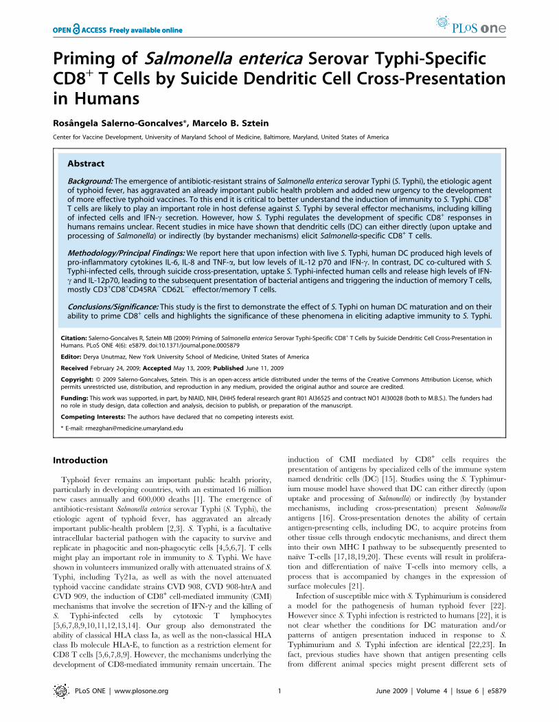
S. Typhi drives DC maturationTo efficiently present antigens to naıve T cells, immature DC
must be activated to mature, a process accompanied by up-
regulation of MHC and costimulatory molecules [24]. To
investigate whether S. Typhi drives DC maturation, we evaluated
the DC surface expression of CD80 and CD83 costimulatory
molecules after different stimulatory conditions. Unstimulated DC
(media) were used as negative controls. We observed that DC
pulsed with live S. Typhi increased their surface expression of
CD80 and CD83 in a dose-dependent manner (Fig. 1A) with levels
comparable to those induced following exposure to LPS, a potent
DC maturation factor [25] (Fig. 1B). Increases, albeit at lower
levels, were also apparent when DC were pulsed with heat-
inactivated S. Typhi demonstrating that neither bacterial infection
nor viability was required for the observed effects. These results
are in agreement with others using S. Typhimurium in a mouse
model [26]. However, the exposure of DC to S. Typhi-infected
blasts induced comparable or higher levels of CD80 and CD83
expression on DC than those induced by exposure of DC to live S.
Typhi or LPS (Fig. 1B). Of interest, the expression level of CD80
and CD83 molecules on DC pulsed with not-infected blasts were
similar to those observed in DC cultured in media alone (Fig. 1B).
In conclusion, the appearance of DC exhibiting higher levels of
CD83 and CD80 molecules after incubation with S. Typhi
antigens demonstrated that S. Typhi drives DC maturation in
humans.
S. Typhi stimulation augments the ability of DC togenerate pro-inflammatory cytokines in vitro
Another hallmark of DC maturation is the production of
cytokines, such as IL-12 and IFN-c, which are critical for the
induction of CMI [27]. Because IL-12 and IFN-c are essential for
resistance to Salmonella infection in mice [28,29], as they are likely
to be in humans [5,6,9,30,31], it was of importance to evaluate the
effect of S. Typhi on cytokine production by DC. To this end, DC
were pulsed with heat-killed S. Typhi or wild-type S. Typhi at a
10:1 MOI, uninfected or S. Typhi-infected blasts at a 1:4 blast to
DC ratio during 48 hours. DC pulsed with heat-killed or live S.
Typhi produced high levels of pro-inflammatory cytokines such as
IL-6, IL-8 and TNF-a but only low levels or no IL-12 p70 (Fig. 2).
This low or lack of IL-12 secretion does not seems to be the result
of IL-10 production by these cells. DC pulsed with heat-killed or
live S. Typhi elicited levels of IL-10 only slightly different than
those detected in culture supernatants of DC incubated in medium
alone (Fig. 2).
Although it has been shown that DC can produce concomi-
tantly IFN-c and IL-12 [32], in our culture conditions, i.e., when
DC were pulsed either with heat-killed S. Typhi or with live S.
Typhi, IFN-c was below the level of sensitivity of the CBA assay
(Fig. 2). Surprisingly, a cytokine pattern for efficient T cell priming
(i.e., high levels of IL-12 p70 and IFN-c) was only observed after
co-culture of DC with S. Typhi-infected blasts (Fig. 2). A low ratio
of S. Typhi-infected cells per DC (1:4) was sufficient to
concurrently stimulate high levels of IL-12 p70 and IFN-c. This
phenomenon was found to be dose-dependent (data not shown)
and observed in all 5 subjects studied. Minimal or no cytokine
production was detected in control cultures with S. Typhi-infected
blasts alone, i.e., in the absence of DC (Fig. 2). These results
suggested that IL-12 p70 and IFN-c production is a function of
uninfected DC being stimulated by S. Typhi antigens from
infected blasts. Moreover, this is the first demonstration of IL-12
p70 production by DC upon stimulation with Salmonella antigens.
S. Typhi induces apoptosis in human blasts and DCBased on the above observations, we next investigated the role
of apoptosis in the generation of antigens that could then be
acquired by uninfected DC. It is known that bacteria-induced
apoptotic cells can serve as a reservoir of antigens for cross-
presentation by DC [16]. Previous studies have shown that some
Salmonella serovars, including S. Typhimurium and S. Typhi, can
induce death in infected macrophages by apoptotic mechanisms
[33,34]. Here, cell apoptosis was monitored by Annexin V and PI
fluorescent dyes and by staining cells for cleaved caspase-3. Blasts
were infected with S. Typhi at MOI of 10:1 and analyzed for
apoptosis after 3 hours of incubation. Uninfected blasts were used
as negative controls. Cells treated with staurosporine or formal-
dehyde were used as positive controls for apoptosis and necrosis,
respectively. Cells were stained with Annexin V and PI dyes or
anti-caspase-3 antibody as well as isotype-matched control mAbs
and analyzed by flow cytometry. We observed that S. Typhi-
infected blasts undergo increased apoptosis, as detected by
annexin V binding in the absence of PI staining, as compared to
uninfected blasts (media) (Fig. 3A). Caspase-3-positive cells were
also detected in S. Typhi-infected blasts (Fig. 3B). Both methods of
detection identified similar percentages of apoptotic cells in S.
Typhi-infected blasts. Similarly, we observed that DC pulsed with
live S. Typhi increased apoptosis, as detected by caspase-3 staining
(Fig. 3C). Increases, albeit at lower levels, were also apparent when
DC were pulsed with heat-inactivated S. Typhi (Fig. 3C).
Apoptosis was inhibited ,30% when the incubation was carried
out in the presence of ZVA-D, a powerful, irreversible and cell
permeable inhibitor for caspases (Fig. 3C). Together these results
further support the possibility that Salmonella-induced apoptosis is
responsible for loading uninfected DC with S. Typhi antigen.
DC efficiently uptake S. Typhi-antigens triggering asuicide mechanism
To directly demonstrate the ability of DC to uptake S. Typhi-
infected blasts, we incubated them with infected blasts labeled with
a mAb to CD45, a surface marker present in all human leukocytes.
After a 2 hour incubation, an average of 20% of the DC that were
incubated with S. Typhi-infected blasts were positive for CD45,
indicating capture of blast-derived material (Figs. 4A and 5A
represent 2 out of 3 experiments using 3 different subjects).
Capture was inhibited by 25–57% in the various repeat
experiments when the incubation was carried out in the presence
of CCD, an inhibitor of phagocytosis (Figs. 4A and 5A). To
exclude the possibility that the decrease in the capture of S. Typhi-
infected blasts was due to the direct toxic effect of CCD on DC, we
measured the viability of the DC in presence and absence of CCD.
To this end, we stained DC with ViViD, an amine-reactive
fluorescent dye used to evaluate mammalian cell viability by flow
cytometry. As shown in Figures 4C and 5B, similar DC viabilities
S. Typhi T Cell Cross-Priming
PLoS ONE | www.plosone.org 2 June 2009 | Volume 4 | Issue 6 | e5879
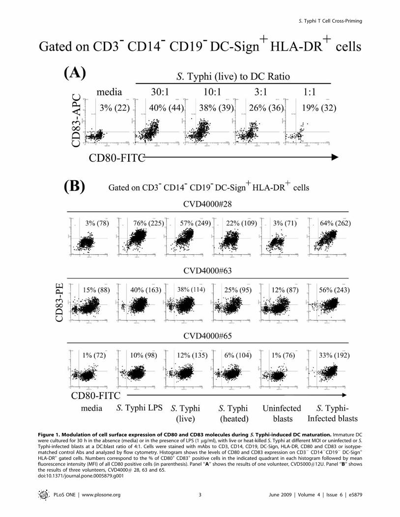
Figure 1. Modulation of cell surface expression of CD80 and CD83 molecules during S. Typhi-induced DC maturation. Immature DCwere cultured for 30 h in the absence (media) or in the presence of LPS (1 mg/ml), with live or heat-killed S. Typhi at different MOI or uninfected or S.Typhi-infected blasts at a DC:blast ratio of 4:1. Cells were stained with mAbs to CD3, CD14, CD19, DC-Sign, HLA-DR, CD80 and CD83 or isotype-matched control Abs and analyzed by flow cytometry. Histogram shows the levels of CD80 and CD83 expression on CD32 CD142CD192 DC-Sign+
HLA-DR+ gated cells. Numbers correspond to the % of CD80+ CD83+ positive cells in the indicated quadrant in each histogram followed by meanfluorescence intensity (MFI) of all CD80 positive cells (in parenthesis). Panel ‘‘A’’ shows the results of one volunteer, CVD5000#12U. Panel ‘‘B’’ showsthe results of three volunteers, CVD4000# 28, 63 and 65.doi:10.1371/journal.pone.0005879.g001
S. Typhi T Cell Cross-Priming
PLoS ONE | www.plosone.org 3 June 2009 | Volume 4 | Issue 6 | e5879

Figure 2. S. Typhi-infected blasts are the most effective stimulus in inducing DC to secrete pro-inflammatory cytokines in vitro.Immature DC were cultured in the absence (media) or in presence of live or heat-killed S. Typhi at a MOI of 10:1, uninfected or S. Typhi-infectedautologous blasts at DC:blast ratio of 4:1. S. Typhi-infected blasts only (without adding DC) were included as controls. After 48 h the supernants wereharvested and cytokine production was measured using the flow cytometry-based BD cytometric bead array (CBA) assay. Bar graphs show mean + SEof 6 experiments using 5 different donors (*, p,0.05 compared with DC pulsed with S. Typhi-infected cells).doi:10.1371/journal.pone.0005879.g002
S. Typhi T Cell Cross-Priming
PLoS ONE | www.plosone.org 4 June 2009 | Volume 4 | Issue 6 | e5879

S. Typhi T Cell Cross-Priming
PLoS ONE | www.plosone.org 5 June 2009 | Volume 4 | Issue 6 | e5879

were observed when DC were cultured with S. Typhi-infected
blasts in the presence or absence of CCD.
Despite the fact that at the beginning of the cultures the
numbers of the DC loaded with either S. Typhi or infected-blasts
were the same, we can not rule-out the possibility that as the
cultures progress, there is an increase in DC death after S. Typhi
infection when compared to those cultured with infected-blasts. In
this case, having fewer DC might adversely impact the cytokines
production by S. Typhi-infected DC. To address this issue, we
measured the apoptosis and viability of DC cultured with either S.
Typhi or S. Typhi-infected blasts. After 2 hours of incubation, we
compared the percentage of cells stained with anti-caspase-3 and
ViViD by flow cytometry. As demonstrated by caspase-3 staining,
DC cells exposed to S. Typhi had equal or less apoptosis than DC
exposed to S. Typhi-infected blasts (Fig. 3C and Figs. 4A and 5A).
Of note, the presence of S. Typhi antigens was detected on
virtually all caspase-3+ cells by Salmonella common structural
antigens (CSA) staining (Fig. 4B). DC cells exposed to S. Typhi
(data not shown) had equal or higher viability than DC exposed to
S. Typhi-infected blasts (Figs. 4C and 5B). Interestingly, not all
caspase-3+ DC were double positive for CD45, showing that the
caspase-3 mediated apoptosis was not due to preferential uptake of
infected-blast cells. This might occur as the result of a bystander
effect in which DC undergoing apoptosis following phagocytosis of
S. Typhi-infected blasts might signal some of the neighboring DC
to rapidly engulf them leading these DC to also undergo apoptosis.
This would offer a mechanism for efficiently making additional
Salmonella antigens available to new DC. This hypothesis is
consistent with previous work showing that in mice Salmonella can
be cytotoxic for liver phagocytes, whether these phagocytes
harbored intracellular bacteria or not [35,36]. Moreover, while
the addition of CCD decreased the amount of DC loaded with
CD45-infected blasts, no changes were observed in the caspase-3+
levels of the CD45-negative DC population. Interestingly, when
DC were cultured with S. Typhi-infected blasts pre-treated with
the apoptosis inhibitor ZVA-D, the percentage of caspase-3+ DC
double positive for CD45 increased from 34–52% while the
general apoptosis level, as well as their viability remained
unchanged (Figs. 4 and 5). These results suggest: (a) an irreversible
induction of apoptosis after ingesting S. Typhi–infected cells and
(b) that the cross-presentation by DC might be related to S. Typhi-
antigen itself rather than by the apoptotic nature of the S. Typhi-
infected blasts. Thus, the apoptosis elicited by S. Typhi antigens
can be the result of at least two pathways leading to cross-
presentation: (a) through a bystander effect on neighboring DC
and (b) through phagocytosis of S. Typhi–infected blasts which
leads to the dead of these DC via irreversible apoptosis (i.e., a
‘‘suicide mechanism’’). In this way, having more S. Typhi-infected
blasts engulfed by DC might result in more Salmonella antigens
available for cross-presentation resulting from more DC undergo-
ing apoptosis.
Generation of cytokine-producing effector cellsThe effect of Salmonella on DC maturation and cytokine
production described above, as well as their capacity to induce
apoptosis in the infected cells, prompted us to investigate the
ability of Salmonella antigen-loaded DC to prime naıve human T
cells. Previous studies have shown that by exposing DC to antigen
and then to naıve T cells in vitro, it is possible to mimic the process
of antigen presentation in vivo [37,38]. We used this approach to
evaluate the ability of DC to either directly (e.g., priming upon
uptake and processing of Salmonella) or indirectly (e.g., by cross-
priming through a bystander mechanism) present Salmonella
antigens to naıve T cells. This was studied by co-culturing ex vivo
PBMC from individuals non-exposed to S. Typhi (naıve subjects)
with DC that had been pulsed with live or heat-killed S. Typhi at
10:1 MOI (priming), or pulsed with uninfected or S. Typhi-
infected blasts at a 1:1 blast to DC ratio (cross-priming). After
30 hours of incubation, the S. Typhi specificity of primed PBMC
was evaluated by their ability to secrete IL-2, IFN-c and TNF-adetected as measured by intracellular staining. To avoid infection
of DC by live bacteria from dying infected-blasts, blasts were
harvested, washed 3 times with RMPI containing gentamicin
(100 mg/ml) and incubated for 1 hour at 37uC to kill and remove
extracellular bacteria before being added to PBMC. Moreover, to
assess the pre-existing background from cross-reactive effector T
cells, a control consisting of PBMC co-cultured with the S. Typhi-
infected blasts in the absence of DC was also included. Although
PBMC in the presence of S. Typhi-infected blasts alone (in the
absence of DC) produced moderate levels of cytokines, these levels
were considerably lower than those observed in PBMC co-
cultured with DC pre-mixed with S. Typhi-infected blasts (Figs. 6
and 7). Thus, these results clearly indicate that DC function as
antigen-presenting cells. Four volunteers were studied to assess the
relative importance of priming and cross-presentation in the early
stages of S. Typhi infection. Results indicate different levels of
cytokine production in the various volunteers. Whilst in 2 of 4
volunteers higher induction of cytokine production was observed
following DC co-cultured with S. Typhi-infected blasts (Figs. 6 and
7), a third volunteer showed similar levels when PBMC were co-
cultured DC exposed to live S. Typhi or with S. Typhi-infected
blasts (data not shown), and in the 4th volunteer the cytokine
production was higher in cultures of PBMC with DC infected with
live S. Typhi (data not shown). Therefore it is likely that both
mechanisms, cross- and direct-priming by DC, might be involved
in stimulating cytokine production from PBMC.
To confirm that DC are engulfing infected-blasts and
subsequently processing them intracellularly, uninfected DC pre-
treated with CCD and co-cultured with infected blasts were used
as antigen-presenting cells in ex vivo PBMC cultures. As expected,
CCD markedly inhibited the DC’s ability to stimulate cytokine
production by naıve PBMC in all 4 volunteers evaluated (Figs. 6
and 7).
To assess the role of apoptosis in antigen presentation we
compared whether blocking apoptosis affected the ability of DC to
stimulate T cells. To this end, uninfected DC pre-treated or not
with ZVA-D and co-cultured with live S. Typhi or infected blasts
pre-exposed or not to ZVA-D were used as antigen-presenting
cells in ex vivo PBMC from individuals non-exposed to S. Typhi
(naıve subjects). A substantial decrease in cytokine production by
Figure 3. S. Typhi induces apoptosis in human cells. Blasts or DC were infected with S. Typhi at a MOI of 10:1 and analyzed for apoptosis3 hours after infection. DC were also exposed to heated-killed S. Typhi or treated with ZVA-D. Uninfected cells (media) were used as negativecontrols. Cells treated with staurosporine (8 mg/ml) or 1% formaldehyde were used as positive controls for apoptosis and necrosis respectively. Cellapoptosis was monitored by Annexin V and PI fluorescent dyes and by staining cells for cleaved caspase-3. (A) Blasts from volunteer A00302-5008were stained with FITC-annexin V and PI or isotype-matched control antibodies and analyzed by flow cytometry. (B) Blasts from volunteersCVD4000#119 and CVD4000#64 and (C) DC from volunteer CVD4000#64 were surface stained for caspase-3 and Salmonella common structuralantigens (CSA). Numbers correspond to the percentage of positive cells in the indicated quadrants or regions in each histogram. Arrows in panels Band C depict the expression of caspase 3 on cells expressing, or not, CSA.doi:10.1371/journal.pone.0005879.g003
S. Typhi T Cell Cross-Priming
PLoS ONE | www.plosone.org 6 June 2009 | Volume 4 | Issue 6 | e5879

PBMC was observed when DC were pre-treated with ZVA-D
before exposure to live S. Typhi (4 of 4 volunteers) (Figs. 6 and 7,
and data not shown). In contrast, when DC from the same
volunteers were exposed to ZVA-D-pre-treated S. Typhi-infected
blasts failed to affect cytokine production by PBMC (Figs. 6 and 7,
and data not shown). While these observations support an
important role for the induction of apoptosis in antigen
presentation, they also suggest that apoptotic material from
different origins might also contribute to this process to varying
degrees. These results reinforce the idea that both mechanisms,
Figure 4. DC efficiently uptake S. Typhi-infected blasts. DC from volunteer CVD4000#64 treated or not with ZVA-D or CCD agents wereincubated with uninfected or S. Typhi-infected CD45-labeled blasts at a DC:blast ratio of 1:5 at 37uC. After 2 hours of incubation, DC were stainedwith ViViD, followed by surface staining with mAbs to HLA-DR, DC-Sign, Salmonella common structural antigens (CSA) and caspase-3 and analysed byflow cytometry. DC were gated based on their scatter characteristics. Single DC were selected by gating on forward scatter height vs. forward scatterarea and then on HLA-DR and DC-Sign. (A) endocytosis and apoptosis on DC was analyzed by studying expression of CD45 and caspase-3,respectively. (B) The presence of S. Typhi antigens on caspase-3+ cells were analyzed by determining their expression of CSA. (C) Viability of DCunder different culture conditions was evaluated by ViViD as a dead cell exclusion marker. Dotted lines represent the cut-offs between positive andnegative cells. Numbers correspond to the percentage of positive cells in the indicated quadrants or regions in each histogram. These results arerepresentative of 1 of 3 volunteers with similar results. Additional data is provided in Fig. 5.doi:10.1371/journal.pone.0005879.g004
S. Typhi T Cell Cross-Priming
PLoS ONE | www.plosone.org 7 June 2009 | Volume 4 | Issue 6 | e5879

Figure 5. DC efficiently uptake S. Typhi-infected blasts. DC from volunteer CVD4000#119 treated or not with ZVA-D or CCD agents wereincubated with uninfected or S. Typhi-infected CD45-labeled blasts at a DC:blast ratio of 1:5 at 37uC. After 2 hours of incubation, DC were stainedwith ViViD, followed by surface staining with mAbs to HLA-DR, DC-Sign, Salmonella common structural antigens (CSA) and caspase-3 and analysed byflow cytometry. DC were gated based on their scatter characteristics. Single DC were selected by gating on forward scatter height vs. forward scatterarea and then on HLA-DR and DC-Sign. (A) endocytosis and apoptosis on DC was analyzed by studying expression of CD45 and caspase-3,respectively. (B) Viability of DC under different culture conditions was evaluated by ViViD as a dead cell exclusion marker. Dotted lines represent thecut-offs between positive and negative cells. Numbers correspond to the percentage of positive cells in the indicated quadrants or regions in eachhistogram. These results are representative of 1 of 3 volunteers with similar results.doi:10.1371/journal.pone.0005879.g005
S. Typhi T Cell Cross-Priming
PLoS ONE | www.plosone.org 8 June 2009 | Volume 4 | Issue 6 | e5879

S. Typhi T Cell Cross-Priming
PLoS ONE | www.plosone.org 9 June 2009 | Volume 4 | Issue 6 | e5879

cross- and direct-priming by DC are important in the development
of S. Typhi-immunity.
S. Typhi-specific memory CD8+ T cell subsets areexpanded following priming by DC
We next investigated which CD8 cell populations within PBMC
preferentially produced IFN-c and TNF-a cytokines under the
different stimulatory conditions described above. We evaluated the
induction of cytokine production in the various memory CD8+ T
cell subsets, defined as follows: central memory T cells (TCM,
CD45RA-CD62L+), effector memory T cells (TEM,
CD45RA2CD62L2) and naıve T cells (Tn, CD45RA+CD62L+)
[14]. We observed that the majority of CD8+ T cells elicited were
composed of classical TEM (Figs. 8 and 9). Minor increases were
also observed in TEMRA. No significant increases were observed in
TCM and T naıve subsets (data not shown).
We next examined the ability of primed T cells to expand. Eight
to twelve days after co-culture of PBMC with DC under the
various culture conditions discussed above, the frequency of S.
Typhi-specificity of PBMC was evaluated by their capacity to
secrete IFN-c by ELISPOT. Significant increases in net
frequencies of IFN-c-SFC were observed between PBMC
previously stimulated with DC pulsed with S. Typhi-infected cells
and all other groups (Fig. 10). Although frequencies above the
threshold of positive IFN-c-SFC responses were observed in
PBMC following stimulation with DC pulsed with live S. Typhi,
no statistically significant differences were observed among these
frequencies and the frequencies observed in cultures with heated S.
Typhi, uninfected blasts and media controls (Fig. 10). Taken
together, these observations suggest that DC cross-priming is very
effective in promoting the expansion of S. Typhi-specific T cells.
Discussion
Here we provide the first direct demonstration that DC,
through suicide cross-presentation, uptake S. Typhi-infected
human cells and release IFN-c and IL-12p70, leading to the
subsequent presentation of bacterial antigens and triggering the
induction of mostly CD3+CD8+CD45RA2CD62L2 memory T
cells.
We observed that upon infection with live S. Typhi, human DC
produced high levels of pro-inflammatory cytokines IL-6, IL-8 and
TNF-a, but low levels of IL-12 p70 and IFN-c. In contrast, DC
co-cultured with S. Typhi-infected cells produced high levels of IL-
12 p70, IFN-c and TNF-a. These interesting and novel findings
suggest that these cytokines play a critical role in controlling cross-
priming in S. Typhi infection in humans. Several lines of evidence
support the contention that high IFN-c and IL-12p70 production
is a consequence of cross-presentation of S. Typhi antigens by DC
rather than being exclusively associated with bacteria infection.
For example, in previous studies, infection of DC with different
Salmonella strains resulted in low levels of IL12p40 secretion, while
the IL-12p70, biologically active form of IL-12 [39], was
undetectable or only detected at levels slightly above those
detected in culture supernatants of DC incubated in medium
[16,26,39]. Additionally, challenge of DC with increasing amounts
of apoptotic cells expressing ovoalbumin, a non bacterial antigen,
resulted in substantial secretion of IL-1b, IL10 and TNF-a but not
IL-12 p70 or IFN-c [40]. It is unclear why DC co-cultured with S.
Typhi-infected cells, but not directly infected with S. Typhi,
produced high levels of IL-12 p70, IFN-c and TNF-a. We
speculate that IL-12 p70 production is induced by ‘‘cell-to-cell
contact’’ model. This hypothesis is supported by recent findings
showing that IL-12 p70 production by DC is induced by a
concomitant contact of TLR and CD40 on the DC with LPS and
CD40L on the T cells, respectively [41]. In our model, DC might
interact with LPS from S. Typhi and CD40L, both of which are
present in the membranes of S. Typhi-infected blasts. Further
studies will be required to dissect the precise mechanisms
underlying the effects of IFN-c and IL-12p70 in cross-presenta-
tion.
Here, we also demonstrate that human DC directly (upon
Salmonella infection) or by active phagocytosis of S. Typhi–infected
cells (a suicide pathway) and/or bystander effect in neighboring
DC are very efficient in priming T cells. These results resemble
previous studies using the S. Typhimurium mouse model that
demonstrated that DC can either directly (upon uptake and
processing of Salmonella) or indirectly (by bystander mechanisms)
present Salmonella antigens [16,42]. Interestingly, we also found
that the suicide pathway might be related to S. Typhi-antigen
cross-presentation by DC rather than by the apoptotic nature of S.
Typhi-infected blasts. This is consistent with a previous model
showing that necrotic cells or even living cells were also cross-
presented by human DC to CD8+ T cells [43]. Moreover, as
shown in other systems, the capacity to act as bystander antigen
presented cells appears to be a unique feature of DC, since
bystander macrophages ingest Salmonella-induced apoptotic cells
but are unable to present peptides from Salmonella antigens for T
cell recognition [32]. We share the view of other investigators that
both mechanisms are important and under specific circumstances
one will predominate over the other [19]. In this regard, it will be
important to study the role of the level and nature (e.g., dead or
live cells, cell fragments) of antigen or environmental factors (e.g.,
induction of heat-shock proteins by stress prior to cell death) in
favoring a particular mechanism [43]. For example, it has been
shown that the level of antigen expressed by peripheral tissues
must be relatively high to facilitate cross-presentation to naive
CD8+ T cells. Below this level, peripheral antigens were unable to
stimulate by cross-presentation and therefore ignored by naive
CD8+ T cells [44].
We also demonstrate for the first time that DC pulsed with
either live S. Typhi, or S. Typhi-infected blasts are effective in
stimulating cytokine production by CD8+ T cells, mostly classical
TEM. This is in agreement with our previous work showing the
predominance of TEM subsets after immunization with CVD 909
typhoid vaccine [14]. It should be emphasized that although cells
Figure 6. DC priming of S. Typhi-specific T cell responses. PBMC from volunteer CVD4000#63 were co-cultured with DC alone (media), or pre-mixed with live or heat-killed S. Typhi at a MOI of 10:1, or uninfected or S. Typhi-infected blasts at a 1:1 blast:DC ratio. In some cases, DC were pre-treated with ZVA-D or CCD before exposure to S. Typhi or S. Typhi-infected blasts respectively. In other cases, blasts were treated with ZVA-D beforeco-culture with DC. After 20 hours of incubation, cells were surface stained with a combination of mAb to CD3, CD4, CD8, CD14 and CD19 as well asViViD. After fixation and permeabilization, cells were intracellularly stained for IL-2, IFN-c, TNF-a and CD69 and analyzed by multichromatic flowcytometry. Lymphocytes were gated based on their light scatter characteristics. Single lymphocytes were gated based on forward scatter height vs.forward scatter area. A ‘‘dump’’ channel was used to eliminate dead cells (ViViD+) as well as CD14+ and CD19+ cells from analysis. This was followed byadditional gating on CD3, CD4 and CD8, to identify cytokine-producing CD8+ T cells. Each cytokine was gated individually. Numbers correspond tothe percentage of positive cells in the indicated regions in each histogram. These results are representative of 1 of 4 volunteers with similar results.Additional data is provided in Fig. 7.doi:10.1371/journal.pone.0005879.g006
S. Typhi T Cell Cross-Priming
PLoS ONE | www.plosone.org 10 June 2009 | Volume 4 | Issue 6 | e5879
S. Typhi T Cell Cross-Priming
PLoS ONE | www.plosone.org 11 June 2009 | Volume 4 | Issue 6 | e5879
and phenotypes may be elicited and may even increase in number
or functional potential with exposure to S. Typhi antigens, these
increases by themselves do not prove that they play a role in
protection. However, since S. Typhi is a human restricted
pathogen and there are significant barriers in performing
challenges with wild type S. Typhi in humans, the system used
here constitutes a reasonable approach which allowed us to
observe the generation and expansion of TM cell subsets that
might be directly relevant for resistance to S. Typhi infection in
humans.
In sum, although other mechanisms might be involved in
presentation of S. Typhi antigens, this study serves as the first
demonstration of the effects of S. Typhi on human DC maturation
and of their ability to cross-prime CD8+ cells and highlights its
significance in eliciting adaptive immunity to S. Typhi.
Materials and Methods
Ethics StatementThe human experimentation guidelines of the US Department
of Health and Human Services and those of the University of
Maryland, Baltimore, were followed in the conduct of the present
clinical research. All blood specimens were collected from
volunteers that participated in University of Maryland Institution-
al Review Board approved protocol numbers HP-00040025 and
HP-00040022 that authorized the collection of blood specimens
for the studies included in this manuscript.
VolunteersThirteen healthy adult volunteers, between 24–53 years old (5
female and 8 male), recruited from the Baltimore-Washington area
and University of Maryland at Baltimore campus, participated in
this study. The PBMC were isolated from the blood of volunteers
by density gradient centrifugation and cryopreserved in liquid N2.
Volunteers were without any antibiotic treatment and had normal
blood counts at the times of blood collection. Before blood
collections, volunteers were explained the purpose of this study
and signed informed consents.
Monoclonal antibodies for surface stainingCells were surface stained with mAbs to CD1a, CD8, CD14,
CD56, CD62L, TCRab, TCRcd, HLA-DR and DC-Sign (BD
Pharmingen, San Diego, CA, USA), CD4, CD27, CD45,
CD45RA, CD80, CD83 (Beckman-Coulter, Miami, FL), CD3,
CD4, CD8, CD19, CD45, CD45RA and CD62L (Molecular
Probes, Eugene, OR).
Generation and infection of human DCDC were generated by standard procedures from peripheral
blood monocytes. The cells were used before or after further
treatment with (a) live or (b) 60uC heated-inactivated wild-type S.
Typhi strain ISP1820 (obtained from Dr. J. Nataro, Center for
Vaccine Development), at different multiplicity of infection (MOI),
(c) uninfected or (d) S. Typhi-infected autologous blasts at different
blast to DC ratios, or (e) LPS from Salmonella Typhi (1 mg/ml,
Sigma) for 30–48 hours. DC cultured in media alone were used as
negative control.
To infect DC, cells were incubated for 3 hours at 37uC in RPMI
(without antibiotics) in the presence of S. Typhi at a MOI of 10:1
[5,6,8,9]. After incubation, DC were harvested, washed 3 times with
RMPI containing gentamicin (100 mg/ml) and incubated for 1 hour
at 37uC to kill and remove extracellular bacteria.
The phenotype of DC was examined by multichromatic flow
cytometry using mAbs to CD14, CD3, CD14, CD19, CD80 and
CD83 cell surface antigens. DC gated on CD32CD142CD192
cells (‘‘dump’’ channel) DC-Sign+ HLA-DR+ were analyzed for
the expression of CD80 and CD83 maturation makers.
Endocytosis assaysDC endocytosis was measured by uptake of CD45 ECD-labeled
blasts by flow cytometry. Fluorescent CD45-labeled blasts were
cocultured with approximately 105 DC at 5:1 blast to DC ratio.
After 2 hours of incubation at 37uC, mixed cells were washed
twice with cold PBS to stop endocytosis, and stained with an amine
reactive viability dye (ViViD)(Molecular Probe) as a dead cell
exclusion marker, followed by surface staining with mAbs to HLA-
DR and DC-Sign. Cells were then washed and fixed in cold 1%
formalin. The quantitative uptake of CD45-labeled blasts by DC
was determined by flow cytometry. DC that either bound together
or very close to blast cells without internalizing them were
identified by doublet discrimination and excluded from analysis
(i.e., only single DC identified by gating on forward scatter height
vs. forward scatter area were analyzed). In some experiments, DC
were treated with cytochalasin D (CCD) (10 mg/ml, Sigma) or Z-
Val-Ala-Asp-FMK (ZVA-D) (100 mM, Sigma) for 1 hour before
and/or during co-culture with blasts. All surface staining was
carried out at 4uC.
Preparation of S. Typhi-infected blast cells as stimulatorsAutologous blasts were generated following standard procedures
[5,6,8,9]. Briefly, blasts were obtained by incubating PBMC with
1 mg/ml phytohemagglutinin (PHA-L) for 24 hours. PHA-activat-
ed PBMC were then washed three times with RPMI 1640, and
cultured with 20 IU/ml human recombinant interleukin-2 (rhIL2)
(Boehringer Gmbh, Mannheim, Germany) for 5 to 6 days. PHA-
stimulated PBMC were then henceforth called ‘‘blasts’’. Blasts
were c-irradiated (4,000 rads) before being used as stimulators for
ELISPOT assays or as a source of infected cells for co-culture with
DC. To confirm that targets were infected with S. Typhi, cells
were stained with anti-Salmonella common structural antigens
(CSA-1)-FITC (KPL, Gaithersburg, MD, USA) and analyzed by
flow cytometry [5,6,8,9].
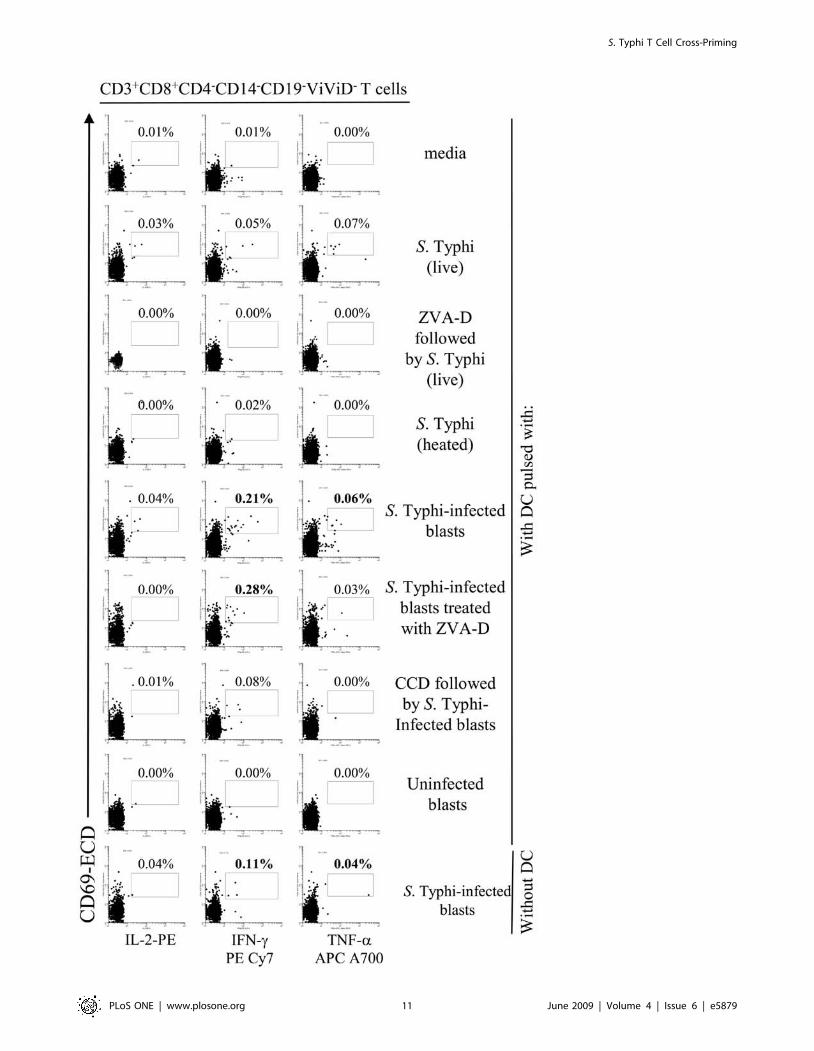
Figure 7. DC priming of S. Typhi-specific T cell responses. PBMC from volunteer CVD4000#65 were co-cultured with DC alone (media), or pre-mixed with live or heat-killed S. Typhi at a MOI of 10:1, or uninfected or S. Typhi-infected blasts at a 1:1 blast:DC ratio. In some cases, DC were pre-treated with ZVA-D or CCD before exposure to S. Typhi or S. Typhi-infected blasts respectively. In other cases, blasts were treated with ZVA-D beforeco-culture with DC. After 20 hours of incubation, cells were surface stained with a combination of mAb to CD3, CD4, CD8, CD14 and CD19 as well asViViD. After fixation and permeabilization, cells were intracellularly stained for IL-2, IFN-c and TNF-a and analyzed by multichromatic flow cytometry.Lymphocytes were gated based on their scatter characteristics. Single lymphocytes were gated based on forward scatter height vs. forward scatterarea. A ‘‘dump’’ channel was used to eliminate dead cells (ViViD+) as well as CD14+ and CD19+ cells from analysis. This was followed by additionalgating on CD3, CD4 and CD8, to identify cytokine-producing CD8+ T cells. Each cytokine was gated individually. During sample acquisition, routinely300,000-500,000 events were collected in the forward and side scatter (FS/SS) lymphocyte gate. This large number of gated lymphocyte events wasnecessary to ensure that a sufficient number of positive cells for a defined subset would be collected for each tube analyzed. Numbers correspond tothe percentage of positive cells in the indicated regions in each histogram. These results are representative of 1 of 4 volunteers with similar results.doi:10.1371/journal.pone.0005879.g007
S. Typhi T Cell Cross-Priming
PLoS ONE | www.plosone.org 12 June 2009 | Volume 4 | Issue 6 | e5879

Figure 8. Induction of IFN-c- and TNF-a-secreting CD8+ T cell subpopulations upon DC priming. PBMC from volunteer CVD4000#63were co-cultured with DC alone (media), or pre-mixed with live or heat-killed S. Typhi at a MOI of 10:1, or uninfected or S. Typhi-infected cells at a 1:4blast:DC ratio. After 20 hours of incubation, cells were surface stained with a combination of mAb to CD3, CD4, CD8, CD14, CD19, CD45RA, andCD62L as well as ViViD. After fixation and permeabilization, cells were intracellularly stained for IFN-c and TNF-a and analyzed by multichromatic flowcytometry. Lymphocytes gated as described in Figure 5 were followed by additional gating on CD3, CD4 and CD8, as well as CDR45RA versus CD62Lto analyze TEM and TEMRA cell subsets. Numbers correspond to the percentage of positive cells in the indicated regions in each histogram. Theseresults are representative of 1 of 4 volunteers with similar results. Additional data is provided in Fig. 9.doi:10.1371/journal.pone.0005879.g008
S. Typhi T Cell Cross-Priming
PLoS ONE | www.plosone.org 13 June 2009 | Volume 4 | Issue 6 | e5879

Figure 9. Induction of IFN-c- and TNF-a-secreting CD8+ T cell subpopulations upon DC priming. PBMC from volunteer CVD4000#65were co-cultured with DC alone (media), or pre-mixed with live or heat-killed S. Typhi at a MOI of 10:1, or uninfected or S. Typhi-infected cells at a 1:4blast:DC ratio. After 20 hours of incubation, cells were surface stained with a combination of mAb to CD3, CD4, CD8, CD14, CD19, CD45RA, and CD62Las well as ViViD. After fixation and permeabilization, cells were intracellularly stained for IFN-c and TNF-a and analyzed by multichromatic flowcytometry. Lymphocytes gated as described in Figure 5 were followed by additional gating on CD3, CD4 and CD8, as well as CDR45RA versus CD62Lto analyze TEM and TEMRA cell subsets. Numbers correspond to the percentage of positive cells in the indicated regions in each histogram. Theseresults are representative of 1 of 4 volunteers with similar results.doi:10.1371/journal.pone.0005879.g009
S. Typhi T Cell Cross-Priming
PLoS ONE | www.plosone.org 14 June 2009 | Volume 4 | Issue 6 | e5879

Quantification of Cytokines by Cytometric bead array(CBA)
Cytokine levels of IFN-c, IL-1b, IL-2, IL-5, IL-6, IL-8, IL-10,
IL-12p70 and TNF-a by DC was measured by flow cytometry-
based BD Cytometric bead array (CBA). Briefly, DC were
cultured in the absence (media) or presence of the various culture
conditions discussed above for 48 hours. The supernatants were
then harvested and kept at -70uC until assayed. CBA assays were
carried-out following the manufacturer’s instructions. The levels of
sensitivity for the various cytokines ranged from 2.5–20 pg/ml.
Apoptosis assaysCell apoptosis was monitored by two assays: (A) by measuring
membrane changes by staining cells with annexin-V and
propidium iodide (PI) (APOAF Annexin V-FITC Apoptosis
detection kit, Sigma) and (B) by measuring cytosol changes
following staining with a rabbit polyclonal antibody to caspase-3
PE (BD-Pharmingen). Annexin V and PI staining were performed
following the manufacturer’s recommendations.
Priming assaysPriming assays were performed as previously described [37,38]
with slight modifications. PBMC from healthy S. Typhi-unvacci-
nated adult volunteers were co-cultured with autologous DC at
PBMC to DC ratios of 15–20:1. The S. Typhi PBMC specificity or
priming was evaluated by their ability to secrete cytokines ex vivo or
in vitro detected by intracellular staining or ELISPOT, respectively.
DC were stimulated as described above for 30-48 hours before
exposure to the PBMC.
Intracellular stainingAfter 1–2 hours of stimulation with S. Typhi antigens, brefeldin-
A (BFA) (2 mg/ml, Sigma), a protein transport blocker, was added
to the PBMC. After 16–18 hours, PBMC were surface and
intracellularly for IFN-c, TNF-a, IL-2 (B-D Pharmingen) and
CD69 (Beckman-Coulter) to perform up to 13-color stainings.
Cells were then resuspended in fixation buffer (1% formaldehyde)
and analyzed as soon as possible by flow cytometry on an LSR-II
instrument (BD Biosciences).
IFN-c ELISPOT assayIFN-c ELISPOT assays were performed as previously described
[5,6,8,9]. Net frequencies of spot forming cells (SFC) were
calculated as previously described [5,6,8,9]. The cut-off for in vitro
expanded effector cells was established as the frequency of SFC/
106 PBMC in co-cultures of effectors with not-infected targets + 2
SE.
Statistical analysisAll tests were performed using SigmaStat software (version 3.10,
SSPS Science software products, Chicago, IL). Comparisons
between groups were performed by the One-way ANOVA test.
Specifically, in flow cytometric experiments, a response was
considered specific if the differential in the number of positive
events between experimental and negative control (media only)
cultures was significantly increased by Chi-square tests. P
values,0.05 were considered significant.
Acknowledgments
We are indebted to the volunteers who allowed us to perform this study.
We also thank Dr. Bernadette McConnell and the staff from the Blood
Bank of the Maryland University hospital for their help in collecting blood
specimens; Mr. Guillermo Sahaniuk, Ms. Regina Harley and Ms. Cathy
Storrer for excellent technical assistance.
Author Contributions
Conceived and designed the experiments: RSG. Performed the experi-
ments: RSG. Analyzed the data: RSG MS. Contributed reagents/
materials/analysis tools: MS. Wrote the paper: RSG MS.
Figure 10. Cross-priming DC are the most effective stimulus in promoting the expansion of S. Typhi-specific T cells. Net frequencies ofIFN-c-spot forming cells (SFC) were assessed by an IFN-c ELISPOT assay using in vitro expanded PBMC as effectors and autologous blasts infected withlive S. Typhi as stimulators. PBMC were co-cultured with their own DC alone or DC that had been pulsed with live or heat-killed S. Typhi, or uninfectedor S. Typhi-infected autologous blasts at a 1:4 blast:DC ratio. Net frequencies of IFN-c SFC were calculated as described in Materials and Methods. Thedashed line represents the cut-off for positive-ELISPOT assays determined as described in Materials and Methods. Bar graphs show means+SE of 3experiments using 4 different donors (*, p,0.05 compared with DC pulsed with S. Typhi-infected cells).doi:10.1371/journal.pone.0005879.g010
S. Typhi T Cell Cross-Priming
PLoS ONE | www.plosone.org 15 June 2009 | Volume 4 | Issue 6 | e5879

References
1. Crump JA, Luby SP, Mintz ED (2004) The global burden of typhoid fever. BullWorld Health Organ 82: 346–353.
2. Mitchell DH (1997) Ciprofloxacin-resistant Salmonella typhi: an emerging
problem. Med J Aust 167: 172.3. Rowe B, Ward LR, Threlfall EJ (1997) Multidrug-resistant Salmonella typhi: a
worldwide epidemic. Clin Infect Dis 24 Suppl 1: S106–109.4. Prost LR, Sanowar S, Miller SI (2007) Salmonella sensing of anti-microbial
mechanisms to promote survival within macrophages. Immunol Rev 219:
55–65.5. Salerno-Goncalves R, Pasetti MF, Sztein MB (2002) Characterization of CD8(+)
Effector T Cell Responses in Volunteers Immunized with Salmonella enterica
Serovar Typhi Strain Ty21a Typhoid Vaccine. J Immunol 169: 2196–2203.
6. Salerno-Goncalves R, Wyant TL, Pasetti MF, Fernandez-Vina M, Tacket CO,et al. (2003) Concomitant Induction of CD4(+) and CD8(+) T Cell Responses in
Volunteers Immunized with Salmonella enterica Serovar Typhi Strain CVD 908-
htrA. J Immunol 170: 2734–2741.7. Sztein MB, Tanner MK, Polotsky Y, Orenstein JM, Levine MM (1995)
Cytotoxic T lymphocytes after oral immunization with attenuated vaccinestrains of Salmonella typhi in humans. J Immunol 155: 3987–3993.
8. Salerno-Goncalves R, Fernandez-Vina M, Lewinsohn DM, Sztein MB (2004)
Identification of a human HLA-E-restricted CD8+ T cell subset in volunteersimmunized with Salmonella enterica serovar Typhi strain Ty21a typhoid vaccine.
J Immunol 173: 5852–5862.9. Salerno-Goncalves R, Wahid R, Sztein MB (2005) Immunization of volunteers
with Salmonella enterica serovar Typhi strain Ty21a elicits the oligoclonalexpansion of CD8+ T cells with predominant Vbeta repertoires. Infect Immun
73: 3521–3530.
10. Sztein MB, Wasserman SS, Tacket CO, Edelman R, Hone D, et al. (1994)Cytokine production patterns and lymphoproliferative responses in volunteers
orally immunized with attenuated vaccine strains of Salmonella typhi. J Infect Dis170: 1508–1517.
11. Tacket CO, Galen J, Sztein MB, Losonsky G, Wyant TL, et al. (2000) Safety
and immune responses to attenuated salmonella enterica serovar typhi oral livevector vaccines expressing tetanus toxin fragment C. Clin Immunol 97:
146–153.12. Tacket CO, Pasetti MF, Sztein MB, Livio S, Levine MM (2004) Immune
responses to an oral typhoid vaccine strain that is modified to constitutivelyexpress Vi capsular polysaccharide. J Infect Dis 190: 565–570. Epub 2004 Jun
2030.
13. Wahid R, Salerno-Goncalves R, Tacket CO, Levine MM, Sztein MB (2007)Cell-mediated immune responses in humans after immunization with one or two
doses of oral live attenuated typhoid vaccine CVD 909. Vaccine 25: 1416–1425.14. Wahid R, Salerno-Goncalves R, Tacket CO, Levine MM, Sztein MB (2008)
Generation of specific effector and memory T cells with gut- and secondary
lymphoid tissue- homing potential by oral attenuated CVD 909 typhoid vaccinein humans. Mucosal Immunology 1: 389–398.
15. Banchereau J, Palucka AK (2005) Dendritic cells as therapeutic vaccines againstcancer. Nat Rev Immunol 5: 296–306.
16. Sundquist M, Rydstrom A, Wick MJ (2004) Immunity to Salmonella from a
dendritic point of view. Cell Microbiol 6: 1–11.17. Albert ML (2004) Death-defying immunity: do apoptotic cells influence antigen
processing and presentation? Nat Rev Immunol 4: 223–231.18. Bevan MJ (2006) Cross-priming. Nat Immunol 7: 363–365.
19. Heath WR, Belz GT, Behrens GM, Smith CM, Forehan SP, et al. (2004) Cross-presentation, dendritic cell subsets, and the generation of immunity to cellular
antigens. Immunol Rev 199: 9–26.
20. Rock KL, Shen L (2005) Cross-presentation: underlying mechanisms and role inimmune surveillance. Immunol Rev 207: 166–183.
21. Salerno-Goncalves R, Sztein MB (2006) Cell-mediated immunity and thechallenges for vaccine development. Trends Microbiol 14: 536–542.
22. Pasetti MF, Levine MM, Sztein MB (2003) Animal models paving the way for
clinical trials of attenuated Salmonella enterica serovar Typhi live oral vaccines andlive vectors. Vaccine 21: 401–418.
23. Dunstan SJ, Ho VA, Duc CM, Lanh MN, Phuong CX, et al. (2001) Typhoidfever and genetic polymorphisms at the natural resistance-associated macro-
phage protein 1. J Infect Dis 183: 1156–1160.
24. Banchereau J, Steinman RM (1998) Dendritic cells and the control of immunity.
Nature 392: 245–252.
25. Arrighi JF, Rebsamen M, Rousset F, Kindler V, Hauser C (2001) A critical role
for p38 mitogen-activated protein kinase in the maturation of human blood-derived dendritic cells induced by lipopolysaccharide, TNF-alpha, and contact
sensitizers. J Immunol 166: 3837–3845.
26. Svensson M, Johansson C, Wick MJ (2000) Salmonella enterica serovar
typhimurium-induced maturation of bone marrow-derived dendritic cells. Infect
Immun 68: 6311–6320.
27. Lanzavecchia A, Sallusto F (2001) The instructive role of dendritic cells on T cell
responses: lineages, plasticity and kinetics. Curr Opin Immunol 13: 291–298.
28. Mastroeni P, Harrison JA, Robinson JH, Clare S, Khan S, et al. (1998)
Interleukin-12 is required for control of the growth of attenuated aromatic-compound-dependent salmonellae in BALB/c mice: role of gamma interferon
and macrophage activation. Infect Immun 66: 4767–4776.
29. Hess J, Ladel C, Miko D, Kaufmann SH (1996) Salmonella typhimurium aroA-
infection in gene-targeted immunodeficient mice: major role of CD4+ TCR-alpha beta cells and IFN-gamma in bacterial clearance independent of
intracellular location. J Immunol 156: 3321–3326.
30. MacLennan C, Fieschi C, Lammas DA, Picard C, Dorman SE, et al. (2004)
Interleukin (IL)-12 and IL-23 are key cytokines for immunity against Salmonella in
humans. J Infect Dis 190: 1755–1757.
31. Stoycheva M, Murdjeva M (2005) Serum levels of interferon-gamma,
interleukin-12, tumour necrosis factor-alpha, and interleukin-10, and bacterialclearance in patients with gastroenteric Salmonella infection. Scand J Infect Dis
37: 11–14.
32. Wick MJ (2003) The role of dendritic cells in the immune response to Salmonella.
Immunol Lett 85: 99–102.
33. Chen LM, Kaniga K, Galan JE (1996) Salmonella spp. are cytotoxic for cultured
macrophages. Mol Microbiol 21: 1101–1115.
34. Monack DM, Raupach B, Hromockyj AE, Falkow S (1996) Salmonella typhimurium
invasion induces apoptosis in infected macrophages. Proc Natl Acad Sci U S A93: 9833–9838.
35. Grant AJ, Sheppard M, Deardon R, Brown SP, Foster G, et al. (2008) Caspase-
3-dependent phagocyte death during systemic Salmonella enterica serovarTyphimurium infection of mice. Immunology 125: 28–37.
36. Richter-Dahlfors A, Buchan AM, Finlay BB (1997) Murine salmonellosis studiedby confocal microscopy: Salmonella typhimurium resides intracellularly inside
macrophages and exerts a cytotoxic effect on phagocytes in vivo. J Exp Med 186:569–580.
37. Mora JR, Bono MR, Manjunath N, Weninger W, Cavanagh LL, et al. (2003)Selective imprinting of gut-homing T cells by Peyer’s patch dendritic cells.
Nature 424: 88–93.
38. Schakel K, Mayer E, Federle C, Schmitz M, Riethmuller G, et al. (1998) A novel
dendritic cell population in human blood: one-step immunomagnetic isolationby a specific mAb (M-DC8) and in vitro priming of cytotoxic T lymphocytes.
Eur J Immunol 28: 4084–4093.
39. Marriott I, Hammond TG, Thomas EK, Bost KL (1999) Salmonella efficientlyenter and survive within cultured CD11c+ dendritic cells initiating cytokine
expression. Eur J Immunol 29: 1107–1115.
40. Rovere P, Vallinoto C, Bondanza A, Crosti MC, Rescigno M, et al. (1998)
Bystander apoptosis triggers dendritic cell maturation and antigen-presentingfunction. J Immunol 161: 4467–4471.
41. Wong KL, Lew FC, Macary PA, Kemeny DM (2008) CD40L-expressing CD8T cells prime CD8alpha(+) DC for IL-12p70 production. Eur J Immunol 38:
2251–2262.
42. Yrlid U, Wick MJ (2000) Salmonella-induced apoptosis of infected macrophages
results in presentation of a bacteria-encoded antigen after uptake by bystanderdendritic cells. J Exp Med 191: 613–624.
43. Guermonprez P, Amigorena S (2005) Pathways for antigen cross presentation.
Springer Semin Immunopathol 26: 257–271.
44. Kurts C, Miller JF, Subramaniam RM, Carbone FR, Heath WR (1998) Major
histocompatibility complex class I-restricted cross-presentation is biased towardshigh dose antigens and those released during cellular destruction. J Exp Med
188: 409–414.
S. Typhi T Cell Cross-Priming
PLoS ONE | www.plosone.org 16 June 2009 | Volume 4 | Issue 6 | e5879