principles of branch formation and branch patterning in hydrozoa
TRANSCRIPT
Principles of branch formation and branch patterning
in Hydrozoa
STEFAN BERKING*
Zoological Institute, University of Cologne, Köln, Germany
ABSTRACT The freshwater polyp Hydra produces buds which separate from the parent. Other
Hydrozoa produce branches which remain connected to the parent, thus forming a colony. Some
Hydrozoa grow by means of an organ that is like a shoot apical meristem. Others display a
sympodial type of growth. In this article, I propose that these different types of branches are
organized by a common pattern-forming system. This system has self-organizing properties. It
causes branch tip formation and is kept active in the tip when the tip finally differentiates into a
hypostome of a polyp. The system does not cause structure formation directly but rather,
determines a tissue property called positional value, in such a way that a gradient of values forms
in the tissue of the bud or branch. The local value determines the local morphodynamic processes,
including differentiation of the hypostome (highest positional value), tentacles and basal disc and
of the exoskeleton pattern along the shoot. A high positional value favors the onset of a new self-
organizing process and by lateral inhibition, such a process prevents the initiation of a further
process in its surroundings. Small quantitative differences in the range of the signals involved
determine whether a bud or a branch forms and whether monopodial and sympodial growth
follows.
KEY WORDS: Hydra, pattern formation, budding, sympodial growth, monopodial growth
Introduction
The best known member of Hydrozoa is the freshwater polypHydra, which can reproduce asexually by budding. The buddevelops somewhere in the middle between the apical and thebasal end of a polyp, out of a group of somatic cells (Fig. 1A, B).A bud develops into a small polyp and separates from the parent.Marine Hydrozoa generally produce colonies, of which someresemble seed plants. As in plants, two parts can be distinguished(Fig. 2B). The basal part, called the hydrorhiza (or stolon), formsa network of hollow tubes that branch and that may reunite. Thispart fixes the organism to a substrate. The apical part consists ofthe hydrocaulus (or shoot), which may branch as well. A tip of sucha branch usually ends in a polyp (or hydranth). The polyps of suchcolonies have a structure similar to Hydra. In some species the tipkeeps its state of a growing tip, like the shoot apical meristem inseed plants. Some authors consistently call this organ a cormus(for example, see Kühn, 1909, 1914; von Schenck, 1965). Themost elaborated branching pattern structures a subgroup ofHydrozoa called Thecata. In this group two types of shoot growthappear, sympodial and monopodial growth.
The questions are, What mechanism of pattern formation is
Int. J. Dev. Biol. 50: 123-134 (2006)doi: 10.1387/ijdb.052043sb
*Address correspondence to: Dr. Stefan Berking. Zoological Institute, University of Cologne, Weyertal 119, 50923 Köln, Germany.Fax: +49-221-470-5171.e-mail: [email protected]
0214-6282/2006/$25.00© UBC PressPrinted in Spainwww.intjdevbiol.com
common to all Hydrozoa? and How do we modify the commonprinciple to account for the observed diversity of branch pattern-ing? In the following discussion I compare observations and ideasconcerning the control of pattern formation and concerning themorphodynamics of budding and branch formation in the variousHydrozoa. Models of pattern formation in Hydrozoa were gener-ally designed to explain results obtained from regeneration andtransplantation experiments. The models assume the existenceof morphogens that are generated by the head and the foot,respectively. These morphogens are proposed to control theformation of these structures and the differentiation of the tissuealong the body axis. The best evolved model of this type is thatdeveloped by Meinhardt (1993). In colonial hydroids, polypsdevelop far away from a basal disc and Kosevich (1991) hasshown that the tissue of the future hypostome, exclusively,controls the patterning of the branch, including the formation ofthe polyp. In colonial hydroids a foot system either does not existor does not influence branch/polyp formation and the patterningof the polyp. Clearly, for developing a concept of pattern controlthat is common to all Hydrozoa, such differences are particularlyimportant. Unquestionably a common principle exists; but whichelements of pattern control and morphodynamics are common to
124 S. Berking
all Hydrozoa and which have evolved by modifications of theexisting ones and by “inventions” later on in the various species?
Results and Discussion
Hydrozoa that produce Branches which separate from theparent animal: vegetative reproduction
The best known member of this group of Hydrozoa thatproduce branches that separate from the parent animal is thefreshwater polyp Hydra, which has a tube-shaped body (Fig. 1).One end consists of the head, with the mouth/anus openingsurrounded by tentacles. The other end, called the foot, in-cludes the basal disc that closes the tube. The middle part is thegastric region. The body wall has two layers, the ectoderm andthe endoderm, separated by an extracellular matrix, the me-soglea. Hydra is famous for its ability to regenerate. Small bodysections of Hydra give rise to complete animals: The headregenerates from the apical end and the foot from the basal end.This indicates a polar organization of the tissue. Tissue piecesobtained from different body levels display different capacitiesto transform into a head or a foot, respectively, when trans-planted to a host animal. The tissue taken from a more apicalposition combines a higher capacity to form a head with a lowercapacity to form a foot. The polarity of the tissue and the gradeddistribution of the noted capacity is determined by a scalartissue property (Gierer et al., 1972) that has been termedpositional value (Wolpert, 1969; Wolpert et al., 1974) or sourcedensity (Gierer and Meinhardt, 1972). By definition, the posi-
tional value (or source density) has its highest value at theapical end of a Hydra and its lowest value in the basal disc.
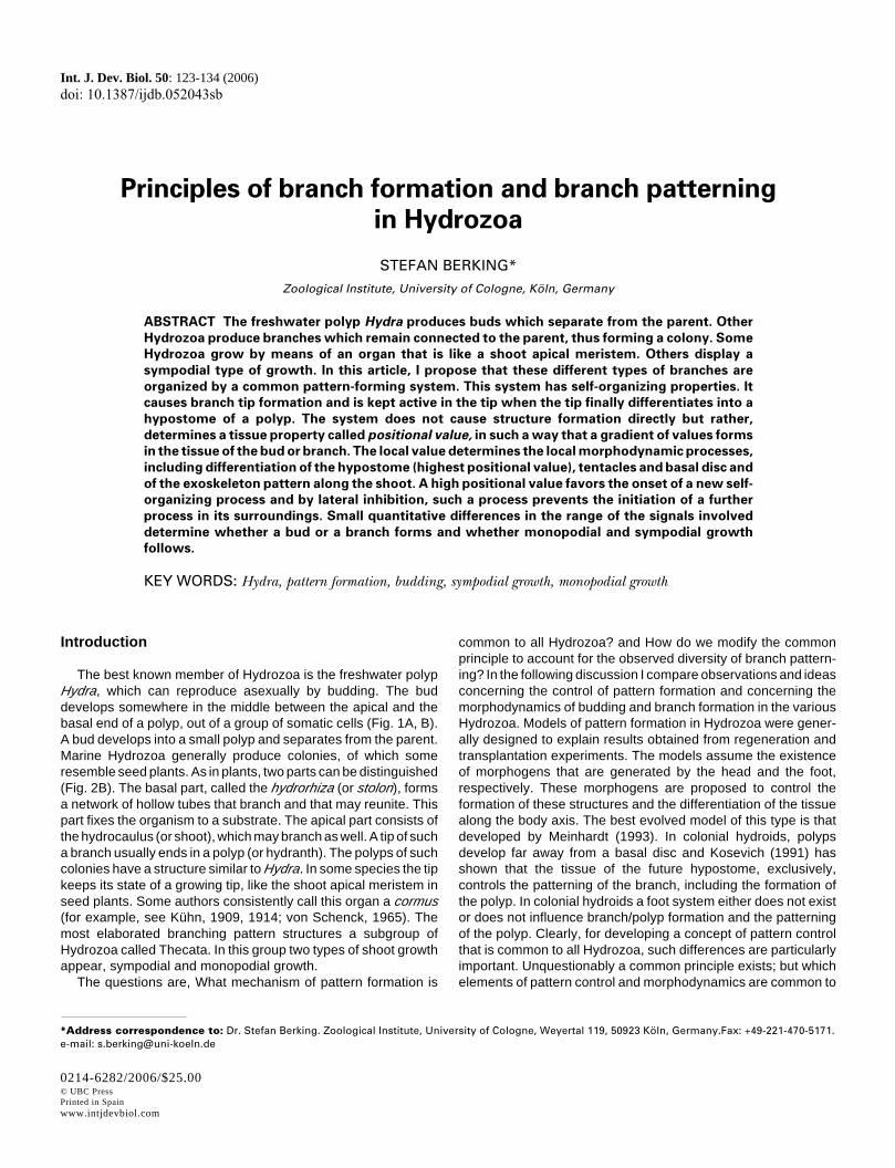
In Hydra, budding visibly starts with the formation of a smallprotrusion of both tissue layers of the parent’s body wall (Fig. 1).The bud grows by recruiting tissue of the surroundings and bycell multiplication (Otto and Campbell, 1977). The tip of the buddevelops into the apical tip of the new polyp’s head. The mostbasal part of the bud develops a basal disc and then the budseparates from the parent. When the tip visibly forms, all bodyparts of the future bud, including the basal disc, are alreadydetermined in form of concentric rings in the parent’s tissue(Sanyal, 1966; Tardent, 1972). Thus the formation of head,gastric region and foot is organized from one point, namely thefuture hypostome of the bud. Obviously, this finding posesproblems to models with symmetric opposing gradients ofhead- and foot-specific morphogens. In the following discus-sion, I propose an alternative that overcomes these problems.
A model for pattern control in HydrozoaWhen an adult animal displays a gradient of positional
values from one end to the other along the body axis, a bud mustalso develop this gradient. The proposition is that at the futurebud’s tip, signals are generated that cause a rise of the posi-tional value up to the maximal value and, some distance away,cause a decrease down to the lowest possible value. In be-tween, a gradient of positional values develops. In a secondstep, secondary systems control the local development, includ-ing the formation of hypostome and basal disc, according to the
A B
C D E
each other and control the increase and the decrease of the positional value (red). The equations used for the simulation may be interpreted as follows:the positional value of a cell is determined by its content in compound A (activator). The activator is produced within the cells, increasing the positionalvalue. The activator can be released from the cells, decreasing the positional value. The released activator stimulates both its release out of cells andits production within cells. Thus two loops of autocatalysis operate. Further, the released activator stimulates the release of two inhibitors(heterocatalysis). In the released form, one of them (B) antagonizes (counteracts) the release of the activator out of the cells, the other (C) antagonizes(counteracts) the production of the activator within the cells. Alternative interpretations of the equations are possible. The self-enhanced release ofthe activator depends on the existence of some activator outside the cells. It is assumed that there is a basal unregulated release, the rate of whichcorrelates with the amount present within the respective cell, that is, with its positional value. (C) In the budding region the morphogens and thepositional value initially display an almost flat distribution. (D) Because of the interactions of the morphogens that have been noted, the positional valuechanges such that (E) a complete bud develops, including head and basal disc.
Fig. 1. Bud formation in Hydra.(A) Sketch of a Hydra bearing ayoung bud. The different texturesindicate the various body regionsof the adult animal and the respec-tive future body regions of the bud(from Sanyal, 1966). On the leftthe respective positional valuesare shown. (B) Shown is a sketchof the process of budding (afterTardent, 1978) and (C-E) a simula-tion of this process in a piece oftissue representing the buddingregion with the model proposed(Berking, 2003). Three morpho-gens control the onset of buddingand bud development: an activa-tor A (shown in green), an inhibitorB (brown) and an inhibitor C (blue).These morphogens interact with
Patterning in Hydrozoa 125
local positional value attained.To get an increase of the positional value at one point,
namely the future bud’s tip and to get a decrease some distanceaway, two signals with a different range are sufficient when bothare generated from that point: One signal increases the posi-tional value; this signal has a short range. The other decreasesthe positional value; this signal has a long range. Gierer andMeinhardt (1972) have solved the problem of how a group ofcells (in this case, cells of the future bud’s tip) is prompted to dosomething different from cells in the surroundings (in this case,cells of the gastric tissue). At the simplest an activator stimu-lates its own release from cells (autocatalysis or self-enhance-ment) and the release of an inhibitor, which in turn antagonizesself-enhancement.
A combination of the two propositions can explain budding,including foot formation at its base and various other featuresof pattern formation (Berking, 2003). The combination made issuch that as few compounds as possible and as few interac-tions as possible, are included: (1) One and the same activator,which by means of autocatalysis (self-enhancement) and lat-eral inhibition determines a patch of cells to generate both theactivator (A) and its antagonist (inhibitor B). (2) The generatedactivator causes an increase of the positional value and (3)where the concentration of A is high, a second inhibitor (inhibi-tor C) is generated that decreases the positional value. Therange of both inhibitors is larger than the range of the activator.With respect to branch formation in the different species dis-cussed in the following, it is particularly important which one ofboth inhibitors B or C has the longer range. I emphasize that thismodel does not postulate the a priori existence of a gradient anda body length axis. Rather, it proposes the existence of certaininteractions or system properties. These interactions generategradients of positional values and thus a body axis, branchesand so on (see Fig. 1).
Patterning of the budSimulation experiments with the proposed model describe
bud formation appropriately (Fig. 1). The tip of the bud developsinto the bud’s hypostome. At the base a foot forms, without foot-specific morphogens displaying a long range. The patterning ofthe bud is exclusively organized from the center, which devel-ops into the new animal’s apical end. Arguments for a continu-
ous increase of the positional value preceding hypostomeformation were derived from the transplantation of regeneratingtissue (Berking, 1979). Further, during head regeneration stain-ing of tissue with a tentacle-specific antibody indicates that thetissue that ultimately forms the hypostome (maximal positionalvalue) initially has properties characteristic of tentacles (Bodeet al., 1988).
Positioning of the budThe pattern-forming system that controls bud development
is generally assumed to remain unchanged during the bud’sfurther development. Among other consequences, the systemdetermines the position at which the former bud produces a budby itself. Two opposing forces control this position: On the onehand, budding tends to start as close as possible to the existinghead; and on the other hand, budding is prevented in the vicinityof the parent’s head (Burnett, 1961; Webster and Hamilton,1972; Shostak, 1974; and, with respect to marine animals andpolyp formation on stolons, Braverman, 1971; Plickert et al.,1987). The models describe this feature correctly (Meinhardt,1993; Berking, 2003).
A high positional value favors the self-enhanced release ofthe activator, causing a bud to form as close as possible to thehead. In contrast, the inhibitor involved in lateral inhibitionallows the onset of budding only at a certain distance from thehead. In other words, one self-organizing process prevents theonset of a further one close to it.
In Hydra the bud develops from tissue of the gastric regionof a polyp. In most marine Hydrozoa, a bud or branch does notdevelop from a polyp but rather from the stolon or from the shoot(hydrocaulus). The reason for that difference appears to be thatthe polyp Hydra is comparatively large. In Hydra the bud field of1 mm in diameter gives rise to a young bud about 1 mm long.Most polyps of Hydrozoa that form marine colonies are evensmaller. The bud of Hydra grows up to 1 cm in length bymultiplication of epithelial cells, which occurs almost randomlyin the body column but excludes the very ends (David andCampbell, 1972). This causes the initial steep gradient ofpositional values to become flat. However, the range of mor-phogens is not stretched accordingly. Morphogens generatedat the apical end barely reach the basal disc. This allowsbudding from the gastric region of the polyp. In animals with
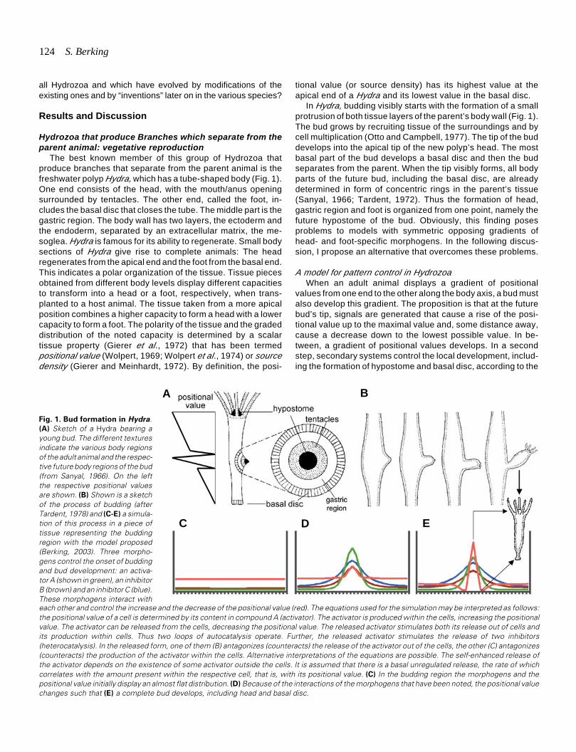
Fig. 2. Frustule formation in Haleremita (Hydrozoa) and
monopodial growth in colonial athecates (Hydrozoa). (A)
A polyp bearing two frustules and two frustules that havebeen separated from the parent. The tip of the frustulebecomes the future polyp’s head. Several days after separa-tion, the first tentacles develop (Kühn, 1914). (B) Sketch of amonopodially growing colony with a terminal polyp (Athecata).Right part: A polyp has formed on top of a stolon (dotted). Leftpart: The distance between the first formed polyp (1) and itsorigin at the stolon has increased by intercalary elongation ofthe tissue tube (coeno-sarc) and by terminal elongation of apossibly existing perisarc covering, that is, just proximal to thepolyp (hydranth). A hydrotheca does not exist. At the time thehydrocaulus (both coenosarc and a possible perisarc covering)had reached a certain length, laterally a bud has formed thatdeveloped at its end into a polyp (2). After further elongationof the main hydrocaulus, a third polyp (3) has formed and soon (after Kühn, 1914).
A B
126 S. Berking
small polyps, the bud field can be expected to match almost thesize of the polyp and the range of the morphogens, in particularof the inhibitor that antagonizes the onset of budding, exceedsthe size of the polyp (Braverman, 1971; Plickert et al., 1987).
Frustule formationPolyps of certain Hydrozoa produce so-called frustules (Fig.
2A). A frustule is a bud without head structures. After separatingfrom the parent, the frustule moves for days or weeks over thesubstrate; then a mouth and tentacles develop. Finally the frustulehas transformed into a normal polyp. It appears that initially at thetip of the frustule the maximal positional value attained is too lowto cause head structures to form. Simulation experiments showedthat when both the positional value and the level of inhibitor B arereduced, a frustule forms (Berking, 2003). To explain why such aprocess does not occur in Hydra, we must assume that in thetissue between the budding region and the basal disc, where thepositional value is low enough for frustule formation, the concen-tration of inhibitor B is too high.
Certain manipulations cause a bud of Hydra to develop into abranch
Two experiments are discussed next. (1) When animals bear-ing a young bud are sectioned just apical to the bud, the budtransforms into a branch without signs of foot formation (Fig. 3A).At the same time head regeneration is prevented (Weimer, 1928;Rulon and Child, 1937; Sanyal, 1966; Tardent, 1972). (2) Trans-planting small pieces of hypostomal tissue to a certain positionalong the body length axis causes the outgrowth of an axis(branch or bud), as does transplanting a head with the hypostomaltip in front (Fig. 3B): When a head is transplanted to between thehead and the budding region, a branch develops. When a head istransplanted to between the budding region and the foot, a buddevelops, which detaches from the parent (Berking, 1979). Com-puter simulation shows (Fig. 3C) that a slight increase of theconcentration of inhibitor B in the bud and in the tissue surround-ing the bud stimulates branch formation without a foot and withoutseparation from the parent (Berking, 2003). With respect to theexperiments mentioned, head-regenerating tissue in close vicin-ity supplies the bud with additional inhibitor B and tissue trans-planted close to the head is expected to get a higher level ofinhibitor B than does tissue far away from the head. In contrast,a developing bud also generates the (hypothetical) inhibitors.Thus a developing bud can antagonize head regeneration andbud formation in its close vicinity.
The model also describes head and foot regeneration. Head
regeneration is simple to understand; it is similar to the onset ofbudding or branch formation. Foot regeneration is more difficult tounderstand. Simulation experiments showed that the very samemorphogens control both head and foot regeneration. The tissueproperty (positional value) adjacent to the former wound deter-mines whether the positional value increases or decreases. Inparticular, the net export of the inhibitor that causes a decrease ofthe positional value was found to be decisive (Berking, 2003). Thisfeature of the model is shown in a simulation of pattern formationin Dynamena (see Fig. 5 and Fig. 6). The system is kept active inthe tip when it differentiates into a hypostome and is finallyswitched off when the tip differentiates into a basal disc.
In summary: A pattern-forming system including at least threemorphogens is proposed to control the quantity of a tissueparameter, namely the positional value and thus generates abody axis starting from homogenous conditions. The local valuedetermines local development. The model can describe buddingas well as head and foot regeneration. In tissue of high positionalvalue, the onset of a new self-organizing process is favored.Whether a bud or branch forms depends on lateral influences andon the property of the tissue that may form the basal disc. Bylateral inhibition, one self-organizing process prevents the onsetof a further, nearby self-organizing process.
Hydrozoa producing branches which persist at the parentanimal: colony formation
Most Hydrozoa form colonies. Colonies generally consist oftwo parts: a net of tubes (termed stolons or hydrorhizas) fixed toa substrate and polyps (hydranths) on top of these stolons (Fig.2, Fig. 4). The polyps look similar to a Hydra. The simplestcolonies produce polyps directly on the stolon. Complex coloniesform shoots made of repetitive elements on which polyps form ina regular pattern.
Shoots form in two ways. Either the stolon tip transforms intoa shoot tip, resulting in a polyp at its end, or a shoot forms on topof a stolon. Such a tip displays shoot quality from the outset. Asatisfactory explanation for the transformation of a stolon tip intoa shoot tip and the reverse—observed, for example, inPlumulariidae, which are thecate Hydrozoa with a monopodialtype of growth (von Schenck, 1965)—appears to be a challengefor models of pattern control. The problem is that if morphogensspecific to head and stolon tip are assumed to control the twodifferent tips, we must explain how the self-enhanced generationof stolon-specific morphogens ceases while that of a polyp’s headstarts and takes over (and the reverse). In the model I propose inthis article, one system with three morphogens controls the
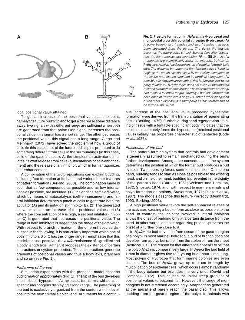
Fig. 3. Branch formation in Hydra. (A) Sectioning distal toa young bud causes the bud to develop into a branch. (B)
Transplanting a head with the tip of the hypostome in front,to various body regions causes branch formation if thetissue is transplanted between budding region and head.The implant causes bud formation if it is placed betweenbudding region and basal disc. (C) The simulation wasperformed in two steps. At first budding was allowed to start
A B C
as shown in Fig. 1. Then the concentration of the inhibitor B was slightly increased and the simulation was carried on. The increase of the inhibitorB should represent the sectioning in (A) and the transplantation in (B). In both cases a secondary axis is forced to form or continues its growth at anunusual high concentration of inhibitor B, which is found in close vicinity of an existing or regenerating head (for details, see Berking, 2003).
Patterning in Hydrozoa 127
various specific developments via the positional value. Simulationexperiments have shown that a quantitative change of oneparameter, such as the local concentration of one of the hypotheti-cal morphogens, is enough to change the developmental fate ofa growing tip. During that change, the self-enhanced generationof morphogens is maintained.
Hydrozoa displaying sympodial growthThe growth pattern described in the following discussion char-
acterizes several species of a suborder of Hydrozoa, the Thecata.These animals form colonies in which all parts are covered withperisarc, a rigid, chitinous, extracellular matrix. Only the polyp canexpand out of the tube-like endings of the perisarc covering.Shoot formation generally starts from the top of a hydrorhiza (Fig.4A). After a short period of growth, the shoot tip develops into ahydranth. The next step of growth is similar to that of a sympodiallygrowing plant. Some distance from the original apical end, whichdifferentiates terminally (in hydrozoa into a polyp) in a lateralposition, the tip of a new branch emerges. This tip takes over theelongation of the shoot.
In several Campanulariidae, repetitive elements consist of twosequences of annuli separated by a smooth, slightly bent tube andfollowed by the finely structured housing of the polyp, thehydrotheca (Fig. 4A, B). Although the perisarc forms a stiff tube,the indentation between two annuli allows a bending of the tubesimilar to a joint in arthropods, which are also completely coveredby a stiff exoskeleton excluding the joints.
Both the stolon and the shoot tubes lengthen only at their tips(Kühn, 1914; Hyman,1928), by secreting the material that laterforms the perisarc (for a review of this process, see Waite, 1990).
Hence the pattern of the perisarc emerges exclusively at that site.Close to the apex of the elongating tube, this secreted material israther soft and flexible. Its shape is precisely that of the underlyingtissue. Some dozens of micrometers proximal to the apex, theperisarc material hardens and from that time onward has a fixedshape. Thus the pattern of the perisarc is a time recording of thetissue activity in the tip.
Experiments performed with Laomedea flexuosa indicate thatthe cells in the tip shape the perisarc by the following activities(Kossevitch et al., 2001). (1) The cells of the tip move activelyforward, displaying a so-called growth pulsation with a periodicityof several minutes (Beloussov, 1973; Hale,1960, 1964;Wyttenbach, 1974). (2) The cells tend to produce a bulb with alarger diameter than the hardened perisarc tube allows. (3) Thecells of the tip secrete perisarc material, which hardens at acertain distance to the very tip of the apex. The regulation of thisdistance largely determines the pattern of the perisarc. When thehardening occurs closer to the apex, the diameter of the ring-shaped border between the hard and the soft perisarc decreases(Fig. 4F). That narrowing forces the tissue to squeeze through thissmall opening. If conditions remain unchanged, the perisarc tubeelongates with a reduced diameter. A widening of the diametertakes place when the border of hardening moves to a moreproximal position.
The second proposition, the tendency of the tissue tube toattain a larger diameter, is supported by the observation that thetissue tube in the shoot tip has a tight contact to the perisarc overa certain length (about 250 to 350 µM in Laomedea flexuosaHincks, Campanulariidae; diameter of the tissue tube is about 160to 250 µM), whereas in proximal regions the tissue tube diameter
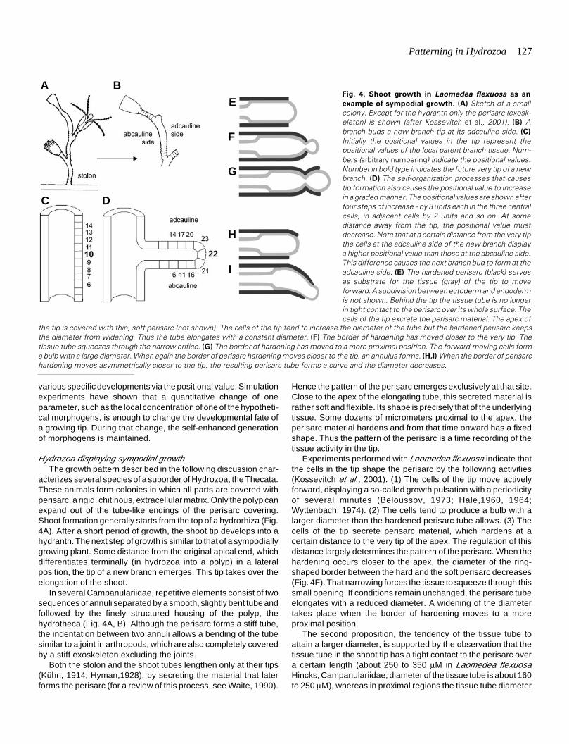
Fig. 4. Shoot growth in Laomedea flexuosa as an
example of sympodial growth. (A) Sketch of a smallcolony. Except for the hydranth only the perisarc (exosk-eleton) is shown (after Kossevitch et al., 2001). (B) Abranch buds a new branch tip at its adcauline side. (C)
Initially the positional values in the tip represent thepositional values of the local parent branch tissue. Num-bers (arbitrary numbering) indicate the positional values.Number in bold type indicates the future very tip of a newbranch. (D) The self-organization processes that causestip formation also causes the positional value to increasein a graded manner. The positional values are shown afterfour steps of increase - by 3 units each in the three centralcells, in adjacent cells by 2 units and so on. At somedistance away from the tip, the positional value mustdecrease. Note that at a certain distance from the very tipthe cells at the adcauline side of the new branch displaya higher positional value than those at the abcauline side.This difference causes the next branch bud to form at theadcauline side. (E) The hardened perisarc (black) servesas substrate for the tissue (gray) of the tip to moveforward. A subdivision between ectoderm and endodermis not shown. Behind the tip the tissue tube is no longerin tight contact to the perisarc over its whole surface. Thecells of the tip excrete the perisarc material. The apex of
the tip is covered with thin, soft perisarc (not shown). The cells of the tip tend to increase the diameter of the tube but the hardened perisarc keepsthe diameter from widening. Thus the tube elongates with a constant diameter. (F) The border of hardening has moved closer to the very tip. Thetissue tube squeezes through the narrow orifice. (G) The border of hardening has moved to a more proximal position. The forward-moving cells forma bulb with a large diameter. When again the border of perisarc hardening moves closer to the tip, an annulus forms. (H,I) When the border of perisarchardening moves asymmetrically closer to the tip, the resulting perisarc tube forms a curve and the diameter decreases.
A B
C D
E
F
G
H
I
128 S. Berking
is much smaller than the inner lumen of the perisarc tube. Further,the shoot and the stolon occasionally form a bulb at the woundafter cutting. The third proposition, the differential control ofhardening, has been studied by means of chemicals that interferewith the hardening process (Kossevitch et al., 2001).
Patterning of the branchA young tip that is isolated by sectioning develops a complete
branch, including the polyp’s housing at its end (Kosevich, 1991).The resultant perisarc tube does not contain tissue over its wholelength; rather, the tissue of the tip develops into only the mostdistal part of the hydranth (positioned in the polyp’s housing). Thisdevelopment shows that the tissue of the growing tip autono-mously determines the perisarc pattern of the branch along itslength axis. There is no indication of the existence of a foot/stolontip system that might influence branch formation, including polyppatterning. The experiment further shows that under normalconditions most of the tissue proximal to the tip is pulled out of theparent shoot by the actively forward-moving tip tissue. Transplan-tation experiments confirm that distal and proximal tissue has noinfluence on the patterning of the shoot (Kosevich, 1996).
The simplest explanation of these observations appears to bethat by means of a self-organizing process, the positional valuecontinuously increases in the growing tip up to the maximal valuepossible, as proposed for budding in Hydra. The perisarc isshaped according to the attained value.
The branches of most sympodially growing thecate Hydrozoadisplay an obvious bilateral symmetry. In Campanulariidae thesmooth part of the branch is usually bent and the tip of a newbranch develops close to the curve (Fig. 4A, B). There are twopossible causes of this asymmetry: Either the branch displaysqualitatively different stripes of tissue along its length axis, orwithin the circumference exists a quantitatively differing tissueparameter. From the evolutionary point of view, we hesitate topropose that so-called Radiata have two qualitatively differentbody sides, like the dorsal and the ventral side in bilaterians. Thusa more conventional proposition is preferred, that is, a gradeddistribution of positional values not only in the longitudinal axis butalso in the transverse axis of the branch.
Transplantation experiments indicate that in the tissue of thebranch tip the cause of the future branch asymmetry is presentfrom the outset (Kossevitch, 2002). I argue that this asymmetrydepends on the origin of the branch tissue. Tissue distal to the newbranch position (that is, closer to the existing hydranth) has ahigher positional value than does tissue proximal to the branch tip(Fig. 4C). Thus, at the outset, the tissue of the new branch tipdisplays a gradient of positional values along the transverse axis.The highest value is present at the future adcauline side of thebranch, namely the side that faces the hydrocaulus of the parent.The tissue that later is pulled out from the parent’s hydrocaulus toform the new branch displays the same feature. This initialtransverse gradient appears not to vanish completely duringoutgrowth that includes a change of the positional value along thelongitudinal axis (Fig. 4D).
The smooth part of a branch forms when the distance betweenthe position of hardening and the forward-moving tip remainsconstant (growth pulsations are ignored). Kossevitch et al. (2001)argue that this is caused by a constant secretion of perisarcprecursors (Fig. 4E). A row of annuli forms when secretion is not
constant but rather oscillates in such a way that the tip cells areentrained to secrete synchronously at a high rate for a short timeperiod (Kossevitch et al., 2001). At least the “hardener” - probablycertain phenols (Knight, 1968, 1970; Holl et al., 1992; Kossevitchet al., 2001) - must be secreted in an oscillating manner. Atransitory high concentration of hardener moves the hardeningborder close to the very tip, producing a ring-shaped furrow. Whenthe cells are exhausted and refractory, the border moves back toa proximal position, causing a widening of the developing perisarctube. Repetitions of the two events, a phase of secretion followedby a refractory phase, cause annuli formation.
Annuli formation starts when the tip cells have attained acertain positional value. The cells that reach first the critical valuesecrete and entrain the others to do the same. The oscillatingsecretion ends when a certain higher positional value is attained,causing the transition from the annulated part to the smooth part(Fig. 4A, B). The oscillated secretion starts a second time, causingthe formation of the distal annulated zone. Cells that first reach thecritical positional value for that transition are positioned at theadcauline side of the branch. They start ahead of the others, withincreased secretion, whereas the others in the circumference arestill refractory. This causes an asymmetric polymerization of theperisarc tube that results in the observed bending of the smoothpart (Fig. 4H, I). In accordance with this proposition, the strongestbending usually appears just before the onset of annulation.
Occasionally the first furrow is not ring-shaped but, rather,restricted to the adcauline part (Kosevich, 2004). In members ofthe Campanulinae family (for example, in Campanulina lacerata),the hydranth pedicel is often not a row of annuli (distal annulatedparts) but a spiral in which one turn matches the diameter of anannulus. Spirals also develop in certain members of the familySertulariidae (Kosevich, 2004). This observation fits the proposi-tion that a cell secretes certain precursors necessary for perisarchardening for a short time, followed by a refractory period and thatthis cell triggers its neighbors also to secrete the same com-pounds.
Positioning of the branchThe tip of a new branch develops close to the curve of the
smooth part of the branch (Fig. 4B). To learn how the axial positionis determined, Kosevich (for review, see Kosevich, 2004) artifi-cially elongated the distal annulated part and found that the newbranch tip formed not in the smooth part but rather in the distalannulated zone. This indicates that the axial position of a newbranch is controlled similarly to the axial position of a bud in Hydra,by two opposing forces: On the one hand, a high positional valuefavors the self-organization process to start and on the otherhand, the existing head/hydranth exerts an inhibitory influencethat antagonizes the onset of that process in its vicinity. Thesepropositions also explain the position of the branch tip in thecircumference: Along the branch length axis, the adcauline tissuedisplays a higher positional value than does the abcauline one(Fig. 4D).
Compared to athecates, thecate Hydrozoa display a muchhigher variability of form and the number of species is also muchhigher. The exoskeleton and the spatial and temporal control of itshardening have apparently permitted this strong increase inspecies number. The arthropods, the bilaterians with the highestvariability of body pattern and the highest number of species, also
Patterning in Hydrozoa 129
have an exoskeleton. We may thus suggest that the success ofarthropods in evolution is also largely based on their ability todevelop diverse body patterns, an ability that appears to belargely caused by a spatial and temporal control of the hardeningof the arthropod exoskeleton. Even the joints in thecate Hydrozoaand arthropods display similarities.
In summary, the model proposed for Hydra is applied toLaomedea: In a branch tip the positional value increases duringgrowth. The perisarc is shaped according to the attained value.Perisarc precursors are generated by the tip cells in differentmodes, namely constant or oscillating. The bilateral symmetry ofthe branch is a result of the gradient of positional values in thetissue that originally forms the bud tip. The position of the branchis proposed to be controlled by two opposing forces: (1) A locallyhigh positional value favors the onset of the self-organizingprocess that leads to branch formation and (2) by lateral inhibitionthe existing self-organizing process in the hydranth prevents theformation of a new one in its vicinity.
Hydrozoa displaying monopodial growthMonopodial growth in Athecata and Thecata is very different:
In Athecata the shoot ends in a polyp and elongates by intercalarygrowth caused by cell multiplication (Fig. 2B). When a certainlength is reached, a branch forms in lateral position. The end of thebranch develops into a polyp. Both the shoot and the branchelongate and form a further branch, when a certain length isreached and so on.
This type of growth has strong similarities to growth andbudding in Hydra and in particular to experimental branch forma-tion in Hydra. It doesn’t appear necessary to posit a pattern-forming system specific for Athecata displaying such a type ofgrowth.
In Thecata an intercalary elongation of the shoot does notexist, because of the perisarc covering of the whole colonyincluding the polyp (Fig. 5). The elongating end of the shoot isoccupied by a stem tip that behaves like a shoot apical meristemin seed plants. It grows by maintaining its character while right andleft polyps emerge in a repetitive manner like leaves in higherplants. When a stem of Dynamena pumila grows in length,generally three primordia form out of the initial single one. The twolateral ones develop into polyps; the central one elongates,subdivides into three primordia and thus starts a new cycle ofinternode formation.
Patterning of the branchTo explain how the new “organ” stem tip becomes organized,
we can assume the existence of an additional pattern-formingsystem, including a set of new morphogens. This rises severalquestions: How can the evolution of this system be explainedwithin the group of Thecata? How do primordia that developdifferently appear at the apex of a stem at a certain distance fromeach other? Either primordia use identical inhibitors, or differentinhibitors cross-react to some extent in the other systems—theycannot keep their distance in different ways. And we must alsoexplain how primordia that follow a different development becomearranged properly. Generally, the stem tip develops in the centerand right and left polyps develop; other arrangements are rarelyobserved (Fig. 5, Fig. 6). A general requirement for all models ofpattern formation is also to explain such rare arrangements.
The following discussion shows that all these problems can besolved by assuming that the system used for bud formation inHydra and branch formation in Laomedea (and other sympodiallygrowing Thecata) also controls pattern formation in Dynamena(and other monopodially growing Thecata). The most important
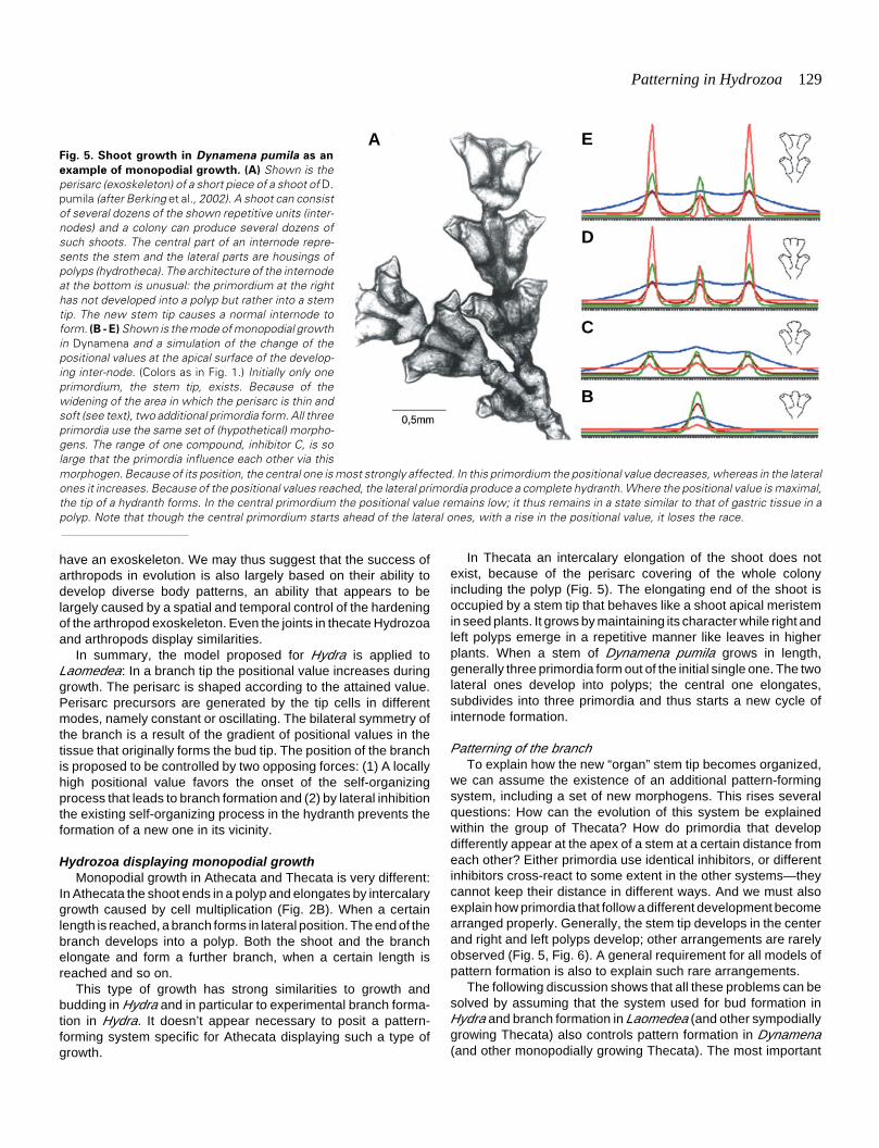
Fig. 5. Shoot growth in Dynamena pumila as an
example of monopodial growth. (A) Shown is theperisarc (exoskeleton) of a short piece of a shoot of D.pumila (after Berking et al., 2002). A shoot can consistof several dozens of the shown repetitive units (inter-nodes) and a colony can produce several dozens ofsuch shoots. The central part of an internode repre-sents the stem and the lateral parts are housings ofpolyps (hydrotheca). The architecture of the internodeat the bottom is unusual: the primordium at the righthas not developed into a polyp but rather into a stemtip. The new stem tip causes a normal internode toform. (B - E) Shown is the mode of monopodial growthin Dynamena and a simulation of the change of thepositional values at the apical surface of the develop-ing inter-node. (Colors as in Fig. 1.) Initially only oneprimordium, the stem tip, exists. Because of thewidening of the area in which the perisarc is thin andsoft (see text), two additional primordia form. All threeprimordia use the same set of (hypothetical) morpho-gens. The range of one compound, inhibitor C, is solarge that the primordia influence each other via thismorphogen. Because of its position, the central one is most strongly affected. In this primordium the positional value decreases, whereas in the lateralones it increases. Because of the positional values reached, the lateral primordia produce a complete hydranth. Where the positional value is maximal,the tip of a hydranth forms. In the central primordium the positional value remains low; it thus remains in a state similar to that of gastric tissue in apolyp. Note that though the central primordium starts ahead of the lateral ones, with a rise in the positional value, it loses the race.
A
B
C
D
E

130 S. Berking
A B C D E F G H
I J K L
differences among the pattern-forming systems of these animalsare the activity ranges of the (hypothetical) morphogens in therespective tissues.
The development of a repetitive unit, an internode, starts witha small stem tip in which the cells of the apex have a certainpositional value and the activity of the (proposed) pattern-formingsystem increases this value (Fig. 5B to E). The cells actively moveforward, using the already hardened perisarc as substrate (asobserved in Laomedea) and produce a spherical bulb at the apex,indicating that the tissue moves faster than the border of perisarchardening. The widened apical surface of the bulb is covered onlyby thin, soft perisarc. This is the moment at which (generally) outof one primordium, three primordia form. Applying the modelproposed for pattern formation in Hydra, the following sequenceof events is suggested (Berking et al., 2002): From the apextissue, the loss of the (hypothetical) inhibitors is larger than fromtissue covered by thick and rigid perisarc. This results in a self-enhanced release of the activator not only in the center of the tipbut also in the surroundings. By autocatalysis and lateral inhibi-tion, additional primordia form. All primordia use the same set ofmorphogens. The inhibitor that decreases the positional value(inhibitor C) is proposed to have a range sufficient to reach theprimordia in the neighborhood.
Obviously, the primordium in the center is most stronglyaffected. Its positional value decreases, whereas in the lateralprimordia the positional value increases (Fig. 5E). The lateralprimordia develop into polyps; the central primordium remains ina state similar to that of gastric tissue. Histological studies showthat in the course of growth the central primordium becomesincreasingly more isolated from the developing polyps that are inthe lateral position (Berking et al., 2002). Isolation reduces theinfluence of the lateral primordia on the central primordium and in
the central one it allows the positional value to increase again: Thenext cycle of internode formation has started.
I propose that all primordia, taken together, are organized inthe same way by the same set of morphogens. The centralprimordium develops into a stem tip simply because of its position.In the central primordium, the positional value increases anddecreases once in each internode cycle, without reaching themaximal and the minimal value. Such a mechanism has aninherent instability, which is proposed to explain the high numberand the nature of malformations observed in D. pumila (up to 1%of all internodes): Quantitative changes of external influencescause a quantitative change of the «lateral dominance.» A smalllateral dominating influence allows the central primordium todevelop into a polyp (Fig. 6F). A very strong such influencecauses the central primordium to develop into a stolon (Fig. 6E,L). The point of interest here is that with respect to thesemorphotypes a rule governs the spatial arrangement of primordiathat develop differently. When three or more primordia form, thetip of the central primordium usually develops a lower - rarely anequal, but never a higher - positional value than the tip of a lateralprimordium. This strongly indicates that primordia form so closeto each other that they influence each other’s developmental fate.No exception to the rule has yet been found, although far morethan 200,000 internodes have been studied (Marfenin et al., 1995:Berking et al., 2002).
Positioning of the branchIn Dynamena, as argued for Hydra and Laomedea, I propose
that two opposing forces determine where branch formationstarts: A high positional value favors the onset of the self-organizing processes and one self-organizing process preventsthe onset of a further one close to it. These propositions explain
Fig. 6. Morphotypes of Dynamena pumila and internodes of species related to Dynamena. (A to H) A selection of morphotypes or malformationsof D. pumila (Marfenin et al., 1995; Berking et al., 2002). Shown are the exoskeletons. In one case the central primordium develops a stolon insteadof a stem tip (E). The respective simulation (L) shows that a stolon forms if the three primordia remain in tight contact with each other for a long time.Note that in the central primordium the positional value (red line) reaches almost zero. (I) Lytocarpia myriophyllum (J) Sertularella gayi (I,J afterCornelius et al., 1995). (K) Sketch of a growing tip of a species of Sertularia, displaying the two hydranths (gray) at one, the “ventral” side (Kühn, 1909).

Patterning in Hydrozoa 131
that in Dynamena and related species, polyps and stem tip keepa regulated distance from each other. These propositions alsoexplain that in successive internodes the polyps form as closelyas possible to each other. Usually polyps form in rows along thelongitudinal axis (Fig. 5). This pattern is caused by a particularfeature of the shoot tips: In the tip, cell proliferation is very rare (forreview, see Kosevich, 2004). Thus the new primordia that form inlateral position include tissue that derives from tissue proximal tothe tip (as observed in Laomedea, sympodial growth). This tissuehas a differential origin in the circumference. Tissue at a certainposition derives from or is influenced by the adjacent hydranthprimordium; tissue at other positions is not. Thus in successiveinternodes polyps form in longitudinal rows. They form in tissuethat at the outset has a higher positional value than does othertissue lying at the same distance to the existing stem tip. The verysame proposition has been made for branch positioning in thecateswith sympodial growth (see earlier discussion). With respect toDynamena, to my knowledge such an export of tissue out of ahydranth into the surroundings has not been demonstrated tooccur but has been shown for Hydractinia echinata (Müller, 1964).
It appears that in contrast to Hydra and sympodially growingThecata, monopodially growing Thecata have developed anadditional way of controlling where primordia form and how manyform. In all Thecata, the positional value controls the area of thetip that is not covered by hardened perisarc. Only in Thecata withmonopodial growth do new primordia form within this area (sec-ondary branches and gonozoids, both of which form late, areignored). The size of this area determines how many additionalprimordia can form. Thus a self-organizing process controls, via
the positional value and via the resulting morphodynamics, theposition and the number of additional self-organizing processes.Several malformations appear to support this proposition: Whenthe apical area that is covered with thin, soft perisarc remains sosmall that additional primordia cannot begin to form, the stemsimply elongates (Fig. 6A). When there is a large loss of inhibitors,a hydranth forms out of the tip (Fig. 6A). When the area is larger,two primordia form. Because of the interaction noted, one primor-dium remains a stem and the other develops into a hydranth (Fig.6B). In one malformation (Fig. 6D), two primordia formed (bot-tom): One developed into a stem tip, the other into a hydranth. Ina next step three formed; the two hydranth-forming primordia areclose to each other and symmetrically positioned “above” the oldone. The new ones formed at the right and the left margins of thehypothetical polyp-derived tissue imported into the widened stemtip. The position of the two hydranth primordia is determined bythe two opposing conditions noted, (1) the local high positionalvalue of the tissue and (2) the mutual lateral inhibition of the twodeveloping primordia. Accordingly, in a third step the hydranthsform at a greater distance from each other. The hydranths form inan axial line because of the export of tissue with a high positionalvalue (Fig. 6I), indicating that the imported tissue displays ahigher positional value than does the stem tip. An alternatepositioning of hydranths (Fig. 6C, J) is caused by the two opposinginfluences if the area is just too small to allow three primordia todevelop simultaneously. How close to each other three or moreprimordia form is determined by the two opposing forces: (1) thehigh positional value of the imported tissue and (2) the range of theinhibitor involved in distance control. If the lateral inhibition has a
Fig. 7. Principles of branch formation and branch patterning in Hydrozoa.

132 S. Berking
long range, primordia form opposite to each other. If the range isshort, hydranths form close to each other. Two genera (Diphasia,Sertularia) have developed shoots with a “ventral” side (Kühn,1914), a “Zoidfläche” (Weismann, 1883) at which hydrothecaform very close to each other, indicating a comparatively shortrange of distance control (Fig. 6K).
The similarity to plants is obvious. Meinhardt et al. (1998)proposed that leaf primordia keep their distance from eachother by signals that involve autocatalysis and lateral inhibition.In plants, most of the tissue that forms a leaf derives from themeristem, the growing stem tip. In Thecata, most of the tissuethat forms the new primordium derives not from the stem tip butfrom the proximal position of the tissue tube. Therefore inHydrozoa two opposing forces control the position of primordiaformation, whereas in plants lateral inhibition appears to playthe most important role (Meinhardt et al., 1998). This differenceappears to explain why certain arrangements of polyps havenot been found–for example (to my knowledge), a spiral ar-rangement, such as is seen in several plants.
Researchers have proposed that mechanical influencescause three primordia form out of one primordium (Beloussov,1975; Beloussov and Grabovsky, 2003). These authors sug-gest that differential development of the tissue in the growing tipis preceded by a differential shaping of that tissue. The shapingis caused by a shift in the region of maximal active stretching ofthe tip’s tissue in base-to-apex direction within one growthpulsation. Thus, out of one center of pulsation several primordiadevelop that may display different pulsation properties. Thepattern of these new primordia is determined by the mechanicalproperties of the tissue of the tip’s surface. If the mechanicalforces trigger the onset of the various specific developmentssuch as polyp, stem, or stolon formation, we must explain howa different mechanical stretching of tissue can cause a differentonset of gene activity. However, the interactions proposed herethat may use diffusible substances could hardly directly alterthe physical form of the tissue layer. The step between theproposed signaling and the resultant structure, such as polyp,stolon, or stem, certainly is an alteration of cell activities,including a change of growth pulsation in the epithelial sheet ofthe tip surface.
Stolon FormationThe stolon tip has features in common with the tip of a shoot:
The tissue of the tip is covered by perisarc, with which it hastight contact—excluding the very tip, where the perisarc is softand thin. The stolon does not form structures such as annuli. Itelongates without forming a structure terminally. It producesstolon branches laterally and shoot branches from the roof.Stolons fix to a surface and thus become asymmetric. In somespecies they accompany the shoot, which results in the forma-tion of very complex colonies. A detailed discussion of stolonformation and stolon patterning is not intended here; I discussonly one question: Is stolon formation controlled by a systemdifferent from that in shoot branch formation? I propose that onesystem controls both shoot and stolon formation. A stolon tipforms when the positional value falls to the lowest value pos-sible. However, other than with basal disc regeneration inHydra, the pattern-forming system is not switched off. A pos-sible cause for that switched-on mode may be a sufficiently high
basal, unregulated production of the agent that determines thepositional value. This production level results in endless growthwithout a change in the positional value at the tip and thuswithout structure formation.
Conclusion
Branch formation in Hydrozoa is organized by a pattern-forming system with self-organizing properties that makes use ofat least three morphogens (Fig. 7). The system does not controlstructure formation directly but, rather, regulates the quantity of atissue parameter (for example, within cells the concentration of acompound involved in regulation) in such a way that a gradient inthe tissue forms. The local quantity of this parameter, termedpositional value, determines the local developmental fate, suchas hypostome (highest positional value), tentacles and basal disc.A gradient and a body length axis are not postulated to exist apriori.
The system controls branch tip formation and is kept active inthe tip even when it differentiates in a hypostome of a polyp.Where the system is active, cell proliferation is rare. A highpositional value favors the onset of a new self-organizing processand by lateral inhibition a self-organizing process prevents theformation of a further one in the vicinity. Small differences in theactivity range/strength of the signals involved in self-organizationallow in certain species a decrease of the positional value to thelowest value possible that causes the formation of a basal discand separation from the parent (budding). In monopodially grow-ing thecate Hydrozoa, the activity that can decrease the positionalvalue is proposed to have a longer range than the one thatcontrols the distance between primordia (Fig. 7). In sympodiallygrowing thecate Hydrozoa, the reverse is proposed to be true.This is suggested to be the essential difference between theprimary pattern-forming systems of monopodially growing th-ecate Hydrozoa and those of sympodially growing thecate Hydro-zoa. Interestingly, several species display transitional forms be-tween a purely sympodial and a purely monopodial growth pattern(Kühn, 1914). Basal disc regeneration in Hydra is controlled bythe same system, including the very same morphogens. Whenthe lowest positional value is reached, a basal disc forms and thesystem is switched off. In stolon formation the system is notswitched off, because of a certain basal production of morpho-gens that causes an endless maintenance of the activity and anendless elongation of the stolon.
In seed plants the shoot apical meristem is well studied.However, the control of its origin in embryogenesis and itsmaintenance during growth is still a matter of research. If theshoot apical meristem is formed and maintained in a way similarto that proposed for monopodially growing thecate Hydrozoa, themeristem must be understood as a leaf primordium that laterallydeveloping leaf primordia block from developing into a leaf.
The developmental morphodynamics of branch formation inHydrozoa can be subdivided into two parts: Hydrozoa elongatetheir branches by cell recruitment and intercalary cell prolifera-tion. Species in which an exoskeleton cover the branches com-bine this intercalary tissue growth with a terminal elongation of theperisarc tube. As in Arthropoda, the exoskeleton of Hydrozoaincludes stiff and jointlike parts. In Hydrozoa the formation ofthese structure elements is controlled by the actual positional

Patterning in Hydrozoa 133
value of the tissue in the growing tip. The positional value controlsthe position of perisarc hardening. Cells that cause the zone ofhardening to move close to the very tip trigger their neighbors todo the same. Then the cells become refractory, which causes thezone of hardening to move to a proximal position. This causes aring-shaped furrow to form, followed by a widening of the perisarctube. In monopodially growing Thecata, the branch primordiaform within the area that is not covered by thick, hardenedperisarc. If this area is small, new primordia do not form. Inmonopodially growing Thecata, morphodynamics controlled bythe local positional value apparently control the onset of new self-organizing processes.
Models and molecular approaches
Advanced molecular techniques have yet to be developed forHydrozoa. Most studies are still based on expression patterns ofcandidates of control genes and putative markers of certainspecific developments. Results obtained are compared with thoseobtained following treatments with certain chemicals or by othertechniques such as sectioning and transplantation. The linksbetween these various approaches are models. Models cansummarize experimental data in that the system properties of thestudied processes become transparent. Certainly they cannotreplace studies at the cellular and the molecular level, but theycan help researchers find and evaluate the critical components.One example is given here; a more detailed discussion is pub-lished elsewhere (see Berking, 1998, 2003).
With respect to Hydra, Wnt signaling has been suggested to bean element of the “head organizer” (Hobmayer et al., 2000). Thearguments are that HyWnt and other members of the Wnt path-way (Hy βcat and HyTcf) are expressed in the hypostomal tip,early in head regeneration and also early in budding. However,HyWnt and in particular Hy βcat (HyTcf was not tested) are alsoexpressed in foot-regenerating tissue, although more weakly andonly transiently (Hobmayer, personal communication, in Berking,2003). The question therefore is, Can this weaker, transientexpression during foot regeneration be ignored, or is it important?The same set of morphogens control both head and foot regen-eration. The tissue adjacent to the wound determines the fate atthe wound. If the positional value decreases down to the lowestvalue possible, a foot forms and morphogen generation ceases.Hence Wnt signaling can have a more fundamental relevancethan originally proposed. It is not head specific but, rather, is agood candidate for an element of the pattern-forming system thatcontrols the positional value.
AcknowledgmentsI thank K. Herrmann and I. Kosevich for helpful discussions.
References
BELOUSSOV, L.V., (1973). Growth and morphogenesis of some marine Hydrozoaaccording to histological data and time-lapse studies. In Recent trends inresearch in coelenterate biology (Eds. T. Tokioka and S. Nishimura). TheProceedings of the second international symposium on Cnidaria. Publs Setomar. biol. Lab. 20: 315-366.
BELOUSSOV, L.V. (1975). Possible ontogenetic mechanism governing formationof the body plans in animal embryos. Verh. Dtch. Zool. Ges. 89: 219-229.
BELOUSSOV, L.V. and GRABOVSKY, V.I. (2003). A geometro-mechanical model for
pulsatile morphogenesis. Comput. Methods Biomech. Biomed. Engine 6: 53-63.
BERKING, S. (1979). Analysis of head and foot formation in Hydra by means of anendogenous inhibitor. W. Roux’s Arch. 186: 189-210.
BERKING, S. (1998). Hydrozoa metamorphosis and pattern formation. Curr. Top.Dev. Biol. 38: 81-131.
BERKING, S. (2003). A model for budding in Hydra: pattern formation in concentricrings. J. Theor. Biol. 222: 37-52.
BERKING, S., HESSE, M. and HERRMANN, K. (2002). A shoot meristem-likeorgan in animals. Monopodial and sympodial growth in Hydrozoa. Int. J. Dev.Biol. 46: 301-308.
BODE, P.M., AWAD, T.A., KOIZUMI, O., NAKASHIMA, Y., GRIMMELIKHUIJZEN,C.J.P. and BODE, H.R. (1988). Development of the two-part pattern duringregeneration of the head in Hydra. Development 102: 223-235.
BRAVERMAN, M. (1971). Studies on hydroid differentiation. VII. The hydrozoanstolon. J. Morph. 135: 131-152.
BURNETT, A.L. (1961). The growth process in Hydra. J. Exp. Zool. 146: 21-83.
CORNELIUS, P.F.S., MANUEL, R.L. and RYLAND, J.S. (1955). Hydroids, SeaAnemones, Jellyfish and Comb Jellies. In Handbook of the Marine Fauna ofNorth-West Europe (Eds. Hayward, P.J., Ryland, J.S.) Oxford University Press,Oxford New York Tokyo.
DAVID, C.N. and CAMPBELL, R.D. (1972). Cell cycle kinetics and development ofHydra attenuata. I. Epithelial cells. J. Cell Sci. 11: 557-568.
GIERER, A. and MEINHARDT, H. (1972). A theory of biological pattern formation.Kybernetik 12: 30-39.
GIERER, A., BERKING, S., BODE, H., DAVID, C.N., FLICK, K., HANSMANN, G.,SCHALLER, H. and TRECKNER, E. (1972). Regeneration of Hydra fromreaggregated cells. Nature, New Biol. 239: 98-101.
HALE, L.J. (1964). Cell movements, cell division and growth in the hydroid Clytiajohnstoni. J. Embryol. Exp. Morph. 12: 517-538.
HALE, L.J. (1960). Contractility and hydroplasmic movements in the hydroid Clytiajohnstoni. Quart. J. Microscop. Sci. 101: 339-350.
HOBMAYER, B., RENTZSCH, F., KUHN, K., HAPPEL, C.M., CRAMER VONLAUE, C., SNYDER, P., ROTHBÄCHER, U. and HOLSTEIN, T.W. (2000). WNTsignalling molecules act in axis formation in the diploblastic metazoan Hydra.Nature 407: 186-189.
HOLL, S.M., SCHAEFER, J., GOLDBERG, W.M., KRAMER, K.J., MORGAN, T.D.and HOPKINS, T.L. (1992). Comparison of black coral skeleton and insectcuticle by a combination of carbon-13 NMR and chemical analyses. Arch.Biochem. Biophys. 292: 107-111.
HYMAN, L. (1928). Miscellaneous observations on Hydra, with special reference toreproduction. Biol. Bull. 54: 65-108.
KNIGHT, D.P. (1968). Cellular basis for quinone tanning of the perisarc in theThecate hydroid Campanularia (= Obelia) flexuosa Hinks. Nature 218: 584-586.
KNIGHT, D.P. (1970). Sclerotization of the perisarc of the caliptoblastic hydroid,Laomedea flexuosa. 1. The identification and localization of dopamine in thehydroid. Tissue & Cell 2: 467-477.
KOSEVICH, I.A. (1991). Comparison of functioning of the sprout and stolon growthapices in the colony of Obelia loveni (Allm.) (Hydrozoa, Campanulariidae).Vestn. Mosk. Univ., Ser.16: Biol., no 2: 44-52.
KOSEVICH, I.A. (1996). Regulation of formation of the elements of the hydroidpolyps colony. Russian J. Dev. Biol. 27: 95-101, Translated from Ontogenez(1996) 27: 114-121.
KOSEVICH [KOSSEVITCH], I.A., HERRMANN, K. and BERKING, S. (2001).Shaping of colony elements in Laomedea flexuosa Hinks (Hydrozoa, Thecaphora)include a temporal and spatial control of skeleton hardening. Biol. Bull. 201: 417-423.
KOSEVICH [KOSSEVITCH], I.A. (2002). Role of the skeleton in determination ofthe branching points in hydroid colonies. J. Obshchej Biologii. 63: 40-49.
KOSEVICH, I.A. (2004). Branching in colonial hydroids. In Branching Morphogen-esis (Ed. Davies, J). Landes Bioscience.
KÜHN, A. (1909). Sprosswachstum und Polypenknospung bei Thecaphoren.Studien zur Ontogenese und Phylogenese von Hydroiden. Zool. Jb. Anat. 28:387-476.
KÜHN, A. (1914). Entwicklungsgeschichte und Verwandschaftsbeziehungen der
134 S. Berking
Hydrozoen. 1. Teil: Die Hydroiden. Erg. Fortschr. Zool. 4: 1-284.
MARFENIN, N.N., MARGULIS, R.J. and MEIER, E.M. (1995). Morphologicalvariability of the colonial hydroid Dynamena pumila, with classification of foundmorphotypes. Russian Academy of Sciences. Proceedings of the ZoologicalInstitute St. Petersburg. 261: 71-89 (Russian).
MEINHARDT, H. (1993). A model of biological pattern formation of hypostome,tentacles and foot in Hydra: How to form structures close to each other, how toform them at a distance. Dev. Biol. 157: 321-333.
MEINHARDT, H., KOCH, A.J. and BERNASCONI, G. (1998). Models of patternformation applied to plant development, In Symmetry in Plants. (Eds. D. Barabeand R.V. Jean) World Scientific Publishing, Singapore: 723-758.
MÜLLER, W. A. (1964). Experimentelle Untersuchungen über Stockentwicklung,Polypendifferenzierung und Sexualchimären bei Hydractinia echinata. Roux’sArch. Dev. Biol. 155: 181-268.
OTTO, J. J. and CAMPBELL, R. D. (1977). Budding in Hydra attenuata: bud stagesand fate map. J. exp. Zool. 200(3): 417-428.
PLICKERT, G., HERINGER, A. and HILLER, B. (1987). Analysis of spacing in aperiodic pattern. Dev. Biol. 120: 399-411.
RULON, O. and CHILD, C.M. (1937). Observations and experiments on develop-mental pattern in Pelmatohydra oligactis. Physiol. Zool. 10: 1-13.
SANYAL, S. (1966). Bud determination in Hydra. Indian J. Exp. Biol. 4: 88-92.
SHOSTAK, S. (1974). Bipolar Inhibitory gradients’ influence on the budding regionof Hydra viridis. Am. Zool. 14: 619-632.
TARDENT, P. (1972). Experimente zum Knospungsprozess von Hydra attenuataPall. Rev. Suisse Zool. 79: 355-375.
TARDENT, P. (1978). Coelenterata, Cnidaria. In Morphogenese der Tiere (Ed.Seidel, F.). Stuttgart New York: Gustav Fischer Verlag.
von SCHENCK, D.A. (1965). Die Kormentektonik der Plumulariiden (Coelenterata,Hydrozoa). Rev. Suisse Zool. 72: 885-1021.
WAITE, J.H. (1990). The phylogeny and chemical diversity of quinone-tanned gluesand varnishes. Comp. Biochem. Physiol. 96B: 19-29.
WEBSTER, G. and HAMILTON, S. (1972). Budding in Hydra: The role of cellmultiplication and cell movement in bud initiation. J. Embryol. exp. Morph. 27:301-316.
WEIMER, B.R. (1928). The physiological gradients in Hydra I. Re-constitution andbudding in relation to length of piece and body level in Pelmatohydra oligactis.Phys. Zoöl. 1: 183-230.
WEISMANN, A. (1883). Die Entstehung des Sexuallebens bei den Hydromedusen.Jena.
WOLPERT, L. (1969). Positional information and the spatial pattern of cellulardifferentiation. J. theor. Biol. 25: 1-47.
WOLPERT, L., HORNBRUCH, A. and CLARKE, M.R.B. (1974). Positional informa-tion and positional signalling in Hydra. Am. Zool. 14: 647-663.
WYTTENBACH C.R. (1974). Cell movements associated with terminal growth incolonial hydroids. Am. Zool. 14: 699-717.