principles of complex oncotherapy and surgical oncology. …€¦ · · 2015-11-12methods of...
TRANSCRIPT

Principles of complex oncotherapy and surgical
oncology.
Methods of surgical reconstruction and
oncological rehabilitation
Director General of National Institute of Oncology, Budapest - Hungary
Prof. Miklós Kásler MD, PhD, DSc, Dr.h.c., FRCS


Top 10 cancers in Hungary (2010)
3
0 50 100 150
Brain
Larynx
Pancreas
Kidney, except renal pelvis
Stomach
Rectum
Bladder
Colon
Prostate
Bronchus & lung
ASR (W) rate per 100 000
Mortality Incidence
Male0 20 40 60 80 100 120 140
Cervix uteri
Stomach
Malignant melanoma of skin
Rectum
Ovary
Pancreas
Corpus uteri
Colon
Bronchus & lung
Breast
ASR (W) rate per 100 000
Mortality Incidence
Female
0 20 40 60 80 100 120
Malignant melanoma of skin
Kidney, except renal pelvis
Stomach
Pancreas
Rectum
Bladder
Prostate
Colon
Breast
Bronchus & lung
ASR (W) rate per 100 000
Mortality IncidenceMortality Incidence
Hungarian
Cancer
Registry

Treatment of cancerCancer treatment programmes
• „The main goals of a cancer treatment programme is to cure or considerably prolong the life of patients and to ensure the best possible quality of life to cancer survivors.
• The most effective and efficient treatment programmes are those that:
a) are provided in a sustained and equitable way;
b) are linked to early detection; and
c) adhere to evidence-based standards of care and a multidisciplinary approach.”

Edward A. Sausville; Dan L. Longo Principles of Cancer Treatment. In. Harrison’s Principles of
Internal Medicine 18th.
• „The goal of cancer treatment is first to eradicate the cancer.
• If this primary goal cannot be accomplished, the goal of cancer treatment shifts to palliation, the amelioration of symptoms, and preservation of quality of life while striving to extend life.
• One of the challenges of cancer treatment is to use the various treatment modalities alone and together in a fashion that maximizes the chances for patient benefit.
• Cancer treatments are divided into four main types: surgery, radiation therapy, chemotherapy (including hormonal therapy and molecularly targeted therapy), and biologic therapy (including immunotherapy and gene therapy).
• The modalities are often used in combination, and agents in one category can act by several mechanisms.
• Surgery and radiation therapy are considered local treatments, though their effects can influence the behavior of tumor at remote sites.
• Chemotherapy and biologic therapy are usually systemic treatments.
• Oncology, the study of tumors including treatment approaches, is a multidisciplinary effort with surgical-, radiotherapy-, and internal medicine–related areas of expertise.”
Principles of complex oncotherapy


Surgical Oncology
• „Over the past 4 decades cancer care has undergone a revolution.
• No longer is surgery the only treatment for most solid malignancies but adjuvant therapies with highly focussed radiotherapy, targeted molecular therapies and multimodal chemotherapy are the standard of care.
• These multimodal treatment regimes have had a great impact on cancer survival rates, as have improved diagnostics.
• Today, general surgeons can no longer work in isolation and must be part of a multidisciplinary team.
• The surgeon must be more than just a technician and must understand the contributions made by other disciplines
and how this may impact on the type and timing of surgery:
he/she must be a Surgical Oncologist. „

The Surgical Oncologist
• The oncologic surgeon is a physician of the
XXIst century, combining the modern theory of
diagnostical and therapeutic oncology and
the manual skills using high-technology.
• Surgical oncology is basic part of the
multidisciplinary tumor care, performed by the
Surgical Oncologist.

Why now?
• Increasing incidence of malignancies
• Biological hypothesis
• Population screening
• Modern evidence based oncology >> better overal survival (OS)
• Social improvements >> better OS >> need of better quality of life (eg. breast oncoplasty)
• Sofisticated extended- as well minimal-invasive operative techniques, according to multidisciplinary stage based but individual surgery
• High-technology (laparoscopy, DaVinci etc.)
• Industrial competition

Characteristics of a Surgical Oncologist
• Specialized- or superspecialized education and training (eg. breast oncoplastic surgeon)
• Special exams (UEMS/EBSQ surgical oncologist, breast surgeon)
• Specialized comprehensive centers
• Multi- and interdisciplinarity (core member of MDTs)
• Quality controll
• Clinical trials
• Education

The European Union of Medical Specialists (UEMS) and the European
Board of Surgery Qualification (EBSQ)


Principles of Surgical Oncology
DeVita,Hellman, and Rosenberg's Cancer: Principles and Practice of Oncology
8th., Lippincott Williams&Wilkins

1. Radicality („en block”)
1.1. Primary tumour
1.2. Metastases2. Monoblock surgery
2.1. Lymphogenic spreading
2.2. Haematogenic spreading3. “No touch isolation technique”
3.1. Surgical approach (minimal-invasive surgery)
3.2. Surgical technique
3.3. Surgical instruments (laser)4. Structure and function sparing
4.1. Organs of non vital importance
- neoadjuvant-/adjuvant therapy
4.2. Organs of vital importance
- neoadjuvant-/adjuvant therapy 5. Reconstruction of function and aesthetic, quality of life
Principles in surgical oncology
15

Extended primary and salvage
surgery with reconstruction
16
Prerequisites Operability (general condition of the patient)
Resecability (complete removal of the cancer)
Reconstruction (restoration of structure and function)

Characteristics of Surgical Oncology
• Evidence based medicine, quidelines
• Multidisciplinary care
• Comprehensive centers
• Integration of continuous development of basic sciences (eg. molecular genetics, medical and radiological oncology)
• Therapeutic and palliative surgical oncology
• Psycho-oncological care
• Specialised international societies (eg. ESSO, EUSOMA)
• Quality indicators based on international consesus
• Regular audit of the results

Evidence based medicine, quidelines
Kásler: Oncotherapeutic Protocol, Springer, 1994, 1995
Kásler: Principles of Oncoptherapy, B+F, 2001
Németh: Radiotherapy, Medicina, 2004
Kásler: Fundamentals of Oncotherapy, 2011
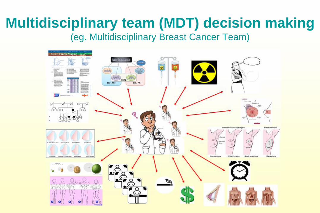
Multidisciplinary team (MDT) decision making (eg. Multidisciplinary Breast Cancer Team)

The role of the oncological team in cancer patient management
1. Determination of the diagnostic algoritm
1.1. Evaluation of the available findings
1.2. Completion of the available findings
- necessary? (has any therapeutic impact?)
- for what purpose? (what information does it yield?)
- sensitivity and specificity of the examination of choice? (CT, MRI, PET CT, etc..)
- in what order, where ?
2. Determination of the therapeutic algoritm
- determination of the necessary therapeutic components(surgery, radiotherapy, medical therapy)
- type of the components (surgical, radiotherapeutic, drug)
- order of the components
- time of the delivery of the components
- site of the delivery of the components
3. Control of therapy: what, how, when, where ?
4. Rehabilitation – palliation20


Specialised Oncological Comprehensive Centers
• „Many studies have shown that increasing hospital volume for major cancer surgery also has positive impact on patient survival.
• In one study of 5,013 patients in the Surveillance, Epidemiology, and End Results registry of patients high hospital volume was linked with lower mortality for patients undergoing pancreatectomy (P = .004), esophagectomy (P <.001), liver resection (P = .04), and pelvic exenteration (P = .04).
• In patients undergoing esophagectomy, operative mortalitywas 17.3% in low-volume hospitals compared with 3.4% in high-volume hospitals.
• For patients undergoing pancreatectomy, the correspon-
ding rates were 12.9% vs. 5.8%.”
DeVita,Hellman, and Rosenberg's Cancer: Principles and Practice of Oncology 8th.,
Lippincott Williams&Wilkins

The National Institute of Oncology, Hungary

http://www.breastcentresnetwork.org
http://www.onkol.hu/en/Breast%20and%20Sarcoma

The main goals of Surgical Oncology
• To perform adequate radical surgery in a multidisciplinary context, according to the treatment plan of the MDT.
• To perform adequate radical surgery
- by the least invasive way
- to preserve maximal function
- to produce maximal aesthetic result
- by the least morbidity and mortality
- on the most cost effective way

To perform adequate radical surgery
- by the least invasive way
• Laparoscopic surgery
• Endoscopic surgery (eg. NOTES Natural Orifice
Translumenal Endoscopic Surgery)
• Radioguided Occult Leasion Localisation (ROLL)
• Skin-, Areola-, Nipple Sparing Mastectomies
• Total Mesorectal Exstirpation (TME)
• Video-assisted thoracoscopic surgery(VATS) lobectomy
• But Cytoreductive Peritonectomy with Intraperitoneal
Hyperthermic Chemotherapy

Laparoscopic stoma operation

Laparoscopic intestinal anastomosis

before intraoperative after
2 mins after 4-5 mins after 6-8 mins after
US guided intraoperative RFA

To perform adequate radical surgery
- to preserv maximal function
• Sentinel Lymph Node Biopsy (eg. breast cancer, melanoma malignum)
• Neoadjuvant chemo- or radio-chemotherapies (eg. sphincter
preserving rectal surgery)
• Intersphincteric rectal surgery
• Isolated limb chemoperfusion (eg. sarcomas)
• Radiofrequency Tumor Ablation (eg. liver metastases)
• Endoscopic mucosal resection (eg. oesophageal, gastric cancer)
• Free jejunal graft for oesophageal reconstruction(microsurgery)
• Onco-vascular surgery
• Oncological bone surgey

Analysis of optimal application of surgical lasers and their
impact on tissues
C) Carbonized layer dotted
with vacuoles. Deeper, a few
necrotic zones can be
detected. Squamous
epithelium shows sporadic
intracellular oedema, 250x
- The features of laser destruction and wound healing under different parametersdifferent duration (0,1-0,2-0,5 sec. continuous, focused) given capacity (5-10-15-20-25 W), given angle of incidence (30o, 45o, 60o and 90o)
different capacity (5-10-15-20-25 W), given duration (0,1; 0,2; 0,5 sec.; continuous),
given angle of incidence (30o, 45o, 60o and 90o)
different angle of incidence (30o, 45o, 60o and 90o), given duration (0,1; 0,2; 0,5 sec.; continuous), given
capacity (5-10-15-20-25 W)
- The effect of the combined application of CO2 and Nd-YAG laser
on tissues and on wound healing
- The effect of surgical incisions with CO2 laserbeam,
electrocautery and scalpel on tissues and on wound healing
A) Typical picture of a laser
crater penetrating to the
mascular layer, 50x
Kásler M. et al: Orv. Hetil. 128. 1945-1946. 1987.
Gáspár L., Kásler M.: Fogorv. Szemle, 84. 243-246. 1991.
Gáspár L., Kásler M.: J. Clin. Laser Med. Surg. 9. 381-383. 1991.
Gáspár L., Kásler M.: Szén-dioxid-laser a klinikai gyakorlatban. Budapest, Tungsram Kiadó, 1990.
B) The rim of crater in the
epithelial and connective
tissue layers. In deeper layers
cross-striated muscles, most of
them sectioned longitudinally,
100x

Advantages:
- „no touch” technique: ablative, sterile, atraumatic
- No bleeding, wound surface covered by coagulum: ablative, protected from superinfection
- Absolute targeting accuracy
- Short surgical intervention time
- Only pathological tissue is removed
- Oedema-free surgical margin
- Painless wound
- Quick and undisturbed wound healing
- Minimal or no cicatrization
- Preserves structure, function and aesthetic quality
- Can be repeated in case of relapse
The introduction of surgical lasers in Hungary (from 1980)
32
Kásler M. et al: Orv. Hetil. 128. 1945-1946. 1987.
Bánhidy F., Kásler M. et al: Magy. Onkol. 38. 187-190, 1984.
Gáspár L., Kásler M.: Szén-dioxid-laser a klinikai gyakorlatban.
Budapest, Tungsram Kiadó, 1990.
Gáspár L., Kásler M.: Fogorv. Szemle, 84. 243-246. 1991.
Gáspár L., Kásler M., Orosz M.: J. Clin. Laser Med. Surg. 9. 209-213.
1991.
Gáspár L., Kásler M.: J. Clin. Laser Med. Surg. 9. 381-383. 1991.
Bánhidy F., Kásler M.: Gegenüberstellung der Ergebnisse von laserchirurgischer und mikrolaryngoskopischer
Behandlung von Larynxpraekanzerosen. In: Aktuelles in der Otorhinolaryngologie, 1983. Georg Thieme. Stuttgart,
New York, (szerk.: Mayer E., H. Zrunek M.) 1983.
Krémer I., ..., Kásler M.: Anaesthesiológia és Intensiv Therapia, 13. 61-69. 1983.
Bánhidy F., Kásler M., et al.: Über die CO2-Laser-Therapie von Stenosen von Larynx und Halstrachea. Wiss.
Zeitschr. 33, 243, 1984.
Bánhidy F., Kásler M.: Proc.Soc.Photo, 473. 259. 1984.
Gáspár L., ..., Kásler M., et al.: Bőrgyógy. Venerol. Szle. 67. 207-212. 1991.
Gáspár L., Kásler M.: Laserek az orvosi gyakorlatban. Budapest, Berlin, Heidelberg, New York, London, Paris,
Tokyo, Hong Kong, Barcelona, Springer-Verlag, 1993.
Kásler M. et al: Magy. Onkol. 52, 301-312, 2008.

The introduction of surgical lasers in Hungary (from 1980)
1.1 Patient group: 7977 patients had been operated between 1980 and 2007
Histological type
• benign 3640
• precancerous 298
• malignant 4108
7977
Localization
• skin 6137
• oral cavity 474
• larynx 848
• pharynx 518
1.2 Method:
– Anaesthesia
• Local anaesthesia (1%-os Lidocain):
- skin alterations
- benign and precancerous lesions in the oral cavity and mesopharynx
• Narcosis: malignant lesions of the oral cavity, pharynx and larynx
– Laser mode:
• coagulation: superficial layers of the epithelium
• vaporization: lesions infiltrating the epithelium
• excision: extended infiltrating lesions
– Laser protection:
• the patient and the operating staff (protective clothes)
• isolation with physiological saline
– histological control
– Control: 1-3-6 months, 1 year
33
Bánhidy F., Kásler M.: Gegenüberstellung der Ergebnisse von laserchirurgischer und mikrolaryngoskopischer Behandlung von Larynxpraekanzerosen.
In: Aktuelles in der Otorhinolaryngologie, 1983. Georg Thieme. Stuttgart, New York, (Editor: Mayer E., H. Zrunek M.)
Bánhidy F., Kásler M. et al: Die Möglichkeit der Anwendung des chirurgischen Lasers in der HNO. In: Aktuelles in der Otorhinolaryngologie, 1984. Georg
Thieme, Stuttgart-New York (Editor: Mayer E., Zrunek M.)
Bánhidy F., Kásler M. et al: Magy. Onkol. 38. 187-190, 1984.
Bánhidy F., Kásler M.,et al.: Über die CO2-Laser-Therapie von Stenosen von Larynx und Halstrachea. Wiss. Zeitschr. 33, 243, 1984.
Gáspár L., ..., Kásler M. et al: Bőrgyógy. Venerol. Szle. 67. 207-212. 1991.

Structure- and function preserving laser surgeries (423 oral cavity, 518 pharynx, 848 larynx - first in Hungary)
Lower lip
leucoplachia
B) After vaporization and coagulationA) Before surgery C) Healed surgical site
T2 oropharyngeal
A) Before surgery B) After surgery C) Healed surgical site
Papillomatosis
Before surgery After surgery Before CO2 surgery After surgery
Residual
laryngeal
tumour
34

To perform adequate radical surgery
- to preserve maximal aesthetics
• Breast Conserving Surgery
• Oncoplastic breast conserving surgery
- volumen replacement, displacement
• Skin-, Areola-, Nipple Sparing Mastectomies
• Immediate / delayed postmastectomy breast
reconstructions

To perform adequate radical surgery
- by the least morbidity and
mortality
• Sentinel Lymph Node Biopsy
• Double stapling technique for low anterior rectal resections
• Radiofrequency Tumor Ablation
• Transanal endoscopic microsurgery (TEM)
• VATS lobectomy
• Robotic surgery

DaVinci Surgical Robotic System

To perform adequate radical surgery
- on the most cost effective way
• Cases detected by population screening, early
stage
• One day breast surgery
• Immediate one stage breast reconstruction
• Gastro-intestinal stanting
• Robotic Surgery

Quality of life in oncological surgery
• Able to reconstruct the missing parts of the
human body
• Surgical reconstruction of function
– Skin and musculo-sceletal, organ failure
• Reconstruction of aesthetic apperance
restoration or improvement
• Demand of immediate non delayed
reconstruction

PALLIATIVE CARE OF CANCER PATIENTS
Provides relief from pain and other distressing symptoms;
Affirms life and regards dying as a normal process;
Intends neither to hasten nor to postpone death;
Integrates the psychological and spiritual aspects of patient care;
Offers a support system to help patients live as actively as possible
until death;
Offers a support system to help the family cope during the patient’s
illness and in their own bereavement;
40


Principles of Surgical Oncology
DeVita,Hellman, and Rosenberg's Cancer: Principles and Practice of Oncology
8th., Lippincott Williams&Wilkins

Strategy of the reconstructive surgery
• Local flaps
- Taken from the direct vicinity
• Pedicle flaps
- Skin and myocutaneous flaps taken from the vicinity
- Skin flaps taken from more distant regions of the body
- Transposition flap – tube flap
• Free transplantation
- Full thickness skin graft
- Split thickness skin graft
- Transplantation of skin flap and myocutaneous flap
43

Left ingional exulcerated sof tissue sarcoma,
radical surgical resection and reconstruction
with TFL flap

FACIAL COSMETIC UNITS AND JUNCTION LINES
45

HOW TO USE AESTHETIC UNITS
Gary C. Burget (Plastic and Reconstructive Surgery, August 1985)
“Just as we can utilize skin lines, wrinkle lines and hair to cover our scars or to minimize theirunattractiveness and obviousness, we can choose, whenever possible, to make our defectsand our reconstructions fit topographic units.”
46

ESSENTIAL PARAMETERS OF TISSUE
DEFECT
Size
Depth
Contidion of the subcutaneous tissue (perichondrium,
periosteum, open cavity)
Free surgical margins
47

CLOSING TYPES OF FACIAL DEFECTS
Primary closing without tension and deformity
Skin grafting
Local flaps
Distant flaps composed of more hitology types (composite
flaps)
48

RECONSTRUCTION METHODS
• Free skin grafting:
in cases of superficial defects
• Flaps:
– If deep-seated tissues are to be covered
– If the wound base is of „poor quality”
– If the surface to be reconstructed is exposed to great mechanic
force
– If disturbing contour hiatus would develop
– If open cavity in body is to be closed
49
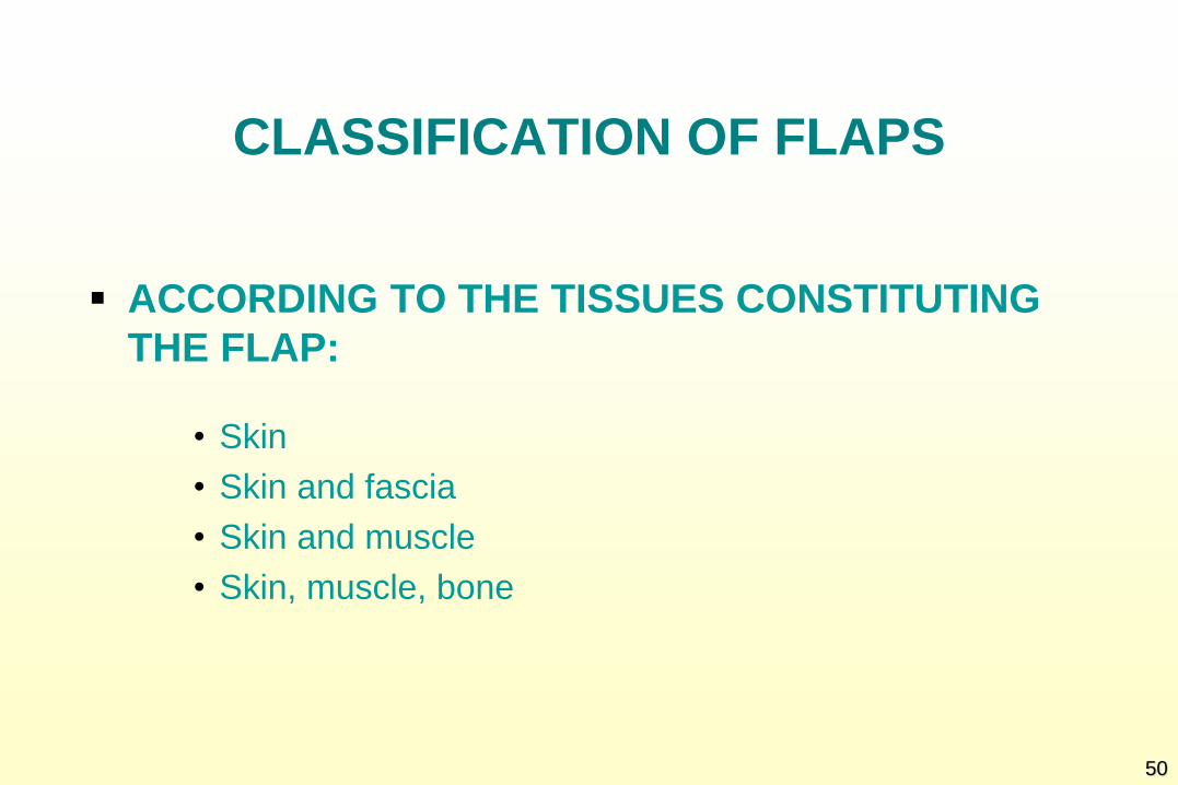
CLASSIFICATION OF FLAPS
ACCORDING TO THE TISSUES CONSTITUTING
THE FLAP:
• Skin
• Skin and fascia
• Skin and muscle
• Skin, muscle, bone
50

ACCORDING TO THE MOBILIZATION METHOD
OF THE FLAP:
– advancement flaps
– rotation flaps
– transposition flaps
– subcutaneously pedicled flaps
– interpolation flaps
– free flaps
– combined reconstruction
51
CLASSIFICATION OF FLAPS

TEMPORAL DEFECT – full thickness skin graft
DEFECT OF THE WING OF THE NOSE - full thickness skin graft

PREAURICULAR DEFECT
advancement flap
53

TRANSPOSITION FLAPS
54

DEFECT OF THE INNER PALPEBRA - transposition flap
55
MIDDLE FACE DEFECT - transposition flap

SUBCUTANEOUSLY PEDICLED FLAP
56

DEFECT OF THE EXTERNAL PALPEBRA - subcutaneously pedicled flap
LOWER LIP DEFECT
subcutaneously pedicled flap
UPPER LIP DEFECT
subcutaneously pedicled flap

INTERPOLATION FLAP
58

DEFECT OF THE WING OF THE NOSE - interpolation flap
59

FREE FLAP RECONSTRUCTION
radial forearm flap
60

FREE FLAP RECONSTRUCTION
anterolateral thigh flap
61

DOUBLE DEFECT OF THE WING OF THE NOSE - expander + interpolation flap

FOREHEAD DEFECT - COMBINED RECONSTRUCTION
63
COMBINED RECONSTRUCTION

64
Introduction of new techniques for reconstructive surgery in Hungary
Pectoralis major (PM) myocutaneous flap modified by the author – first time in Hungary
Localizations: - floor of the mouth, mandible, tongue, root of the tongue
- Tonsillolingual area, lateral pharyngeal defects
- dorsal pharyngeal wall, circular pharyngeal defects
(laryngectomy) Intratracheal narcosis
1. Patient group: 1981-2011: 248 surgeries1981-1985: 50 surgeries
Sketch of a PM flap
modified by us
2. Results: based on 248 operations performed from 1981 to 2011
- To the height of the os zygomaticum:
• a defect of any size (4-6x8-11x1-6 cm)
• suitable for closing a defect of any location
- Excellent viability:
total necrosis: 0%
Partial: 5%
- healing disorder of the donor site: 9%
3. disadvantages:
- Excess tissue volume and excess contour in case of a defect not deeper than
1 cm
- Functional deficiency after the reconstruction of the mandible
Kásler M.: Fül-orr-gégegyógy. 34, 157-163, 1988.
Kásler M., Bánhidy F., Trizna Z.: Arch. Otolaryngol Head Neck Surg. 118. 931-932. 1992.
Kásler M., Bánhidy F.: Eur. J. Surg. Oncol. 19. 587-591. 1993.
Fülöp M., ..., Kásler M.: Magy. Onkol. 52: 261-267, 2008.

• Resection of the tongue
• Resection of mandible, floor of the mouth and tongue
• Resection of the mandible and floor of the mouth
• Resection of mandible, floor of the mouth, tongue, tonsils, base of the tongue
• Resection of base of the tongue and horizontal laryngeal resection
• Laryngectomy, subtotal pharyngectomy
• Laryngo-pharyngectomy
• Resections of larynx cancers
65
Application of PM island flap

66
Introduction of new techniques for reconstructive surgery in Hungary
For closing extensive defects non invading deeper layers
3. Surgical techniques used for reconstruction: • nasolabial transposition flap (NL) 39 patients 100%
• buccal transposition flap (B) 11 patients 100%
• forearm free flap – transposed with microvascular anastomosis (A) 288 patients 87%
• fibular free flap – transposed with microvascular anastomosis 39 patients 82%
•
377 patients
2. Size of the defect:• surface greater that 2x4 cm (min. 1 cm)
• depth less than 2-3 cm (min. 1 cm)
1. Localization of the defect :• oral cavity-mesopharyngeal soft tissue: anterior- mid third (NL, B)
lateral wall, palate (B, NL)
• mandible –oral cavity- mesopharyngeal soft tissue: corpus + anterior oral cavity, angulus+lateral oral cavity and oropharynx (F)
• meso-hypopharynx: dorsal wall, dorsal-lateral wall (PM, A)
• hypopharynx – larynx: circular defect (A)
4. Wound healing
Oberna F., …, Kásler M.: Magy. Onkol. 45. 169-172. 2001.
Fülöp M., …, Kásler M.: Magy. Onkol. 45. 177-180. 2001.
Oberna F., …, Kásler M.: Oral Surg. Oral Med. Oral Pathol. Oral Radiol. Endod. 99. 550-55. 2005.
Fülöp M., ..., Kásler M.: Magy. Onkol. 52: 261-267, 2008.
Kásler M. et al: Magy.Onkol. 52: 279-281, 2008.

67
1. Patient group: 39 patients
Mean age: 58,2 years (40-73)
- Localization: tongue, lateral and anterior part of the floor of the mouth
- Stage: T2N0M0 -T4N0M0, respectively T2N2M0
- Size of tissue defect: 3-5x2-3 cm
2. Results:
- flap necrosis complete: 0%
partial: 4%
- Anatomical-functional-aesthetic restitution
ad integrum
- Avoidance of disadvantages concomitant to
other techniques
A) Superficial tissue loss on the floor of the mouth
B) Donor site
C) Suturing of nasolabial flap mobilized from two sides to the tissue defect
D) Oral cavity after complete wound healing
Oberna F., ..., Kásler M.: Magy. Onkol. 45. 169-172. 2001.
Oberna F., …, Kásler M.: Oral Surg. Oral Med. Oral Pathol.
Oral Radiol. Endod. 99. 550-55. 2005.
Fülöp M., ..., Kásler M.: Magy. Onkol. 52: 261-267, 2008.
Application of nasolabial transposition flap

68
Application of bucca transposition flap
2. Results:
- Flap necrosis 0%
- Localization: oral cavity, lateral-dorsal wall of the oropharynx, soft palate
- Stage: T2-T4N0M0, resp. T2N2M0 (1 recurrence, 1 residuum)
- Size of tissue defect: 4x2-7x3,5x <1 cm
- Postop. irradiation: 50-66 Gy
1. Patient group: 11 patients
A) Bucca flap transposed to the
palate.
B) The transposed buccal flap on
day 21.
C) The transposed buccal flap after 3
months
Oberna F., ..., Kásler M.: Magy. Onkol. 45. 169-172. 2001.
Oberna F., …, Kásler M.: Oral Surg. Oral Med. Oral Pathol.
Oral Radiol. Endod. 99. 550-55. 2005.
Fülöp M., ..., Kásler M.: Magy. Onkol. 52: 261-267, 2008.

69
2. Results:
- Primary wound healing: 87 %
- Flap necrosis (initially venous circulatory disturbance):
complete 13%
partial 2%
Acceptable anatomical and functional reconstruction:100 %
- acceptable: 87%
- restitutio ad integrum: 7%
- Complications:
- Dysphagia 2%
- Aspiration 2%
- Wound healing disorder at donor site: 4,5%
- Secondary wound healing 3%
- Digital motility disorder 1%
Application of forearm free flaps with microvascular anastomosis
for oral cavity defects
1. Patient group: 288 patients, 1993-2011
– average age: 51 years (26-77), male/female: 10:1
– location: any site in the head-neck
– Size of the tissue defect: 4-8x6-10x <1 cm
A) Donor site
B) During preparation
D) A flap taken in the tongue incorporated
C) A defect
Fülöp M., …, Kásler M.: Magy. Onkol. 45. 177-180. 2001.
Fülöp M., ..., Kásler M.: Magy. Onkol. 52: 261-267, 2008.

70
1. Patient group: 1995-2011, 39 patients- Mean age: 50,6 years (38-66) male/female: 32:7
- Localization:
different areas of the mandible and surrounding soft tissue
- Stage: T3(6)-4(33)N0(8)-1(31)M0
A) Donor site B) A flap prepared
Fibula flap preparation
C) Modelling of the bone segment to the mini plate
D) Fixation of dental prosthetis
E) Implanted dental prosthetis
F) Aesthetical result
2. Results:
- Flap necrosis (complete): 18% (venous circulation disorder)
- Complication of donor area 0%
- knee and ankle stability is perfect
- Full recovery: 82%
- Avoiding laryngo-pharyngectomy
- Anatomical restitution ad integrum
- Functional (occlusion, chewing, speech) and aesthetical restitution is satisfactory,dental rehabilitation is possible
Fülöp M., …, Kásler M.: Magy. Onkol. 45. 177-180. 2001.
Fülöp M., ..., Kásler M.: Magy. Onkol. 52: 261-267, 2008
Kásler M. et al: Magy.Onkol. 52: 279-281, 2008.
Application of fibula free flaps with microvascular anastomosis
for mandible and soft tissue defects
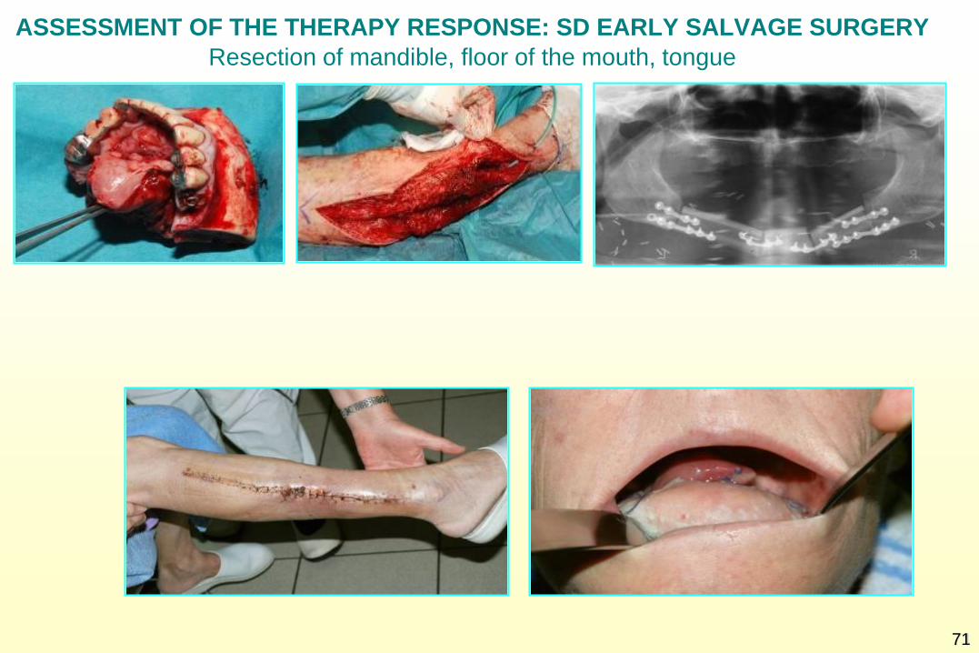
ASSESSMENT OF THE THERAPY RESPONSE: SD EARLY SALVAGE SURGERY
71
Resection of mandible, floor of the mouth, tongue

LATE SALVAGE OPERATION
Reconstruction: m. latissimus dorsi free flap
72

Postmastectomy breast reconstruction

Skin envelope (area, quality)
Structural elements of the
reconstructed breasts
Symmetry
NAC
Volume (autologeous, implant)
Soft tissue coverage of the implant
IMF
Scars
Mátrai Z, incl. Kásler M. et al. Challenges in oncologic plastic surgery of the breast.
Magy Onkol. 2011, 55: 40-52.

A=r2+r√r2+H2
Example: footprint height 12 cm, width
12 cm, projection 8 cm
A=6x3.14x10=188.4 cm2
Skin envelope (area)

Excision of the (SSM) = reduced breast projection
ASM, NSM = preserved breast projection
V=(R2+r2+Rr)1/3H
NAC

Mátrai Z., incl. Kásler M. et al.: The place of skin-sparing mastectomy in oncoplastic breast
surgery. Magy. Onkol. 2011, 55, 252-267.


Mátrai Z., incl. Kásler M. et al.: Role of nipple-sparing mastectomy in modern breast surgery. Orv. Hetil.
2011;152:1233-49.


Symmetry

To reach the best symmetry: create the same
structure
Skin
Parenchyma/
muscle
Implant Implant
Parenchyma/
muscle
Skin

Otherwise the symmetry is suboptimal
Skin Skin
Parenchyma/
muscle
Implant
Parenchyma/
muscle


Bilateral skin-sparing mastectomies and delayed-immediate
reconstructions, delayed nipple reconstrcution

Bilateral skin-sparing mastectomies and delayed-immediate
reconstructions, delayed nipple reconstrcution

Areola-sparing mastectomies and immediate breast
reconstructions with silicone implants and Ultrapro mesh
for lower pole strengthening


Latissimus dorsi myocutaneous flap reconstruction

Combined breast
recontruction: LD
myocutaneous soft
tissue reconstruction
on the right side and
delayed-immediate
reconstr. with
expander and
silicone implant on
the left side by a
BRCA mutation
holder very young
cancer patient.


Mátrai Z., incl. Kásler M. et al. Special aspects of breast cancer surgery in the elderly. Orv Hetil.
2014; 155: 931-8.


Skin-sparing mastectomy and implant preservation in a former augmented breast on
the left side. The soft tissue coverage of the implant is inadequate.

Results after endoscopically assisted LD muscle flap
transopsition through a 6 cm long axillary incision.

There is no scar on the donor side

The results after symmetrisation
Mátrai Z., incl Kásler M. et al. Minimally invasive breast surgery. Orv Hetil. 2014,155: 162-169.
Mátrai Z, incl. Kásler M. et al. Modern breast reconstruction with endoscopically assisted
latissimus dorsi flap harvesting. Orv Hetil. 2014,155:106-113.
Mátrai Z, incl. Kásler M. et al. Special considerations in breast cancer treatment of an
augmented breast. Orv Hetil. 2011,152: 1679-91.

Skin envelope (quality)

Skin-sparing mastectomy on the left side. Sun
damaged poor quality skin, unsuit for reconstruction.


Endoscopically assisted LD muscle flap transposition
through axillary incision

The muscle underlay had succesfully revascularised
the residual skin

Symmetrisation using silicone implants on both
sides and a mastopexy on the right side

End result after nipple reconstruction and areola tatooing


Pedicled TRAM flap reconstruction, after flap delay


Bilateral TRAM flap reconstruction


Autologeous fat transplantation

Mátrai Z., incl. Kásler M et al.:Autologeous fat transplantation in the modern reconstructive surgery of breast
cancer. Orv Hetil. 2012, 153: 1816-1831.

Thank You for Your attention!
Bai X. et al. CD59 mediates
cartilage patterning during sponta
neous tail regeneration. Sci Rep.
2015 Aug 4;5:12798.