probing metastable sm2+ and optically stimulated tunnelling emission in … · accepted manuscript...
TRANSCRIPT

General rights Copyright and moral rights for the publications made accessible in the public portal are retained by the authors and/or other copyright owners and it is a condition of accessing publications that users recognise and abide by the legal requirements associated with these rights.
Users may download and print one copy of any publication from the public portal for the purpose of private study or research.
You may not further distribute the material or use it for any profit-making activity or commercial gain
You may freely distribute the URL identifying the publication in the public portal If you believe that this document breaches copyright please contact us providing details, and we will remove access to the work immediately and investigate your claim.
Downloaded from orbit.dtu.dk on: Jul 04, 2020
Probing metastable Sm2+ and optically stimulated tunnelling emission in YPO4: Ce,Sm
Prasad, Amit Kumar; Kook, Myung Ho; Jain, Mayank
Published in:Radiation Measurements
Link to article, DOI:10.1016/j.radmeas.2016.11.012
Publication date:2017
Document VersionPeer reviewed version
Link back to DTU Orbit
Citation (APA):Prasad, A. K., Kook, M. H., & Jain, M. (2017). Probing metastable Sm
2+ and optically stimulated tunnelling
emission in YPO4: Ce, Sm. Radiation Measurements, 106, 61-66.
https://doi.org/10.1016/j.radmeas.2016.11.012

ACCEPTED MANUSCRIPT
1
Probing Metastable Sm2+ and Optically Stimulated Tunnelling Emission in YPO4: Sm, Ce
A. K. Prasad*, M. Kook, M. Jain
Center for Nuclear Technologies, Technical University of Denmark, DTU Risø Campus, Roskilde, Denmark
(* Corresponding author: [email protected])
Abstract: When the model dosimetry system YPO4: Sm3+, Ce3+ is exposed to X-rays, the charge
state of the dopants changes, becoming Sm2+ and Ce4+ via electron and hole trapping, respectively;
the original charge states can be achieved through electron transfer back from Sm2+ to Ce4+ via
optical stimulation. The work presented here adds further details to the energy levels of the
metastable Sm2+ defect and the electron transfer processes by undertaking measurements of a) Sm2+
excitation spectrum through the internal 5D0 → 7F2 emission at 7 K, b) relaxation lifetime of Sm2+
(5D0 state) and its temperature dependence to provide insights into thermal quenching, and c) the
kinetics of localised recombination from Sm2+ to Ce4+ on nanoseconds to seconds time scales using
sub-band-edge excitation.
Keywords: radio-photoluminescence, low temperature spectroscopy, localised recombination,
excited-state tunnelling, thermal quenching, Sm2+ and Ce3+ relaxation lifetime
1. Introduction
Doping wide band-gap materials with rare earth ions introduces defect levels within the bandgap
which makes them useful in many applications, such as charge storage phosphors for use in
luminescence dosimetry (Chakrabarti et al, 1989, Meijerink et al, 1991), display phosphors and
optical memory (Thiel et al, 2002, Ding et. al., 2016), and persistent luminescent materials
(Matsuzawa et. al., 1996, Hölsa et al., 2001). The extensive knowledge accumulated over the last

ACCEPTED MANUSCRIPT
2
four to five decades in identifying the behaviour and energy levels of lanthanide dopants within the
band gap of a host material gives us a clear idea about whether they will act as an electron or a hole
trap on exposure to ionizing radiation (for summary, see Dorenbos et al., 2003, 2011). In this
respect, YPO4 co-doped with Sm3+ and Ce3+ ions shows excellent promise as a model system for
fundamental studies: on irradiation with X–rays, these defects form the metastable states Sm2+ and
Ce4+ whose energy levels can be precisely characterised for controlled charge transfer (Dorenbos et
al., 2003, Poolton et al, 2012).
Previous work to understand the luminescence charge transfer properties of YPO4:Ce3+, Sm3+ have
used synchrotron radiation (4-20 eV) and both optical and thermoluminescence spectroscopy
(Pieterson et al., 2001; Poolton et al, 2010, 2012; Dorenbos et al., 2011; Mandowski and Bos, 2010;
Bos et al., 2010). These studies have also allowed determination of the electron and hole trap
energies within the band-gap. However, understanding the precise nature of energy transfer from
Sm2+ to Ce4+ processes requires detailed information concerning the excited state energy levels of
the metastable Sm2+ defect, and this is still very much work-in-progress. The first indications of the
defect’s excited state energy levels came from the optically stimulated luminescence (OSL)
excitation spectra measured via the Ce3+ emission (Bos et al, 2010). As this involves excited-state
tunnelling from Sm2+ to Ce4+, the transitions were significantly broadened due to interference with
4f 5d (SmA, SmB) bands (which are the most effective conduit for the tunnelling), and only limited
spectroscopic information was obtainable. By using a low flux, 1.92 eV laser stimulation at 10 K,
Poolton et al (2012) were able to exclusively probe isolated Sm2+ emission 4f-4f transition, and
thereby determine the ground state energy levels in more detail. In the same study a 2.33 eV laser
probe for the emission at 10 K confirmed the simultaneous presence of both stable Sm3+ ions and
metastable Sm2+ ions in the YPO4 host.
Regarding charge transfer, the OSL in YPO4: Sm, Ce has previously been studied using the 2.79 eV
excitation which photo-ionises the Sm2+ followed by recombination via the conduction band
(delocalised charge transfer) at the Ce4+ (Poolton et al., 2010). In contrast, thermal decay (TL) has
shown a tunnelling emission corresponding to a continuum of localised recombination probabilities,
superimposed on the TL peaks produced by delocalised recombination (Dobrowolska et al., 2014).
Detailed understanding of the tunnelling kinetics is tedious using the TL signal because of a
continuum of temperature dependent excitation probabilities and an overlap between localised and

ACCEPTED MANUSCRIPT
3
delocalised processes. This problem can be avoided using an energy specific, sub-bandgap
excitation leading to a direct charge transfer from Sm2+ to Ce4+.
The missing details of the YPO4:Ce3+, Sm3+ model system are: knowledge about the exact energy
levels of the excited state of metastable Sm2+, and the relaxation rates from the excited-to-ground
states of the ion. Furthermore, the charge transfers kinetics using Sm2+ excited state tunnelling is
not understood at the moment; there is a general need for such information on model systems, as it
is important for testing and refining our theoretical understanding of stimulated localised
recombination emission (e.g. see Jain et al. 2012, 2015).
In this article, we move towards a more complete picture of the YPO4: Ce, Sm model system by
measuring for the first time the detailed excitation spectra of metastable Sm2+ at 7 K via the Sm2+
emission itself; this allows us to determine the precise location of the ion’s excited states. We also
measure the temperature dependent relaxation lifetime and dose-dependency of this internal Sm2+
emission (Stokes shifted OSL or radio-photoluminescence), without relying on any charge transfer
to Ce4+. Finally, we study the optically stimulated tunnelling recombination from the excited states
of Sm2+ to the Ce4+. The method of isolating the luminescence processes of individual metastable
defects (rather than relying on the charge transfer between defects) is an important outcome of this
work which can be applied to the other lanthanide co-dopants, as well as to complex natural
materials, such as feldspars.
2. Instrumentation and sample details
The yttrium phosphate sample used in this study was co-doped with 0.5% cerium and 0.5%
samarium, and prepared by solid state reaction (for details, see Bos et al, 2008). All measurements
were made using the Risø station for CryOgenic LUminescence Research (COLUR) at DTU
Nutech. This system consists of a Horiba Fluorolog-3 spectrofluorometer, upgraded to include a
temperature controlled (7-300 K) closed-loop cryostat, an X-ray irradiation facility (40 kV, 100 µA
copper anode with 3 ms action X-ray shutter), and multiple ports for laser excitation and photo-
detection, for use in a number of dual-probe type experiments. The sample is in vacuum, attached
directly to the cryostat cold finger.

ACCEPTED MANUSCRIPT
4
Luminescence excitation spectra were measured with a 450 W Xenon CW lamp and detected with a
photomultiplier tube (PMT; S1 response). A series of appropriate long-pass filters were placed
between the sample and the emission monochromator to remove second order excitation light.
Time resolved luminescence measurements were measured using time-correlated single photon
counting (TCSPC), with a 657 nm (~ 1.89 eV) laser diode as the source. The laser has a maximum
power of 40 mW, and works in either CW mode, or can be modulated up to 155 MHz with rise/fall
times of < 3 ns, with a minimum pulse width ~ 50 ns. Dose-dependent OSL curves were obtained
with the laser in CW mode and directly recorded using a PMT (with U340 filters), external to the
Fluorolog monochromators; this exclusively samples the Ce3+ emission.
3. Results and Discussion
3.1 Low temperature luminescence properties of Sm2+
X-irradiation of YPO4: Ce, Sm (9.2 eV bandgap) leads to the formation of free electrons and holes.
The original charge states of the dopants (Sm3+, Ce3+) change to metastable state (Sm2+, Ce4+). On
capturing the electrons and holes they return to their original (Sm3+, Ce3+) configuration after
several days via non-radiative ground-state tunnelling, or via external stimulation by heat or light
resulting in TL and OSL, respectively. Fig. 1(a) shows the laser excited Sm2+ emission spectra at 7
K for different X-ray irradiation durations. The emission arises exclusively from the internal 4f (5D0
→ 7F2,3,4) transitions at 1.70, 1.64 and 1.54 eV, respectively, all of which increase in intensity with
dose. The dose-dependent change in the PL intensity confirms that we are directly probing the
metastable Sm2+; such signal in dosimetry is commonly named as radio-photoluminescence
(Schulman et.al., 1951). Only some Sm3+ are converted to Sm2+ and the inset to Fig. 1(a) shows the 4G5/2 → 6H5/2 ,7/2, 9/2, 11/2 transitions of Sm3+, excited in a non-dosed sample under 3.06 eV laser
excitation; the separate spectral emission features of Sm2+ and Sm3+ are clearly distinguished. These
results are in good agreement with those of Poolton et al. (2012) confirming the reliability of our
measurements.
The excitation spectrum of Sm2+ was recorded at 7 K after 3 hours of X-ray irradiation with the
detection fixed at 1.70 eV (the 5D0 → 7F2 transition) and the excitation energy varying from 1.75 to
2.05 eV (Fig.1b). As Sm2+ has a trap depth of 2.3 eV (Bos et al., 2010; Poolton et al., 2010)
excitation in the energy range 1.75 - 2.1 eV has insufficient energy to evict charge from the centre

ACCEPTED MANUSCRIPT
5
via the conduction band. The observed peaks, therefore, represent the internal transitions within
Sm2+, an interpretation further strengthened by the fact that the signal does not decay with time
(steady state). The clearly resolved peaks in the present excitation spectrum contrast with those in
previous work where the transitions were observed via Ce3+ emission by excited state tunnelling
(Dorenbos et al., 2011, Bos et al, 2010, Poolton et al, 2010, 2012); this charge transfer process in
the earlier work led to extensive broadening of the transitions, making most of the features almost
indistinguishable. The resolution of the spectral features obtained here allow for the first time a
clear identification of the excited levels of Sm2+ in YPO4 host. These excitation peaks arise from
internal 4f-4f transitions of Sm2+ and, in combination with the previous work regarding analysing
the emission lines (Poolton et al, 2012), we tentatively assign 7F0 → 5D0,1,2,3 at 1.837, 1.879, 1.898
and 1.977 eV respectively, and 7F1 → 5D0 at 1.785 eV. Low intensity phonon replicas (~20 meV)
associated with each electronic transition are also observed.
The time resolved luminescence of the main Sm2+ peak emission excited using the 1.89 eV laser at
7 K, is shown in Fig.1(c); the inset shows the relevant excitation and emission energy levels. The
data, when fitted with a single exponential function, yield lifetimes of 751 ± 16, 729 ± 11 and 704 ±
10 ns for the emission peaks at 1.54, 1.64 and 1.70 eV respectively (corresponding to 5D0 → 7F4,3,2).
These lifetime estimates are consistent within 2σ uncertainty.
3.2 Temperature dependent luminescence properties of Sm2+
The temperature dependence of the Sm2+ PL emission excited by the 1.89 eV laser is shown in Fig.
2(a). This shows that the peaks at 1.70 eV and 1.64 eV (5D0 → 7F2,3) are broadened as the
temperature is increased in the range of 100-300 K, and the peaks become rapidly quenched at
temperatures higher than 130 K. This thermal quenching behaviour of Sm2+ has been reported
before by Poolton et al. 2012; our data mimics the trend reported by these authors.
Thermal enhancement and thermal quenching of a signal can occur due to interplay of many
different mechanisms (see for e.g. Pal et al., 2013). In comparison to PL intensity, the lifetime is a
more robust parameter to understand thermal quenching due to a non-radiative pathway. Therefore,
in order to accurately define the kinetics of thermal quenching in our signal we examined the
lifetime dependence of Sm2+ on temperature.
Fig. 2(b) shows the luminescence decay of the Sm2+ emission at 1.7 eV (5D0 → 7F2) at different
temperatures, analysed in each case with single exponential function (after background subtraction).

ACCEPTED MANUSCRIPT
6
The average lifetime of the Sm2+ emission at 1.7 eV (5D0 → 7F2) in the temperature range 10-140 K
yields 706 ± 5 ns, whereas at higher temperature the lifetime decreases very rapidly, being ~ 98 ± 1
ns at 220 K. Fig. 2(c) shows the temperature dependence of the integrated PL intensity for emission
peaks between 1.69-1.72 eV (denoted as “1.7 eV”) and the temperature dependence of the lifetime
for the 1.7 eV emission peak. We note that the 1.625-1.65 eV (denoted as “1.64 eV”) emission
showed the same trend as 1.7 eV peak (not shown here for clarity).
The lifetime data show an excellent reproducibility (Fig. 2(c)). The lifetime and the PL data show a
broadly similar trend with temperature, suggesting that Mott-Seitz mechanism can explain the
overall thermal quenching of the Sm2+ photoluminescence in the YPO4: Ce, Sm system; in this
mechanism, the increased probability of non-radiative relaxation pathway at higher temperature
leads to a simultaneous reduction in signal intensity and a lower lifetime (e.g., Ueda et. al., 2015,
Pal et al. 2013, Calderon et al, 1990, Pagonis et al., 2010). We note that tunnelling loss through the
SmA and SmB bands becomes detectable above 200 K (Fig. 5 of Poolton et al., 2012), and is,
therefore, not likely to be responsible for the decreasing trend observed in our data up to at least 200
K. A slight deviation in the PL and lifetime data may originate due to thermal partitioning of
electrons in the ground states (7F multiplet) of the Sm2+, which will vary with temperature and
likely affect the PL intensity; but this effect will not alter the excited state lifetime.
Based on the Mott-Seitz mechanism, the temperature dependence of lifetime can be described by
the following function (Pal et al. 2013; Calderon et al, 1990):
����� =
���+ �
���� ����� � (1)
Where ���� is the measured temperature dependent lifetime, 1/�� is probability of radiative
transition, where �� corresponds to the measured lifetime before the onset of thermal quenching,
1/��� is the frequency factor for the non-radiative transition probability, �� is the threshold energy
for non-radiative transition and � is Boltzmann’s constant. The fitting of equation (1) to the lifetime
data (Fig. 2(c)) yielded a quenching energy of 0.20 ± 0.005 eV, �� = 706 ± 5 ns, and 1/��� = 5.4 ±
1.8 x 10¹¹ s-1.
Similarly, the reduction in the PL intensity with temperature can be analysed using the thermal
quenching equation, which can be derived from Eq (1) (Chitambo et al., 2007).
ACCEPTED MANUSCRIPT
7
���� = ������ !"#����/��� (2)
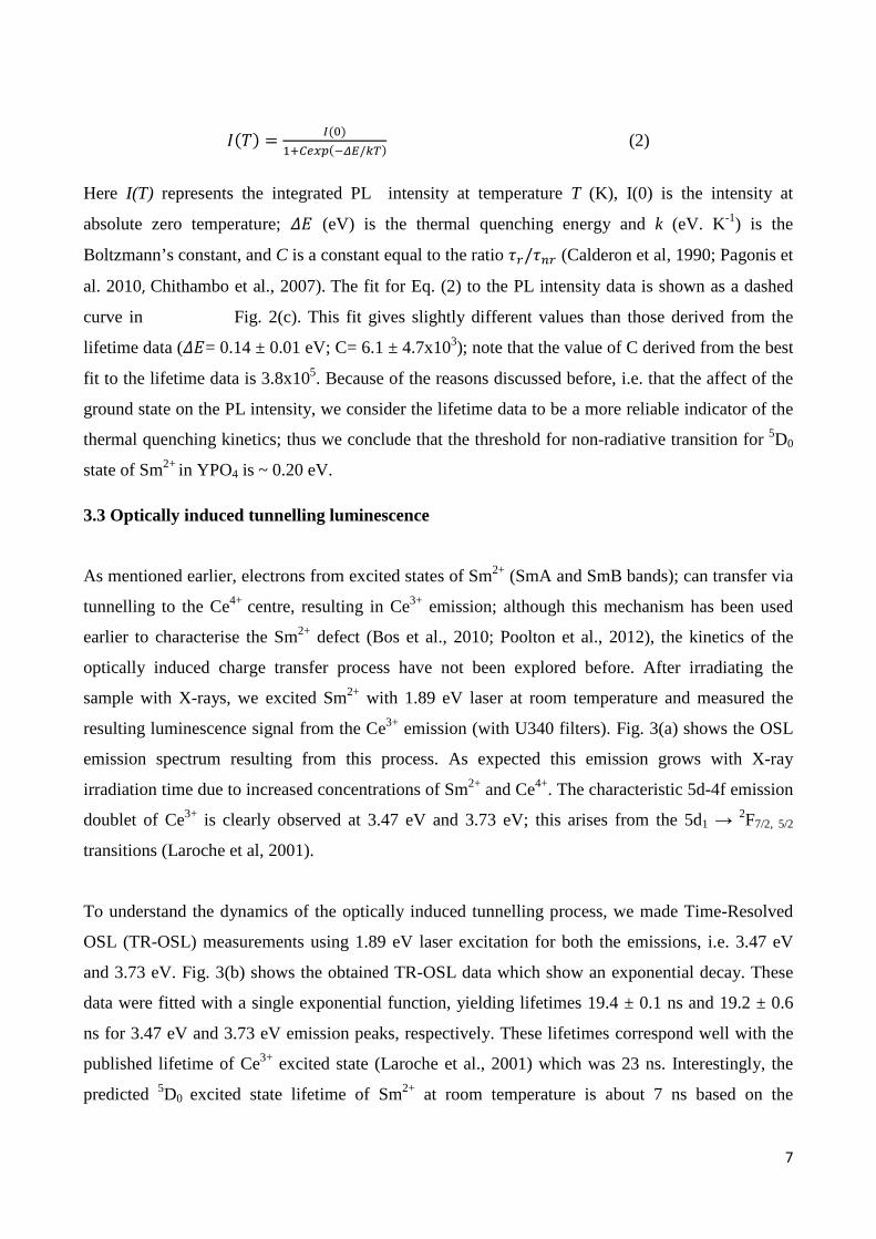
Here I(T) represents the integrated PL intensity at temperature T (K), I(0) is the intensity at
absolute zero temperature; �� (eV) is the thermal quenching energy and k (eV. K-1) is the
Boltzmann’s constant, and C is a constant equal to the ratio ��/��� (Calderon et al, 1990; Pagonis et
al. 2010, Chithambo et al., 2007). The fit for Eq. (2) to the PL intensity data is shown as a dashed
curve in Fig. 2(c). This fit gives slightly different values than those derived from the
lifetime data (��= 0.14 ± 0.01 eV; C= 6.1 ± 4.7x10³); note that the value of C derived from the best
fit to the lifetime data is 3.8x10⁵. Because of the reasons discussed before, i.e. that the affect of the
ground state on the PL intensity, we consider the lifetime data to be a more reliable indicator of the
thermal quenching kinetics; thus we conclude that the threshold for non-radiative transition for 5D0
state of Sm2+ in YPO4 is ~ 0.20 eV.
3.3 Optically induced tunnelling luminescence
As mentioned earlier, electrons from excited states of Sm2+ (SmA and SmB bands); can transfer via
tunnelling to the Ce4+ centre, resulting in Ce3+ emission; although this mechanism has been used
earlier to characterise the Sm2+ defect (Bos et al., 2010; Poolton et al., 2012), the kinetics of the
optically induced charge transfer process have not been explored before. After irradiating the
sample with X-rays, we excited Sm2+ with 1.89 eV laser at room temperature and measured the
resulting luminescence signal from the Ce3+ emission (with U340 filters). Fig. 3(a) shows the OSL
emission spectrum resulting from this process. As expected this emission grows with X-ray
irradiation time due to increased concentrations of Sm2+ and Ce4+. The characteristic 5d-4f emission
doublet of Ce3+ is clearly observed at 3.47 eV and 3.73 eV; this arises from the 5d1 → 2F7/2, 5/2
transitions (Laroche et al, 2001).
To understand the dynamics of the optically induced tunnelling process, we made Time-Resolved
OSL (TR-OSL) measurements using 1.89 eV laser excitation for both the emissions, i.e. 3.47 eV
and 3.73 eV. Fig. 3(b) shows the obtained TR-OSL data which show an exponential decay. These
data were fitted with a single exponential function, yielding lifetimes 19.4 ± 0.1 ns and 19.2 ± 0.6
ns for 3.47 eV and 3.73 eV emission peaks, respectively. These lifetimes correspond well with the
published lifetime of Ce3+ excited state (Laroche et al., 2001) which was 23 ns. Interestingly, the
predicted 5D0 excited state lifetime of Sm2+ at room temperature is about 7 ns based on the

ACCEPTED MANUSCRIPT
8
parameters derived from Equation (1), implying that the 5D0 could potentially be feeding to the
tunnelling active states (SmA and SmB bands) and thereby Ce4+, while the Ce3+ excited state
relaxation is ongoing; this process could give rise to an initial non-exponentially (peak shape) in the
TR-OSL. However, based on our TR-OSL data, it can be inferred that both the 5D0 and the
tunnelling active states in the Sm2+ must empty very rapidly (at least an order of magnitude faster
than the Ce3+ relaxation rate) at room temperature so as to give clean single exponential decay
representative of Ce3+. Thus at room temperature there must be much faster emptying of the 5D0
state (possibly to the tunnelling bands) than that predicted by thermal quenching model, and an
insignificant retrapping from the tunnelling active states to the 5D0 state.
Since the excitation with 1.89 eV is sub-conduction band, the only mechanism of charge transfer is
tunnelling from Sm2+ excited state to Ce4+. Understanding the excited state tunnelling process is of
interest in order to understand the behaviour of both artificial and natural dosimeters (e.g. see Jain et
al., 2012). However, this understanding is usually hampered by complexity of the materials of
interest. For example, decades of research has been devoted to tackling the anomalous fading
problem in feldspars, which arises due to tunnelling loss from the dosimetric trap (see Jain and
Ankjærgaard, 2011 for a comprehensive discussion). Our model YPO4: Ce, Sm gives us an
opportunity to isolate the excited state tunnelling process and use it to test our theoretical
understanding of tunnelling in randomly distributed defects. With this objective we measured the
CW-OSL using the 1.89 eV laser excitation after different X-ray irradiation durations. Fig. 4(a)
shows the CW-OSL decay curves recorded using a separate PMT with U340 filters; the
corresponding emission spectra are plotted in Fig. 3(a).
The truncated-distribution tunnelling OSL model of Jain et al (2015) predicts that the shape of the
OSL decay curve should depend both on the hole density (e.g. due to increasing dose), and on the
truncation of the nearest neighbour distribution (e.g. due to prior heating or ground state tunnelling).
We apply their model equation to our data in Fig. 4 (a) obtained after different X-ray irradiation
times:
$%& ∝ − *�*+ = 3-�.′z�0′���1ln�0′4� − 567 exp;−.′1ln�0′4� − 56<= (3)

ACCEPTED MANUSCRIPT
9
Here n is the instantaneous, n0 the initial trapped electron concentration,.> is the number density of
the trapped holes, b [s-1] is the attempt to tunnel frequency, 0> is a variable, which is a linear
function of time (�� + zt), 5 = ln�?/��, where s is frequency factor and p is optical excitation
probability per unit time. The equation (3) fits well to all the OSL curves in Fig. 4(a) confirming
that the excited state tunneling model describes well the observed OSL signal produced by localized
recombination. Two fitting parameters that deserve closer scrutiny here are the dimensionless hole
density .>, which should increase with dose, and the truncation parameter �� which should increase
with irradiation time because of ground state tunneling. Both these parameters are plotted in Fig.
4(b); their trend with X-ray irradiation time (or dose) confirms the model predictions (Equation 3).
4. Conclusion:
Using low temperatures and sub-conduction-band excitation, new details of the metastable Sm2+
defect in co-doped, irradiated YPO4: Ce, Sm have been measured, including details of the excited
states, measurement of the internal 4f-4f time decay constants, and dynamics of the optically
induced excited-state tunnelling pathways for electron recombination at Ce4+. The methods are ideal
for exploring metastable states in other lanthanide co-doped wide band-gap materials, and for
understanding charge recombination processes in complex natural dosimeters such as feldspars.
5. Acknowledgment
Prof. A. J. J. Bos is thanked for providing YPO4: Ce, Sm sample and Dr. N. R. J. Poolton is thanked
for participation during the various stages of this study. Dr. Torben Lapp is thanked for his support
in the commissioning of low temperature facility.

ACCEPTED MANUSCRIPT
10
References Bos, A. J. J., Dorenbos, P., Bessière, A, Viana, B., (2008) Lanthanide energy levels in YPO4. Radiation Measurements 43, 222 – 226 Bos, A. J. J., Poolton, N. R. J., Wallinga, J., Bessière, A., Dorenbos, P. (2010). Energy levels in YPO4:Ce3+, Sm3+ studied by thermally and optically stimulated luminescence. Radiat. Meas. 45, 343-346. Calderon, T, Millan, Jaquet, F., Sole, G., J. (1990) Optical properties of Sm2+ and Eu2+ in natural fluorite crystals. Nucl. Tracks Radiat. Meas.,17, 557-561 Chakrabarti, K., Mathur, V.K., Thomas, L.A., Abbundi, R.J., (1989). Charge trapping and mechanism of stimulated luminescence in CaS: Ce; Eu. J. Appl. Phys. 65, 2021–2023. Chithambo, M.L., 2007. The analysis of time-resolved optically stimulated luminescence. II: Computer Simulations and Experimental Results. J. Phys. D.: Appl. Phys. 40, 1880-1889. Dorenbos, P., (2003) Systematic behaviour in trivalent lanthanide charge transfer energies J. Phys.: Condens. Matter 15, 8417–843 Dorenbos, P., Bos, A. J. J., Poolton, N. R. J. (2011) Electron transfer processes in double lanthanide activated YPO4, Optical Materials 33, 1019–1023 Ding, D., Pereira, L. M. C., Bauters, J. F., Heck, M.J. R., Welker, G., Vantomme, A., Bowers, J. E., Dood, M.J. A., Bouwmeester, D.(2016) Multidimensional Purcell effect in an ytterbium-doped ring resonator. Nature Photonics 10, 385–388 Dobrowolska, A., Bos, A. J. J., Dorenbos, P. (2014) Electron tunnelling phenomena in YPO4: Ce, Ln (Ln= Er, Ho, Nd, Dy). Journal of Physics D: Applied Physics 47, 33
Hölsa, J., Jungner, H., Lastusaari, M., Niittykoski, J., (2001). Persistent luminescence of Eu2+ doped alkaline earth aluminates, MAl2O4: Eu2+. J. Alloys Compd. 323, 326–330. Jain, M., Sohbati, R., Guralnik, B., Murray, A., Kook, M., Lapp, T., Prasad, A.K., Thomsen, K.J., Buylaert, P., (2015). Kinetics of infrared stimulated luminescence from feldspars. Radiat. Meas. 81, 16-22. Jain, M., and Ankjærgaard, C., (2011). Towards a non-fading signal in feldspar: Insight into charge transport and tunnelling from time-resolved optically stimulated luminescence. Radiation Measurements, 46, 292–309.
Jain, M., Guralnik, B., Andersen, M. T. (2012) Stimulated luminescence emission from localized recombination in randomly distributed defects. Journal of physics: Condensed matter 24, 385402.

ACCEPTED MANUSCRIPT
11
Laroche, M., Girard, S., Margerie, J. Moncorge, J., Bettinelli, M., Cavalli,E.,(2001) Experimental and theoretical investigation of the 4fn ↔4fn-15d transitions in YPO4 :Pr3+ and YPO4 :Pr3+
, Ce3+. J. Phys.: Condens. Matter, 13, 765–776 Meijerink, A., Schipper, W.J., Blasse, G., (1991). Photostimulated luminescence and thermally stimulated luminescence of Y2SiO5 : Ce3+, Sm 3+. J. Phys. D: Appl. Phys. 24, 997–1002 Mahbubulalam, A. S. M, and Bartolo, B. Di, (1967). Deexcitation and lifetime of 5D0 state in optically pumped SrF2:Sm2+. Physics Letters, 25A. Mandowski, A., Bos, A. J. J. (2011) Explanation of anomalous heating rate dependence of thermoluminescence in YPO4:Ce3+, Sm3+ based on the semi-localized transition (SLT) model Radiation Measurements 46 1376-1379 Matsuzawa, T., Aoki, Y., Takeuchi, N., Murayama, Y., (1996). New long phosphorescence phosphor with high brightness, SrAl2O4: Eu2+, Dy 3+. J. Electrochem. Soc. 143, 2670–2673. Pal, P., Penhouët, T., D’Anna ,V., Hagemann, H.(2013) Effect of temperature and pressure on emission lifetime of Sm2+ ion doped in MFX (M=Sr, Ba; X=Br, I) crystals. J. Lumin., 142, 66–74
Pieterson, L. V., Reid, M. F., Burdick, G. W., and Meijerink , A.(2001). 4fn ↔4fn-15d transitions of the light lanthanides: Experiment and theory. Phys. Rev. B 65, 045113
Poolton, N. R. J., Bos, A. J. J., Dorenbos, P. (2012). Luminescence emission from metastable Sm2+ defects in YPO4: Ce, Sm.J. Phys.: Condens. Matter 24,225502 Poolton, N. R. J., Bos, A. J. J., Jones, G.O., Dorenbos, P (2010). Probing electron transfer processes in YPO4: Ce, Sm by combined synchrotron–laser excitation spectroscopy. J. Phys.: Condens. Matter 22, 185403 Pagonis, V., Ankjærgaard, C., Murray, A., Jain, M., Chen, R., Lawless, J., Greilich, S. (2010). Modelling the thermal quenching mechanism in quartz based on time-resolved optically stimulated luminescence. Journal of Luminescence, 130(5), 902-909 Schulman, J.H., Ginther, R.J., Klick, C.C., Alger, R.S., Levy, R.A. (1951). Dosimetry of X-rays and gamma-rays by radiophotoluminescence. J. Appl. Phys. 22, 1479-1487. Thiel, C.W., Sun, Y., Cone, R.L., (2002). Progress in relating rare-earth ion 4f and 5d energy levels to host bands in optical materials for hole burning, quantum information and phosphors. J. Mod. Opt. 49, 2399–2411.
Ueda, J., Dorenbos, P., Bos, A. J. J., Meijerink, A., Tanabe, S. (2015) Insight into the Thermal Quenching Mechanism for Y3Al 5O12:Ce3+ through Thermoluminescence Excitation Spectroscopy. J. Phys. Chem. C, 119, 25003−25008

ACCEPTED MANUSCRIPT
12
Figure Captions Figure 1 (a) Emission features of metastable Sm2+ in YPO4: Ce, Sm, under 1.89 eV stimulation after 30, 45
and 60 minutes of X-ray irradiation (black, blue and red curves, respectively). The inset shows
emission corresponding to Sm3+ in un-irradiated material, using 3.06 eV laser.
(b) Excitation spectrum of Sm2+ in YPO4: Ce, Sm after 3 hours of X-ray irradiation, recorded for
emission fixed at 1.7 eV. (c) Time-resolved measurement of Sm2+ emissions under 1.89 eV laser
stimulation, fitted with a single exponential function. The inset shows the relevant energy levels.
All measurements are made at 7 K.
Figure 2
Temperature dependence of the Sm2+ emission under 1.89 eV laser excitation, after 3 hours of X-
ray irradiation: (a) emission spectra, (b) Time-resolved measurement of 5D0 → 7F2, transitions fitted
with an exponential function, and (c) lifetime and integral PL intensity as a function of
measurement temperature; the data are fitted with equation (1) and (2), respectively (see text).
Figure 3
Characteristics of optically stimulated luminescence emission arising from excited state tunnelling
from metastable Sm2+ to Ce4+ states. The signals are detected from Ce3+ emission produced by
electron recombination at the Ce4+center. The data are recorded at room temperature under 1.89 eV
laser excitation. (a) OSL emission spectra produced after different X-ray irradiation durations (or
dose), and (b) Time-resolved OSL emissions (3.47 and 3.73 eV) fitted with a single exponential
function.
Figure 4
(a) OSL decay curves produced after different X-ray irradiation durations (or dose); these data are
fitted with the truncated distribution tunnelling model (Equation 3), (b) Dimensionless hole density
( ρ´) and the truncation parameter τ0 derived from fitting of OSL decay curves in Fig. 4(a), plotted
as a function of X-ray irradiation duration (see text).

ACCEPTED MANUSCRIPT
1.75 1.80 1.85 1.90 1.95 2.00 2.050
5
10
15
20
25
30
35 Sm2+
7 F0 →
5 D3
7 F0 →
5 D2
7 F0 →
5 D1
7 F0 →
5 D0
7 F1 →
5 D0
I (au
)
Excitation energy (eV)
1.55 1.60 1.65 1.70
0
5
10
15
5 D0→
7 F4
5 D0→
7 F3
Sm2+
1.5 1.6 1.7 1.8 1.9 2.0 2.1 2.2 2.3
Emission energy(eV)
Emission energy (eV)
Sm3+
5 D0→
7 F2
PL
(au)
0 400 800 1200 1600
0.1
1 Sm2+ [email protected] [email protected] [email protected]
Nor
mal
ised
PL
Time (ns)
(c)
(b)
(a)
Figure 1
5D
4F

ACCEPTED MANUSCRIPT
1.60 1.62 1.64 1.66 1.68 1.70 1.720
5
10
15
20
25 Sm2+
5 D0→
7 F3
5 D0→
7 F2
PL
(au)
Emission energy (eV)
7K 30K 60K 90K 120K 150K 180K 210K 300K
(a)
(b)
(c)
Figure 2
0 400 800 1200 1600 2000 2400
0.01
0.1
1 Sm2+
10K 150K 160K 170K 180K 190K 200K 210K 220K
Nor
mal
ised
PL
Time (ns)
0 30 60 90 120 150 180 210 240 2700
100
200
300
400
500
600
700 Sm
2+, [email protected] eV
Lifetime
Life
time
(ns)
Temperature (K)
0.0
0.2
0.4
0.6
0.8
1.0
PL
Nor
mal
ised
PL

ACCEPTED MANUSCRIPT
3.3 3.4 3.5 3.6 3.7 3.8 3.90
1
2
3
4Ce3+
5d1→
2 F7/
2
5d1→
2 F5/
2
OSL after X-ray irradiation (minutes)
2 5 10 20 40 80
O
SL
(au)
Emission energy (eV)(b)
(a)
Figure 3
0 10 20 30 400.1
1Ce3+ [email protected] eV
Nor
mal
ised
PL
Time (ns)

ACCEPTED MANUSCRIPT
Figure 4
(a)
(b)
0 20 40 60 80
1
2
3
4
5
Dim
ensi
onle
ss h
ole
dens
ity, p
'
X-ray irradiation time (minutes)
0
10
20
30
40
50
60τ 0(
s)
0.1 1 10 100 10000
1
2
3
4
Ce3+
OSL after X-ray irradiation (minutes)
2 5 10 20 40 80
OS
L (a
u)
Time (s)

ACCEPTED MANUSCRIPT
Highlights: Probing Metastable Sm2+ and Optically Stimulated Tunnelling Emission in YPO4: Sm, Ce
• The detailed excitation spectrum of metastable Sm2+ at 7 K through its internal emission.
• Thermal quenching and temperature dependent relaxation lifetime of Sm2+. • Kinetics of optically stimulated tunnelling recombination.