probing the biogeochemical introduction behavior of technetium using a novel nuclear...
TRANSCRIPT

Probing the BiogeochemicalBehavior of Technetium Using aNovel Nuclear Imaging ApproachG A V I N L E A R , † , # J O Y C E M . M C B E T H , † , ∇
C H R I S T O P H E R B O O T H M A N , †
D A R R E N J . G U N N I N G , † B E V E R L Y L . E L L I S , ‡
R I C H A R D S . L A W S O N , ‡
K A T H E R I N E M O R R I S , § I A N T . B U R K E , §
N I C H O L A S D . B R Y A N , |
A N D R E W P . B R O W N , ⊥ F R A N C I S R . L I V E N S , |
A N D J O N A T H A N R . L L O Y D * , †
Williamson Centre for Molecular Environmental Science,School of Earth, Atmospheric and Environmental Sciences,The University of Manchester, M13 9PL, U.K., Department ofNuclear Medicine, Manchester Royal Infirmary, Oxford Road,M13 9WL, U.K., School of Earth and Environment, Universityof Leeds, LS2 9JT, U.K., Centre for Radiochemistry Research,School of Chemistry, The University of Manchester,M13 9PL, U.K., and Institute for Materials Research,University of Leeds, LS2 9JT, U.K.
Received October 20, 2008. Revised manuscript receivedMarch 13, 2009. Accepted April 1, 2009.
Dynamic γ-camera imaging of radiotracer technetium (99mTc)was used to assess the impact of biostimulation of metal-reducingbacteria on technetium mobility at 10-12 mol L-1 concentrationsin sediments. Addition of the electron donor acetate wasused to stimulate a redox profile in sediment columns, fromoxic to Fe(III)-reducing conditions. When 99mTc was pumpedthrough the columns, real-time γ-camera imaging combined withgeochemical analyses showed technetium was localized inregionscontainingbiogenicFe(II). Inparallelexperiments,electronmicroscopy with energy-dispersive X-ray (EDX) mappingconfirmed sediment-bound Tc was associated with iron, whileX-ray absorption spectroscopy (XAS) confirmed reduction ofTc(VII) to poorly soluble Tc(IV). Molecular analyses of microbialcommunities in these experiments supported a direct linkbetween biogenic Fe(II) accumulation and Tc(VII) reductiveprecipitation, with Fe(III)-reducing bacteria more abundant intechnetium immobilization zones. This offers a novel approachto assessing radionuclide mobility at ultratrace concentrationsin real-time biogeochemical experiments, and confirms theeffectiveness of biostimulation of Fe(III)-reducing bacteria inimmobilizing technetium.
IntroductionTechnetium-99 (99Tc) is a �-emitting product of nuclearfission and a significant contaminant in effluents fromnuclear facilities and sediments affected by legacy wastesfrom nuclear fuel cycle operations (1-5). Technetium has along half-life of 2.1 × 10⁵ years (99Tc) and high environmentalmobility as the pertechnetate anion (Tc(VII); TcO4
-) whichdominates in oxic conditions (4). These factors make thepresence of Tc in the environment a matter of ongoingconcern. There has been considerable interest in the bio-geochemical behavior of technetium in contaminated sedi-ments, as anaerobic bacteria can potentially reduce solubleTc(VII) to the low-valence, poorly soluble form Tc(IV)(3, 6-10). Tc(IV) is expected to form hydrous TcO2-like phasesor, at very low concentrations, to sorb as Tc(IV) to mineralphases, effectively immobilizing the radionuclide. Reductioncan be achieved enzymatically (11-13) or through indirectmechanisms mediated by Fe(II) and sulfide formed byanaerobic bacteria respiring Fe(III) and sulfate, respectively(3, 6, 8, 11, 14, 15). Stimulation of anaerobic microbialprocesses through the controlled addition of electron donorinto the subsurface offers considerable potential for thesustained remediation of Tc(VII) in situ (16). However, despitethe obvious interest in harnessing the potential for metal-reducing bacteria to reduce Tc(VII) in sediments throughbiostimulation, many of the studies in the literature havebeen carried out using 99Tc with concentrations typically inthe 10-6 molar range. Indeed, there is little information onthe effectiveness of bioreduction at the very low concentra-tions of technetium typically observed at contaminated sites(<1 Bq L-1 or 10-11 to 10-12 mol L-1). Here, a noninvasive,sensitive, real-time technique utilizing the isotope 99mTc hasbeen used to probe the effectiveness of technetium im-mobilization at ca. 10-12 molar concentrations in sedimentsprimed for the reduction of Tc(VII). This will explore thepotential for in situ bioremediation strategies at in situ Tcconcentrations.
The imaging of 10-12 mol L-1 Tc is common in medicalimaging applications (4, 17) using 99mTc, an isomer state of99Tc with a half-life of only 6 h. This short-lived isotope hasalso been used as a conservative tracer in hydrological andcivil engineering experiments (18-22) but has not yet beenapplied to bioremediation studies. In sediment environments,indigenous anaerobic microbial communities and associatedchanges in mineralogy and geochemistry potentially have aprofound impact on technetium mobility (3, 6-8, 15). Theaim of this study was therefore to use γ-camera imaging of99mTc migration through biostimulated heterogeneous sedi-ment columns, combined with geochemical, mineralogical,and microbial characterization of the sediments to identifythe links between geomicrobiological activity and the mobilityof the radionuclide over a range of environmentally relevantconcentrations from 10-4 to 10-12 mol L-1.
Experimental SectionSafety. 99Tc is a radioactive beta-emitter (half-life 2.13 × 10⁵
years; Emax ) 294 keV) and should be handled in a properlyequipped radiochemistry laboratory. The possession and useof radioactive materials is subject to statutory controls.
Microcosm and Sediment Column Construction. Sedi-ments were obtained from the U.S. Department of Energy(DoE) Environmental Remediation Sciences Division (ERSD,formerly NABIR) Field Research Center (FRC) located in OakRidge, Tennessee. This extensively studied site is contami-nated with a range of organic and inorganic pollutants
* Corresponding author phone: +44 161 275 7155; fax: +44 161306 9361; e-mail: [email protected].
† Williamson Centre for Molecular Environmental Science, Schoolof Earth, Atmospheric and Environmental Sciences, The Universityof Manchester.
‡ Department of Nuclear Medicine, Manchester Royal Infirmary.§ School of Earth and Environment, University of Leeds.| Centre for Radiochemistry Research, School of Chemistry, The
University of Manchester.⊥ Institute for Materials Research, University of Leeds.# Present address: School of Biological Sciences, University of
Auckland, Auckland 1001, New Zealand.∇ Present address: Bigelow Laboratory for Ocean Sciences, West
Boothbay Harbor, Maine, 04575.
Environ. Sci. Technol. XXXX, xxx, 000–000
10.1021/es802885r CCC: $40.75 XXXX American Chemical Society VOL. xxx, NO. xx, XXXX / ENVIRONMENTAL SCIENCE & TECHNOLOGY 9 A
Dow
nloa
ded
by U
NIV
OF
LE
ED
S on
Oct
ober
28,
200
9 | h
ttp://
pubs
.acs
.org
P
ublic
atio
n D
ate
(Web
): A
pril
29, 2
009
| doi
: 10.
1021
/es8
0288
5r

including radionuclides derived from the Y-12 plant that weredisposed of in waste ponds over a ca. 30 year period (formore details on the site and sampling locations, and acomprehensive list of publications, please refer to http://www.esd.ornl.gov/orifrc/). Our study used sediments col-lected anoxically from below the water table in the back-ground area (uncontaminated and located approximately 4km from the source area) and in the contaminated Area 2(located approximately 250 m from the source of the majorcontaminant plume). The sediments are unconsolidated,clay-rich saprolite, comprising interbedded shale, siltstone,and limestone, with high porosity and low permeability.
Sediment microcosms were prepared anaerobically using10 g of FRC background area sediment (homogenized fromcore collected at 3-3.3 m depth in borehole FB610 locatednear well FW301) and 50 mL of basal freshwater medium(23) in triplicate 100 mL serum bottles. Bottles were amendedwith 10 mM acetate to stimulate bioreduction and incubatedin the dark for 64 days at 20 °C. Triplicate oxic controls werealso prepared. In addition, biogenic magnetite microcosmswere prepared by adding resting cells of Geobacter sulfurre-ducens to basal freshwater medium containing 50 mM poorlycrystalline iron oxide and incubating for 14 days (24, 25).
To visualize the transport of 99mTc-associated activitythrough a redox zoned sediment column in real time, columnswere constructed under aerobic conditions using sedimentfrom FRC contaminated Area 2 which was homogenized fromcore material collected at 6.7-7 m depth in borehole FB073near well FW203. Sediment was slurried 1:1 (w/v) withcircumneutral basal freshwater medium and packed into glasscolumns (length, 9 cm; I/D 1 cm; volume, 7 cm3) blocked atone end with glass wool. The sediment slurry had beenamended with 50 mM Na-acetate as an electron donor tostimulate Fe(III)-reduction, and the final saturated porositywas ca. 0.3 mL of water per mL of sediment. Columns wereincubated in the dark at room temperature until visualinspection showed development of a gray-green colorationcharacteristic of biological Fe(III)-reduction in the lower partof the column, indicating the development of a redox gradient(∼21 °C, 6 weeks).
γ-Camera Imaging of 99mTc-Associated Activity. Columnsand microcosms were transported to the Manchester RoyalInfirmary (UK) for imaging with a GE Millennium Multi-Purpose Rectangular (MPR) field γ-camera (GE MedicalSystems, Milwaukee, WI) with a high-resolution collimator(2 mm holes, accuracy of( 0.5 cm). Sealed microcosms werespiked with 0.1 mL of deionized water containing 10 MBqof 99mTc activity as pertechnetate (final concentration ca. 10pM). Columns were injected with 0.5 mL of deionized watercontaining ca. 25 MBq (spike concentration 2.5 nM) of 99mTcthrough a rubber septum in a T-piece opening into thecolumn fluid headspace. Columns were pumped withcircumneutral N2 sparged basal freshwater medium at a flowrate of 7 mL hr-1 to deliver the 99mTc spike into the column.After injection, gamma camera images of both microcosmsand column experiments were taken at 20 min intervals forthe first hour and then at hourly intervals until 14.5 h. Witha total packed sediment volume of 7 mL, and a saturatedporosity of 0.3 mL of water per mL of sediment, 47 porevolumes of basal freshwater medium (100 mL) passed throughthe column during the experiment. Once imaging wascomplete, samples were stored at 4 °C for 5 days to allow99mTc to decay to background levels. Count data analysis andexport of images from γ-camera data acquisition wereconducted using Xeleris workstation imaging software (GEMedical Systems, Milwaukee, WI). Count data from verticalsections through the center of each column for hourly timepoints were decay corrected.
Analysis of Sediment Chemistry and Microbiology.Following γ-camera analysis and 99mTc decay, columns were
sealed in an oxygen-free environment at-80 °C. When frozen,columns were placed within an argon filled box (oxygen free),and the frozen sediment core was split into horizontalsections of ca. 0.7 cm under argon gas using a band saw.These sediment “discs” were subsequently analyzed forgeochemical parameters and molecular (DNA) markers. Weakacid (0.51 HCl) extractable Fe(II) and total Fe(III) concentra-tions were measured spectrophotometrically in triplicateusing the ferrozine assay (Supporting Information Table SI-4notes; 26, 27).
DNA was extracted from the sediment column at depthsof 0.7, 2.1, 3.4, and 9 cm (the bottom of the sediment column)using a PowerSoil DNA Isolation kit (Mo Bio Laboratories,Inc., CA). Nucleic acids were resuspended in sterile, nuclease-free water and stored at -20 °C until analysis. To assess thediversity of the bacterial communities present within sedi-ment zones, a conserved region of the 16S rRNA gene wasamplified by PCR. Primers used in this study are summarizedin Table SI-3. Initial use of the primers 8Forward (28, 29) and519Reverse (30) yielded no product utilizing a range of PCRregimes. Consequently, a nested PCR was undertaken usingthe primers (a) 8F and 1492R (29) and then (b) 530F (31) and943R (32) as follows: (i) 95 °C for 5 min; (ii) 35 cycles of 95°C for 30 s, either (a) 57.4 °C or (b) 62.9 °C for 30 s, and then72 °C for 5 min, (iii) 72 °C for 10 min. PCR was also undertakento assess the genetic diversity of the Fe(III)-reducing familyGeobacteraceae using the 16S rRNA gene primers GEO494F(33) and GEO825R (34) as follows: (i) 95 °C for 5 min; (ii) 35cycles of 95 °C for 30 s, 58 °C for 30 s, 72 °C for 30 s, and then(iii) 72 °C for 3 min. The presence of amplification productswas verified by electrophoresis in 1% agarose gels, stainedwith ethidium bromide. PCR products were purified usinga QIAQuick Purification kit (Qiagen Ltd., Crawley, UK) andcloned into a pCR2.1 vector, using a TA Cloning kit (InvitrogenLtd., Paisley, UK) and competent Escherichia coli cells (OneShot TOP10, Invitrogen Ltd.), according to manufacturersinstructions. Approximately 30 recombinant clones wereselected by ampicillin resistance and blue/white colonyscreening before the presence of the 16S rRNA gene fragmentwas verified by PCR and agarose gel electrophoresis. Cloneswere separated into Operational-Taxonomic-Units (OTUs)based upon the similarity of Restriction-Fragment-Length-Polymorphism (RFLP) profiles prepared using the restrictionendonucleases Sau3A1 and Msp1, and sequenced as de-scribed previously (6, 7). Sequences were analyzed againstthe NCBI (USA) nucleotide-nucleotide BLAST database(http://www.ncbi.nlm.nih.gov) and matched to known 16SrRNA sequences (35). Sequences have been submitted to theNCBI GenBank database (accession numbers EF113250-EF113299, Tables SI-1 and SI-2).
To assess changes in the numbers of Fe(III)-reducingbacteria affiliated with the family Geobacteraceae, qPCR wasundertaken using appropriate primers (GEO494F (33) andGEO825R (34)). A standard curve for the qPCR reaction wascreated by plotting the cycle threshold values of the qPCRperformed on a dilution series of DNA obtained fromGeobacter sulfurreducens (GenBank Accession NumberU13928) against the log of DNA template concentration.Dilution series concentrations ranging from 2 ng µL-1 to 0.2pg µL-1, were analyzed in triplicate and had an R2 value of0.87. PCR amplification was performed in triplicate on aStratagene MX3000P qPCR machine using 0.15 µM of eachprimer and qPCR SYBR Green Master Mix (Stratagene) withan initial step of 94 °C for 10 min followed by 40 cycles of94 °C for 30 s, 58 °C for 30 s, and 72 °C for 30 s. A dissociationcurve was run between 94 and 58 °C to check primerspecificity. Cycle threshold was determined automatically.
TEM Imaging, EDX Elemental Mapping, and XAS Analy-sis. Electron microscopy and spectroscopic analysis wereundertaken on parallel samples labeled with much higher
B 9 ENVIRONMENTAL SCIENCE & TECHNOLOGY / VOL. xxx, NO. xx, XXXX
Dow
nloa
ded
by U
NIV
OF
LE
ED
S on
Oct
ober
28,
200
9 | h
ttp://
pubs
.acs
.org
P
ublic
atio
n D
ate
(Web
): A
pril
29, 2
009
| doi
: 10.
1021
/es8
0288
5r
99Tc concentrations to compliment the γ-camera imagingdata, and to probe the fate of technetium within reducedsediments at a molecular scale. FRC sediments (1 g wetweight) from Area 2 were prereduced in batch experimentsin the presence of 2 mL of basal freshwater mediumcontaining 20 mM acetate. After 1 month of incubation atroom temperature, 101 ( 4% of the sediment bound Fe waspresent as acid extractable Fe(II) as determined by ferrozineanalyses (26, 27). Approximately 20 mg of prereduced FRCsediment in 50 µL of basal freshwater medium was thentransferred under anaerobic conditions, reacted with 60 kBq(15 µM) ammonium pertechnetate, and left for 2 weeks afterwhich time ca. 25% of the Tc had been removed from solution.After reaction, TEM specimens were prepared by washingsamples twice in deoxygenated ethanol to dehydrate them,resin embedding by washing twice more using 100% PR whiteresin T, and finally adding resin and curing the samples at70 °C for 24 h. The Tc-containing sediment was thin-sectionedto a thickness of 70-100 nm using an ultramicrotome andthen mounted onto a standard TEM support grid. Specimenswere examined using a Philips CM200 field emission gunscanning TEM fitted with an ultrathin window energydispersive X-ray detector (Oxford Instruments ISIS EDX) todetermine the distribution of Tc and Fe in the sample. Tc Land Tc K peaks were used for data analysis. Though the Kpeak is definitive for Tc it only provided a weak signal; theL peak was stronger but showed a potential interference withthe S peak. Analysis of the sediment samples showed nodetectable S in the elemental scans, thus the maps preparedusing both the Tc L and K peaks are representative of Tc-richareas in the sample.
X-ray absorption spectroscopy (XAS) was used to inves-tigate the redox state and coordination environment of Tcin the samples, which were prepared and analyzed usingmethods detailed in previous studies (7, 10). Briefly, ca. 1 gwet weight samples of FRC column sediments in 10 mL ofbasal freshwater medium were spiked with ca. 0.14 mM 99Tc(85 kBq) and incubated until ca. 99% of the Tc had beenremoved from solution as measured using scintillationcounting. The samples were frozen until mounted for analysisin anoxic sediment holders, transported to the DaresburySynchrotron Radiation Source, and analyzed using X-rayabsorption near edge structure (XANES; 4-8 scans). Dataanalysis was identical to that described in previous work(10, 36, 37).
Error Analysis. Unless otherwise noted, samples wereanalyzed in triplicate; error shown is standard error.
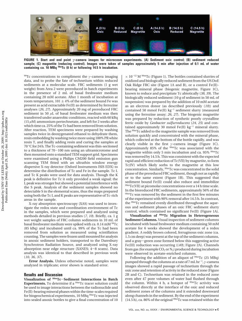
Results and DiscussionVisualization of 99mTc-Sediment Interactions in BatchExperiments. To determine if a 99mTc tracer solution couldbe used to image interactions between the radionuclide andFe(II)-bearing mineral phases within the time-scales requiredfor biogeochemical experiments, 10 MBq 99mTc was injectedinto sealed anoxic bottles to give a final concentration of 10
× 10-12 M 99mTc (Figure 1). The bottles contained slurries ofoxidized and biologically reduced sediment from the US DoEOak Ridge FRC site (Figure 1A and B), or a control Fe(II)-bearing mineral phase (biogenic magnetite, Figure 1C),known to reduce and precipitate Tc abiotically (38, 39). Thebiologically reduced sediment (10 g of sediment in 50 mL ofsuspension) was prepared by the addition of 10 mM acetateas an electron donor (as described previously (10)) andcontained 50 mmol Fe(II) kg-1 sediment slurry (measuredusing the ferrozine assay; 26, 27). The biogenic magnetitewas prepared by reduction of synthetic poorly crystallineferric oxide by Geobacter sulfurreducens (24, 25) and con-tained approximately 30 mmol Fe(II) kg-1 mineral slurry.The 99mTc added to the magnetite sample was removed fromsolution quickly and concentrated with the mineral phase,which collected at the bottom of the bottle rapidly, and wasclearly visible in the first γ-camera image (Figure 1C).Approximately 85% of the 99mTc was associated with themagnetite within only 5 min incubation and ca. 95% 99mTcwas removed by 14.5 h. This was consistent with the expectedrapid and efficient reduction of Tc(VII) by magnetite, to formTc(IV) which likely sorbs to the magnetite at this lowconcentration. Similarly, 99mTc was concentrated in the solidphase of the prereduced FRC sediment, though not as rapidlyor to the same extent (Figure 1B). This suggested thatsediment bound Fe(II) could also reduce and immobilize99mTc(VII) at picomolar concentrations over a 14 h time scale.In the bioreduced FRC sediments, approximately 56% of the99mTc was removed by the sediment in the first few minutesof the experiment with 90% removal after 14.5 h. In contrast,the 99mTc remained evenly distributed throughout the aque-ous and sediment phases of an oxic FRC sediment slurrycontrol, which contained no significant Fe(II) (Figure 1A).
Visualization of 99mTc Migration in HeterogeneousSediment Columns. Visual inspection of sediment columnsincubated with basal freshwater medium containing 50 mMacetate for 6 weeks showed the development of a redoxgradient. A reddy brown colored, ferruginous oxic zone (ca.1.5 cm deep) was present at the top of the sediment columns,and a gray-green zone formed below this suggesting activeFe(III) reduction was occurring ((40); Figure 2A). Channelsfrom gas (for example CO2 or N2) produced during incubationwere observed in acetate-enriched columns.
Following the addition of an aliquot of 99mTc (25 MBq)pumped through the columns at a rate of 7 mL hr-1, γ-cameraimages showed a rapid passage of technetium through theoxic zone and retention of activity in the reduced zone (Figure2B and C). Technetium was retained in the reduced zoneeven after 47 pore volumes of water had flushed throughthe column. Within 4 h, a hotspot of 99mTc activity wasobserved directly at the interface of the oxic and reducedsediment zones of the columns and it extended downwardalong channels in the sediment. By the end of the experiment(14.5 h), ca. 86% of the original 99mTc was retained within the
FIGURE 1. Start and end point γ-camera images for microcosm experiments. (A) Sediment oxic control; (B) sediment reducedsample; (C) magnetite (reducing control). Images were taken of samples approximately 5 min after injection of 0.1 mL of watercontaining ca. 10 MBq 99mTc (0 h) or following 14.5 h incubation.
VOL. xxx, NO. xx, XXXX / ENVIRONMENTAL SCIENCE & TECHNOLOGY 9 C
Dow
nloa
ded
by U
NIV
OF
LE
ED
S on
Oct
ober
28,
200
9 | h
ttp://
pubs
.acs
.org
P
ublic
atio
n D
ate
(Web
): A
pril
29, 2
009
| doi
: 10.
1021
/es8
0288
5r
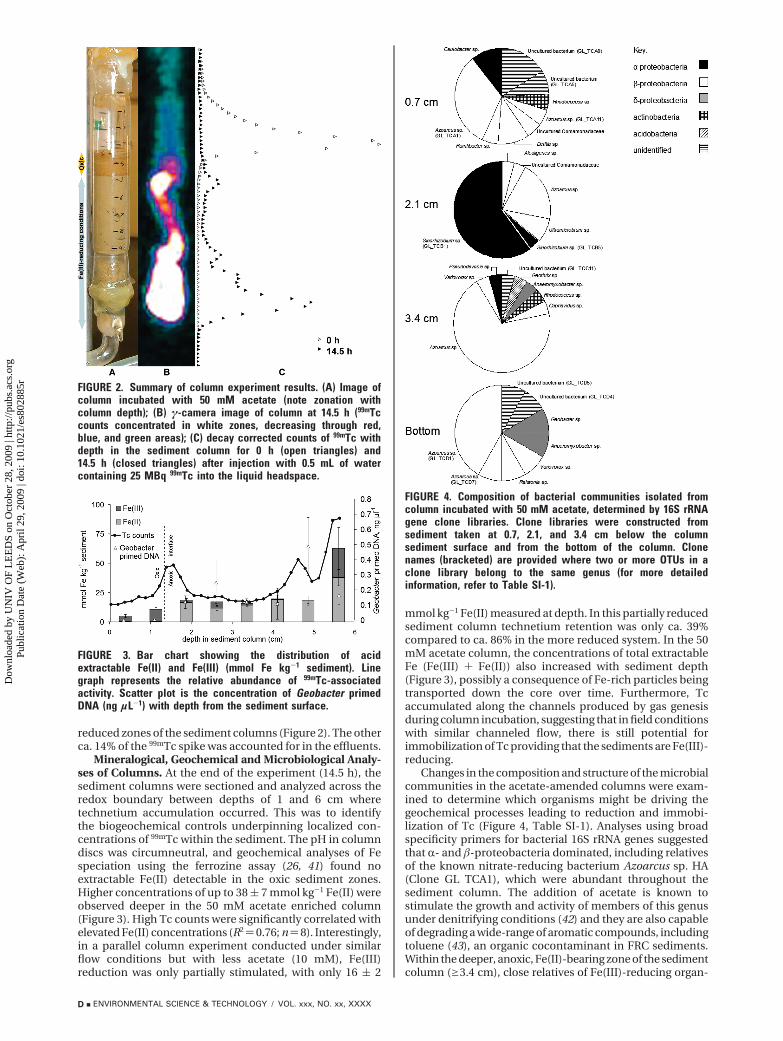
reduced zones of the sediment columns (Figure 2). The otherca. 14% of the 99mTc spike was accounted for in the effluents.
Mineralogical, Geochemical and Microbiological Analy-ses of Columns. At the end of the experiment (14.5 h), thesediment columns were sectioned and analyzed across theredox boundary between depths of 1 and 6 cm wheretechnetium accumulation occurred. This was to identifythe biogeochemical controls underpinning localized con-centrations of 99mTc within the sediment. The pH in columndiscs was circumneutral, and geochemical analyses of Fespeciation using the ferrozine assay (26, 41) found noextractable Fe(II) detectable in the oxic sediment zones.Higher concentrations of up to 38 ( 7 mmol kg-1 Fe(II) wereobserved deeper in the 50 mM acetate enriched column(Figure 3). High Tc counts were significantly correlated withelevated Fe(II) concentrations (R2)0.76; n)8). Interestingly,in a parallel column experiment conducted under similarflow conditions but with less acetate (10 mM), Fe(III)reduction was only partially stimulated, with only 16 ( 2
mmol kg-1 Fe(II) measured at depth. In this partially reducedsediment column technetium retention was only ca. 39%compared to ca. 86% in the more reduced system. In the 50mM acetate column, the concentrations of total extractableFe (Fe(III) + Fe(II)) also increased with sediment depth(Figure 3), possibly a consequence of Fe-rich particles beingtransported down the core over time. Furthermore, Tcaccumulated along the channels produced by gas genesisduring column incubation, suggesting that in field conditionswith similar channeled flow, there is still potential forimmobilization of Tc providing that the sediments are Fe(III)-reducing.
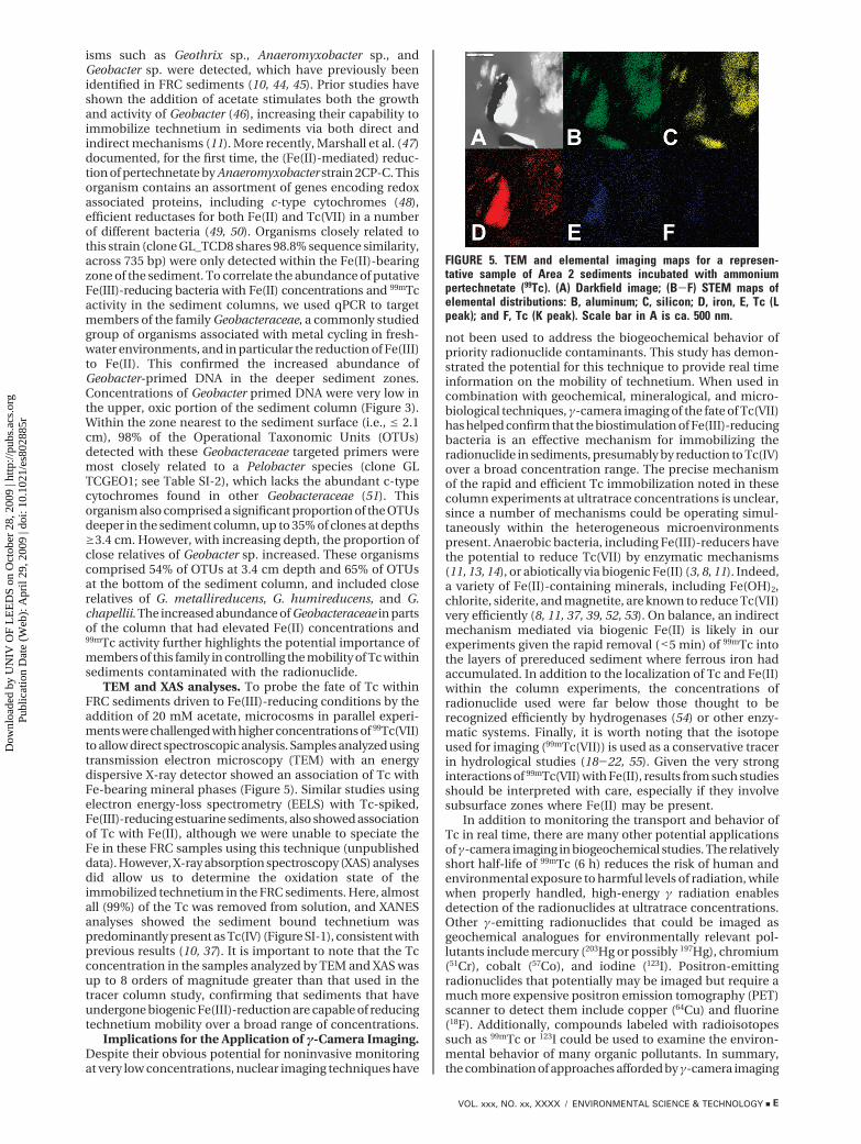
Changes in the composition and structure of the microbialcommunities in the acetate-amended columns were exam-ined to determine which organisms might be driving thegeochemical processes leading to reduction and immobi-lization of Tc (Figure 4, Table SI-1). Analyses using broadspecificity primers for bacterial 16S rRNA genes suggestedthat R- and �-proteobacteria dominated, including relativesof the known nitrate-reducing bacterium Azoarcus sp. HA(Clone GL TCA1), which were abundant throughout thesediment column. The addition of acetate is known tostimulate the growth and activity of members of this genusunder denitrifying conditions (42) and they are also capableof degrading a wide-range of aromatic compounds, includingtoluene (43), an organic cocontaminant in FRC sediments.Within the deeper, anoxic, Fe(II)-bearing zone of the sedimentcolumn (g3.4 cm), close relatives of Fe(III)-reducing organ-
FIGURE 2. Summary of column experiment results. (A) Image ofcolumn incubated with 50 mM acetate (note zonation withcolumn depth); (B) γ-camera image of column at 14.5 h (99mTccounts concentrated in white zones, decreasing through red,blue, and green areas); (C) decay corrected counts of 99mTc withdepth in the sediment column for 0 h (open triangles) and14.5 h (closed triangles) after injection with 0.5 mL of watercontaining 25 MBq 99mTc into the liquid headspace.
FIGURE 3. Bar chart showing the distribution of acidextractable Fe(II) and Fe(III) (mmol Fe kg-1 sediment). Linegraph represents the relative abundance of 99mTc-associatedactivity. Scatter plot is the concentration of Geobacter primedDNA (ng µL-1) with depth from the sediment surface.
FIGURE 4. Composition of bacterial communities isolated fromcolumn incubated with 50 mM acetate, determined by 16S rRNAgene clone libraries. Clone libraries were constructed fromsediment taken at 0.7, 2.1, and 3.4 cm below the columnsediment surface and from the bottom of the column. Clonenames (bracketed) are provided where two or more OTUs in aclone library belong to the same genus (for more detailedinformation, refer to Table SI-1).
D 9 ENVIRONMENTAL SCIENCE & TECHNOLOGY / VOL. xxx, NO. xx, XXXX
Dow
nloa
ded
by U
NIV
OF
LE
ED
S on
Oct
ober
28,
200
9 | h
ttp://
pubs
.acs
.org
P
ublic
atio
n D
ate
(Web
): A
pril
29, 2
009
| doi
: 10.
1021
/es8
0288
5r
isms such as Geothrix sp., Anaeromyxobacter sp., andGeobacter sp. were detected, which have previously beenidentified in FRC sediments (10, 44, 45). Prior studies haveshown the addition of acetate stimulates both the growthand activity of Geobacter (46), increasing their capability toimmobilize technetium in sediments via both direct andindirect mechanisms (11). More recently, Marshall et al. (47)documented, for the first time, the (Fe(II)-mediated) reduc-tion of pertechnetate by Anaeromyxobacter strain 2CP-C. Thisorganism contains an assortment of genes encoding redoxassociated proteins, including c-type cytochromes (48),efficient reductases for both Fe(II) and Tc(VII) in a numberof different bacteria (49, 50). Organisms closely related tothis strain (clone GL_TCD8 shares 98.8% sequence similarity,across 735 bp) were only detected within the Fe(II)-bearingzone of the sediment. To correlate the abundance of putativeFe(III)-reducing bacteria with Fe(II) concentrations and 99mTcactivity in the sediment columns, we used qPCR to targetmembers of the family Geobacteraceae, a commonly studiedgroup of organisms associated with metal cycling in fresh-water environments, and in particular the reduction of Fe(III)to Fe(II). This confirmed the increased abundance ofGeobacter-primed DNA in the deeper sediment zones.Concentrations of Geobacter primed DNA were very low inthe upper, oxic portion of the sediment column (Figure 3).Within the zone nearest to the sediment surface (i.e., e 2.1cm), 98% of the Operational Taxonomic Units (OTUs)detected with these Geobacteraceae targeted primers weremost closely related to a Pelobacter species (clone GLTCGEO1; see Table SI-2), which lacks the abundant c-typecytochromes found in other Geobacteraceae (51). Thisorganism also comprised a significant proportion of the OTUsdeeper in the sediment column, up to 35% of clones at depthsg3.4 cm. However, with increasing depth, the proportion ofclose relatives of Geobacter sp. increased. These organismscomprised 54% of OTUs at 3.4 cm depth and 65% of OTUsat the bottom of the sediment column, and included closerelatives of G. metallireducens, G. humireducens, and G.chapellii. The increased abundance of Geobacteraceae in partsof the column that had elevated Fe(II) concentrations and99mTc activity further highlights the potential importance ofmembers of this family in controlling the mobility of Tc withinsediments contaminated with the radionuclide.
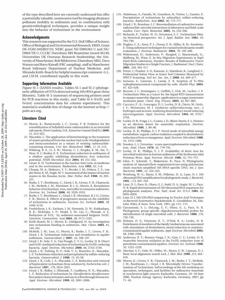
TEM and XAS analyses. To probe the fate of Tc withinFRC sediments driven to Fe(III)-reducing conditions by theaddition of 20 mM acetate, microcosms in parallel experi-ments were challenged with higher concentrations of 99Tc(VII)to allow direct spectroscopic analysis. Samples analyzed usingtransmission electron microscopy (TEM) with an energydispersive X-ray detector showed an association of Tc withFe-bearing mineral phases (Figure 5). Similar studies usingelectron energy-loss spectrometry (EELS) with Tc-spiked,Fe(III)-reducing estuarine sediments, also showed associationof Tc with Fe(II), although we were unable to speciate theFe in these FRC samples using this technique (unpublisheddata). However, X-ray absorption spectroscopy (XAS) analysesdid allow us to determine the oxidation state of theimmobilized technetium in the FRC sediments. Here, almostall (99%) of the Tc was removed from solution, and XANESanalyses showed the sediment bound technetium waspredominantly present as Tc(IV) (Figure SI-1), consistent withprevious results (10, 37). It is important to note that the Tcconcentration in the samples analyzed by TEM and XAS wasup to 8 orders of magnitude greater than that used in thetracer column study, confirming that sediments that haveundergone biogenic Fe(III)-reduction are capable of reducingtechnetium mobility over a broad range of concentrations.
Implications for the Application of γ-Camera Imaging.Despite their obvious potential for noninvasive monitoringat very low concentrations, nuclear imaging techniques have
not been used to address the biogeochemical behavior ofpriority radionuclide contaminants. This study has demon-strated the potential for this technique to provide real timeinformation on the mobility of technetium. When used incombination with geochemical, mineralogical, and micro-biological techniques, γ-camera imaging of the fate of Tc(VII)has helped confirm that the biostimulation of Fe(III)-reducingbacteria is an effective mechanism for immobilizing theradionuclide in sediments, presumably by reduction to Tc(IV)over a broad concentration range. The precise mechanismof the rapid and efficient Tc immobilization noted in thesecolumn experiments at ultratrace concentrations is unclear,since a number of mechanisms could be operating simul-taneously within the heterogeneous microenvironmentspresent. Anaerobic bacteria, including Fe(III)-reducers havethe potential to reduce Tc(VII) by enzymatic mechanisms(11, 13, 14), or abiotically via biogenic Fe(II) (3, 8, 11). Indeed,a variety of Fe(II)-containing minerals, including Fe(OH)2,chlorite, siderite, and magnetite, are known to reduce Tc(VII)very efficiently (8, 11, 37, 39, 52, 53). On balance, an indirectmechanism mediated via biogenic Fe(II) is likely in ourexperiments given the rapid removal (<5 min) of 99mTc intothe layers of prereduced sediment where ferrous iron hadaccumulated. In addition to the localization of Tc and Fe(II)within the column experiments, the concentrations ofradionuclide used were far below those thought to berecognized efficiently by hydrogenases (54) or other enzy-matic systems. Finally, it is worth noting that the isotopeused for imaging (99mTc(VII)) is used as a conservative tracerin hydrological studies (18-22, 55). Given the very stronginteractions of 99mTc(VII) with Fe(II), results from such studiesshould be interpreted with care, especially if they involvesubsurface zones where Fe(II) may be present.
In addition to monitoring the transport and behavior ofTc in real time, there are many other potential applicationsof γ-camera imaging in biogeochemical studies. The relativelyshort half-life of 99mTc (6 h) reduces the risk of human andenvironmental exposure to harmful levels of radiation, whilewhen properly handled, high-energy γ radiation enablesdetection of the radionuclides at ultratrace concentrations.Other γ-emitting radionuclides that could be imaged asgeochemical analogues for environmentally relevant pol-lutants include mercury (203Hg or possibly 197Hg), chromium(51Cr), cobalt (57Co), and iodine (123I). Positron-emittingradionuclides that potentially may be imaged but require amuch more expensive positron emission tomography (PET)scanner to detect them include copper (64Cu) and fluorine(18F). Additionally, compounds labeled with radioisotopessuch as 99mTc or 123I could be used to examine the environ-mental behavior of many organic pollutants. In summary,the combination of approaches afforded by γ-camera imaging
FIGURE 5. TEM and elemental imaging maps for a represen-tative sample of Area 2 sediments incubated with ammoniumpertechnetate (99Tc). (A) Darkfield image; (B-F) STEM maps ofelemental distributions: B, aluminum; C, silicon; D, iron, E, Tc (Lpeak); and F, Tc (K peak). Scale bar in A is ca. 500 nm.
VOL. xxx, NO. xx, XXXX / ENVIRONMENTAL SCIENCE & TECHNOLOGY 9 E
Dow
nloa
ded
by U
NIV
OF
LE
ED
S on
Oct
ober
28,
200
9 | h
ttp://
pubs
.acs
.org
P
ublic
atio
n D
ate
(Web
): A
pril
29, 2
009
| doi
: 10.
1021
/es8
0288
5r
of the type described here are currently underused but offera potentially valuable, noninvasive tool for imaging ultratracepollutant mobility in sediments and, in combination withgeomicrobiological techniques, provides a unique insightinto the behavior of technetium in the environment.
AcknowledgmentsThis research was supported by the U.S. DoE Office of Science,Office of Biological and Environmental Research, ERSD, GrantDE-FG02-04ER63743; NERC grant NE/D005361/1 and NE/D00473X/1; CCLRC Daresbury SRS Beamtime Award 46140;Universities UK Overseas Research Studentship; the Uni-versity of Manchester; Bob Bilsborrow (Daresbury SRS), DaveWatson and Barry Kinsall (FRC sampling), staff at ManchesterRoyal Infirmary Department of Nuclear Medicine, andMiranda Keith-Roach for helpful manuscript comment. G.L.and J.M.M. contributed equally to this work.
Supporting Information AvailableFigure SI-1 (XANES results), Tables SI-1 and SI-2 (phyloge-netic affiliation of OTUs detected using 16S rRNA gene clonelibraries), Table SI-3 (summary of sequencing primers usedfor PCR reactions in this study), and Table SI-4 (Fe(II) andFe(tot) concentration data for column experiment). Thismaterial is available free of charge via the Internet at http://pubs.acs.org.
Literature Cited(1) Morris, K.; Butterworth, J. C.; Livens, F. R. Evidence for the
remobilization of Sellafield waste radionuclides in an intertidalsalt marsh, West Cumbria, U.K. Estuarine Coastal Shelf Sci. 2000,51, 613–625.
(2) Macaskie, L. The application of biotechnology to the treatmentof wastes produced from the nuclear fuel cycle: biodegradationand bioaccumulation as a means of treating radionuclide-containing streams. Crit. Rev. Biotechnol. 1991, 11, 41–112.
(3) Wildung, R. E.; Li, S. W.; Murray, C. J.; Krupka, K. M.; Xie, Y.;Hess, N. J.; Roden, E. E. Technetium reduction in sediments ofa shallow aquifer exhibiting dissimilatory iron reductionpotential. FEMS Microbiol. Ecol. 2004, 49, 151–162.
(4) Lieser, K. H. Technetium in the nuclear-fuel cycle, in medicineand in the environment. Radiochim. Acta 1993, 63, 5–8.
(5) Champ, M. A.; Makeyev, V. V.; Brooks, J. M.; Delaca, T. E.; vander Horst, K. M.; Engle, M. V. Assessment of the impact of nuclearwastes in the Russian Arctic. Mar. Pollut. Bull. 1998, 35, 203–221.
(6) Burke, I. T.; Boothman, C.; Lloyd, J. R.; Livens, F. R.; Charnock,J. M.; McBeth, J. M.; Mortimer, R. J. G.; Morris, K. Reoxidationbehavior of technetium, iron, and sulfur in estuarine sediments.Environ. Sci. Technol. 2006, 40, 3529–3535.
(7) Burke, I. T.; Boothman, C.; Lloyd, J. R.; Mortimer, R. J. G.; Livens,F. R.; Morris, K. Effects of progressive anoxia on the solubilityof technetium in sediments. Environ. Sci. Technol. 2005, 39,4109–4116.
(8) Fredrickson, J. K.; Zachara, J. M.; Kennedy, D. W.; Kukkadapu,R. K.; McKinley, J. P.; Heald, S. M.; Liu, C.; Plymale, A. E.Reduction of TcO4
- by sediment-associated biogenic Fe(II).Geochim. Cosmochim. Acta 2004, 68, 3171–3187.
(9) Keith-Roach, M. J.; Morris, K.; Dahlgaard, H. An investigationinto technetium binding in sediments. Mar. Chem. 2003, 81,149–162.
(10) McBeth, J. M.; Lear, G.; Morris, K.; Burke, I. T.; Livens, F. R.;Lloyd, J. R. Technetium reduction and reoxidation in aquifersediments. Geomicrobiol. J. 2007, 24, 189–197.
(11) Lloyd, J. R.; Sole, V. A.; Van Praagh, C. V. G.; Lovley, D. R. Directand Fe(II)-mediated reduction of technetium by Fe(III)-reducingbacteria. Appl. Environ. Microbiol. 2000, 66, 3743–3749.
(12) Lloyd, J. R.; Nolting, H.-F.; Sole, V. A.; Bosecker, K.; Macaskie,L. E. Technetium reduction and precipitation by sulfate-reducingbacteria. Geomicrobiol. J. 1998, 15, 45–58.
(13) Lloyd, J. R.; Cole, J. A.; Macaskie, L. E. Reduction and removalof heptavalent technetium from solution by Escherichia coli. J.Bacteriol. 1997, 179, 2014–2021.
(14) Lloyd, J. R.; Ridley, J.; Khizniak, T.; Lyalikova, N. N.; Macaskie,L. E. Reduction of technetium by Desulfovibrio desulfuricans:biocatalyst characterization and use in a flowthrough bioreactor.Appl. Environ. Microbiol. 1999, 65, 2691–2696.
(15) Abdelouas, A.; Fattahi, M.; Grambow, B.; Vichot, L.; Gautier, E.Precipitation of technetium by subsurface sulfate-reducingbacteria. Radiochim. Acta 2002, 90, 773–777.
(16) Lloyd, J. R.; Renshaw, J. C. Bioremediation of radioactive waste:radionuclide-microbe interactions in laboratory and field-scalestudies. Curr. Opin. Biotechol. 2005, 16, 254–260.
(17) Richards, P.; Tucker, W. D.; Srivastava, S. C. Technetium-99m:An historical perspective. Int. J. Appl. Radiat. Isot. 1982, 33,793–799.
(18) Hughes, C. E.; Airey, P. L.; Duran, E. B.; Miller, B. M.; Sombrito,E. Using radiotracer techniques for coastal hydrodynamic modelevaluation. J. Environ. Radioactiv. 2004, 76, 195–206.
(19) Widestrand, H.; Andersson, P.; Byegard, J.; Skarnemark, G.;Skalberg, M.; Wass, E. In Situ Migration Experiments at AspoHard Rock Laboratory, Sweden: Results of Radioactive TracerMigration Studies in a Single Fracture. J. Radioanal. Nucl. Chem.2001, 250, 501–517.
(20) Perret, J.; Prasher, S. O.; Kantzas, A.; Hamilton, K.; Langford, C.Preferential Solute Flow in Intact Soil Columns Measured bySPECT Scanning. Soil Sci. Soc. Am. J. 2000, 64, 469–477.
(21) Jurisson, S.; Gawenis, J.; Landa, E. R. Sorption of Tc-99mradiopharmaceutical compounds by soils. Health Phys. 2004,87, 423–428.
(22) Borroto, J. I.; Dominguez, J.; Griffith, J.; Fick, M.; Leclerc, J. P.Technetium-99m as a tracer for the liquid RTD measurementin opaque anaerobic digester: application in a sugar wastewatertreatment plant. Chem. Eng. Process. 2003, 42, 857–865.
(23) Caccavo, F., Jr.; Lonergan, D. J.; Lovley, D. R.; Davis, M.; Stolz,J. F.; McInerney, M. J. Geobacter sulfurreducenssp. nov., ahydrogen and acetate-oxidizing dissimilatory metal reducingmicroorganism. Appl. Environ. Microbiol. 1994, 60, 3752-3759.
(24) Lovley, D. R.; Fraga, J. L.; Coates, J. D.; Blunt-Harris, E. L. Humicsas an electron donor for anaerobic respiration. Environ.Microbiol. 1999, 1, 89–98.
(25) Lovley, D. R.; Phillips, E. J. P. Novel mode of microbial energymetabolism: organic carbon oxidation coupled to dissimilatoryreduction of iron or manganese. Appl. Environ. Microbiol. 1988,54, 1472–1480.
(26) Stookey, L. L. Ferrozine - a new spectrophotometric reagent foriron. Anal. Chem. 1970, 42, 779–781.
(27) Lovley, D. R.; Phillips, E. J. P. Availability of ferric iron formicrobial reduction in bottom sediments of the freshwater tidalPotomac River. Appl. Environ. Microb. 1986, 52, 751–757.
(28) Eden, P.; Schmidt, T.; Blakemore, R.; Pace, N. Phylogeneticanalysis of Aquaspirillum magnetotacticum using polymerasechain reaction-amplified 16S rRNA-specific DNA. Int. J. Syst.Bacteriol. 1991, 41, 324–325.
(29) Weisburg, W. G.; Barns, S. M.; Pelletier, D. A.; Lane, D. J. 16Sribosomal DNA amplification for phylogenetic study. J. Bacteriol.1991, 173, 697–703.
(30) Lane, D. J.; Pace, B.; Olsen, G. J.; Stahl, D. A.; Sogin, M. L.; Pace,N. R. Rapid determination of 16S ribosomal RNA sequences forphylogenetic analyses. Proc. Natl. Acad. Sci. U.S.A. 1985, 82,6955–6959.
(31) Lane, D. J. 16S/23S rRNA sequencing. In Nucleic Acid Techniquesin Bacterial Systematics; Stackebrandt, E.; Goodfellow, M., Eds.John Wiley & Sons: New York, 1991; pp 115-175.
(32) Giovannoni, S. J.; DeLong, E. F.; Olsen, G. J.; Pace, N. R.Phylogenetic group-specific oligodeoxynucleotide probes foridentification of single microbial cells. J. Bacteriol. 1988, 170,720–726.
(33) Holmes, D. E.; Finneran, K. T.; O’Neil, R. A.; Lovley, D. R.Enrichment of members of the family Geobacteraceae associatedwith stimulation of dissimilatory metal reduction in uranium-contaminated aquifer sediments. Appl. Environ. Microbiol. 2002,68, 2300–2306.
(34) Anderson, R. T.; Rooney-Varga, J. N.; Gaw, C. V.; Lovley, D. R.Anaerobic benzene oxidation in the Fe(III) reduction zone ofpetroleum-contaminated aquifers. Environ. Sci. Technol. 1998,32, 1222–1229.
(35) Altschul, S. F.; Gish, W.; Miller, W.; Myers, E. W.; Lipman, D. J.Basic local alignment search tool. J. Mol. Biol. 1990, 215, 403–410.
(36) Morris, K.; Livens, F. R.; Charnock, J. M.; Burke, I. T.; McBeth,J. M.; Boothman, C.; Lloyd, J. R. Microbially Driven Transfor-mations of Technetium. InProceedings of the 4th workshop onspeciation, techniques, and facilities for radioactive materialsat synchrotron light sources, Karlsruhe, Germany, 18-20 Sept2006; Nuclear Energy Agency: Karlsruhe, Germany, 2007; pp71-81.
F 9 ENVIRONMENTAL SCIENCE & TECHNOLOGY / VOL. xxx, NO. xx, XXXX
Dow
nloa
ded
by U
NIV
OF
LE
ED
S on
Oct
ober
28,
200
9 | h
ttp://
pubs
.acs
.org
P
ublic
atio
n D
ate
(Web
): A
pril
29, 2
009
| doi
: 10.
1021
/es8
0288
5r
(37) Morris, K.; Livens, F. R.; Charnock, J. M.; Burke, I. T.; McBeth,J. M.; Begg, J. D. C.; Boothman, C.; Lloyd, J. R. An X-ray absorptionstudy of the fate of technetium in reduced and reoxidisedsediments and mineral phases. Appl. Geochem. 2008, 23, 603–617.
(38) Maes, A.; Geraedts, K.; Bruggeman, C.; VanCluysen, J.; Rossberg,A.; Hennig, C. Evidence for the interaction of technetium colloidswith humic substances by X-ray absorption spectroscopy.Environ. Sci. Technol. 2004, 38, 2044–2051.
(39) Cui, D. Q.; Eriksen, T. E. Reduction of pertechnetate in solutionby heterogeneous electron transfer from Fe(II)-containinggeological material. Environ. Sci. Technol. 1996, 30, 2263–2269.
(40) Lovley, D. R. Dissimilatory Fe(III) and Mn(IV) reduction.Microbiol. Rev. 1991, 55, 259–287.
(41) Lovley, D. R.; Phillips, E. J. P. Rapid assay for microbially reducibleferric iron in aquatic sediments. Appl. Environ. Microb. 1987,53, 1536–1540.
(42) Reinhold-Hurek, B.; Hurek, T. The Genera Azoarcus, Azovibrio,Azospira and Azonexus. In The Prokaryotes, 3rd ed.; Dworkin,M., Ed.; Springer: Singapore, 2006; Vol. 5 (Proteobacteria: Alphaand Beta Subclasses); pp 873-891.
(43) Zhou, J.; Fries, M. R.; Chee-Sanford, J. C.; Tiedje, J. M.Phylogenetic analyses of a new group of denitrifiers capable ofanaerobic growth of toluene and description of Azoarcustolulyticus sp. nov. Int. J. Syst. Bacteriol. 1995, 45, 500–506.
(44) Shelobolina, E. S.; O’Neill, K. R.; Finneran, K. T.; Hayes, L. A.;Lovley, D. R. Potential for in-situ bioremediation of a low-pH,high-nitrate uranium-contaminated groundwater. Soil SedimentContam. 2003, 12, 865–884.
(45) North, N. N.; Dollhopf, S. L.; Petrie, L.; Istok, J. D.; Balkwill,D. L.; Kostka, J. E. Change in bacterial community structureduring in situ biostimulation of subsurface sediment cocon-taminated with uranium and nitrate. Appl. Environ. Microbiol.2004, 70, 4911–4920.
(46) Holmes, D. E.; O’Neil, R. A.; Vrionis, H. A.; N’Guessan, L. A.;Ortiz-Bernad, I.; Larrahondo, M. J.; Adams, L. A.; Ward, J. A.;Nicoll, J. S.; Nevin, K. P.; Chavan, M. A.; Johnson, J. P.; Long,P. E.; Lovley, D. R. Subsurface clade of Geobacteraceae thatpredominates in a diversity of Fe(III)-reducing subsurfaceenvironments. ISME J. 2007, 1, 663–677.
(47) Marshall, M. J.; Dohnalkova, A. C.; Kennedy, D. W.; Plymale,A. E.; Thomas, S. H.; Loffler, F. E.; Sanford, R. A.; Zachara, J. M.;
Fredrickson, J. K.; Beliaev, A. S. Electron donor-dependentradionuclide reduction and nanoparticle formation by Anaer-omyxobacter dehalogenans strain 2CP-C. Environ. Microbiol.2009, 11, 534–543.
(48) Thomas, S. H.; Wagner, R. D.; Arakaki, A. K.; Skolnick, J.; Kirby,J. R.; Shimkets, L. J.; Sanford, R. A.; Loffler, F. E. The MosaicGenome of Anaeromyxobacter dehalogenans Strain 2CP-CSuggests an Aerobic Common Ancestor to the Delta-Proteo-bacteria. PLoS ONE 2008, 3, e2103.
(49) Marshall, M. J.; Plymale, A. E.; Kennedy, D. W.; Shi, L.; Wang,Z.; Reed, S. B.; Dohnalkova, A. C.; Simonson, C. J.; Liu, C.;Saffarini, D. A.; Romine, M. F.; Zachara, J. M.; Beliaev, A. S.;Fredrickson, J. K. Hydrogenase- and outer membrane c-typecytochrome-facilitated reduction of technetium(VII) by Sh-ewanella oneidensis MR-1. Environ. Microbiol. 2008, 10, 125–136.
(50) Seeliger, S.; Cord-Ruwisch, R.; Schink, B. A Periplasmic andExtracellular c-Type Cytochrome of Geobacter sulfurreducensActs as a Ferric Iron Reductase and as an Electron Carrier toOther Acceptors or to Partner Bacteria. J. Bacteriol. 1998, 180,3686–3691.
(51) Haveman, S. A.; Holmes, D. E.; Ding, Y.-H. R.; Ward, J. E.;DiDonato, R. J., Jr.; Lovley, D. R. c-Type Cytochromes inPelobacter carbinolicus. Appl. Environ. Microbiol. 2006, 72, 6980–6985.
(52) Vandergraaf, T. T.; Ticknor, K. V.; George, I. M. Reactions betweentechnetium in solution and iron-containing minerals under oxicand anoxic conditions. In Geochemical Behavior of DisposedRadioactive Waste; Barney, G. S., Navratil, J. D., Schulz, W. W.,Eds.; American Chemical Society: Washington, DC, 1984; Vol246, pp 25-43.
(53) Cui, D. Q.; Eriksen, T. E. Reduction of pertechnetate by ferrousiron in solution: Influence of sorbed and precipitated Fe(II).Environ. Sci. Technol. 1996, 30, 2259–2262.
(54) Lloyd, J. R.; Harding, C. L.; Macaskie, L. E. Tc(VII) reduction andaccumulation by immobilized cells of Escherichia coli. Bio-technol. Bioeng. 1997, 55, 505–510.
(55) Aarkrog, A.; Chen, Q.; Dahlgaard, H.; Nielsen, S. P.; Trapeznikov,A.; Pozolotina, V. Evidence of 99Tc in Ural river sediments. J.Environ. Radioactiv. 1997, 37, 201–213.
ES802885R
VOL. xxx, NO. xx, XXXX / ENVIRONMENTAL SCIENCE & TECHNOLOGY 9 G
Dow
nloa
ded
by U
NIV
OF
LE
ED
S on
Oct
ober
28,
200
9 | h
ttp://
pubs
.acs
.org
P
ublic
atio
n D
ate
(Web
): A
pril
29, 2
009
| doi
: 10.
1021
/es8
0288
5r