probing the structural basis of the catalytic activity of hiv-1 pr through total chemical protein...
TRANSCRIPT

Probing the structural basis of the catalytic activity of HIV-1 PRthrough total chemical protein synthesis
Maria Millera,*, Manuel Baca1b, J.K. Mohana Raoa, Stephen B.H. Kentb
aMacromolecular Structure Laboratory, ABL-Basic Research Program, NCI-Frederick Cancer Research Facility and Development Center,P.O. Box B, Frederick, MD 21702 USA
bThe Scripps Research Institute, 10666 N. Torrey Pines Road, La Jolla, CA 92037, USA
Received 2 October 1996; accepted 29 November 1996
Abstract
Historically, total chemical synthesis had been used to prepare native proteinase from the human immunodeficiency virus(HIV-1 PR) for structural studies by X-ray crystallography. More recently, several functionally-relevant analogues of HIV-1 PRhave also been obtained by total chemical synthesis. The results of structural and biochemical studies of the backboneengineered analogues put in question the established belief of the importance of an internal, tetrahedrally coordinated watermolecule (water 301) in mediating catalytically important flap–substrate interaction. An enzyme analogue in which the peptidebond between residues Gly51 and Gly52 was replaced by a thioester moiety displayed normal enzymatic activity, while thecrystal structure of its complex with the inhibitor MVT101 (solved at 2.5 A˚ resolution as mirror image, D-enantiomer) did notshow the presence of water 301. The enzyme analogue in which the ability to donate hydrogen bonds to substrate was deleted(by substitution of Ile50 –N(H)– by a sulfur atom) inboth flapswas 2500-fold less active. By contrast, the covalent dimer formof the enzyme with the Gly49–Ile50 peptide bond –N(H)– atom specifically replaced by an –O– atomin one flaponly retainednormal enzymatic activity. The combined data from these studies strongly indicate that flap–substrate hydrogen bonds fromonly one flap are sufficient for full enzymatic activity of the HIV-1 PR, and raise the possibility that the retroviral enzyme maymake use of only one flap in catalysis. This result may have profound implications for drug design targeted at HIV-1 PR.q 1998 Elsevier Science B.V.
Keywords:HIV-1 proteinase; Structural engineering; Chemical protein synthesis; Catalytic mechanism; Structure-based drugdesign
1. Introduction
Successful chemical synthesis of complex poly-peptides marked a milestone in the development ofthe field of protein chemistry. It opened not only
numerous novel avenues for biotechnology and drugdesign, but also new possibilities for the study ofphysico-chemical and biological properties of the pro-tein molecule itself. One of the best examples is thecase of the retroviral aspartyl proteinase from humanimmunodeficiency virus HIV-1 (HIV-1 PR). It wasthe first biologically active enzyme obtained in repro-ducible fashion by total chemical synthesis [1], andthe first example of the preparation of a complete
Journal of Molecular Structure (Theochem) 423 (1998) 137–152
THEOCH 5022
0166-1280/98/$19.00q 1998 Elsevier Science B.V. All rights reserved.PII S0166-1280(96)05022-1
* Corresponding author.1 Present address: Department of Protein Engineering, Genentech,
Inc., 460 Pt. San Bruno Blvd., So. San Francisco, CA 94080, USA.

enzyme molecule containing non-coded amino acids.Obtaining milligram quantities of a folded homo-genous protein of that size was precedent-setting,and the synthesis came at a time when efficient pro-cedures for recombinant expression of HIV-1 PR hadnot yet been developed. Because of this confluence ofcircumstances, total chemical synthesis of the HIV-1PR molecule has had a profound influence on ourunderstanding of this important molecule.
In early 1989, a low resolution X-ray structure ofunliganded HIV-1 PR had been reported by the Merckgroup [2]. This was the first reported structure of thisimportant target for drug design and revealed theessential features of the catalytic apparatus and thepresumed substrate binding cleft at the interfacebetween the two subunits. However, aspects of thisoriginal HIV-1 PR structure did not conform toexpectations based on known structural data fromthe eukaryotic (the ‘‘pepsin-like’’) aspartic proteinases.Notably, there were significant differences withrespect to a structural model of the HIV-1 PR basedon homology modeling [3], and to the just then com-pleted structure of the Rous sarcoma virus (RSV)proteinase [4]. These discrepancies were particularlypronounced in the dimer interface region involvingthe N- and C-terminal of each subunit polypeptidechain, and cast doubt on the mechanism originallyproposed [2] for the autocatalytic release of theHIV-1 PR from the virally encoded gag-pol poly-protein.
Because of these questions, and because of thepotential importance of the HIV-1 PR molecule forstructure-based drug design, in early 1989 a collabora-tive effort was undertaken between two researchgroups, one at the California Institute of Technology(chemical protein synthesis) and the other at the NCI-Frederick (X-ray crystallography). The goal of thisjoint effort was to solve the molecular structure ofthe HIV-1 PR both unliganded and in complex withsubstrate-based inhibitors. The 99 amino acid residuepolypeptide chain of the HIV-1 PR monomer wasprepared by total chemical synthesis using highlyoptimized stepwise solid phase peptide synthesis(SPPS) [5]. The sequence of the SF2 isolate of thevirus was used, with the cysteine residues at positions67 and 95 of the chain replaced with the isosteric L-a-amino-n-butyric acid (i.e. replacing an –SH of thecysteine residue with a CH3) [6]. The synthetic
proteinase was crystallized (Fig. 1(a)) and X-ray dif-fraction studies provided the first correct crystal struc-ture of the free enzyme [6], and the first co-crystalstructure of HIV1-PR complexed with an inhibitor,the substrate-derived MVT101 [7] (Fig. 1(b)). Syn-thetic HIV-1 PR was also used to determine the struc-tures of two other complexes with substrate-derivedinhibitors; these were the first structures of theenzyme with canonical examples of the two mostimportant classes of inhibitors of this enzyme, con-taining respectively hydroxyethylamine [8] andhydroxyethylene [9] isosteres.
These three sets of coordinates were made freelyavailable to the research community and were used inmodeling and theoretical studies [10–14], and also inmany laboratories worldwide to solve by molecularreplacement X-ray structures of the HIV-1 PR com-plexed with a variety of inhibitors. In these ways, thedata obtained from the synthetic enzyme played a keyrole in the intense world-wide drug design effortfocused on this molecule, with the aim of developinganti-AIDS therapeutics. The crystal structures of syn-thetic and recombinant HIV-1 PR complexed withdifferent active-site directed ligands [15–17] estab-lished the mode of inhibitor binding by the dimericretroviral enzyme (Fig. 1(c)). In some respects thiswas different from the binding mode observed forinhibitors of the pepsin-like cell-encoded aspartic pro-teinases (for a review, see [18]). The most intriguingdifference between the two classes of enzymes was inthe mode of interaction of the tip of the flap(s) with theinhibitor. The flaps are mobile, extendedb-sheetregions that close down over the substrate, desol-vating it, and contributing to the formation of thespecificity pockets involved in substrate recognition.The cell-encoded aspartyl proteinases consist of asingle polypeptide chain folded into two homologousdomains and have only one flap. By contrast, theretroviral aspartyl proteinases are homodimeric andhave two flaps, one from each monomer.
Based on the results of the structural studies of theHIV-1 PR, of similar studies on the cell-encodedaspartyl proteinases, and on the general body ofknowledge of this class of proteolytic enzymes, a con-sensus has been reached on the mechanism of cata-lysis. The accepted mechanism has been describedbest by Suguna et al. [19], for the aspartic proteinasefrom rhizopus chinensis, and involves nucleophilic
138 M. Miller et al./Journal of Molecular Structure (Theochem) 423 (1998) 137–152

attack of a water molecule presumed to exist betweenthe side chain carboxyl groups of the two catalyticallyessential aspartic acid residues. In the case of theretroviral proteinases, one catalytic Asp is contributedfrom each monomer [3]. The side chain carboxylsfunction as general acid–base catalysts for the attackby the water molecule. The broad features of thiscatalytic mechanism for the aspartyl proteinases arenoncontroversial. The proposed mechanism is ingeneral agreement with the few, but carefully done,kinetic studies of this class of enzyme [20,21]. Alter-native mechanisms, such as covalent catalysis, havenot been rigorously excluded but are not currentlyfavored by the broad preponderance of evidence [22].
A mechanism essentially similar to that of Sugunaet al. [19] has been discussed by Pearl [23] who
pointed out that in the aspartyl proteinases the distor-tion of the scissile peptide bond, an out-of-planerotation ‘‘stabilized by interactions of the substratewith the extended binding cleft’’, gives rise to the‘‘apparent electrophilicity of the catalysis’’. This dis-tortion would occur on binding and may be promotedby the observed H-bonding interactions between theinterior side of the flap(s) and the substrate carbonylson either side of the scissile bond. In crystal structuresof both types of aspartic proteinases (Fig. 2), hydro-gen bonds are observed between the protein backbonenear the tip of the flap(s) and the carbonyl oxygensflanking the pseudo-scissile bond of bound substrate-based peptide inhibitor. As shown in Fig. 2, the singleflap of the cell-encoded aspartyl proteinases closesdown ‘‘flat’’ over the scissile bond, i.e., with the
Fig. 1. (a) Crystal of HIV-1 PR prepared by total chemical synthesis, obtained as described in Ref. [6]. (b) The structure of the complex ofsynthetic HIV-1 PR with the MVT101 inhibitor. The picture was drawn using program MOLSCRIPT. (c) Stereo view of the MVT101 (solidline) inhibitor in the active site in its highly occupied state [34]. Hydrogen bonds are shown as dashed lines.
139M. Miller et al./Journal of Molecular Structure (Theochem) 423 (1998) 137–152

plane of the reverse turn at the tip of the flap (nearly)parallel to the plane of the inhibitor peptide; direct H-bonding interactions occur between backbone peptidebonds in this flap and the substrate carbonyls. By con-trast, the two flaps of the HIV-1 PR both close over thesubstrate-derived inhibitor, ‘‘edge on’’ (i.e. at<908)to the plane of the substrate polypeptide chain.
Besides this major difference in the inhibitor–enzyme interactions, it should be mentioned that theamino acid sequences in the flap region of these twoclasses of enzymes are quite different.2 Moreover,while the amide NH moieties of peptide bonds nearthe tip of the pepsin-like proteinase flap donate hydro-gen bonds directly to the substrate, the correspondinghydrogen bonds in the HIV-1 PR are contributed onefrom each flap, and are mediated by a specific,tetrahedrally coordinated internal water molecule,‘‘water 301’’, poised between the flaps and theinhibitor. Since first being observed in the HIV-1
PR–MVT101 complex [7] (see Fig. 1(c)), this water301 has been observed in a wide variety of co-crystalstructures of the HIV-1 PR with inhibitors [17], andhas also been observed in solution by NMR studies ofenzyme–inhibitor complexes [24].
The way in which the two flaps of the HIV-1 PRwere observed to interact with the inhibitor wasregarded as a key feature differentiating the mechan-isms of the retroviral HIV-1 PR from the corre-sponding cell-encoded enzymes [25–27]. This keymechanistic difference has become a major targetfor structure-based drug design [28]. Several classesof inhibitors have been explicitly designed to take upthe specific water 301-mediated H-bonding inter-actions with both flaps of the HIV-1 PR, and in thisway are designed to exhibit specific inhibition of theretroviral enzyme over the cell encoded aspartylproteinases.
Here we describe several examples making use of atotal chemical protein synthesis approach in order toobtain functionally relevant enzyme analogs designedto investigate the structural basis for the enzymaticactivity of HIV-1 PR. HIV-1 PR composed of D-amino acids was synthesized in order to show that apolypeptide composed entirely of D-amino acids isable to properly fold to form a functional molecule
Fig. 2. Comparison of flap(s)–‘‘substrate’’ interactions in cell encoded and HIV-1 proteinases.
2 The observed role of the flaps in co-crystal structures of the HIV-1 PR with substrate-derived inhibitors is similar to that proposed inthe homology model developed by Pearl and Taylor [3]. Withrespect to this and other features of the (now) known molecularstructure of the HIV-1 PR, this work of Pearl and Taylor [3] standsout as one of the best examples of the prediction of the structure of afolded protein molecule.
140 M. Miller et al./Journal of Molecular Structure (Theochem) 423 (1998) 137–152

and to investigate the chiral specificity of peptide sub-strates and inhibitors. Also, co-crystals of bothenantiomers of the enzyme, if obtained in the centro-symmetric space group, would provide very highquality data for structural studies [29]. Additionalbackbone engineered analogues of the HIV-1 PRwere prepared to delete a prominent enzyme(flap)–substrate hydrogen bond either from one or bothflaps. The kinetic and structural properties of thesesynthetic enzyme analogues were investigated.These studies were aimed to answer the questionwhether the water-mediated hydrogens bonds fromthe flaps to substrate (inhibitor) observed by X-ray
crystallography in the enzyme complexed withsubstrate-derived inhibitors are really important forthe enzymatic activity of the HIV-1 PR.
2. Results
2.1. ([COS]51–52)2HIV-1 PR
The development of the chemical ligation method forthe total synthesis of proteins [30] made possible thepreparation of several backbone engineered HIV-1 PRanalogues, each with precise single atom substitution
Fig. 3. A scheme for the total chemical synthesis of ([COS]51–52)HIV-1 PR as described in Ref. [31].
141M. Miller et al./Journal of Molecular Structure (Theochem) 423 (1998) 137–152

Fig. 4.
142 M. Miller et al./Journal of Molecular Structure (Theochem) 423 (1998) 137–152

in the polypeptide backbone. A fully active analogueof the enzyme was first prepared by this method, asoutlined in Fig. 3. The ligation of the two unprotectedpeptides: HIV-1 PR(1–51)aCOSH and BrAcetyl(53–99)HIV-1 PR gave a 99 residue polypeptide corre-sponding to the HIV-1 PR monomer, with the peptidebond between Gly51–Gly52 replaced by an isostericthioester bond. The homodimeric form of the enzyme,([COS]51–52)2HIV-1 PR, was fully active, thus demon-strating that replacement of the Gly51–Gly52 amidebond by a thioester had no ill effect on the foldingor catalytic function of the enzyme.
By the same approach, the D-enantiomer of theenzyme, D-([COS]51–52)2HIV-1 PR was preparedand its enzymatic properties were determined[31,32]. The two mirror image enzyme moleculeswere equally active, but showed reciprocal chiralspecificity in that the L-enzyme cleaved only theL-substrate whereas the D-enzyme cleaved only thecorresponding D-substrate. Similarly, the enantio-meric forms of the inhibitor MVT101 [32] were effec-tive only against the corresponding enantiomer of theenzyme; i.e., L-MVT101 inhibited the L-HIV-1 PR
catalyzed reaction but not the D-HIV-1 PR catalyzedreaction, while D-MVT101 inhibited D-HIV PR buthad no effect on the reaction catalyzed by L-enzyme.
Crystallographic studies were undertaken with thetotal D-enzyme. Synthetic D-([COS]51–52)2HIV-1 PRco-crystallized with the D-MVT101 hexapeptide inthe space group P212121, a=67:5, b=92:8,c=29:4 A. There were two monomers of the enzymeand one inhibitor in the crystallographic asymmetricunit. X-ray diffraction data to a resolution of 2.5 A˚
were collected from a single crystal. The structurewas solved by molecular replacement [33], using thecoordinates of the protein dimer from the structure ofthe complex of native backbone L-HIV-1 PR andMVT101 inhibitor [34] (in the absence of anomalousdispersion it is not possible to distinguish between Land D enantiomers using X-ray diffraction tech-niques). The same refinement protocol, that wasused in the refinement of the L-complex [7,34](using X-PLOR and PROFFT packages) was followedso the comparison between the two structures wouldbe more meaningful. The structure was refined to anR-factor of 0.188 in the resolution range of 10–2.5 A˚ .
Fig. 4. (continued) (a) Stereo view of the Ca tracing of the ligated D-PR structure (see also Ref. [35]). Bound D-MVT101 inhibitor (thick line) isshown in its electron density map. (X-ray diffraction data to a resolution of 2.5 A˚ were collected from a single crystal using a Siemens areadetector. The rotation and translation solutions were obtained using the program MADIRA developed by J.K. Mohana Rao and followed by arigid body refinement. The structure was refined toR-factor of 0.188 in the resolution range of 10–2.5 A˚ ). (b) Structure of D-(HIV-1 PR/MVT101) complex displayed as mirror image, for comparison with the L-enzyme. (c) Native backbone L-(HIV-1 PR/MVT101) complex, asdescribed in Ref. [34]. Two possible alternative orientations with 70% and 30% occupancy are shown for the bound MVT101 hexapeptide. (d)Distance differences between equivalent Ca atoms of natural backbone L-PR and the mirror image of ligated D-PR.
143M. Miller et al./Journal of Molecular Structure (Theochem) 423 (1998) 137–152

A stereo view of the backbone of the D-enantiomorphof HIV-1 PR and the bound hexapeptide inhibitor inits electron density map is shown in Fig. 4(a).
Comparison of the crystal structure of inhibitor–D-HIV-1 PR complex [35] with that of the natural-backbone synthetic L-amino acid enzyme [7,34]showed that the two molecules were in all respectsthe mirror image of each other, including the centersof asymmetry not directly determined by the chiralityof Ca atoms in the polypeptide backbone The rootmean square (rms) differences between equivalentCa atoms when the mirror image of the D-PR dimer(Fig. 4(b)) was superimposed on the L-enantiomermodel [34] (Fig. 4(c)) was 0.73 A˚ for 198 targetpairs (Fig. 4(d)). The largest deviations are forresidues involved in the crystal contacts in one orboth the crystal forms. This result demonstrates thatthe structure of ([COS]51–52)2HIV-1 PR is essentiallythe same as that of the native backbone enzyme.
Despite the overall similarity, the crystal structureof the ([COS]51–52)2HIV-1 PR/MVT-101 complexrevealed some important differences to the nativebackbone enzyme. Although the general mode ofinhibitor binding is the same, there are notable devia-tions in the flap region of the enzyme. Instead of asingle tetrahedrally coordinated water molecule, twodistinct water molecules were observed to mediatehydrogen bonds between the Gly49–Ile50 amidebond and inhibitor P2 and P19 carbonyl groups.These two water molecules, were characterized byhigh thermal vibration factors and only partial(<50%) occupancy. By contrast, the single water(Wat-301) in the structure of the natural backboneenzyme is highly ordered. The disorder of Wat-301in the ([COS]51–52)2HIV-1 PR/MVT101 complexcould not be attributed to any artifacts caused bycrystal packing, since crystal structure of a recom-binant HIV-1 PR/inhibitor complex in the samespace group showed well defined density for Wat-301 (K. Appelt, personal communication). In additionto discrepancies in bound water, the flap regions of([COS]51–52)2HIV-1 PR could not be unambiguouslymodeled to the electron density. The flap from mono-mer 1 was involved in crystal contacts, while the flapfrom monomer 2 was exposed to solvent and appearedto be disordered. As shown in Fig. 5, there wereseveral breaks in the electron density of the flaps,even when contoured at 0.8j level.
A further observation to arise from comparison ofthe flap conformations in natural backbone versus([COS]51–52)2HIV-1 PR complexed with MVT-101relates to the N–Ca dihedral angle (f) for Ile50 atthe tip of each flap. In the original HIV-1PR/MVT-101 complex, this angle was observed to be−658 inboth subunits [7,34]. However, in the structure of the([COS]51–52)2HIV-1 PR/MVT-101 complex, the Ile50
N–Ca dihedral angle was−1108 in monomer 1 and−958 in monomer 2. To check that this discrepancywas not an artifact of crystal packing in the([COS]51–52)2HIV-1 PR/MVT-101 complex, we alsomeasured the Ile50 N–Ca dihedral angle in solution.For this, we prepared ([COS]51–52)2HIV-1 PR(using L-amino acids) containing a single site-specific 15N label (95% enrichment) in the Ile50
residue in each subunit [36]. We measured the Ile50
N–Ca dihedral angle for the solution complex of thismolecule bound to MVT-101 by NMR (Fig. 6) [36].A series of J-modulated15N-edited HSQC experi-ments were recorded to obtain an estimate of the3JHNa coupling constant for residue Ile50 in eachsubunit of the enzyme. This coupling constant wasmeasured as 9.0 Hz, consistent withf = −1008 or−1408 (658) for Ile50 in each flap. The valueof −1008 correlates closely with values of−1108 and−958 observed in the crystal structure. Thus, these datasuggest that the conformation at the tips of the flapsobserved in the ([COS]51–52)2HIV-1 PR/MVT-101crystal structure is the same as that which exists insolution.
The structural differences observed in the flaps ofnative backbone HIV-1 PR compared to the backboneengineered ([COS]51–52) HIV-1 PR analogue arealmost certainly related to the replacement of theGly51–Gly52 amide bond with a thioester. In the back-bone-engineered ([COS]51–52)2HIV-1 PR, theGly52NH is replaced by the sulfur atom of the thio-ester isostere. Consequently, the hydrogen bondnormally present between the NH of Gly52 and thecarbonyl of Gly49 is disrupted. In the absence of thishydrogen bond, the Gly49–Ile50 amide bond rotates<708 out of the plane of the flap and this necessarilyaffects the Gly49–Ile50 N–H vector and thus water 301binding. In the place of the ordered, tetrahedrallycoordinated water 301 we observed two poorlydefined water molecules.
Intriguingly, the ([COS]51–52)2HIV-1 PR is a fully
144 M. Miller et al./Journal of Molecular Structure (Theochem) 423 (1998) 137–152

active enzyme. The observation of a fully activeHIV-1 PR analoguewithout water 301 in the crystalstructure with a substrate-derived inhibitor casts doubton the role of the crystallographic water 301 in HIV-1PR enzymatic activity.
2.2. ([COS]49–50)2HIV-1 PR
To further investigate the role of flap–substratehydrogen bonding in the activity of HIV-1 PR, wesought to specifically eliminate hydrogen bondsfrom the Ile50 NH in each flap to substrate P2 and
P19 carbonyl groups. To this end, a backbone-engineered HIV-1 PR analogue, ([COS]49–50)2HIV-1PR, was prepared [37] in which the amide –CONH–bond between residues Gly49 and Ile50 in eachmonomer was replaced by a thioester –COS–(Fig. 7). Single atom substitution of the –NH– byan –S– removed the hydrogen bond donatingability at that position at the tip of each flap inthis HIV-1 PR analogue. Analysis of the kineticparameters for cleavage of a peptide substrate by([COS]49–50)2HIV-1 PR indicated an identicalKm
to the native backbone enzyme, but revealed adramatic 2500-fold reduction in the turnover
Fig. 5. (2Fo− Fc) electron density maps contoured at 0.8j level for the model in flap regions.
145M. Miller et al./Journal of Molecular Structure (Theochem) 423 (1998) 137–152

number (kcat). This decrease in the catalytic activitycorresponds to a 5 kcal mol−1 increase in the activa-tion energy for the rate limiting step in catalysis and isconsistent with the effect of deleting two hydrogenbonds.
The large decrease in activity resulting from thesingle atom substitution not only suggests an impor-tant role for flap–substrate hydrogen bonds, but alsoindicates that these critical interactions cannot be
substituted by any neighboring groups in the flapregions of enzyme molecule. The results obtainedwith ([COS]49–50)2HIV-1 PR were consistent withboth flaps contributing equally to the enzymaticactivity. Nonetheless, we wondered whether, by ana-logy with the cell-encoded aspartyl proteinases,hydrogen bonding from the tip of only one flap wasrequired for normal enzymatic activity. Although thisis not what one would infer from the large body of
Fig. 6.15N NMR measurements, (for details see [36]):1D15N-edited1H 600 MHz HSQC spectrum of (15N-ILE 50[COS]51–52)HIV-1 proteinasehomodimer. The single resonance at 8.05 ppm indicates that only one residue (i.e. Ile50 in each subunit) of the HIV-1 PR analogue is labeledwith 15N.
146 M. Miller et al./Journal of Molecular Structure (Theochem) 423 (1998) 137–152
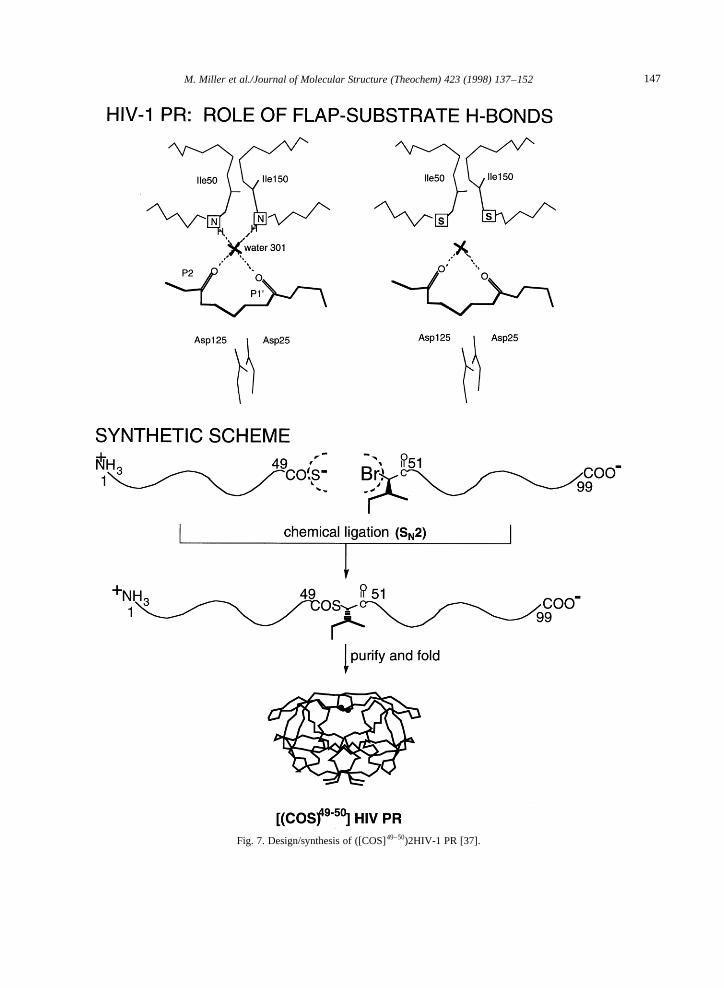
Fig. 7. Design/synthesis of ([COS]49–50)2HIV-1 PR [37].
147M. Miller et al./Journal of Molecular Structure (Theochem) 423 (1998) 137–152

HIV-1 PR inhibitor structures [17], it is a scenarioworth considering when one recalls that the pepsin-like proteinases possess only a single functional flap.We considered this an avenue worth pursuing andaccordingly set out to further explore this idea.
2.3. ([COO]49–50,[CONH]149–150)HIV-1 PR
Based on the need to further investigate the role ofspecific flap–substrate hydrogen bonds in the catalyticmechanism of HIV-1 PR, we designed an analogue of
Fig. 8. Design/synthesis of ([COO]49–50.[CONH]149–150)HIV-1 PR [38].
148 M. Miller et al./Journal of Molecular Structure (Theochem) 423 (1998) 137–152

HIV-1 PR in which hydrogen bond donor was deletedfrom the tip of one flap only. A covalent dimeric formof HIV-1 PR was prepared by total chemical synthesisin which the Gly49–Ile50 peptide bond –NH– atomwas specifically replaced, in one flap only, by anester –O– atom (Fig. 8). This single atom substitutionremoves the hydrogen bond donating ability of theGly49–Ile50 backbone linkage. The normal peptidebond was maintained in the second subunit of theenzyme (numbered Gly149–Ile150). As a control mole-cule, a second covalent dimer analogue was simulta-neously prepared [38] which differed from the singleflap ester analogue only in possessing the nativeamide [CONH] linking Gly49–Ile50 in both flaps.The control molecule was fully active, with kineticparameters (kcat, Km) similar to those of either recom-binant or chemically synthesized HIV-1 PR. Surpris-ingly, the analogue containing a backbone ester bondat Gly49–Ile50 in one flap was also highly active, andkinetic analysis indicated thatkcatwas reduced only bya factor of 2 relative to the control enzyme molecule.This was a remarkable result. Based on a 5 kcal mol−1
effect for deletion of both flap–substrate hydrogenbonds, a logical assumption is that deletion of oneof these bonds would lead to a 2.5 kcal mol−1 effect,or < 50-fold reduction in the intrinsic catalyticactivity. The observation of only a 2-fold effect inkcat is not consistent with this expectation. We inter-preted the 2-fold reduction ofkcat as consistent withthe suggestion that hydrogen bonding from the tip ofonly one flapis important to catalytic activity.
Additional evidence which calls into question thesignificance of hydrogen bonding from both flapscame from the study of inhibitor binding. DMP-323[28], an HIV-1 PR inhibitor specifically designed todisplace water 301 and hydrogen bond with the tip ofboth flaps, is actually bound 3-fold tighter to themonoester enzyme analogue relative to the controlenzyme. This is despite the inability of the monoesteranalogue to form one of these two enzyme–substratehydrogen bonds. Thus even for inhibitor binding,hydrogen bonding from the tip of the second flapmakes no net contribution to function.
3. Discussion
In this paper we have described a series of experi-ments performed on the HIV-1 PR molecule that were
designed to elucidate specific aspects of the molecularbasis of its action as a proteolytic enzyme. We haveused the capabilities provided by total chemical syn-thesis to prepare unique variant forms of the enzymemolecule in order to explore the role of the flaps inHIV-1 PR catalysis and inhibitor binding. Full kineticcharacterization (including studies with key inhibitors)was carried out on each analogue; in addition, struc-tural studies were carried out on selected enzymechemical variants. Taken together, the kinetic andstructural data on these backbone engineered enzymemolecules present a strong case that something isamiss with the conventional two-flap model of HIV-1PR action. The ([COS]51–52)HIV-1 PR retains fullcatalytic activity, but does not seem to coordinatewater 301 in the co-crystal structure with MVT101;the ‘‘double (backbone H-bond) deletion’’ variant,([COS]49–50)HIV-1 PR, displays a loss of catalyticactivity that shows that the Ile50 –NH– makes animportant contribution to enzyme action; yet, the‘‘single flap (backbone H-bond) deletion’’ tethereddimer HIV-1 PR retains full catalytic activity!
Based on this combined structural and biochemicalcharacterization of a number of HIV-1 PR analoguesengineered in the flap region, we find no evidence tosupport a mechanistic role for water 301. Water 301was seen by crystallography also in catalytically non-relevant complexes [39], while changes to the enzymemolecules which result in the absence of this watermolecule caused no change in the catalytic properties.We have further observed that removing the flap–sub-strate hydrogen bonds apparently mediated by water301 has drastically different consequences accordingto whether one or both of these hydrogen bonds isdisrupted. Deleting both of these hydrogen bonds(one from each flap) has a major deleterious effecton enzymatic activity. Deletion of just one of thesehydrogen bonds has essentially no effect. Theseresults, surprising given the highly symmetricalnature of the HIV-1 PR homodimer observed crystal-lographically, contrast with the hitherto assumed rele-vance, both for inhibitor design [17,28] and catalysis[26], of the hydrogen bonds from both flaps to the P2and P19 substrate/inhibitor carbonyls. What we pro-pose based on these results is that the two flaps of theHIV-1 PR homodimer do not contribute equally to thecatalytic process.
While this difference may be confined to the level
149M. Miller et al./Journal of Molecular Structure (Theochem) 423 (1998) 137–152

of a single hydrogen bond, we wish to raise thepossibility that HIV-1 PR may in fact function in amanner analogous to the pepsin-like proteinases.Even before the three dimensional structures of theretroviral proteinases were predicted [3] and eluci-dated [2,4,6], Tang et al. [40] put forward the hypoth-esis that (1) the sequence of the retroviral proteinasecorresponds to a domain of an aspartic proteinasemolecule, (2) the retroviral proteinases must exist asdimers, and, (3) the pepsin-like enzymes are fusionproteins arising out of an ancestral protein similar tothe retroviral proteinase. It is still not clear whetherretroviral proteinases, evolving from an ancestralgene, are direct precursors to the cellular enzymesor whether they are independently derived from theancestral gene by deletion events. In order to carry outits enzymatic functions, the aspartic proteinase mustcomprise two domains or two monomers, and musthave two characteristic Asp–Thr–Gly signaturesequences at the active site. However, if the asparticproteinases from higher organisms could functionwith only one flap, may one conclude that the retro-viral proteinase or a similar archetypal one also usesonly one flap? If the convergence of the catalyticmechanism of the native enzymes belonging to thetwo classes, viz., two adjacent Asp residues from dif-ferent domains or monomers, is any indication, onecan extrapolate it to the entire process of the catalysisand conclude that perhaps only one flap is necessaryand sufficient for enzymatic activity for both classesof aspartyl proteinases.
Over all, the data discussed in this paper supportthis hypothesis. The experimental observations pre-sented here provide evidence that interactions fromone flap are sufficient for full enzymatic activity ofHIV-1 PR and are consistent with a model in whichthe retroviral enzyme uses only a single flap closeddown over the substrate in the catalytically productivecomplex (see Fig. 2) similar to that of pepsin-likeenzymes. This would involve direct flap–substrateinteractions, with a quite different (flat vs. edge on)orientation of the flap with respect to the substrate.While not in itself an outrageous suggestion (after all,the cell encoded enzymes work perfectly well withonly one flap), at first glance, it is not easy to seehow such a mode of action can be reconciled withthe extensive crystal structure data and with NMRevidence that both HIV-1 PR flaps close down over
substrate-derived inhibitors, and that both flaps areinvolved in specific interactions of potential impor-tance for enzyme action.
The appearance of two fully symmetrical flaps inX-ray structures of catalytically non-relevant HIV-1PR complexes with symmetrical inhibitors is notsurprising and is not in disagreement with the singleflap hypothesis. In the case of complexes with sub-strate-based inhibitors the same observation may bean artifact caused by the bidirectionality of the inhi-bitor in the crystal lattice. Unfortunately X-ray datafor HIV-1 PR with bound inhibitors are available inmost cases only at low to medium resolution. In thecrystal structure of HIV-1 PR/MVT101 complex,MVT101 hexapeptide, initially reported as bound inone orientation [7], on refinement of 2 A˚ data in factexhibits a two-fold static disorder [34]. However, noattempt to correlate the structure of the flap with theorientation of the peptide chain was made. The firstindication of ‘‘nonequivalence’’ of the two flaps ofthe homodimer came from the crystal structure of the([COS]51–52)2HIV-1 PR (solved as a the mirror imageof D-enantiomer) described here. At 2.5 A˚ resolutionMVT101 inhibitor was modeled in one orientation. Ifthis indeed is a case in this crystal form, then thesecond flap is visibly disordered (see Fig. 5(b)). Theother possibility is that the poorly defined electrondensity corresponds to the flap which closes over theinhibitor bound in the alternative orientation withlower occupancy. The ‘‘nonfunctional’’ flap wouldbe then in this crystal lattice completely disorderedand undetectable, as were both flaps in the crystalstructure of the unliganded form of RSV PR [4,41].
We have recently collected X-ray data extending to2 A resolution on co-crystals of D-ligated HIV-1 PRcomplexed with D-MVT101 inhibitor and we will tryto verify these possibilities with carefully designedrefinement procedures. The chemical synthesis ofother flap-region analogues of the HIV-1 PR is alsobeing undertaken to test the role of the flaps in catalysis.
4. Conclusions
The conventional ‘‘two-flap’’ model of HIV-1 PRcatalysis has been subjected to a critical test, and onthe face of it has been put in doubt by the structuraland kinetic properties of the variant forms of the
150 M. Miller et al./Journal of Molecular Structure (Theochem) 423 (1998) 137–152

enzyme prepared by total chemical synthesis. The‘‘single flap’’ mode of catalytic action of the HIV-1PR has to be subjected to more stringent experimentalinvestigation, in order to clarify if it is the normalmode of action or an alternative mechanism in theevent of a mutation in one of the flaps of the PRdimer. In either case, the results reported here havesignificant implications for understanding the mol-ecular origins of substrate specificity in the HIV-1PR and the problem of escape mutants, and will con-tribute to the design of improved inhibitors. Morework remains to be done to explore these importantquestions.
Acknowledgements
We thank Drs. Jane Dyson and Afshin Karimi fortheir assistance in the design and execution of theNMR experiment. This research is sponsored in partby the National Cancer Institute (DHHS) undercontract with ABL and by NIH grants R01 48897and P01 GM 48870 (SBHK). The contents of thispublication do not necessarily reflect the views orpolicies of the Department of Health and HumanServices, nor does the mention of trade names, com-mercial products, or organizations imply endorsementby the U.S. government.
References
[1] J. Schneider, S.B. Kent, Cell 54 (1988) 363.[2] M.A. Navia, P.M. Fitzgerald, B.M. McKeever, C.T. Leu, J.C.
Heimbach, W.K. Herber, I.S. Sigal, P.L. Darke, J.P. Springer,Nature (London) 337 (1989) 615.
[3] L.H. Pearl, W.R. Taylor, Nature (London) 329 (1987) 351.[4] M. Miller, M. Jaskolski, J.K.M. Rao, J. Leis, A. Wlodawer,
Nature (London) 337 (1989) 576.[5] S.B. Kent, Annu. Rev. Biochem. 57 (1988) 957.[6] A. Wlodawer, M. Miller, M. Jaskolski, B.K. Sathyanarayana,
E. Baldwin, I.T. Weber, L.M. Selk, L. Clawson, J. Schneider,S.B.H. Kent, Science 245 (1989) 616.
[7] M. Miller, J. Schneider, B.K. Sathyanarayana, M.V. Toth,G.R. Marshall, L. Clawson, L. Selk, S.B.H. Kent, A.Wlodawer, Science 246 (1989) 1149.
[8] A.L. Swain, M.M. Miller, J. Green, D.H. Rich, J. Schneider,S.B. Kent, A. Wlodawer, Proc. Natl. Acad. Sci. USA 87 (1990)8805.
[9] M. Jaskolski, A.G. Tomasselli, T.K. Sawyer, D.G. Staples,R.L. Heinrikson, J. Schneider, S.B. Kent, A. Wlodawer, Bio-chemistry 30 (1991) 1600.
[10] A. Caflisch, P. Niederer, M. Anliker, Proteins: Struct. Funct.Genet. 13 (1992) 223.
[11] A. Gustchina, I.T. Weber, Proteins: Struct. Funct. Genet. 10(1991) 325.
[12] D.M. York, T.A. Darden, L.G. Pedersen, M.W. Anderson,Biochemistry 32 (1993) 1443.
[13] W.E. Harte, Jr., S. Swaminathan, D.L. Beveridge, Proteins:Struct. Funct. Genet. 13 (1992) 175.
[14] A. Gustchina, C. Sansom, M. Prevost, J. Richelle, S.Y.Wodak, A. Wlodawer, I.T. Weber, Protein Eng. 7 (1994)309.
[15] P.M.D. Fitzgerald, Curr. Opin. Struct. Biol. 3 (1993) 868.[16] K. Appelt in P.S. Anderson, G.L. Kenyon, G.R. Marshall
(Eds.), Perspectives in Drug Discovery Design, ESCOM,Leiden, 1993, p. 23.
[17] A. Wlodawer, J.W. Erickson, Annu. Rev. Biochem. 62 (1993)543.
[18] D.R. Davies, Annu. Rev. Biophys. Biophys. Chem. 19 (1990)189.
[19] K. Suguna, E.A. Padlan, C.W. Smith, W.D. Carlson, D.R.Davies, Proc. Natl. Acad. Sci. USA 84 (1987) 7009.
[20] T. Hofmann, R.S. Hodges, M.N. James, Biochemistry 23(1984) 635.
[21] T. Hofmann, A.L. Fink, Biochemistry 23 (1984) 5247.[22] B.M. Dunn, A.L. Fink, Biochemistry 23 (1984) 5241.[23] L.H. Pearl, FEBS Lett. 214 (1987) 8.[24] S. Grzesiek, A. Bax, L.K. Nicholson, T. Yamazaki, S.J. Stahl,
C.J. Eyermann, D.A. Torchia, C.N. Hodge, P.Y. Lam, P.K.Jadhav, C.-H Chang, J. Am. Chem. Soc 116 (1994) 1581.
[25] J.K.M. Rao, J.W. Erickson, A. Wlodawer, Biochemistry 30(1991) 4663.
[26] A. Gustchina, I.T. Weber, FEBS Lett. 269 (1990) 269.[27] J. Erickson, J.K.M. Rao, C. Abad-Zapatero, A. Wlodawer, in:
H. Krausslich, S. Oroszlan, E. Wimmer (Eds.), Current Com-munications in Molecular Biology, Cold Spring HarborLaboratory Press, Cold Spring Harbor, 1989, p. 191.
[28] P.Y. Lam, P.K. Jadhav, C.J. Eyermann, C.N. Hodge, Y. Ru,L.T. Bacheler, J.L. Meek, Science 263 (1994) 380.
[29] L.E. Zawadzke, J.M. Berg, Proteins: Struct. Funct. Genet. 16(1993) 301.
[30] M. Schnolzer, S.B. Kent, Science 256 (1992) 221.[31] R.C. Milton, S.C. Milton, M. Schnolzer, S.B.H. Kent, Tech-
niques in Protein Chemistry IV, Academic Press, New York,1993, p. 257.
[32] R.C. Milton, S.C. Milton, S.B. Kent, Science 256 (1992)1445.
[33] The Molecular Replacement Method, a Collection of Paperson the Use of Non-Crystallographic Symmetry, Gordon andBreach, New York, 1972.
[34] M. Miller, M. Geller, M. Gribskov, S.B.H. Kent, Proteins:Struct. Funct. Genet., 27 (1997) 184.
[35] S.B. Kent, M. Baca, J. Elder, M. Miller, R. Milton, S. Milton,J.K. Rao, M. Schnolzer, in: K. Takahashi (Ed.), AsparticProteinases, Plenum, New York, 1995, p. 425.
151M. Miller et al./Journal of Molecular Structure (Theochem) 423 (1998) 137–152

[36] M. Baca, Ph.D. Thesis, The Scripps Research Institute, LaJolla, CA, 1994.
[37] M. Baca, S.B. Kent, Proc. Natl. Acad. Sci. USA 90 (1993) 11638.[38] M. Baca, T.W. Muir, M. Schnolzer, S.B.H. Kent, J. Am.
Chem. Soc. 117 (1995) 1881.[39] J.W. Erickson, in: P.S. Anderson, G.L. Kenyon, G.R. Marshall
(Eds.), Perspectives in Drug Discovery, Design, ESCOM,Leiden, 1993, p. 109.
[40] J. Tang, M.N.G. James, I.N. Hsu, J.A. Jenkins, T.L. Blundell,Nature (London) 271 (1978) 618.
[41] M. Jaskolski, M. Miller, J.K.M. Rao, J. Leis, A. Wlodawer,Biochemistry 29 (1990) 5889.
152 M. Miller et al./Journal of Molecular Structure (Theochem) 423 (1998) 137–152